~i iii ii~ i,iiii! '84184
,,,,~iii~
iiii, ~'ii!i,i
~ i~iii!iiii ~!~i~'!'!
' ~!!ii!PiR'~TO~o~Li!iiiiii,,,,i,,~!iiiiii,,~i~!!i!~i~'"i
~! ii~,!!~i'~i
~ ,i,!!i~i
84
ii~,,i~,~!!
!~il ,, i! i~i~i
i~ ~ i~i !i!~ ii !,!iiii iiiiiii!iiL!~ ~
The Use of RNA Probes for the Analysis of Gene Expression
Dominique Be~in
Abstract
of these two methods to quantitatively detect mRNA targets are discussed:
Index Entries: RNA; riboprobe; phage; RNA polymerase; transcription; hybridization.
1. Introduction
The isolation and characterization of RNA
polymerases from the Salmonella phage SP6 and
the Escherichia coli phages T7 and T3 have revo- lutionized all aspects of the study of RNA
metabolism (1-6). Indeed, it is now possible to
generate unlimited quantities of virtually any RNA molecule in a chemically pure form. This technology is based on a number of properties of the viral transcription units. First, and in con- trast to their cellular counterparts, the enzymes are single-chain proteins that were easily purified from phage-infected cells and are now produced by recombinant DNA technology. Second, they very specifically recognize their own promoters (7 and references therein), which are contiguous 17- to 20-bp-long sequences rarely encountered in bacterial, plasmid, or eukaryotic sequences. Third, the enzymes are highly processive, allow- ing the efficient synthesis of very long transcripts from DNA templates. In this article, I discuss the preparation of the DNA templates, the transcrip- tion from the templates of labeled synthetic RNA molecules, commonly called riboprobes, and their use in Northern and RNase protection assays.
2. Materials
These protocols require the use of standard molecular biology materials and methods for carrying out subcloning, polymerase chain reaction (PCR), and gel electrophoresis, in addition to those listed herein. More rigorous precautions are required for working with RNA than are commonly used for most DNA studies, because it is important to avoid RNase contami- nation. The two major sources of unwanted RNases are the skin of investigators and micro- bial contamination of solutions. Most RNases do not require divalent cations and are not irre- versibly denatured by autoclaving. Gloves should be worn and frequently changed. Sterile plasticware should be used, although some mechanically manufactured tubes and pipet tips have been used successfully without steriliza- tion. Glassware should be incubated at 180~ in a dried baking oven for several hours. In addition, divalent cations (such as Mg 2§ or Ca 2+) accelerate base-mediated RNA hydroly- sis. RNA can be stored in sterile water, but is most stable when stored in 1 mM KOAc, pH 5.0, with 0.1 mM EDTA.
Address to which all corespondence and reprint requests should be addressed. Department of Pathology, University of Geneva Medical School, CMU, 1 rue Michel-Servet, CH-1211 Geneva, Switzerland, e-mail: [email protected]
2.1. Preparation of Riboprobes
1. Water: Although a number of protocols recom- mend treatment of the water used for all the solutions with diethylpyrocarbonate, I find this to be unnecessary. Double-distilled water is used for the preparation of stock solutions that
can be autoclaved (121~ 15-30 min), and
sterilized water is used otherwise.
2. TE: 10 mM Tris-HC1, pH 8.1, and 1 mM ethyl- ene-diamine tetraacetic acid (EDTA).
3. 10X TB: 0.4M Tris-HC1, pH 7.4, 0.2M NaC1, 60 mM MgC12, and 20 mM spermidine. If ribo- nucleotides are used at concentrations >0.5 mM each, MgC12 concentration should be increased to provide a free magnesium concentration of 4 mM. 4. 0.2M Dithiothreitol (DTT): The solution is
stored in small aliquots at-20~ Aliquots are
used only once. EDTA can be included at 0.5 mM to stabilize DTT solutions.
5. Ribonucleotides: Neutralized solutions ofribenucle- otides are commercially available, or can be made
up from dry powder
(see
Note 1). Ribonucleotidesolutions can be stored at-20~ for several months. 6. RNA polymerase stocks: The three RNA poly- merases, SP6, T3, and T7, are available com- mercially. Store at -20~
7. RNA polymerase dilution buffer: 50 mM Tris-HC1, pH 8.1, 1 mM DTT, 0.1 mM EDTA, 500 ktg/mL bovine serum albumin (BSA) and 5% glycerol. Diluted enzyme is unstable and should be stored on ice for no more than a few hours. 8. Stop-mix: 1% sodium dodecyl sulfate (SDS), l0 mM EDTA and 1 mg/mL tRNA. The tRNA may be omitted.
9. TEN: 10 mMTris-HC1, pH 8.1, 1 mM EDTA, and 100 mM NaCl.
10. Sample buffer (polyacrylamide/urea gels): 80%
deionized formamide
(see
Note 2), 2M urea,0.1X TBE (8.9 mM Tris base, 8.9 mM boric acid, 0.2 mM EDTA), and 0.01% each of xylene cyanol and bromophenol blue. Use 1-2 lxL/~L of RNA in aqueous solution.
2.2. Northern Blot
Hybridization
1. Formamide: Pure formamide is slowly hydro- lyzed by water vapor to ammonium formate
and therefore must be deionized
(see
Note 2).Store at -20~
2. Glyoxal: A 30% glyoxal solution (6M) is deion- ized by several incubations at room tempera- ture with a mixed bed resin (AG501-X8, Bio-Rad), until the pH is 6.0-7.0 and the conductivity of 3% glyoxal in water is below 30 I.tSiemens. Store at -20~ in small aliquots. 3. Denaturation buffer: 1M glyoxal in 50% dimethyl sulfoxide (DMSO) and 10 mM Na2HPO4, pH 6.8. Ethidium bromide can be included at a concen- tration of 50 ~tg/mL to visualize the rRNAs during electrophoresis.
4. 5X RNA sample buffer (RSB): 50% glycerol, 10 mM Na2HPO4, pH 6.8, and 0.4% bromophenol blue. Autoclave and store at-20~ in small aliquots. 5. Transfer membrane: Nitrocellulose (Schleicher and Schuell, Keene, NH) and nylon membranes (Hybond N, Amersham, Arlington Heights, IL or Biodyne A, Pall Biosupport, Glencove, NY) have been used successfully.
6. 50X Denhardt's solution: Dissolve 2 g of bovine serum albumin (fraction V) in 80 mL of sterile water. Bring the pH to 3.0 with 2N HC1, boil for 15 min, and cool on ice for 10 min. Bring the pH to 7.0 with 2N NaOH and add sterile water to 100 mL. Autoclave a 100-mL solution of 2% polyvinylpyrrolidone (K90, Fluka Chemic, AG, Buchs, Switzerland) and 2% Ficoll 400 (Pharmacia, Piscataway, NJ), and add to the 2% albumin solution.
7. Hybridization solution: 50% deionized forma- mide, 0.8M NaC1, 50 mM Na-PIPES, pH 6.8, 2 mM EDTA, 0.1% SDS, 2.5X Denhardt's solution, and 0.1 mg/mL sonicated and heat- denatured salmon sperm DNA. The hybridiza- tion solution is heated, filtered over 0.45 l.tm Nalgene sterilization units or Millipore nitro- cellulose filters, and kept at the hybridization temperature during prehybridization. The filtration step decreases nonspecific attachment of the probe to the membrane and degasses the solution.
8. 20X Standard saline citrate (SSC): 3M NaC1 and 0.3M sodium citrate, pH 7.0.
2.3. gNase Protection
1. Hybridization mixture for RNase protec-
tion: 80% deionized formamide
(see
Note 2),0.4M NaC1, 40 mM Na-PIPES, pH 6.8, and 1 mM EDTA.
2. RNase digestion buffer: 300 mM NaC1, 10 mM Tris-HC1, pH 7.4, and 4 mM EDTA.
3. Pancreatic RNase: Make a 10 mg/mL solution in TE containing 10 mM NaC1. Vials contain- ing lyophilized RNases should be carefully open in a ventilated hood to avoid contamina- tion. Boil for 15 min and slowly cool to room temperature. Store in aliquots at -20~
4. T1 RNase: Make a 1 mg/mL solution in TE. Adjust the pH to 7.0. Store in aliquots at -20~ 5. Proteinase K: Dissolve the enzyme at 20
mg/mL in water and store in aliquots at -20~
3. Methods
3.1. Preparation of Riboprobes
3.1.1. Linearized Plasmid Templates
for Runoff Transcription
1. Subclone the desired gene fragment in a tran-
scription vector
(see
Note 3).2. Isolate plasmid DNA by alkaline lysis from a 30-100 mL saturated culture. Plasmid DNAs are purified by CsC1/ethidium bro- mide centrifugation or by precipitation with
polyethyleneglycol
(see
Note 4).3. Linearize 2-20 ~g of plasmid DNA with an
appropriate restriction enzyme
(see
Note 5).Verify the extent of digestion by electrophore- sis of an aliquot (0.2-0.5 ~tg of DNA) on an agarose minigel in the presence of ethidium
bromide (0.5 l.tg/mL)
(see
Note 6).4. Purify the restricted DNA by two extractions with phenol/chloroform/isoamyl alcohol (25/24/1 by vol), and remove residual phenol by one extraction with chloroform (24/1 by vol). Precipitate the DNA with ethanol. If little DNA is present (below 5 Ixg), add 10 ~tg of glycogen (Boehringer Mannheim, Mannheim, Germany) as a carrier; this carrier has no adverse effect in the transcription reactions. After washing the ethanol pellet, the DNA is dried in air and resuspended in TE at 1 ~tg/lxL.
3.1.2. Synthetic and PCR-Derived Templates
The major limitation in using restriction enzymes to clone inserts and to linearize plasmid templates is that appropriate sites are not always available. Furthermore, the transcripts will almost always contain 5' and 3' portions that differ from
those of endogenous RNA. One possibility to cir- cumvent these difficulties is based on the tran- scription of small DNA fragments obtained by annealing of synthetic oligodeoxynucleotides
(6,8).
An alternative and more general approach generates the transcription templates via PCR amplification of plasmid DNA. In theory, such templates could direct the synthesis of virtually any RNA sequence.1. Design the 5' primer, which has a composite sequence: its 5' portion is constituted by a mini- mal T7 promoter, and its 3' portion corresponds
to the beginning of the transcript
(see
Note 7).Six to ten nucleotides (nt) are usually sufficient to prime DNA synthesis on the plasmid template. 2. Design the 3' primer, which is usually 17-20-nt
long and defines the 3' end of the transcript
(see
Note
8).3. PCR amplify 2-50 ng of plasmid DNA in a total volume of 100 [tL. Verify that a DNA fragment of the expected size has been ampli- fied, and estimate the amount of DNA by com- parison with known standards.
4. Purify the DNA as described in Subheading 3.1.1., step 4 and resuspend in TE at the appro- priate concentration. The PCR-derived tem- plates are transcribed at lower DNA concentra- tions than plasmids to maintain the molar ratio of enzyme to promoter; I use 3 lxg/mL for a
100-bp
fragment.3.1.3. Basic Transcription Protocol
for Radioactive Probes
1. Assemble the transcription mixture to a total volume of 10 [tL by adding in the following order: water (as required), 1 BL of 10X TB, 0.5 ~tL of bovine serum albumin (2 mg/mL), 0.5 ~tL of 0.2M DTT, 0.25 ~tL of placental RNase inhibitor (40 U/I.tL), 1 p.L of a 5 mM solution of each ribonucleotide, i.e., ATP, GTP, CTP, 5 ~tL of ~-[32p]-UTP (400 Ci/mmol, I0 mCi/mL), 1 ~tL of restricted plasmid DNA tem- plate, and 0.25-0.75 ~tL of RNA polymerase
(see
Notes 9 and 10).2. Incubate 40 min at 37~ for T7 and T3 poly- merases or 40~ for SP6 polymerase. After addition of the same number of units of enzyme
(see
Note 10), incubate for a further 40 min.1 5 6 ...
:
:::
:
3. Degrade the template with RNase-free DNase (1 U/I.tg of DNA) for 20 min at 37~
4. Stop the reaction by adding 40 ~L of stop-mix. 5. After two extractions with phenol/chloroform, in which the organic phases are back-extracted with 50 I.tL of TEN, the combined aqueous phases are purified from unincorporated nucle- otides by spun-column centrifugation. The spun-column is prepared by filling disposable columns (QS-Q, Isolab, Hrrth, Germany) with a sterile 50% slurry of G-50 Sephadex (Pharmacia) in TEN followed by centrifugation for 5 min at 1200 rpm (200g). The samples are carefully deposited on top of the dried resin, and the col- umn is placed in a sterile conical centrifuge tube. After one centrifugation for 5 min at 1200 rpm, 200 l.tL of TEN is deposited on top of the resin and the column recentrifuged. The eluted RNA (200-400 pL) is ethanol precipitated and
resuspended in water
(see
Note 11).6. Measure the incorporation efficiency by count-
ing an aliquot
(see
Note 12 for calculations andfor modifications of the basic protocol). 7. The size of the transcript may be verified by
electrophoresis in polyacrylamide/urea gels
(see
Note 13).3.2.
Northern Blot Hybridization
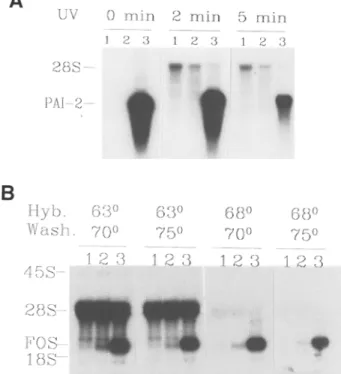
The use of riboprobes in RNA blot hybridiza- tions follows the same general principles as that for DNA probes. The major disadvantage in using double-stranded DNA probes results from self-annealing, which decreases the avail- ability of DNA probes to bind to the immobilized target; this is particularly critical with heterolo- gous probes, in which the reannealed probe may displace incompletely matched hybrids. Self-annealing of course does not occur with single-stranded RNA probes. Most difficulties encountered with riboprobes stem from the increased thermal stability of RNA:RNA hybrids. Thus, cross-hybridization of GC-rich probes to rRNAs can generate unacceptable backgrounds. This problem is often solved by increasing the stringency of hybridization, as illustrated in Fig. lB. Alternatively, the template may have to be shortened to remove GC-rich regions from the probe.
1. Denature the sample in 8 lxL of denaturation
buffer for 15-30 min at 50~ Add 2 IxL of
5X RSB and electrophorese in 0.7-2% agar- ose gels in i0 mM Na2HPO 4, pH 6.8. The buffer should be circulated with a peristaltic pump, so that the pH near the electrodes remains neutral.
2. Transfer the RNAs by capillarity onto a mem-
brane
(see
Note 14).3. Fix the RNA by incubating the blots at 80~ under vacuum. This step is essential to remove glyoxal covalently fixed to guanine residues in the RNA.
4. UV-crosslinking is often used to improve RNA retention, although irradiation may increase
background hybridization
(see
Fig. 1A andNote 15).
5. Prehybridize the blots for 3-12 h in hybrid- ization solution. Use 200 l.tL of the solution per cm 2 of membrane.
6. Dilute the probe in hybridization solution (25-50 I.tL/cm 2 of membrane), then add to the membrane. I frequently hybridize 2-3 filters per bag. Hybridize at the appropriate temper-
ature (58-68~
see
Fig. 1B and Note 16), usu-ally for 12-18 h
(see
Note 17).7. Wash the membranes twice for 10-20 min at the hybridization temperature with 100 lxL/cm 2 of 3X SSC, and 2X Denhardt's solution, and then three times with 0.2X SSC, 0.1% SDS, and 0.1% sodium pyrophosphate at the appropriate
temperature
(see
Fig. 1B and Note 17).8. Expose to autoradiographic film. As long as the membranes are not allowed to dry, they can be further washed at increased stringencies to reduce background.
3.3. RNase Protection
This assay is based on solution hybridization and on the resistance of R N A : R N A hybrids against single-strand specific RNases. A 32p-labeled probe is synthesized that is partially complementary to a portion of the target RNA. It is hybridized in excess to the target so that all complementary sequences are driven into the labeled RNA:RNA hybrid. Unhybridized probe and any single-stranded region of the hybridized probe are then removed by RNase digestion. The
A
B
UV 2 8 S - PAl-2-- H y b . W a s h . 4 5 S - 2 8 S - FOS I 8 S 0 m i n 2 m i n 5 r a i n 1 2 3 1 2 3 1 E 3!
6oo 7 0 o 1 2 { } 6 3 0 6 8 0 6 8 o 7 5 o 7 0 o 7 5 0 1 2 3 1 2 3 1 2 3Fig. 1. Northern blot hybridization with riboprobes. (A) Effect of UV crosslinking. Northern blot hybrid- ization of PAI-2 mRNA in murine total cellular RNA with an homologous cRNA probe. Lanes 1 and 2:5 gg of placental RNA (15.5 and 18.5 d gestation), that do not contain detectable levels of PAI-2 mRNA. Lane 3:
1 gg of LPS-induced macrophage RNA, an abundant
source of PAI-2 mRNA
(29).
All samples were elec-trophoresed and transferred together. After cutting the membrane, each filter was UV-treated as described. The filters were hybridized at 58~ washed at 70~ and exposed together. Cross-hybridization of the probe to 28S rRNA is more pronounced after UV irradiation, and specific hybridization is decreased after 5 min of UV exposure. (B) Effect of hybridization temperature.
Northern blot hybridization of
c-fos
mRNA in rat totalcellular RNA with a murine
v-fos
cRNA probe. Lane1: uninduced cells. Lane 2: partially induced cells. Lane 3: fully induced cells (M. Prentki and D. Belin, unpublished). All samples were electrophoresed and transferred together. The filters, which were not UV cross-linked, were hybridized and washed in parallel at the indicated temperatures. The four filters were exposed together. Cross-hybridization of the probe to 28S rRNA was essentially abolished by hybridizing at 68~ Some specific signal was lost with the 75~ stringency wash. "protected" probe is then detected and quantitated on a denaturing polyacrylamide gel. It can be used to map the ends of RNA molecules or exon-intron
boundaries. It also provides an attractive and highly sensitive alternative to Northern blot hybridization for the quantitative determination of mRNA abundance.
RNase protection has a number of advantages. First, solution hybridization tolerates high RNA input (up to 60 gg of total RNA), and is not affected by the efficiency of transfer on mem- branes or by the availability of membrane-bound RNAs. Second, the signal to noise ratio is much more favorable, since cross-hybridizing RNAs yield only short protected fragments. Third, a sig- nificant fraction of mRNAs is often partially degraded during RNA isolation; in Northern blots, this generates a trail of shorter hybridizing spe- cies, which reduces the sensitivity of detection. Finally, the detection of hybridized probes on sequencing gels is much more sensitive because the width of the bands is less than a tenth of those of intact RNAs in agarose gels.
Only two features of Northern blots are lost in RNase protection assays: c o m p l e t e size determination of target RNAs and multiple use of each sample.
1. Linearize the plasmid DNA template as descri-
bed in Subheading 3.1.1.
(see
Note 18).2. Transcribe the template as described in Sub- heading 3.1.3. The amount of labeled ribo-
nucleotide may be varied
(see
Note 19).3. An optional step is to purify the full-length tran-
scripts by electrophoresis
(see
Note 20). Separatethe transcript on a preparative 5-6% polyacryla- mide/urea gel (gel thickness: 0.4-1.0 mm). Cover
the wet gel with Saran Wrap TM and expose for
30 s to 2 min at room temperature to localize the full-length transcript. Cut the exposed band on the film with a razor blade. After aligning the cut film on the gel, excise the gel band with a sterile blade. The cut gel should be reexposed to verify that the correct band has been excised.
4. Elute the RNA from the gel either by diffusion or electroelution.
a. Incubate the gel fragment in an Eppendorf tube in 500 gL of 0.5M ammonium acetate, 1% SDS, and 20 gg/mL tRNA for 1-3 h at
37~ or overnight at 4~ The eluate and
residual gel can be counted to ensure that more than 60% of the RNA is eluted. After two
extractions with phenol/chloroform, recover the eluted RNA by ethanol precipitation. b. Electroelute for 1-2 h at 30 V/cm in 0.1X
TBE in a sterile dialysis bag, after which invert the polarity for 30 s to detach the eluted RNA from the membrane. Purify the eluate by two phenol/chloroform extractions and ethanol precipitation with a known amount of tRNA carrier. This procedure is very sensitive to RNase degradation. 5. Resuspend the probe in water at 1-2 ng/~tL.
Add 1 ktL of probe to 29 ].tL of hybridization mixture for each assay. The exact amount of probe is not critical, since it is in excess of its
specific target
(see
Notes 19 and 21).6. Lyophilize or ethanol precipitate the sampie
RNAs
(see
Note 22). Resuspend in 30 l.tL ofcomplete hybridization mixture including probe, heat for 2 min at 90~ and incubate overnight,
usually at 45~
(see
Notes 23 and 24).7. Cool the samples on ice and add 300 ktL of RNase digestion buffer. Digest for 1 h at 25~ with pancreatic RNase, which cleaves after uracil and cytosine residues, with T1 RNase, which cleaves after guanine residues,
or with both RNases
(see
Notes 25 and 26).8. Add 20 BL of 10% SDS, anddegrade the enzyme(s) with 0.5 ktL (10 ktg) of proteinase K for 10-20 min at 37~ Extract once with phenol/chloro- form, and precipitate the RNAs with ethanol
with 10 ~tg of carrier tRNA
(see
Note 27).9. Resuspend the RNAs in sample buffer, dena- ture the hybrids for 2 min at 90~ and electro- phorese in polyacrylamide/urea sequencing gels. Alternatively, the hybrids may be analyzed on nondenaturing polyacrylamide gels. Fix the gels with 20% ethanol and 10% acetic acid to remove
the urea, dry, and autoradiograph
(see
Note 28).4. Notes
1. Powdered ribonucleotides should be resuspen- ded in water, neutralized to pH 7.0 with 1M NaOH or HC1, and adjusted to the desired con- centration by measuring the UV absorbance of appropriate dilutions:
a. 100 mM ATP: 1 540 absorbance units at 259 nm. b. 100 mM GTP: 1370 absorbance units at 253 nm. c. 100 mMCTP: 910 absorbance units at 271 nm. d. 100 mM UTP: 1000 absorbance units at 262 nm.
.
.
The integrity of ribonucleotide triphosphate solutions can be verified by thin-layer chroma- tography on PEI-cellulose (PEI-CEL300). The resin is first washed with water by ascending chromatography to remove residual UV- absorbing material and dried. Ribonucleotides 10-30 nmol are deposited on the resin, which is then resolved by ascending chromatography with 0.5M KH2PO 4, adjusted to pH 3.5 with H3PO 4. After drying, the ribonucleotides are detected by UV-shadowing at 254 nm.
To deionize formamide, incubate at -80~ until 75-90% of the solution has crystallized. Discard the liquid phase, thaw and incubate at 4~ for several hours with a mixed bed resin (AG501-X8, Biorad). Use a TeflonTM-covered magnet that has been freed of RNase by treat- ment with 0.1M NaOH for 10 min, and rinsed with water and with crude formamide. Check the conductivity, which should be below 20 ~Siemens. Filter the solution onto sterile paper (Whatman, LS-14) over a sterile funnel. The absorbance at 270 nm should be below 0.2. All the vectors that are commercially available
consist of high copy number
Escherichia coli
plasmids derived from ColE 1. The original plasmids (pSP64 and pSP65, Promega, Madison WI) contained only one SP6 promoter located
upstream of multiple cloning sites (MCS)
(2).
In the second generation of plasmids, two pro- moters in opposite orientation flank the MCS to allow transcription of both strands of inserted
DNA fragments. The pGEM TM series (Promega)
contain SP6 and T7 promoters
(5),
whereasthe pBluescript TM series (Stratagene Cloning
Systems, Inc., La Jolla, CA) contain T7 and T3 promoters. The choice of plasmid is mostly a matter of personal preference, although it can be influenced by the properties of individual sequences. For instance, we have frequently observed premature termination with SP6 transcripts. The nature of the termination sig-
nals is not completely understood
(4,9),
andtheir efficiency can be more pronounced when one ribonucleotide is present at suboptimal
concentration
(see
Note 13). The problem hassometimes been solved by recloning the inserts in front of a T7 or T3 promoter. The partial rec- ognition ofT7 (T3) promoters by T3 (T7) poly-
merases may result in the transcription of both strands when the ratio of enzyme to promoter is very high. This can be a source of artifacts, particularly when the templates are linearized inside the cloned inserts.
4. It is possible to use plasmid DNA from "mini- preps," although transcription efficiency can be reduced, particularly with SP6 polymerase. The RNA present in the minipreps is digested with pancreatic RNase (20 ~tg/mL), which is removed during purification of the linearized templates. Spun-column centrifugation of the digested minipreps can improve transcrip- tion efficiency.
5. Since RNA polymerases can initiate transcrip- tion unspecifically from 3' protruding ends
(4,10),
restriction enzymes that generate 5' protruding or blunt ends are usually preferred. If the only available site generates 3' protrud- ing ends, the DNA can be blunt-ended by exonucleolytic digestion with T4 DNA poly- merase or with the Klenow fragment of DNA polymerase I. Restriction with enzymes that cut the plasmids more than once may also be used, provided that the promoter is not separated from the insert.6. The plasmids must be linearized as exten- sively as possible. Since circular plasmids are efficient templates, their transcription by the highly processive enzymes may yield RNA molecules that can be up to 20 kb long and thus incorporate a significant portion of the limiting ribonucleotide.
7. There are constraints on the 5'-sequence of tran- scripts since the sequence immediately down- stream of the start site is necessary for the transition from an abortive initiating cycling mode to the elongation mode. The first 6 nt have a strong influence on promoter efficiency; in particular, the presence of uracil residues are
usually detrimental
(6,8).
It may be necessary,therefore, to include in the 5'-end of the tran- scripts 5-6 bases that differ from those present in natural RNAs. My colleagues and I have used a number of composite T7 promoters, whose efficiency is summarized below. In addition to the promoter sequence (-17 to -1: 5'-TAATACGACTCACTATA) at the 5' end, the first 6 nt of the templates are:
Efficient promoters Inefficient promoters
GGGAGA (T7 consensus) G'Iq'GGG (5% efficiency) GGGCGA (pBS plasmids) GCTITG (1% efficiency) GCCGAA
Composite 5' primers with the SP6 promo- ter sequence have also been used success- fully. There are also constraints on the 5' sequence of the transcripts, but the optimal sequences (GAATA, GAACA, and GAAGA) are different than those with T7 polymerase
(11-13).
8. The 3' end of the transcripts should be exactly defined by the 5' end of the downstream primer. However, template-independent addition of 1- 2 nt during transcription usually generates populations of RNAs with different 3' ends. The proportion of each residue at the 3' end may depend on context and is influenced by the rela- tive concentration of each ribonucleotide, lim- iting ribonucleotide being less frequently
incorporated
(2,6,8,14).
9. The order of addition of components may be changed. Remember that dilutions of RNasin are very unstable in the absence of DT'I', and that DNA should not be added to undiluted 10X TB. The temperature of all components must be at least at 25~ to avoid precipitation of the DNA:spermidine complex. Since the radioac- tive nucleotides, which constitute half of the reactions, are provided in well-insulated vials, they can take up to 10-15 min after thawing to reach an acceptable temperature. I routinely incubate all components, except the RNasin and the polymerase, at 30~ for 15-20 min. When more than one probe is to be synthesized, a reaction mixture with all components is added to the DNA template. It is possible to reduce the total volume to 8 ~tL, in order to conserve materials; however, since the enzymes are very sensitive to surface denaturation, these incuba- tions are done in 400-ktL vials.
10. For SP6 polymerase, use 5-10 U/~tg ofplasmid DNA (size: 3-4 kbp). For T7 and T3 poly- merases, use 10-20 U/~tg of plasmid DNA.
11. The purification step (Subheading 3.1.3.,
step
4) may not be required, and some investigators use the transcription mixtures directly in hybridization assays. However, low back- grounds, consistency, and quantitation of the
newly synthesized RNAs probably justify the additional time and effort.
12. More than 50% of the labeled ribonucleotide is routinely incorporated, and often greater than 80%. An input of 50 ~tCi may therefore yield up to 4 x 10⁷ Cerenkov-cpm of RNA. This represents 100 pmol of UMP and, assuming no sequence bias (i.e., 25% of residues are uracil residues), 130 ng of RNA (specific activity: 3 • 108 Cerenkov-cprn/~g).
Similar results are obtained with labeled CTP and GTP, although GTP rapidly loses its incorporation efficiency on storage. ATP is not routinely used because of its higher apparent
K m for SP6 polymerase on linear templates (2,15). UTP is particularly stable and can be
used even after several weeks, once radioactive decay is taken into account. With each labeled ribonucleotide, the initial concentration must be >12.5 ~tM to ensure efficient incorporation and to prevent polymerase pausing. Transcripts destined to be translated must incorporate a 5' 7mG cap structure. This is usually achieved by performing the transcription in the presence of 500 ~tM of a cap analog dinucleotide and 50 }.tM GTP. Since the cap analog can also be used during elongation, labeled GTP should not be used to calculate incorporation effi- ciency. A variety of other labeled or of unla- beled probes may also be made for use in in
situ hybridization (see refs. 16-20).
For the synthesis of large quantities of RNA (>10 RNA transcripts/DNA template mol- ecule), each ribonucleotide is added at a final concentration of 0.5-1 mM; trace amounts of labeled UTP should be included to calculate transcription efficiency and to verify the size of the transcript. Total volume is increased to reduce the concentration of plasmid DNA to 30-50 ktg/mL. Additional modifications of the standard protocol have been described, and include the use of HEPES-KOH, pH 7.5, at 120 mM (SP6 polymerase), 200 mM (T7 polymerase), or 300 mM (T3 polymerase) (21). Furthermore, the T4 gene 32 protein can increase transcription efficiency when added at 10 ~tg/~g of template DNA (D. Caput, per- sonal communication). It has been recently reported that a reduction of premature termina-
tion and an increased synthesis of large full- length transcripts (size >1 kb) can be obtained by performing the transcription at 30~ or at room temperature (4,22).
13. Large transcripts that do not enter the poly- acrylamide gel are diagnostic of incompletely linearized templates. Small transcripts are indicative of extensive pausing or premature termination. In addition to the natural T7 ter- mination signal, a site located in the coding region of the parathyroid hormone (PTH) gene has been extensively characterized (23,24). Mutant T7 RNA polymerases that show reduced termination at natural sites have been constructed by Drs. D. H. Lyakhov and W. T. McAllister (SUNY, Brooklyn, NY).
14. Hybridization of dot-blots is often used to quantitate mRNA levels with a large number of samples. To accurately quantitate specific hybrids, it is necessary to include as negative control total RNA from cells that do not express the transcript under examination.
15. The intensity provided by transilluminators, a common source of UV light, varies with time, and excessive crosslinking can severely reduce hybridization efficiency (Fig. 1A). Further- more, UV-crosslinking can increase cross-hy- bridization to rRNAs, possibly by inducing covalent cross-linking with the probe. UV irra- diation should thus be limited to membranes subjected to multiple rounds of hybridization. An apparatus commercially available from Stratagene provides a means to control the dose of UV. The appropriate dose of UV is probably different for different membranes and is broader for wet than for dry membranes. 16. The major variables in optimizing signal-to-
noise ratio are the temperature of hybridization and the temperature of the stringency washes (Fig. 1B). Many probes can be hybridized at 58~ although the temperature must be increased to 68~ for certain pairs of probe and target RNAs. Stringency washes are usually per- formed at 70 or 75~ It is difficult to increase stringency much further, since most water baths cannot maintain accurate temperatures at or above 80~ For the detection of DNA targets, hybridization is usually performed at 42~ and the stringency washes are at 65~
Probes and Gene ExpresSion
: : ::
: :...
17. RNA probes are usually 200-800-nt long. A
With 600-nt-long RNA probes, a hybridization ~. 2:1
plateau is achieved in 20 h at 58~ with 40,~ 346
2.5 ng/mL of probe. This represents an input
of 7.5 x 10⁵ Cerenkov-cpm/mL. The probe con- 236
centration can be increased to 10 ng/mL, and ~65
the hybridization time decreased to 5 h without
increasing background. ~:~
18. The probes should be 100-400-nt long, and should include at least 10 nt that are not complementary to the target RNA. Residual template DNA generally produces a trace of
full-length protected probe that must be distin- ~
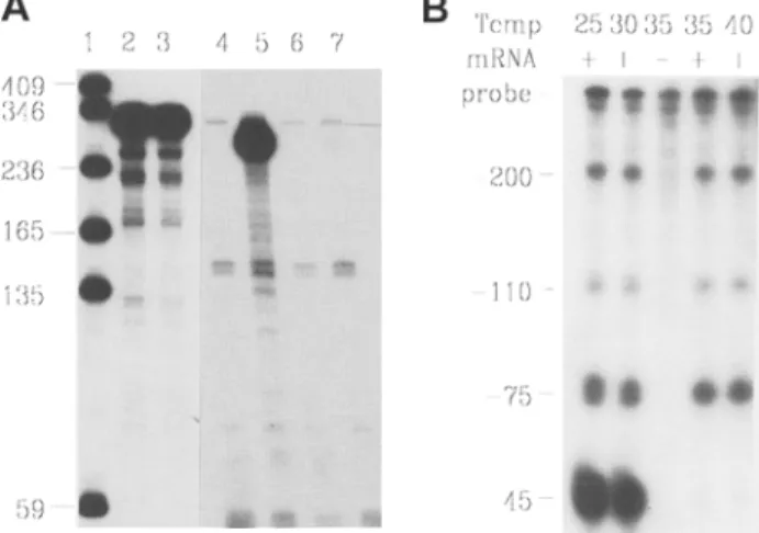
guishable from the fragment protected by the target RNA (Fig. 2).
19. It is often useful to decrease the specific activ- ity of the probe: more RNA is synthesized at the resulting higher ribonucleotide concentration, the probes are less susceptible to radiolysis, and less radioactivity is used. The guideline in Table 1 can be used to alter the specific activity of the probes according to the sensitivity required. 20. Purification is often necessary for maximal sen- sitivity or for mapping purposes. The transcrip- tion reaction can be directly loaded on the preparative gel after DNase digestion of the template, provided that enough EDTA is present in the sample buffer to chelate all the magnesium. Omitting the DNase digestion of the template results in higher amounts of fully protected probe in the assay.
21. The probe must be in excess of the target RNA
(see Fig. 2A). An input of 1-2 ng of a 300-nt probe in a total volume of 30 gL will drive the hybridization of target RNA to completion
(4-8 T~/2) in 12-16 h. Shorter hybridizations
can be performed but require higher probe
input (R o) to achieve the same extent of satura-
tion, i.e., to maintain the R o x T~/2 value.
22. To facilitate the RNase digestion step, each sample should contain the same amount of total RNA. For very low abundance target RNA, the amount of sample RNA may be increased up to 60 gg. Inequalities should be eliminated by addition of tRNA. A negative control sample, containing only tRNA, is always
included (Fig. 2A, lane 4; Fig. 2B, lane 3). 24.
23. The temperature of hybridization must be reduced to detect small or very AU-rich protected frag-
4 , 5 6 7 i i
~m
:iii~iii:
}B
Tcmp 2 5 3 0 3 5 35 40 mRNA f I + I,, 91
2,oo-OO ' 0 6
1 1 0 ~ ~ ~O0 O0
4 5Fig. 2. RNase protection assay. (A) Discrimination between target-specific signal and complete probe pro- tection by residual DNA. Detection of PN-I mRNA in
total RNA from murine tissues (25). The probe (310-
nt long) was gel purified. Lane 1: Size markers. Lane 2: purified probe. Lane 3: probe hybridized and pro- cessed without RNase digestion. This part of the gel was autoradiographed for 6 h. Lane 4: control hybrid- ization with 10 ~tg of tRNA; traces of fully protected probe are visible. Lane 5:10 [tg of RNA from seminal vesicles, an abundant source of PN-I mRNA; the spe- cific protected fragment is 260-nt long. Lane 6:10 ~tg of liver RNA, which does not contain detectable levels of PN-I mRNA. Lane 7:10 gg of testis RNA. which contains trace levels of PN-I mRNA. This part of the gel was autoradiographed for 24 h. (B) Effect of hybridization temperature on the detection of short complementary RNAs. The 5' ends of phage T4 gene 32 transcripts in total RNA from bacteria carrying a gene 32 expression cassette were mapped by hybrid- ization to a cRNA probe containing 400 nt of gene 32
upstream sequences (23). The probe was not gel puri-
fied, and hybridizations were performed at the indi- cated temperatures. Fully protected probe results from incomplete DNase digestion of the template, and is also visible in the control hybridization without target RNA. The 44-nt-protected fragment is no longer detected above 30~
ments. For instance, a 44-nt fragment of a phage T4 gene 32 transcript (containing 35 A/U and 9 C/G) was only protected by perform-
ing the hybridization at 25-30~ (Fig. 2B) (26).
Altematively, the probe and target RNAs are made up in 9 ~tL of TE, heated for 2 min at 90~ and chilled on ice. After the addition of 1 gL of
ii!iiMoii i iiieia o : iiiiiil 84
5 i
i i!i!iiiiiiii!:! i! iiiii!
ii!!iiii!!! !!iill !!!ill:
iii iiiiiiiiii! !i !ii i i99 !!ii!
Table 1
Specific Activity of RNase Protection Probes Target RNA
abundance Unlabeled UTP [32p]-UTP Probe/sample a
High 100 ~tM 2.5 ~tM, 10 ~tCi 6-12 Kcpm
Moderate 10 t.tM 2.5 t.tM, 10 ~tCi 60-120 Kcpm
Low - - 12.5 I.tM, 50 I . t C i 300-600 Kcpm
aThe amount of probe required per sample for the detection of the target (see Note 21).
3M NaC1, 0.2M Tris-HCI, pH 7.4, and 20 mM EDTA, the hybridization is carried out for 30--60 min at 70~ Since the probe concentra- tion is higher, there is no formamide, and the incubation temperature is higher, the hybridiza- tion is driven to completion more rapidly. The RNase digestion can be performed in 50-100 ~tL
(27; J. Curran, personal communication). 25. The amount of RNase is determined by the
total amount of RNA present in the samples, including that contributed by the probe. I usu- ally add 0.5 Bg of pancreatic RNase and/or 0.25 ~g of T1 RNase per microgram RNA. In most cases, digestion with pancreatic RNase alone is sufficient. When the probe and the target RNAs are from different species, the extent of homology can be sufficient to generate discrete protected fragments, particularly if digestion is performed with RNase T1 only. The tempera- ture of digestion can be increased to 30-37~ although this often leads to partial cleavages within the RNA:RNA hybrids.
26. To ensure that the probe remains intact during hybridization, it may be useful to include a parallel control that is hybridized and processed without RNase treatment (Fig. 2A, lanes 2 and 3). 27. RNases may also be inactivated by the addition
of 330 IxL of 4M guanidinium thiocyanate, 25 mM sodium citrate, pH 7.0, and 1M 13-mer- captoethanol. Add 20 l.tg of tRNA, precipitate the RNAs with 660 ~tL of isopropanol, and cen-
trifuge immediately for 15 min at 13,000g (28;
P. A. Menoud, personal communication). 28. Minor shorter protected fragments are often
detected, and they may complicate the inter-
pretation of mapping assays (29). To distin-
guish between digestion artifacts and rare target RNAs that are only partially complementary to the probe, a synthetic sense transcript fully
complementary to the probe may be used as a
control target RNA (30). A sense RNA can be
included as a reliable external control (31).
Acknowledgments
I thank P. Vassalli for his early encouragement to use riboprobes for detecting rare mRNAs. Over the last few years, many colleagues, students, and technicians have contributed to the methods out- lined in this article, including M. Collart, N. Busso, J.-D. Vassalli, H. Krisch, S. Clarkson, J. Huarte, S. Strickland, P. Sappino, M. Pepper, A. Stutz, G. Moreau, D. Caput, M. Prentki, W. Reith, J. Curran, P. A. Menoud, A. Nichols, P. Gubler, F. Silva, V. M o n n e y , D. Gay-Ducrest, and N. Sappino. Research is supported by grants from the Swiss National Science Foundation and by the Canton de Gen~ve.
References
1. Butler, E. T. and Chamberlin, M. J. (1984) Bacte-
riophage SP6-specific RNA polymerase. J. Biol.
Chem. 257, 5772-5788.
2. Melton, D. A., Krieg, P. A., Rebagliati, M. R., Maniatis, T., Zinn, K., and Green, M. R. (1984) Efficient in vitro synthesis of biologically active RNA and RNA hybridization probes from plasmids
containing a bacteriophage SP6 promoter. Nucleic
Acids Res. 12, 7035-7056.
3. Davanloo, P., Rosenberg, A. H., Dunn, J. J., and Studier, F. W. (1984) Cloning and expression of the
gene for bacteriophage T7 RNA polymerase. Proc.
Natl. Acad. Sci. USA 81, 2035-2039.
4. Krieg, P. A. and Melton, D. A. (1987) In vitro RNA
synthesis with SP6 RNA polymerase. Methods
Enzymol. 155, 397-415.
5. Yisraeli, J. K. and Melton, D. A. (1989) Synthesis of long, capped transcripts in vitro by SP6 and T7 RNA
polymerases. Methods Enzymol. 180, 42-50.
6. Milligan, J. F. and Uhlenbeck, O. C. (1989) Synthe-
sis of small RNAs using T7 RNA polymerase. Meth-
7. Breaker, R. B., Banerji, A., and Joyce, G. F. (1994) Continuous in vitro evolution of bacteriophage RNA polymerase promoters. Biochemistry 33,
11,980-11,986.
8. Milligan, J. F., Groebe, D. R., Witherell, G. W., and Uhlenbeck, O. C. (1987) Oligoribonucleotide synthe- sis using T7 RNA polymerase and synthetic DNA templates. Nucleic Acids Res. 15, 8783-8798. 9. Roitsch, T. and Lehle, L. (1989) Requirements for
efficient in vitro transcription and translation: a study using yeast invertase as a probe. Biochim. Biophys.
Acta 11109, 19-26.
10. Schenbon, E. T. and Mierendorf, R. C. (1985) A novel transcription property of SP6 and T7 RNA polymerases: dependence on template structure.
Nucleic Acids Res. 13, 6223-6234.
11. Nam, S. C. and Kang, C. (1988) Transcription initia- tion site selection and abortive initiation cycling of phage SP6 RNA polymerase. J. Biol. Chem. 263, 19,123-19,127.
12. Solazzo, M., Spinelli, L., and Cesareni, G. (1987) SP6 RNA polymerase: sequence requirements downstream from the transcription start site. Focus 10, 11-12.
13. Stump, W. T. and K. B. Hall (1993) SP6 RNA poly- merase efficiently synthesizes RNA from short double-stranded DNA templates. Nucleic Acids Res. 21, 5480-5484.
14. Moreau, G. (1991). Ph.D. thesis, University of Geneva. RNA binding properties of the Xenopus LA proteins.
15. Taylor, D. R. and Mathews, M. B. (1993) Transcrip- tion by SP6 RNA polymerase exhibits an ATP dependence that is influenced by promoter topology.
Nucleic Acids Res. 21, 1927-1933.
16. Sappino, A.-P., Huarte, J., Belin, D., and Vassalli, J.-D. (1989) Plasminogen activators in tisue remod- eling and invasion: mRNA localization in mouse ova- ries and implanting embryos. J. Cell. Biol. 109, 2471-2479.
17. Jostarndt, K., Puntschart, A., Hoppeler, H., and Billeter, R. (1994) The use of [33p]-labeled riboprobes for in situ hybridizations: localization of myosin light chain mRNAs in adult human skeletal muscle. Histochem. J. 26, 32--40.
18. Dtirries, U., Bartsch, U., Nolte, C., Roth, J., and Schachner, M. (1993) Adaptation of a non-radioac- tive in situ hybridization method to electron micros- copy: detection of tenascin mRNA in mouse cerebellum with digoxigenin-labeled probes and gold-labeled antibodies. Histochemistry 99, 251-262.
19. Kriegsmann, J., Keyszer, G., Geiler, T., Gay, R. E., and Gay, S. (1994) A new double labeling technique for combined in situ hybridization and immunohis- tochemical analysis. Lab. Invest. 71, 911-917. 20. Egger, D., Troxler, M., and Bienz, K. (1994) Light
and electron microscopic in situ hybridization: non-radioactive labeling and detection, double hybridization, and combined hybridization-immuno- cytochemistry. J. Histochem. Cytochem. 42, 815-822. 21. Pokrovskaya, I. D. and Gurevich, V. V. (1994) In
vitro transcription: preparative RNA yields in analyti- cal scale reactions. Anal. Biochem. 220, 420--423. 22. Krieg, P. A. (1991) Improved synthesis of full length
RNA probe at reduced incubation temperatures.
Nucleic Acids Res. 18, 6463.
23. Belin, D., Mudd, E. A., Prentki, P., Yi-Yi, Y., and Krisch, H. M. (1987) Sense and antisense transcrip- tion of bacteriophage T4 gene 32. J. Mol. Biol. 194, 231-243.
24. Mead, D. A., Szczesna-Skorupa, E., and Kemper, B. (1986) Single-stranded DNA blue T7 promoter plas- mids. Prot. Eng. 1, 67-74.
25. Vassalli, J.-D., Huarte, J., Bosco, D., Sappino, A.-P., Sappino, N., Velardi, A., Wohlwend, A., Erno, H., Monard, D., and Belin, D. (1993) Pro- tease-nexin I as an androgen-dependent secretory product of the murine seminal vesicle. EMBO J. 12, 1871-1878.
26. Macdonald, L. E., Durbin, R. K., and McAllister, W. T. (1994) Characterization of two types of termina- tion signals for bacteriophage T7 RNA polymerase.
J. Mol. Biol. 238, 145-158.
27. Curran, J., Marq, J.-B., and Kolakofsky, D. (1992) The Sendai virus nonstructural C proteins specifi- cally inhibit viral mRNA synthesis. Virology 189, 647-656.
28. Hod, Y. (1992) A simplified ribonuclease protection assay. Biotechniques 13, 852,853.
29. Lau, E. T., Kong, R. Y. C., and Cheah, K. S. E. (1993) A critical assessment of the RNase protection assay as a means of determining exon sizes. Anal. Biochem. 2119, 360-366.
30. Belin, D.,Wohlwend, A., Schleuning, W.-D., Kruithof, E. K. O., and Vassalli, J.-D. (1989) Facultative polypetide translocation allows a single mRNA to encode the secreted and cytosolic forms of plasminogen activators inhibitor 2. EMBO J. 8, 3287-3294.
31. Scott, P. A. E., Smith, K., Bichmel, R., and Harris, A. L. (1977) reliale external control for RNase pro- tection assays. Nucleic Acids Res. 95, 1305,1306.