European Heart Journal (1989) 10, {Supplement F), 153-158
Effect of intracoronary and intravenous
propranolol on
human coronary arteries
O. M. HESS, A. BORTONE, A. GAGLIONE, H. NONOGI, J. GRIMM AND H. P. KRAYENBUEHL
Department of Internal Medicine, Medical Policlinic, Cardiology, University Hospital, Zurich, Switzerland
KEY WORDS: Coronary vasomotion, quantitative coronary arteriography, bicycle exercise, intracoronary and intravenous propranolol, coronary artery disease, nitroglycerin.
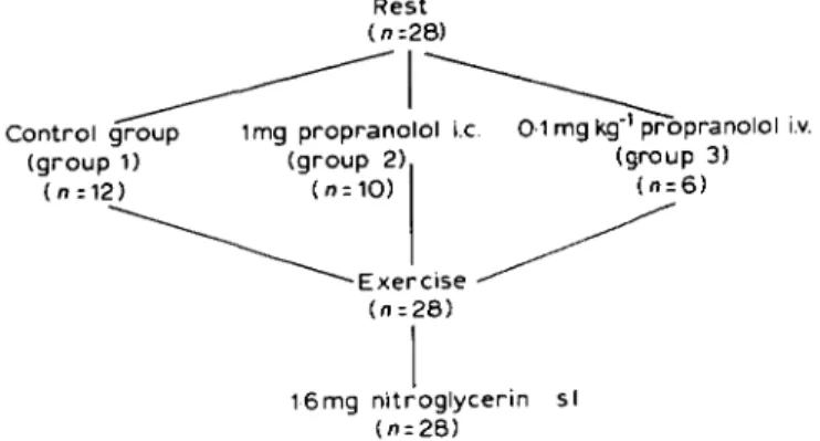
The effect of intracoronary and intravenous propranolol on coronary vasomotion was evaluated in 28 patients with coronary artery disease. Luminal area of a normal and a stenotic coronary vessel segment was determined at rest, during submaximal bicycle exercise and 5min after 1-6 mg sublingual nitroglycerin administered at the end of the exercise test involving biplane quantitative coronary arteriography. Patients were divided into three groups: group 1 (n = 12) served as the control group, group 2 consisted of 10 patients with intracoronary administration of 1 mg propranolol and group 3 of six patients with intravenous administration of 0-1 mg kg ''propranolol prior to the exercise text.
In the control group there was coronary vasodilation (+23%, P <001) of the normal and coronary vasoconstriction (—29%, P < 0-001) of the stenotic vessel segment during bicycle exercise. After sublingual administration of 1-6 mg nitroglycerin there was vasodilation of-both normal (+40%, P <0-001 vs rest) and stenotic (+12%, NS vs rest) vessel segments. In group 2 intracoronary propranolol was not accompanied by a change in coronary vessel area but both normal (+13%, P<0-05) and stenotic (+22%, P<0-05) vessel segments showed coronary vasodilation during bicycle exercise. After sublingual nitroglycerin there was further vasodilation of both normal (+31 %, P < 0-001 vs rest) and stenotic (+45%, P < 0-01 vs rest) arteries. In group 3 intravenous administration of propranolol was associated with a decrease in coronary luminal area of both normal (—24%, P <0-001) and stenotic (—31%, P <0-001) vessel segments. During dynamic exercise there was coronary vasodilation of both vessel segments when compared with the data after intravenous injection of propranolol but there was no change in luminal area (normal vessel —2%, NS vs rest; stenotic vessel —3%, NS vs rest) when compared with the resting data. After sublingual administration of 1-6mg nitroglycerin both normal (+21%, P<001) and stenotic (+36%, P<0001) vessel segments showed coronary vasodilation.
It is concluded that supine bicycle exercise in patients with coronary artery disease is associated with vasodilation of the normal and vasoconstriction of the stenotic coronary arteries. Intravenous administration of propranolol is followed by coronary vasoconstriction of both normal and stenotic coronary arteries, probably due to secondary mechanisms because it is not observed after intracoronary injection of propranolol and it is overridden by bicycle exercise and sublingual nitroglycerin.
Introduction and exercise-induced myocardial ischaemia. The _, . . . t . , . . beneficial effect of betablocking agents has been Betablockers are frequently used ,n the treat- a t t r j b u t e d t o t h e r e d u c t i o n i n ^yocardial oxygen
ment of pat.ents w.th coronary artery d.sease c o n s u m p t i o n c a u s e d b y a r e d u c t i o n i n m y o c a r.
Address for correspondence: Otto M. Hess, M.D., d i a l contractility, heart rate and left ventricular
Medical Policlinic, Cardiology, University Hospital, afterload. Previous Studies have shown1 • ' that
Raemistrasse 100, CH-8091 Zurich, Switzerland. myocardial blood flow is decreased after 0195-668X/89/0F0153 + 06 $02.00/0 © 1989 The European Society of Cardiology
intravenous administration of propranolol, probably due to a decrease in coronary luminal area. Rafflenbeul and coworkers'3' reported a
decrease in coronary luminal area of the large and the small epicardial arteries after in-travenous administration of propranolol. It has been postulated that beta-adrenergic blockade potentiates coronary artery vasoconstriction by the unopposed alpha-adrenergic tone. These findings contrast with the well-documented beneficial effect of propranolol in the treatment of patients with coronary artery disease and classic angina pectoris in whom adverse reactions with potentiation of myocardial is-chaemia are rare. Thus, the purpose of the present study was to examine the effect of intracoronary and intravenous propranolol on coronary vasomotion at rest and during supine bicycle exercise in patients with coronary artery disease.
Patients and methods
Twenty-eight patients (mean age 53 years, range 36 to 67 years) with coronary artery disease underwent coronary arteriography for
diagnostic purposes (Fig. 1). Previous myocar-dial infarction was present in 17 patients and a positive exercise test with ST-segment depres-sion 3=0-1 mV and/or anginal pain in 26. All medication was stopped at least 12 to 24 h before cardiac catheterization. Patients were selected on a consecutive basis when the following criteria were fulfilled: a history of stable angina pectoris with no signs of coronary vasospasm and a clearly visible coronary artery stenosis for quantitative evaluation.
QUANTITATIVE CORONARY ARTERIOGRAPHY
After an interval of at least lOmin after the last diagnostic coronary arteriogram, baseline biplane coronary arteriography for quantitative evaluation was carried out after the patient's feet were attached to the bicycle ergometer'4 5|.
Intracoronary injections of 5 to 7 ml of amidotrizoate (Urografin 76%) were used for quantitative coronary arteriography in the first group of patients, but later during the study 5 to 7 ml of iopamidol (Iopamiro 370) was injected for quantitative coronary arteriography to reduce the effect of the contrast medium on coronary vasodilation. Aortic and pulmonary
Control group (group 1) Rest (n;28) 1mg propranolol i.c (group 2) 0-1 mg kg"1 propranolol i.v. (group 3) 16mg nitroglycerin si (n=28)
Figure 1 Study protocol for the assessment of coronary vasomotion
in 28 patients with coronary artery disease and exercise-induced angina pectoris using biplane quantitative coronary arteriography. The control group consisted of 12 patients (group 1) with no pretreatment prior to the exercise test, group 2 of 10 patients with intracoronary administration of 1 mg propranolol prior to the exercise test and group 3 of six patients with intravenous administration of 0-1 mgkg"1 propranolol prior to the exercise test.
The infusion of propranolol was carried out over 5 min; repeat coronary arteriography was performed after 6-8 min in group 2 and after 9-12 min in group 3. At the end of the exercise test 1-6 mg sublingual nitroglycerin was administered, and S min thereafter biplane coronary arteriography was repeated, i.c. = intracoronary, i.v. = intravenous, s.l. = sublingual.
Intracoronary and intravenous propranolol 155
artery pressure were recorded at rest and at the end of each exercise level immediately before coronary arteriography. Repeat coronary angio-grams were obtained at the end of each exercise level which was begun at 50 to 75 W and was increased every 2 min in increments of 25 to 50 W. The exercise test was terminated because of anginal pain, fatigue or ST-segment depres-sion of more than 0-2 mV. At the end of the exercise test 1-6 mg sublingual nitroglycerin was administered and biplane coronary arteriog-raphy was repeated 5 min thereafter. There were no complications related to the procedure in any of the 28 patients.
Quantitative evaluation of biplane coronary
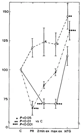
150 125 100 75 P<O05 P<OO1 vs C p<0001 C PR 2 min ex max ex NTG Figure 2 Percent changes in normal coronary vessel
segments during exercise in 12 control patients (D), 10 patients with intracoronary administration of 1 mg propranolol (O) and six patients with intravenous administration of 0-1 mgkg~' propranolol ( • ) . Data are given at rest (C), after intracoronary or intravenous administration of propranolol (PR), during 2 min of exercise (2 min ex.) and during maximal exercise (max ex.) as well as 5 min after l-6mg of sublingual nitroglycerin (NTG). 150 125 100 7 5 . P<005 *• P<001 f ••• P<0001 vs C C PR 2 min ex max ex NTG
Figure 3 Percent changes in coronary stenosis area
during exercise in 12 control patients ( • ) , 10 patients with intracoronary administration of 1 mg propranolol (O) and six patients with intravenous administration of 0-1 mgkg"1 propranolol ( • ) . Data are given at rest (C),
after intracoronary or intravenous administration of propranolol (PR), during 2 min of exercise (2 min ex.), during maximal exercise (max ex.) as well as 5 min after l-6mg sublingual nitroglycerin (NTG).
arteriography was carried out in a blinded fashion. Tracings were made manually from both projections during diastasis or end-diastole. Each vessel segment was analysed four to six times separately and the results were averaged to reduce the sampling error'4-51. A section of the
catheter of known dimensions was traced as a scaling factor. The tracings of the coronary vessel segments were digitized manually and analysed on a PDP 11/34 computer1"-51. The
luminal area of a normal and a stenotic vessel segment was calculated in each patient and expressed in absolute values and in percent of the resting value (see Figs 2 and 3).
STATISTICS
Statistical comparisons of angiographic data in response to intracoronary or intravenous pro-pranolol, supine bicycle exercise and sublingual nitroglycerin were carried out by a two-way analysis of variance for repeated measurements. Comparisons between all three groups were done by a one-way analysis of variance; when the analysis was significant the Scheffe test was applied. In both figures mean values ± 1 standard error are given.
Results
HAEMODYNAMIC MEASUREMENTS
Administration of intracoronary propranolol was not associated with a significant change in heart rate (65 beats min"1 at rest and 64 beats
min"1 after propranolol). However, after
in-travenous administration of propranolol heart rate decreased significantly (P < 0-05) from 76 beats min"1 at rest to 65 beats min"1 after
propranolol. Mean aortic pressure remained unchanged after intracoronary or intravenous administration or propranolol (group 2, 97 mmHg at rest and 98 mmHg after proprano-lol; group 3, 104 mmHg at rest and 98 mmHg after propranolol). Heart rate increased sig-nificantly in all three groups during exercise, whereas mean aortic pressure showed a significant increase only in group 1, from 87 mmHg to 107 mmHg ( P < 0-001) but re-mained unchanged in group 2 (97 vs 93 mmHg, NS) and group 3 (104 vs 113 mmHg, NS). Mean pulmonary artery pressure increased significantly from 26 mmHg to 47 mmHg ( P < 0-001) in group 1, from 17 mmHg to 41 mmHg (P < 0-001) in group 2 and from 20 mmHg to 36mmHg (P<0001) in group 3.
ANGIOGRAPHIC MEASUREMENTS
Percent changes in coronary luminal area of the normal vessel segments are shown in Fig. 2 and of the stenotic vessel segments in Fig. 3. Normal coronary vessel segments showed coro-nary vasodilation during exercise (±23%, P < 0 0 1 ) and after sublingual nitroglycerin (+40%, /><0-001 vs rest) in the control group. Intracoronary administration of propranolol (group 2) was not associated with a significant change in coronary luminal area ( + 6 % , NS) but during bicycle exercise there was coronary
dilation of the normal vessel segments (+13%, />< 0 0 5 vs rest) which further dilated after
sublingual nitroglycerin ( + 3 1 % , f>< 0 0 0 1 vs
rest). Intravenous administration of propranolol (group 3) was accompanied with a significant decrease in coronary luminal area of the normal vessel segment (-24%, /*<0-001), whereas bicycle exercise was associated with coronary vasodilation compared with the angiographic data after intravenous administration of propra-nolol, but remained more or less unchanged compared with the resting data (—2%, NS). After sublingual nitroglycerin there was coro-nary vasodilation of normal vessel segments (+21% P<0-01 vs rest) in group 3.
Stenotic coronary vessel segments (Fig. 3) showed exercise-induced coronary vasoconstric-tion in the control group 1 ( - 2 9 % , /»< 0-001) which was prevented after intracoronary ad-ministration of propranolol (+22%, P < 0 0 5 ) in group 2. Sublingual nitroglycerin was accom-panied in both group 1 (+12%, NS vs rest) and group 2 (+45%, P < 001) by coronary vasodila-tion. Intravenous administration of propranolol (group 3) was followed by coronary vasocons-triction of the stenotic vessel segments ( - 3 1 % , P < 0-001). However, during submaximal exer-cise there was coronary vasodilation of the stenotic vessel segments compared with the data after intravenous propranolol, but there was no change in stenotic vessel segment (—3%, NS vs rest) compared with the data at rest. Admin-istration of sublingual nitroglycerin in group 3 was accompanied by a significant increase in stenotic vessel area (+36%, P < 0-001 vs rest).
Discussion
The reduction in myocardial blood flow after intravenous administration of propranolol112| has
been attributed to a decrease in coronary luminal area131, due to the unopposed
alpha-adrenergic vasomotor tone after blockade of the beta-adrenergic receptors of the epicardial coronary arteries. Experimental data in the conscious dog'6' have shown, however, that the
decrease in coronary cross-sectional area after intravenous administration of propranolol (beta-1 and beta-2 receptor blockade) or atenolol (selective beta-1 receptor blockade) is not prevented by alpha-adrenergic blockade with either phentolamine or prazosin. It was
con-Intracoronary and intravenous propranolol 157
eluded that the decrease in coronary cross-sectional area is probably related to the decrease in heart rate and contractility, but not due to the unopposed alpha-adrenergic tone. Since most patients with classic, exercise-induced angina pectoris respond well to betablocker treatment, the reduction in myocardial oxygen consumption during physical exercise cannot be explained by the occurrence of coronary vasoconstriction. It is a well-known fact that epicardial coronary arteries show vasodilation during dynamic exercise to meet the increased metabolic demands of the myocardium'4'5'. If intravenous
administration of propranolol would cause coronary vasoconstriction during physical exer-cise, more patients with exercise-induced is-chaemia would experience an adverse reaction to betablocker treatment.
The present study shows that intravenous administration of 0-lmgkg"1 propranolol leads
to a decrease in epicardial luminal vessel area of both normal and stenotic coronary arteries (Figs 2 and 3) which is not seen after intracoronary administration of 1 mg propranolol. Heart rate decreased significantly after intravenous ad-ministration of propranolol, whereas blood pressure decreased only slightly but not significantly. However, after intracoronary in-jection of propranolol, both heart rate and blood pressure remained unchanged. These differences in the haemodynamic determinants of myocardial oxygen consumption might ex-plain the decrease in coronary luminal vessel area after intravenous propranolol, because it has to be assumed that not only heart rate but also contractility has decreased, which is another important determinant of myocardial oxygen consumption. However, it cannot be ruled out that the unopposed alpha-adrenergic vasomotor tone caused coronary vasoconstriction after intravenous administration of propranolol, as has been suggested by others'12' but which could
not be confirmed in the conscious dog'6'. The
fact that intracoronary administration of propra-nolol was not associated with coronary vasocon-striction supports the previous experimental data or Vatner and Hintze'61 which showed no direct
effect of the betablockers on coronary vasomo-tion. It might be, however, that the drug had been washed out from the coronary vascular tree before it had blocked the beta-adrenergic receptors completely.
Dynamic exercise represents a physiological stimulus for coronary vasodilation to meet the metabolic demands of the myocardium during high energy expenditure such as bicycle exercise. In a previous report'4', we have shown that
normal coronary arteries dilate during dynamic exercise, but eccentric coronary stenoses show exercise-induced coronary vasoconstriction. The exact mechanism of this exercise-induced ste-nosis narrowing is not clear but might be due to endothelial dysfunction (atherosclerotic altera-tions) with an insufficient production of the endothelium-derived relaxing factor'45' or due to
a passive collapse of the free vessel wall within the stenosis during high flow states (Venturi mechanism) such as bicycle exercise'71.
Intracor-onary administration of 1 mg propranolol pre-vented exercise-induced stenosis narrowing (Fig. 3), and after intracoronary pretreatment with propranolol there was coronary vasodilation (+22%, /><005) of the stenotic vessel segments during dynamic exercise. Intravenous administration of O l m g k g "1 propranolol was
associated with a decrease in luminal area of both normal and stenotic vessel segments (Figs 2 and 3) which was followed by an increase in coronary vessel area during bicycle exercise. At the maximal exercise level coronary luminal area reached its control value of both normal and stenotic coronary arteries. Sublingual ad-ministration of 1-6 mg nitroglycerin further dilated normal and stenotic vessel segments to a vessel area which was similar (NS) to the control group. Apparently, intravenous administration of propranolol is associated with a reduction in luminal vessel area of the epicardial coronary arteries, but the response of the coronary arteries to the dilator stimulus of bicycle exercise is not affected by the injection of propranolol prior to the exercise test. This observation parallels the clinical finding that most patients with exercise-induced angina pectoris do better after betablocker treatment because epicardial coronary arteries are still able to dilate during exercise, resulting in an increase in coronary blood flow.
The exact mode of action which prevents exercise-induced stenosis narrowing after in-tracoronary and intravenous administration of propranolol is not clear but might involve the following mechanisms:
lessens the autoregulatory rise in coronary blood flow during exercise and lessens, therefore, the flow-dependent rise in transstenotic pressure gradient with a reduced flow-induced fall in stenosis distending pressure.
2. The rise in coronary vascular resistance after betablocker administration results in a higher poststenotic pressure and in a smaller transsten-otic pressure gradient with a higher stenosis distending pressure.
3. The local anaesthetic effect of propranolol leads to a reduced influx of calcium into the smooth vascular musculature of the epicardial coronary arteries (calcium-antagonistic action) which is associated with coronary vasodilation during bicycle exercise. This mechanism, how-ever, seems unlikely, since high blood levels of circulating propranolol are necessary for this local anaesthetic effect.
The possible role of these three mechanisms in coronary vasomotion during dynamic exercise cannot be estimated from our present data. However, our findings support the good clinical response to propranolol in most patients with classic, exercise-induced myocardial ischaemia.
References
[1] Kern JM, Ganz P, Horwitz JD et al. Potentiation of coronary vascoconstriction by beta-adrenergic blockade in patients with coronary artery disease. Circulation 1983; 67: 1178-85.
[2] Wolfson S, Gorlin R. Cardiovascular pharmacology of propranolol in man. Circulation 1969; 40: 501-11. [3] Rafflenbeul W, Berger Ch, Jost S, Lichtlen PR. Constriction of coronary arteries and stenoses with propranolol. Circulation 1987; 76: IV-276 (abstr). [4] Gage JE, Hess OM, Murakami T, Ritter M, Grimm
J, Krayenbuehl HP. Vasoconstriction of stenotic coronary arteries during dynamic exercise in patients with classic angina pectoris: reversibility by nitrogly-cerin. Circulation 1986; 73: 865-76.
[5] Gaglione A, Hess OM, Corin WJ, Ritter M, Grimm J, Krayenbuehl HP. Is there coronary vasoconstric-tion after intracoronary beta-adrenergic blockade in patients with coronary artery disease? J Am Coll Cardiol 1987; 10: 299-310.
[6] Vatner SF, Hintze TH. Mechanism of constriction of large coronary arteries by beta-adrenergic receptor blockade. Circ Res 1983; 53: 389-400.
[7] Brown BG, Bolson EL, Dodge HT. Dynamic mechanisms in human coronary stenosis. Circulation 1984; 70: 917-22.