Bioinspired Stimuli-Responsive Color-Changing Systems
Golnaz Isapour and Marco Lattuada*
G. Isapour, Prof. M. Lattuada Department of Chemistry University of Fribourg
Chemin du Musée 9, CH-1700 Fribourg, Switzerland E-mail: [email protected]
different microstructural features, such as diffraction gratings, photonic crystals, selective mirrors, crystal fibers, spiral coils, and surface gratings, just to cite the most commonly encountered ones. These elements use different mechanisms to produce a color. For example, diffraction gratings are periodic structures that split and diffract light into several beams trav-elling in different directions, thus creating a structural color, which depends on the spacing between the structures, as well as the direction of observation. Photonic crys-tals, on the other hand, are 2D or 3D peri-odic structures with low- and high-refractive-index parts, leading to only selective wavelengths to be reflected by the crystal. Many animals and plants use a combination of different structural colorations’ mechanisms, sometimes also including pigmentary coloration, to achieve the desired colors combination.
Understanding the mechanism of structural coloration has led scientists to develop artificial systems that mimic the per-formance of natural ones.[1–4] Colloidal crystals are typically made of monodispersed spherical particles, assembled usu-ally in face-centered cubic structures. They have been inves-tigated both from a fundamental point of view, as well as for their ability to produce a variety of iridescent colors, depending on the particles size and spacing. Alternatively, colloidal crys-tals have been used as templates to prepare inverse opals. First, either a monomer solution or an inorganic material pre-cursor infiltrates in the interstices left empty by the particles and then it is solidified by polymerization or through a sol–gel process. Finally, the template particles are removed and leave empty holes in the solidified matrix. Inverse opals have com-plementary optical properties to those of the original colloidal crystal templates, because the voids left by the particles are now the low-refractive-index structures. Bragg stacks, i.e., peri-odic assemblies consisting of alternating layers of two mate-rials with very different refractive indices and a well-defined thickness, are another example of a man-made structure that mimics the behavior of diffraction gratings. Their optical prop-erties are determined by the ratio between the refractive indices of the two materials and by the thickness of the layers. The use of biomimicry to try and replicate the structural colors of living systems is quite challenging, because much still has to be learned from the perfection and the complexity of the architec-tures used by Mother Nature.
Stimuli-responsive structural colors have an obviously more complex architecture.[4] They require the ability to change the microstructure in response to a stimulus. This implies, for example, changing the particles’ size or the spacing among particles in a colloidal crystal, changing the thickness of some
Stimuli-responsive colors are a unique characteristic of certain animals, evolved as either a method to hide from enemies and prey or to communicate their presence to rivals or mates. From a material science perspective, the solutions developed by Mother Nature to achieve these effects are a source of inspiration to scientists for decades. Here, an updated overview of the lit-erature on bioinspired stimuli-responsive color-changing systems is provided. Starting from natural systems, which are the source of inspiration, a classifi-cation of the different solutions proposed is given, based on the stimuli used to trigger the color-changing effect.
1. Introduction
It is hard not to be mesmerized by the extraordinary ability of certain animals to change colors in response to a threat, to signal their presence to others, or to camouflage with the envi-ronment, in order to hide from potential predators or preys. Examples include some cephalopods and chameleons, which can adapt their colors to the surrounding background in an instant; beetles that change their colors depending of the level of humidity; zebra fish changing their appearance depending on the light conditions, just to name a few. All these creatures share one common feature: they use responsive structural colors.
Two types of coloration exist in nature: pigmentary coloration and structural coloration. Pigmentary coloration, i.e., coloration due to chemical hues or dyes, is probably the most common form of coloration available in natural systems. On the contrary, structural coloration, i.e., coloration caused by microstructural features of a material that interfere with certain wavelengths of light, is less common, but offers some distinctive advantages. Structural colors are highly durable, since they are not affected by pigment photochemical degradation, and are character-ized by striking brilliance, metallic luster, purity of colors, and opalescence.
In principle, any material with a refractive index (RI) higher than that of the surroundings can display a wide palette of structural colors, provided that it possesses the proper micro-structure. Structural colors are created by the presence of
Published in "Advanced Materials 30(19): 1707069, "
which should be cited to refer to this work.
layers in a Bragg stack, or using pigment dislocation, as it occurs in cephalopods. The mimicry of such natural systems has been heavily pursued in the literature,[1,3] because stimuli-responsive structural colors could have potential applications for the creation of smart sensors, of cloaking devices, of intel-ligent textiles, just to mention a few examples. While research in this area has been carried out for the past three decades, a wide range of novel results have appeared and continue to be published in the literature. In our opinion, the quantity and quality of new results are so overwhelming that we decided to dedicate this review to provide a general overview of the research efforts, made especially in the last 5–6 years, in the field of bioinspired systems showcasing stimuli-responsive structural colors. Table 1 summarizes some of the advances in the stimuli-responsive structural colors’ systems highlighted throughout this work, grouped together based on the structural features that they are based on, and clearly indicating, the stim-ulus used to trigger the color change.
The work is structured as follows: first, a brief introduction to some of the fundamental physical concepts necessary to understand the behavior of the systems described in this work is presented. This is followed by a review of natural systems showing switchable structural colors, focusing on explaining their mechanisms, and on those works that have provided new insight into the field. Afterward, man-made systems are reviewed by categorizing them by the stimulus used to induce the color switch. We begin with systems based on hydrogels, because the wide variety of chemistry associated with them allows for a variety of stimuli to be used as triggers for the color change, and because they are an almost omnipresent component in artificial stimuli-responsive systems. After cov-ering all the most important stimuli used as triggers (such as temperature (T), pH, electrical and magnetic fields, light, and mechanical force), natural systems artificially modified will be discussed, before the concluding remarks.
2. Fundamental Physical Concepts
We begin by briefly reviewing some fundamental physical concepts, which will be recalled frequently during the work. Structural colors are obtained when a material possesses microstructural features that can interfere with certain wavelengths of light. These microstructural features require a certain level of periodicity in the structure, at least over a given range of length scales. As schematized in Figure 1a–c, periodicity in a structure can occur in one, two, or three dimensions. The presence of periodicity results in construc-tive interference in the light reflected by the structures, for specific wavelengths, depending on the geometry and optical properties of the materials. This gives rise to specific colors of light being preferentially reflected or transmitted by the material.
In order to exemplify the underlying physics, we will con-sider first what happens in the case of Bragg stacks, i.e., a struc-ture with 1D periodicity, and then to colloidal crystals, which are a common example of a 3D periodic structure. Most of the systems considered here can be rationalized in a similar fashion.[115]
Bragg stacks, i.e., multilayer structures made of two mate-rials (with different refractive indices) deposited in alternating layers, are an example of a system that is commonly used to produce structural colors. By considering the schematics in Figure 1d, where two materials, A and B, with refractive indices nA and nB, respectively, form alternating layers with thicknesses dA and dB, constructive interference between the light reflected by the layers is only possible if the wavelength satisfies the fol-lowing condition 2 cos cos A A A B B B λ=
(
(
θ)
+(
θ)
)
m n d n d (1)where θA and θB are the angles of refraction of the light in the two materials, and m is an integer number. Any change in
Golnaz Isapour obtained her Bachelor’s degree in polymer engineering from Amirkabir University of Technology in Iran. After working in a polymer composite company for a few years, Golnaz moved to Norway to obtain a Master’s degree in polymer chemistry from the University of Oslo. She then joined the group of Prof. Marco Lattuada, at the University of Fribourg. Her Ph.D. project aims at designing novel materials with stimuli-responsive color-changing ability that mimic natural systems, using responsive particles as building blocks.
Marco Lattuada received a Master’s degree in chemical engineering from Politecnico di Milano, Italy. He obtained a Ph.D. in chemical engineering from ETH, Zurich, under the guidance of Prof. Massimo Morbidelli. He then moved to MIT, where he carried out a postdoc for over 2 years in Prof. Alan Hatton’s group. He then moved back to ETH Zurich, where he worked as a senior scientist and group leader for about 6 years, before moving to the University of Fribourg, at the Adolphe Merkle Institute, where he obtained a prestigious Swiss National Science Foundation Professor-founded professorship, to start his independent career. He is currently a Professor in the Department of Chemistry at the University of Fribourg, Switzerland. His research activity is a balanced combination of experimental and modeling work, aiming at engineering smart nano- and microparticles, and at exploiting their self-assembly, with the objective to design novel soft materials.
Table 1. Highlighted stimuli-responsive structural colors systems throughout this work, classified according to their structure and the stimuli used to
trigger the color change.
Structure Materials and setup Application target Stimuli Hydrogel bulk – PS microspheres immobilized into polyacrylamide (PAm) supraballs hydrogels[5]
– High-aspect-ratio silicon nanocolumns attached or free-standing embedded in PAm gel[6]
– Micrometer scale silicon plates coated on one side with Au or Ag (micromirrors) embedded in poly(acrylamide-co-acrylic acid) (PAm-co-PAA)[7]
– Silk-fibroin inverse opals[8]
– Fe3O4@C colloidal nanoparticles embedded in polyacrylamide glycol gel[9]
– Fe3O4@SiO2 colloids colloidal nanoparticles embedded in a composite matrix of PEGMA
and PEGDA[10]
Sensors, artificial muscles[6,7]
Humidity, humidity, and pH[7]
(Thin) Films – Synthetic melanin nanoparticle films[11]
– Stacks of alternately deposited thin films of phosphatoantimonic acid, H3Sb3P2O14,
nanosheets and TiO2 or SiO2 nanoparticles[12]
– An HSbP2O8/TiO2 multilayer structure not only humidity responsive, but also able to
distinguish between chemically similar solvent vapors[13]
– Alternating thin films of PMMA and PNIPAM-co-PGMA[14]
– Macroporous PHEMA/PETPTA films[15]
– Fe3O4@SiO2 colloids dispersed in the mixture of PEGMA, PEGDA, and TPM
(3-trime-thoxysilyl-propyl-methacrylate)[16]
– A 2D nanocomposite made of silk and titanate nanosheets[17]
– Inverse opal hydrogels thin films with PEGDA[18]
Sensors, environmental monitoring, anticounterfeit labeling[14,16]
Humidity, Humidity and temperature[14]
Chiral nematic – A mesoporous chiral nematic composite of cellulose nanocrystals and a poly(urea formal-dehyde)[19]
– Nanocrystalline cellulose in PAm hydrogel[20]
Biosensing, optics, func-tional membranes, soft templates for new materials
Humidity and pressure,[19]
Humidity, pH, ionic strength, solvent polarity[20]
Hydrogel bulk – Inverse opals with PNIPAM microfluidic hydrogel[21]
– Silica nanoparticles embedded in poly(N-isopropylacrylamide-co-N-methylolacrylamide)
[22]
– Microfluidic PNIPAM hydrogel balls containing PS colloidal crystals[23]
– Ag/Au nanoparticles coupled with the inverse opal of P(NIPAM-co-MA) hydrogel[24]
– Inverse opal of MMAA–BIS polymer filled with thermosensitive liquid crystals (LC)[25]
– Inverse opal hydrogel based on HEMA, 4-acryloyl morpholine (ACMO), and NIPAM monomers[26]
– Au nanoparticles assembled on P(NIPAM-co-AAm)[27]
Sensors, active loading and controlled release of drug,[21] energy-saving
multicolor displays,[25]
plasmonic on/off valve systems for flow control systems[27]
Temperature, Temperature and NIR light,[24]
Temperature and voltage,[25] Temperature
and pH[27]
(Thin) Films – 1D free-standing flexible films made of Fe3O4@PVP in a PNIPAM matrix[28]
– Stacks of alternate films of PNIPAM and poly(para-methyl styrene)[29]
– Free-standing films of P(NIPAM–AAc) colloidal crystals modified by HEMA[30]
– 1D photonic stacks of P(NIPAM-co-AA) and TiO2 layers[31]
– Inverse opal film of poly (2-(2-methoxyethoxy)ethyl methacrylate) P(MEO2)[25]
– Casts of PS-b-P2VP solutions[32]
– Diffraction grating composed of nanoemulsions of PNIPAM brush on SPIONS[33]
Sensors, displays[25,30] Temperature, Temperature
and pH,[30,31] Temperature
and solvent[25]
Colloidal particles
– PNIPAM brush-coated silica particles[34]
– PNIPAM microgel photonic crystals[35]
– PNIPAM microgels copolymerized with pH-responsive fluorescein and rhodamine derivatives[36]
– Core–shell microgels of PS-P(NIPAM-co-AA)[37]
Sensors, displays Temperature, Temperature and pH[36,37]
Hydrogel bulk – PS crystalline colloidal arrays incorporated in PAm[38]
– Inverse opal hydrogel of P(HEMA-co-AA)[39] using PS microspheres as a template
– Embedded (PEG)-crosslinked poly((methyl vinylether)-co-maleic acid) (PMVE-co-MAA) hydrogels in titania/graphene oxide (TiO2/GO) 1D photonic crystals[40]
– Inverse opal hydrogel consisting of 2-hydroxyethyl methacrylate, N-isopropylacrylamide, acrylic acid using PS microspheres as a template[41]
– PNIPAM gel particles confined in the inverse opal hydrogel consisting of HEMA, AA, and EGDM[42]
– Fe3O4@PVP immobilized in PNIPAM hydrogel balls[43]
– Fe3O4@C nanoclusters introduced into a PAm–PAA matrix[44]
– Inverse opals of supraballs based on polyionic liquids sensitive to different counterions (e.g., BF4−, PF6−, Tf2N−, NO3−, and ClO4−)[45]
– 2D photonic films and 3D photonic (microfluidic) supraballs to detect 11 kinds of metal ions (Al3+, Zn2+, Cd2+, Li+, Mg2+, Pb2+, Mn2+, Co2+, Ni2+, Fe3+
, and Cu2+)[46]
– Photonic gel composed of SiO2 colloidal crystals embedded in PEGMA/EG gel to
distin-guish alcohol homologs, isomers, and organic solvents[47]
Sensors, displays[42,43] pH, pH and ionic
strength,[38,39,44] pH
and temperature,[41,42]
counterions,[45,46]
alcohol homologs, isomers and organic solvents[47]
Structure Materials and setup Application target Stimuli (Thin) Films – Ultrathin polymer gel film with P2VP gel infiltrated in monolayers of PS[48]
– 2D Au nanosphere array attached onto a polyacrylic acid (PAA) hydrogel film[49]
– 1D photonic crystals flakes consisting of P(HEMA-co-MA) matrix and multilayers of Ag nanoparticles[50]
– Morpho butterfly wing as a template coated by PMA[51]
– Papilio paris wing as a template coated by P(AA-co-Am)[52]
– PMA integrated into the natural gyroid structure of the C. rubi butterfly wing[53]
– CLC polymer containing crown ether moieties sensitive to Ca2+ concentration[54]
Sensors pH, counterion[54]
Colloidal particles
– 2D free-standing hydrogel films of chitosan (with a thickness of 8 μm), with PS colloidal monolayers of 500 nm in diameter attached on both the surfaces[55]
– Ultrathin poly(acrylamide-co-acrylic acid) (PAm-co-PAA) films’ PS colloidal crystals incorporated[56]
– Photosensitive PS/AgBr hybrid colloidal crystals prepared with a co-deposition method for gas sensing[57]
Sensors,[55] antiforgery[56] pH,[55] ionic strength,[56]
UV light, and bromine gas[57]
Etalons – Etalons of P(NIPAM-co-AA) microgels in a photoacid o-nitrobenzaldehyde (o-NBA) solution[58]
– P(NIPAM-co-AA)-based etalon[59]
Displays, controlled/triggered drug delivery system, sensors
UV light,[58] pH, and
temperature[59]
Hydrogel bulk – An ink consisting of Fe3O4@SiO2, a solvation liquid (ethanol), and a photocurable
resin[60]
– Fe3O4@SiO2 incorporated into microspheres (hydrogel balls) made of a photocurable
resin (PEGDA)[61]
– Fe3O4@PVP immobilized in PNIPAM hydrogel balls[43]
– Aqueous suspension of inverse opals of ETPTA-3-(trimethoxysilyl)-propylmethacrylate (TPM) dispersed in an aqueous ferrofluid of Fe3O4 nanocrystals[62]
– An on/off reflection switch based on P(S-co-AA) nanoparticles of different sizes dispersed in an aqueous ferrofluid made of highly surface-charged Fe3O4 nanocrystals.[63]
Triple-responsive Janus supraballs (temperature, magnetic, and fluorescence Triple-responsive) based on PS colloidal crystals, CdS quantum dots, and PNIPAM hydrogels[64]
Forgery protection, structurally colored design materials and printing technology,[60] switchable
color display, signage, bio- and chemical detection, magnetic field sensing,[43,61]
magnetic field strength estimation[62]
Magnetic field, magnetic field and temperature[43]
Films – Films of PETPTA containing Fe3O4@C[65]
– Films of EG/PDMS containing Fe3O4@C[66]
– Agarose hydrogel film containing Fe3O4@SiO2 core/shell colloids[67]
Anticounterfeiting labels, displays[67]
Magnetic field
Colloidal particles
– Fe3O4@SiO2 photonic nanochains[68,69]
– Microfluidic Janus microspheres of the photocurable resin ETPTA coated by a thin ferromag-netic iron layer and alternate layers of silica and titania[70]
– Fe@SiO2 nanoellipsoids[71]
– Fe3O4@PVP in organic media[72]
– Microfluidic PEGDA microspheres containing Fe3O4@SiO2[73]
– Ferrimagnetic Fe3O4@SiO2 nanorod-based liquid crystals[74]
Anticounterfeiting, signage, energy-efficient color dis-plays, sensors
Magnetic field
Fiber – Stretchable/squeezable PDMS fiber containing carbon encapsulated Fe3O4@C embedded
in ethylene glycol droplets[75]
None-powered and function-alized fibers for camouflage
Magnetic field
Colloidal particles
– Negatively charged ZnS– silica core–shell colloidal crystals stabilized by PVP[76]
– Fe3O4@SiO2 core–shell nanoparticles with hydrophobic surface dispersed in a
low-dielec-tric medium[77]
– Hollow Fe3O4@C[78]
– Crystalline colloidal arrays composed of monodispersed particles of the copolymer PMMA–PS[79]
– Colloidal photonic crystal composed of PS nanoparticles and modified ITO electrode with ion exchange resins[80]
Displays Electric field
Chiral nematic – Films of polymer-stabilized CLC made of E7 (Merck) mixed with a prepolymer (NOA65) and a chiral dopant (R5011, Merck)[81]
– Oblique helicoidal (heliconical) structured CLC made of two dimeric, LCs (1′,7′-bis(4-cyanobiphenyl-4′-yl)heptane (CB7CB) and 1-(4-cyanobiphenyl-4′-yl)-6-(4-cyanobiphenyl-4′-yloxy)hexane (CB6OCB), and a standard LC pentylcyanobiphenyle (Merck), doped with a left-handed chiral additive S811 (Merck)[82]
– LCE based on micropatterned inverse opal films based on a mixture of a nematic diacrylate monomer and a monoacrylate mesogenic monomer[83]
– Alternate layers of TiO2 nanoparticles and an LCE base on 4-cyanophenyl-4-(allyloxy)
benzoate, 4-methoxyphenyl-4-(allyloxy)benzoate, and 4-((trimethylsilyl)oxy)phenyl-4-(allyloxy)benzoate, and a top layer a fluorescent dye (rhodamin B)[84]
Optical elements and color information, displays
Electric field, Electric field and temperature[83]
Table 1. Continued.
Structure Materials and setup Application target Stimuli EC device – EC device covering RGB from 450 to 750 nm wavelength range provided from P3HT for
red, PEDOT for blue, PANBS for green, and PDHFA for yellow covering below 450 nm completing the visible color spectrum[85]
– An EC device in the form of fiber consisting of (PEDOT), poly(3-methylthiophene) (P3MT), and poly(2,5-dimethoxyaniline)[86]
– A rainbow-like EC device with two viologens (4,4′-bipyridine derivatives –nonpolymer electroactive component) with five different multiswitchable colors based on four-zoned electrodes[87]
– EC device based on electroactive polyamides with N,N,N′,N′-tetraphenyl-p-phenylenediamine and tetraphenylbenzidine units in the backbone and heptyl viologen (HV) in the supporting electrolyte[88]
– EC device made of TiO2 inverse opals[89]
– EC device made of PS-b-P2VP coated on the ITO bottom electrode with a fluoropolymer spacer filled with 2,2,2-trifluoroethanol (TFE) electrolyte[90]
– EC device consisting of highly ordered Au-core/Ag-shell nanodomes integrated in a porous SiO2 film[91]
– EC films based on surface plasmon polaritons with polyaniline (PANI) and poly(2,2-dimethyl-3,4 propylenedioxythiophene) thin films integrated with Au nanoslit arrays assembled on the electrode[92]
Energy-saving displays,[85,89–91]
electrochromic fibers for wearable devices,[86] smart
windows,[87,88] mechanical
chameleon,[91] applications
ranging from catalysis to Photovoltaics[92]
Electric field
e-skins – e-skin consisting of artificial chromatophores layers, ultrathin silicon diode, and the PDMS layer and the Ag layer equipped with photodetectors and actuators[93]
– Highly stretchable, tactile sensing e-skin equipped with a pressure sensor based on organic EC device (as chromatophore cells), with an elastic pyramidal-microstructured layer (as tactile sensing) and PDMS layer, both layers coated by single-wall carbon nanotubes[94]
Wearable products for consumer, industrial, and military applications, smart robots[94]
Electric field, Electric field and pressure[94]
Hydrogel-based elastomers
– Fe3O4@C embedded in a hydrogel made of soft copolymer of N-hydroxymethyl
acryl-amide and N-vinylcaprolactam[95]
– Fe3O4@C embedded in a hydrogel based on Am and EG[96]
– Fe3O4@C embedded in PAm hydrogel[97]
– Tough photonic hydrogel made of PAA network introduced into the PAm layer of a PDGI/ PAm gel[98]
– Metastable SiO2 colloidal crystals embedded in the PEGMA/EG gel[99]
Sensors, high-precision displays, fingerprinting materials
Mechanical force, Mechanical force and organic solvents,[97]
Mechanical force and temperature and pH[98]
Colloidal-particle-based elastomers
– (PEA-co-MMA) colloidal particles embedded in PEA elastomer[100]
– PS core, poly(ethyl acrylate-co-allyl methacrylate) interlayer, PEA-poly(isobutyl methacry-late) shell[101]
– PS core in (PDEGMEMA-co-PEA) film[102]
– Core–interlayer–shell particles (PS core, poly(ethyl acrylate-co-allyl methacrylate) inter-layer, PEA shell) embedded in poly(butanediol diacrylate)) elastomer film[103]
– Mechanochromic photonic papers made of silica photonic crystals embedded in PEGMA and EG matrix[104]
Sensors, rewritable 3D optical data storage, tunable laser action,[101]
security materials[102,104]
Mechanical force, Mechanical force and light and temperature[101]
PDMS-based elastomers
– Patterned 1D array of gold nanoparticles integrated into PDMS[105]
– TiO2 nanoparticles layer coating on an ≈60 μm thick PDMS membrane[106]
– Highly stretchable, tactile sensing e-skin based on organic EC device, with an elastic pyramidal-microstructured layer and PDMS layer; both layers coated by single-wall carbon nanotubes[94]
Optical strain sensors, visualization devices,[106]
e-skin[94]
Mechanical force
LCE-based elastomers
– LCE film made of A6OCB (6-((4′-cyano-[1,1′-biphenyl]-4-yl)oxy)hexyl acrylate), 4-(4-(hexy-loxy)cyclohexyl)phenyl-4-((6-(acryloyloxy)hexyl)oxy)benzoate and hexane-1,6-diyl diacrylate and single-layer close packed array of silver nanoparticles on top of the film[107]
– LCE bilayers of PS film on top a poly(methylhydrosiloxane) showing surface wrinkling patterns[108]
Smart environmental-responsive devices, thermal-camouflage skin, color-changing actuators
Mechanical force and temperature
Fibers – Optical reflectors made of 2D graphene nanoplatelets on a single glass fiber surface integrated in the epoxy resin[109]
– PS nanoparticles electrophoretic-deposited onto continuous aligned-carbon-nanotube sheets, which are rolled on an elastic PDMS fiber and eventually embedded in a layer of PDMS[110]
– Photonic fiber based on Fe3O4@C super-paramagnetic nanoclusters embedded in a PAm
matrix[111]
– Hollow multilayer fiber photonic fiber made of PDMS and polyisoprene–polystyrene triblock copolymer[112]
– Spandex fibers dip-coated by PS core–PMMA interlayer– PEA shell microspheres[113]
Optical deformation sen-sors, wearable electronic clothes, smart fabrics
Mechanical force
Table 1. Continued.
spacing of the materials or in their refractive index leads to a change in the wavelength of the reflected light that experiences a constructive interference.
In the case of photonic (colloidal) crystals, the explanation is even more intuitive, as one can refer to the familiar Bragg’s law, commonly used to interpret diffraction from an ordered array of spherical particles. Figure 1e is used as a reference. Assuming an ordered configuration of identical spheres, such as the one depicted in the figure, the wavelength λ of light reflected by the array of spheres, which determines the color seen by an observer looking at the crystal, is given by
2 eff2 cos
2
D n
λ= −
( )
θ (2)where D is the spacing between the planes, neff is the effective refractive index of the sphere assembly, and θ is the angle of the incident light. According to Equation (2), the wavelength of the radiation reflected by the crystal depends on the spacing between the layers of spheres, on the refractive index of the particles, and on the incident angle of the radiation. By tuning these parameters, different reflected colors appear. To change on demand the wavelength of the reflected light, one or more of these parameters need to vary.
Artificially stimuli-responsive systems are most often based on soft materials, very often swollen and crosslinked polymer networks, which can change their volume in response to a stim-ulus. This can be used, for example, in a Bragg stack, where at least one of the materials can swell or shrink in response a stim-ulus. Analogous argument can be used when dealing with col-loidal crystals, except that in this case there are two possible sce-narios. In the first case, ordered structures made of colloidal par-ticles can be prepared by tightly packing highly monodispersed
particles together, as depicted in Figure 1e. The reflected color is dictated by the size of the particles, and an increase in the parti-cles size leads to an increase in the spacing D. By infiltrating a soft material in the inter-stices left free by the particles, a change in their spacing can be induced by a change in volume of the soft material. If the parti-cles are themselves made of a soft material, their volume change in response to a stim-ulus leads to the same effect, and to a corre-sponding color variation.[116]
A second route is instead based on pre-paring a colloidal crystal of particles in a liquid state, where ordering is induced by a strong repulsion between the particles. This strong repulsion is most often of electrostatic origin, or of steric origin, or combination of both (electrosteric). A change in the repulsive interactions leads to either a change in the colloidal crystal structure or even to a loss of the order and to the corresponding disappear-ance of the structural color. These changes are caused, for example, by a change in the ionic strength, in the pH, or in the surface charge of the particles, or the application of an external field, introducing a new interaction among the particles.[117] In case of metastable particles, any subtle motion, slight friction, or shearing force can cause a reversible transfor-mation of assembled–disassembled particles’ states.[118]
3. Switchable Structural Coloration in Nature
Structural coloration in animals and plants has been a great source of inspiration for researches in the design of artificial materials able to retain their coloration, without having pig-ments or dyes and with the ability to even switch their colors on demand. There are plenty of examples of creatures with structural coloration on their skin, wings, cuticles, feathers, and leaves.[4,119–122] These topics have been the subject of numerous reviews, and will not be dealt with here. Our focus here is on those systems which are able to change their color rap-idly (within a few seconds) in response to a stimulus. Under-standing the origin of such responsive coloration, the basic physics behind it and the mechanisms of color change in nature help to develop bioinspired strategies for fabrication of smart color-changing materials with similar functionality.[4,120,123]
There are three major mechanisms that are utilized by living creatures to have stimuli-responsive coloration.
3.1. Bragg Stacks Changing Spacing
The first mechanism is the change in spacing, orientation, or refractive index between ordered layers in a Bragg stack. Sev-eral examples are worth mentioning. Morpho butterflies are well known for their brilliant iridescent colors. The coloration origi-nates from hierarchical nanostructures of scales on the wings.
Figure 1. Periodic structures: a) 1D, b) 2D, c) 3D, d) interference patterns of Bragg Stacks,[114] and e) colloidal crystals. a–c,e) Adapted with permission.[4] Copyright 2013, Royal Society of Chemistry. d) Reproduced under the terms of the CC-BY Creative Commons attribution license.[114] Copyright 2013, The Authors; published by MDPI.
The studies on the iridescence in Morpho sulkowskyi butterfly show that the higher visible reflectance is due to a more regular lamellar structure compared to other Morphos butterflies.[124] These authors have shown that the iridescence changes in response to different vapors; the higher the vapor pressure, the higher the differential reflectance spectra (ΔR) between the test vapor and the carrier gas. The different spatial periodicity on the wing regions results in dif-ferent reflectance at difdif-ferent wavelengths (Figure 2a).
Eliason and Shawkey have studied, for the first time, the dynamic color changes in a keratinous biophotonic nanostruc-ture.[128] They have shown the iridescent color of the Theobroma bicolor feather barbule changes rapidly and reversibly from blue–green to yellow–green in response to humidity and as a result of swelling of the keratin cortex.
Beetles are also known to change the structural colors of their cuticle in response to humidity. Tmesisternus isabellae changes the iridescent color—which originates from a multi-layer in the scale interior—from golden to red when exposed to a wet environment. Upon water uptake, the periodic multi-layers swell and constructive interference results in a different reflected wavelength.[129] A similar behavior has been observed in the beetle Charidotella egregia.[130] The nanostructure of the beetle Hoplia coerulea is composed of a solid thin flat slab of chitin and a porous layer made of parallel rectangular rods.[131] When exposed to humidity, the scale changes from bright blue to emerald green due to the increased refractive index contrast between chitin and water as opposed to air (pores) in the dry state.
Figure 2. a) A highly selective vapor-responsive system based on hierarchical photonic structures inspired by M. sulkowskyi iridescent scales.
Differ-ential reflectance spectra ΔR, provide information about the nature and concentration of the vapors: ΔR = 100% × (R/R0− 1), where R is a spectrum collected from scales upon vapor exposure and R0 is a spectrum collected from scales upon exposure to a carrier gas (dry N2). Reproduced with permission.[124] Copyright 2007, Springer Nature. b) The dramatic effect of changing background reflections on wing interference patterns visibility in
Closterocerus coffeellae. Reproduced with permission.[125] Copyright 2010, National Academy of Sciences. c) Chromatophores of (left) dorsal mantle and (right) ventral mantle of the cuttlefish Sepia officinalis. Scale bar is 1.5 mm. Reproduced with permission.[126] Copyright 2008, Springer Nature. d) The panther chameleon (Furcifer pardalis). e) Transmission electron microscopy (TEM) small ≈ 130 nm guanine crystallites. f) Face-centered cubic geometry of the crystallites. d–f) Reproduced under the terms of the Creative Commons Attribution 4.0 International License.[127] Copyright 2015, Macmillan Publishers Limited.
The neon tetra fish change their structural color in response to alteration in light conditions. Their lateral stripe has a blue–green color (λpeak = 490 nm) in the light-adapted state and it turns indigo (λpeak = 400 nm) in the dark-adapted state. It has been found that the origin of the structural color is the stacks of intracellular guanine crystals intercalated with cytoplasm, which reflect light through constructive interference.[132] When the crystal arrays tilt, the spacing of guanine crystals (RI = 1.83) and cytoplasm (RI = 1.33) changes and, as a result, the reflec-tance spectrum changes too. The Anolis lizard is able to rapidly change the brightness of its stripes along its body in response to a mild mechanical force (handling and restraint);[133] it turns from brown to pale blue and green on some parts and to a lighter brown on some other parts along its body. Light expo-sure also acts as a stimulus for this luminance change.
3.2. Background Effect: Camouflage Ability
Wing interference pattern is another interesting phenomenon observed in some insects with transparent wings. Vivid color pat-terns are observed through thin-film interference in the wings against backgrounds with certain light properties, while they turn invisible when the background is changed (Figure 2b).[125] This offers another mechanism to design materials with camou-flage ability, which is one of the main functions of color change.[134] A quantitative study on the color change and pattern of camou-flage in flounders has been reported, in which the degree of spec-tral match between flounder and background with high-resolution optical instrument has been investigated.[135] Flounders attain the background appearance in terms of color, intensity, and pattern.
White coloration has an important contribution in camou-flage of biophotonic structures. It is found that Sepia officinalis, a type of cuttlefish, can produce a bright white color on its skin through “leucophores,” which are special cells containing organic spheres.[136] On top of the white background created by leuco-phores, different cells, called chromatoleuco-phores, provide dark pat-terns that create contrast, thanks to the luminescence of pigment granules, located inside sacks that are retractable and expand-able in a controlled manner by the cuttlefish. Meanwhile, there are colorless cells called “iridophores” containing stacks of thin plates, which act as structural light reflectors. The hierarchy of dermal coloration in S. officinalis, i.e., the white-scattering leu-cophores underneath, iridophores in the middle and chromato-phores on top and all acting in harmony, results in the camou-flage and color change effect in the cuttlefish (Figure 2c).[126,137] The concept of adaptive coloration and mechanisms of reflection, diffusion, and absorption of light by leucophores, iridophores, and chromatophores, and alteration in texture and morphology of the skin in cephalopods (squid, cuttlefish, and octopus) have been thoroughly reviewed so far.[138] Nevertheless, there are still major challenges in the fabrication of smart materials with exactly the same functionality as the one shown by these creatures.
3.3. Colloidal Crystals Spacing Alteration
Last mechanism is the one displayed by chameleons, which have always been a great source of inspiration in design and fabrication
of color-changing systems. A thorough set of experiments com-bining microscopy, photometric videography, and photonic- bandgap modeling on panther chameleons, in particular, has been conducted.[127] It has been found that there are two populations of iri-dophores in the dermal structure of the chameleons, each having dif-ferent morphologies. The underneath layer, which is made of rather disordered guanine nanocrystals, contributes to the near-infrared (IR) reflection of light, while the upper layer, which is made of guanine nanocrystals arranged in a triangle lattice, is responsible for the rapid color change through changing the spacing in the lattice (Figure 2d). Similarly Goda has shown that the rapid color change in the chame-leon sand tilefish Hoplolatilus chlupatyi originates from a change of spacing between the intracellular reflecting platelets (iridophores).[139] It is found that the color changes from blue to red in response to an increase in K+ ion concentration in 0.5 s, and to addition of nor-epinephrine within 1 s. Sapphirinid male copepods change their color both in intensity and wavelength in response to changes in the light conditions. It is shown that this color change originates from changing the spacing between ordered layers of hexagonally shaped guanine crystals. Some types of the copepods can gain camouflage and become transparent against any background.[140]
A common denominator of most biophotonic structures is the presence of guanine crystals. The high reflectivity of many living organisms is due to the extremely high refractive index (RI = 1.83) of guanine crystals. Therefore, understanding guanine-based optics can inspire novel design and fabrication of advanced optical materials. In this respect, the morphology, functionality, and architecture of guanine crystals in organisms and their role in the light manipulation, e.g., scatterers, mirrors, reflectors, and photonic crystals, have been investigated.[141] The morphology of these platelet-forming crystals is beautifully controlled by certain organisms. For instance, in the white widow spider (Latrodectus pallidus), coherent scattering from thick dense layers of guanine crystals results in a white color. Disordered multilayer reflectors of guanine crystals in the Japanese Koi fish (Cyprinus carpio) reflect a silver color. 3D photonic crystal structures in chameleons and well-aligned multilayer reflectors in neon tetra fish are other examples that have been mentioned before.
4. Artificial Materials
We will begin now to review the most important examples recently proposed in the literature of man-made systems dis-playing stimuli-responsive structural color-changing features. The classification is made based on the stimulus used to trigger the color-changing mechanisms.
On top of this, inspired by the three mechanisms used by living creatures, outlined in the previous section, we have clas-sified, whenever possible, all systems according to the same structural principles, to establish a clear connection with the natural source of inspiration.
4.1. Gas- and Liquid-Responsive Systems
Gas-responsive systems that are sensitive to small concentra-tions of certain gas molecules (H2O, NH3, NO2, CO2, etc.), as well as liquid-responsive systems to detect the amount of a
liquid (water or organic dispersants), are common colorimetric sensors used in safety applications, high-tech and quality con-trol of water/air.[5,9,10,18,57,142] Increased colorimetric sensitivity can be achieved with an optimal fabrication process, e.g., with 1D, 2D, or 3D photonic crystals and functionalized materials. In particular, hydrogel-based systems are a class of materials that have been well studied in tunable structural color systems and will be the focus of this work.
Hydrogels are networks of crosslinked hydrophilic polymer chains, in which the chains are connected through a variety of mechanisms, such as physical entanglement, electrostatic forces, and covalent bonds. The polymer network can swell and take up a great volume of water. The possibility of controlling the degree of hydration and the reversible swelling and con-traction of hydrogels make them applicable in various fields, including drug delivery, tissue engineering, sensors, and actua-tors. The chemical nature of the polymer chains dictates the physicochemical properties of the swelling mechanism and its kinetics. As a result, different types of hydrogels have been synthesized for various applications.[2,143,144] Among these, responsive hydrogels have attracted a great deal of attention in the past few decades. External stimuli such as temperature, pH, humidity, light, electric field, magnetic field, electrolytes, or certain small molecules can be used to trigger a responsive hydrogel to change its volume, which is associated with a
change in hydrophobicity, optical, electrical, magnetic, or mechanical properties of the hydrogel.
Inspired by nature, synthetic melanin nanoparticles (NPs)’ thin films have been prepared for the first time, which show a rapid reversible color change in response to a change in the humidity level. When the nanoparticles swell upon water uptake in a humid environment, the thin-film thickness increases and the reflection peak wavelength shifts from 475 to 530 nm when relative humidity (RH) is increased from 10% to 90%.[11]
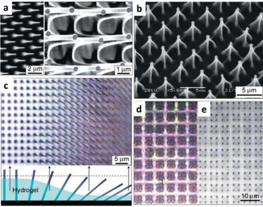
Moving to a more complex architecture, hydrogel-actuated integrated responsive systems (HAIRS) introduce a composite material prepared by integrating high-aspect-ratio (HAR) sil-icon nanocolumns as a hard element with a hydrogel layer as a soft element, which acts like a synthetic muscle. The humidity-responsive polyacrylamide (PAm) gel is grafted to an Si/SiO2 substrate via the polyglycidyl methacrylate (PGMA) anchoring layer (Figure 3). This complex and yet robust microstructure is able to change its optical pattern by reversibly tilting the nano-columns based on the humidity level.[6] The nanocolumns can be free-standing or attached on the substrate. The architecture based on high-aspect-ratio structures and the fabrication of hybrid systems composed of nanostructured surfaces combined with responsive hydrogels have been reviewed in details.[145] The key steps in the design of HAIRS are soft lithography, used to replicate high-aspect-ratio structures, and substrate
Figure 3. Microscopy study of a hydrogel-actuated integrated responsive systems (HAIRS). a) Scanning electron microscopy (SEM) images of
character-istic conical features at the bottom of the nanocolumns formed by the hydrogel layer. b) Optical microscopy image of the drying edge of the nanocolumns in the hydrogel taken perpendicular to the surface. The clarification of the actuation mechanism is shown schematically below the microscopy image. A dashed line in the schematic corresponds to the focal plane in the image. The degree of hydration or swelling of the polymer layer decreases gradually across the sample from left to right. Correspondingly, the nanocolumns gradually change their orientation from perpendicular to tilted. c) An example of a complex nanocolumn system in the hydrogel pattern, showing an array of microtraps, in which every group of four attached nanocolumns is held together by the hydrogel. d,e) Optical microscopy imaging microtraps shown in panel (c) in a d) dry and e) a wet state. The switching of the nanocolumns from bent fourfold clusters to a vertical orientation is clear. Reproduced with permission.[6] Copyright 2007, American Association for the Advancement of Science.
modification, used to anchor the hydrogel. The system can be responsive in or out of liquid depending on the hydrogel of choice.[145] Various design strategies can be applied in the con-cept of HAIRS. Taking advantage of plasmonic coatings on the nanostructured surface, micromirrors can be designed. They represent another application of these hybrid systems, and they are investigated in the development of next-generation active optics components. Optical properties can reversibly switch in response to the environment.[7]
Bragg stacks have been one of the well-known strategies in photonics design, in particular, for electronic devices. A highly humidity-responsive touchless positioning interface has been fabricated by phosphatoantimonic acid (H3Sb3P2O14) nanosheets.[12] In a humid environment, the bulk material is delaminated into 2D nanosheets in the form of a stable liquid crystalline suspension with anisotropic optical properties (Figure 4). The alternative layer in the Bragg stacks is made of either TiO2 or SiO2 nanoparticles, to provide a sufficiently high RI difference. Similarly, a photonic HSbP2O8/TiO2 multi-layer structure has been developed, which is not only humidity responsive, but also able to distinguish between chemically similar solvent vapors.[13] Photonic paper through alternating thin films of poly(methyl methacrylate) (PMMA) and poly(N-isopropyl acryl amide)-co-polyglycidyl methacrylate (PNIPAM-co-PGMA has been prepared.[14] The paper is crosslinked with a pattern so that it only displays the pattern in water (as ink) because of high RI difference of the layers. The color can be tuned by changing the crosslinking density and the ink (water) temperature.
A different strategy has been employed, for the first time, to fabricate colloidal photonic crystals supraballs of about 200 μm in diameter, which consist of polystyrene (PS) microspheres immobilized into PAm hydrogel matrix in an ordered colloidal crystal configuration.[5] Compared to the conventional film-type colloidal photonic crystals, the superball approach takes advan-tage of having a maximal packing fraction of PS microspheres and a long-range order. By increasing the humidity from 25%
to 100%, the PAm hydrogel matrix swells, thus increasing the spacing among the parti-cles. This causes a reflection peak shift from 495 nm (blue) to 654 nm (red).[5]
Siloxane-containing photonic prints have also been fabricated, in which a pattern that develops a visual contrast based on a dif-ferent crosslinking degree or a difdif-ferent hydrophobization with the background is cre-ated with lithographical methods.[16] The pat-tern only displays when the print is dipped in water and disappears when it is dry. The film is made by Fe3O4@SiO2 nanoparticles embedded in an ordered configuration (i.e., as a colloidal crystal in poly(ethylene glycol) diacrylate (PEGDA), poly(ethylene glycol) methacrylate (PEGMA), and 3-trimethoxys-ilyl-propyl-methacrylate (TPM), which is later photopolymerized). The reason for showing and hiding the pattern is that the reflection wavelength difference between the pattern and the background becomes significant because of their different degrees of swelling. Similar approach for preparing photonic papers with a writing–erasing feature has been also proposed.[14] With a different approach, photonic gels have been developed as sensors to distinguish alcohol homologs, isomers, and organic solvents with similar struc-tures and physical properties. The photo nic gels are composed of SiO2 colloidal crystals embedded in PEGMA/EG gel, and are able to detect/differentiate the analyte based on the dynamic reflection spectra.[47]
Cholesteric liquid crystals (CLCs) or chiral nematic liquid crystals (LCs) are a special class of materials used as colorimetric sensors, owing to the helical arrangement of their molecular directors, which selectively reflect the incident light. A variety of CLC materials exist in nature, such as chiral biopolymers, condensed DNA phases, and plant cell walls. Light is separated along the helicoidal axis into right-handed and left-handed circu-larly polarized components. If the pitch of the helix changes by a stimulus, CLCs can be used as smart color-changing systems. CLC-based coatings with the ability to change color have been prepared. When placed in water, within about 30 s they change their color and retake their original color in about 2–3 min when left to dry at ambient conditions.[146] To prepare these responsive patterned CLC coatings, a chiral dopant, nematic mesogens, and polymerizable molecules (benzoic acid derivatives) are mixed together with a photoinitiator (Figure 5). After polymerization, the mesogens are trapped into a hydrogel matrix, which swells and contracts upon contact with water and drying, respectively. By using different concentrations of the chiral dopant in the CLCs, which is the responsible component for the cholesteric molecular order, a different colored coating is prepared.
Cellulose nanocrystals (CNCs) are known to be a natural source to prepare CLCs. CNCs are typically 100–200 nm long and 5–15 nm in width.[147] In water, they form chiral nematic liquid crystalline phase at sufficiently high concentrations, and when they are dried they display iridescent colors. The helical pitch of CNCs can be changed by various external means or by co-assembly them with stimuli-responsive materials. Photonic
Figure 4. SiO2 /H3Sb3 P2O14 Bragg stacks. a) Optical humidity sensing features b) Micro-scopy images of the surface at relative humidity values of 32%, 93%, and 100%, respectively. Reproduced with permission.[12] Copyright 2015, Wiley-VCH.
multilayer films with co-assembly of CNCs and poly(ethylene glycol)(PEG) have been prepared.[148] The films have dif-ferent colors depending on the CNC/PEG ratio and reversibly change their colors in the order of seconds as the environment humidity is changed. For a CNC/PEG ratio of 80/20, as RH is increased from 30% to 100%, the green composite film uni-formly turns to dark red and then transparent where the multi-layer structure is swelled due the highest water uptake. Inspired by the shell structure of the Chrysina genus of beetles, a com-posite structure consisting of a uniaxially oriented polyamide-6 (as a half-wave retarder) sandwiched between CNCs/PEGDA layers has been developed. The composite can go through a simultaneous and reversible 3D deformation and a shift of the Bragg diffraction by a change in the humidity.[149] Similarly, CNC nanocomposite hydrogels with acrylamide (Am)[20] or
mesoporous poly(urea formaldehyde)[19] have been prepared, in which the reflection peak is reversibly shifted when the hydro-gels uptake the solvent medium (ethanol, methanol, acetone, or isopropanol) and swell to various extents.
A different design principle for the preparation of stimuli-responsive colloidal crystals is to use the inverse opals design. A typical fabrication method to make inverse opals is to incor-porate colloidal crystals made of PS microspheres[39,41] or silica nanoparticles[42,150c] in a hydrogel matrix. After removal of the templating particles, an ordered crystal arrangement of pores, i.e., an inverse opal is created. Such porous hydrogels are prac-tical choices to prepare responsive systems that can go through a fast volume transition. Inspired by the Hercules beetle that changes its cuticle’s color in response to humidity, thin-film hydrogels of PEGDA inverse opal have been made. The color
Figure 5. a) Top: schematic representation of the manufacturing method for a patterned cholesteric liquid crystals (CLC) coating. The mixture was
partially polymerized in a red-reflecting CLC phase at T1 during the masked photoexposure step. Subsequent flood exposure at T2 polymerizes a green-reflecting CLC phase and ensures full polymerization. Bottom: activation with an alkaline solution leads to a patterned water-responsive dual colored changing CLC polymer coating. b) Optical images showing three different polymer salt coatings with the letter “F” in ambient conditions (RH = 40%, T = 20 °C) and in the wet state. The coatings were made as follows: left—20 °C mask, 50 °C flood exposure; middle—20 °C mask, 50 °C flood exposure; and right—20 °C mask, 100 °C flood exposure. The size of the images is 17 mm × 9 mm. Reproduced with permission.[146] Copyright 2015, Royal Society of Chemistry.
of the nanoporous structure changes from blue–green to red in response to an increased humidity.[18]
Using a different fabrication approach, a scalable and roll-to-roll compatible doctor-blade technology has been employed to prepare a reversible vapor detector based on macroporous poly(ethoxylated trimethylolpropane triacrylate) (PETPTA) films.[15] They have used monodispersed silica colloids (225 nm in size) as a template, which are then etched away and result in the macroporous polymer film. A layer of poly(2-hydroxyethyl meth-acrylate) (PHEMA) coats the porous film and finally the macro-porous PHEMA/PETPTA composite films are prepared. Upon condensation of vapors in the porous film, the reflection peak shifts because of the swelling of PHEMA and changing the RI of the diffractive medium. An increase in ethanol vapor pressure (diffractive medium) from 0 to 1 P0 (where P0 is the saturation vapor pressure of ethanol at 25 °C, which is equal to 60 mmHg) drives a redshift in the diffraction peak from 500 to 595 nm.
4.2. Temperature-Responsive Systems
In aqueous systems, which are one of the main foci of this review, PNIPAM is the most commonly investigated temper-ature-responsive polymer. It has a lower critical solution tem-perature (LCST) of about 32 °C, and for this reason it has been investigated repeatedly in the last 40 years, especially in the form of polymer brushes, hydrogels, and microgels. Research on the synthesis of monodispersed PNIPAM microgels,[151] colored thin films formed by microgels,[152] microgel size, col-loidal crystal formation, colcol-loidal stability, ionic strength effects on PNIPAM microgels, and their optical properties,[153] as well as the phase behavior, and rheology of the microgels,[154] is well documented in the literature. There have been various strate-gies to modify the thermochromic behavior for sensing applica-tions as well.[155]
Chiappelli and Hayward have prepared Bragg stacks of PNIPAM film as the low RI (≈1.5) layer and poly(para-methyl styrene) as the high RI layer (≈1.6).[29] By adjusting the number of layers in the stacks, fabrication parameters (spin coating, ultraviolet (UV) exposure duration, and intensity) and tailoring the properties in each layer various reflection behaviors can be achieved upon changing the temperature.
An angle-independent thermoresponsive material with an amorphous array of PNIPAM brush-coated silica nanoparti-cles has been prepared.[34] In another study, a high-tempera-ture-induced hydrophobic assembly method has been intro-duced as a simple strategy to create ordered self-assembly of PNIPAM microgels. In the resulting colloidal crystal, the effect of sedimentation time and temperature, solvent quality, and crosslinking degree on the reflected structural color has been investigated.[35] A non-thermosensitive comonomer together with N-isopropylacrylamide (NIPAM) has been used to change the volume transition temperature, and silica photonic crys-tals have been embedded into it to make a thermosensitive photo nic gel.[22] Taking the idea of immobilizing nanocrystals in a PNIPAM hydrogel matrix,[156] microfluidic hydrogel balls containing PS colloidal crystals have been prepared, which beautifully display a thermoresponsive color change due to altering the colloidal crystals spacing.[23]
A thermochromic photonic gel based on 2-hydroxyethyl meth-acrylate (HEMA), 4-acryloyl morpholine (ACMO), and NIPAM monomers has been prepared.[26] The pores are created by peri-odic structures of PS microspheres of 230–235 nm in diameter. The effect of different molar ratios of HEMA:BIS:NIPAM:ACMO on the temperature-driven swelling of the inverse opal has been investigated. Since P(ACMO) is also a temperature-responsive hydrogel (LCST ≈ 95 °C) the reflection peak of the copolymer gel is changed with temperature. It is found that, compared to NIPAM-only gels, ACMO-containing gels show more uniform dλpeak/dT within the temperature range of 10–80 °C over the entire visible light spectra, as well as a smaller hysteresis of the λpeak on the heating and cooling cycle.
According to Bragg’s diffraction law, the reflected structural color is angle dependent and this associates with long-range order of the periodic structures. However, fabricating photo nic materials that show certain colors independent of observation angles is for some applications an advantage or even a first requirement. Carbon black has been used in the PNIPAM inverse opals to correct the overall magnitude of the scat-tering coming off the amorphous nanopores.[150b] As a result, the inverse opal hydrogel displays bright angle-independent structural color. There are several strategies to prepare angle-independent structural colors which can be applied in many responsive systems. Inspired by nature, there have been studies in that area as well;[157] nanostructured amorphous colloidal array with short-range order that displays homogeneous, angle-independent, although in most cases nonbrilliant, yet tunable colors[34,158] or beads containing nanocrystals.[159]
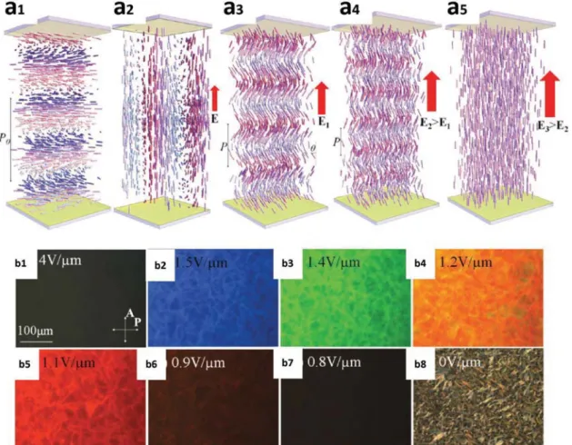
Temperature-responsive inverse opals with PNIPAM hydrogel (LCST ≈ 32 °C) have been prepared, in which tem-plates of silica nanoparticles self-assemble in microfluidic drop-lets of sizes from several to hundreds of micrometers.[21] The silica nanoparticles are packed in crystalline order, which is essential to show structural colors. The presence of macropores left from the SiO2 provides passages for active drug loading and release by changing the temperature. The hydrogel volume change is associated with changing the average refractive index or spacing between nanopores and the reflection peak changes as a result (Figure 6a).
Takeoka has reviewed a new strategy to prepare optical com-posites, which display reversible angle-independent structural colors based on Christiansen effect. This describes the reduced scattering of multiphase microstructures at wavelengths where their refractive indices match.[25] A porous poly (2-(2 methoxy-ethoxy)ethyl methacrylate) P(MEO2) membrane is impregnated and slightly swollen with a mixture of solvents and exhibits different colors depending on the composition or temperature of the solvents (Figure 6b). The same mechanism is observed with N-methyl methacrylamide (MMAA)–N,N′-methylene-bis-acrylamide (BIS) porous polymer filled with thermosensitive LC droplets because of a mismatch between the LC droplets and the porous polymer. By a change of temperature or potentially applying an electric field, refractive index of the LC changes and the transmittance wavelength peak shifts.[25]
A large class of multiresponsive materials has been devel-oped, using functionalized building blocks combined with responsive materials. Two pH-responsive fluorescent dyes were synthesized and then copolymerized with NIPAM to add a
color-changing pH response to the standard PNIPAM thermal response.[36] The system of microgels dispersed in an aqueous solution shows both a temperature-responsive change in tur-bidity, and a pH-responsive change in fluorescence.
Creating hybrid systems consisting of hydrogels and plas-monic nanoparticles (AgNPs or AuNPs) has been a successful strategy to increase the light absorption and introduce unique optical properties in the hydrogels. PNIPAM hydrogels as one of the well-studied temperature-responsive color-changing sys-tems have been coupled with plasmonic nanoparticles, since the latter induce a heat buildup in the hydrogel systems as a result of the light absorption. A design strategy for a thermo-photochromic composite consisting of photonic poly(N-iso-propylacrylamide-co-methacrylic acid) (P(NIPAM-co-MA)) copolymer and plasmonic nanoparticles has been proposed.[24] The gold/silver nanoparticles coupled with the inverse-opal-responsive hydrogel increase the sensitivity of the temperature response and make it possible to show a full color response over the entire visible spectrum in the system (Figure 10a). It is also demonstrated that by increasing the temperature, a molecular conformation transformation of PNIPAM happens that induce conformation rotation from intermolecular hydrogen bond into intramolecular hydrogen bond.[24]
4.3. Ion-Responsive Systems
Among hydrogels-, which are one of the foci of this work, tem-perature is one of the most convenient stimuli to be used to induce a structural color change. However due to the large quantities of water in hydrogels, pH and electrolytes are also
both natural stimuli that can be used to elicit a change in swelling conditions. Hydrogels containing charged moieties display swelling behavior strongly dependent on pH and ionic strength, both of which can be used to tune the electrostatic part of the osmotic pressure that determines the equilibrium state of the hydrogel.
pH-responsive Bragg stacks based on maleic-acid-containing hydrogels (pKa ≈ 1.8) have been reported. By embedding the (PEG)-crosslinked poly((methyl vinylether)-co-maleic acid) (PMVE-co-MAA) hydrogels in titania/graphene oxide (TiO2/ GO) 1D photonic crystals, pH-responsive TiO2/GO Bragg stacks are fabricated.[40] TiO2, a pregel solution and GO were spun alternatively and finally the Bragg stacks was baked to form the gel. The resulting multilayer photonic crystals exhibit six times faster response compared to simple TiO2/GO Bragg stacks.
Bragg stacks with dual-responsive behavior have also been reported. Gao and Serpe have prepared etalons of P(NIPAM-co-AA) microgels in a photoacid o-nitrobenzaldehyde (o-NBA) solution.[58] The system shows a color change induced by UV light exposure, which causes a pH change in the acrylic-acid-containing (pKa ≈ 4.3) system (Figure 8c). The Bragg stacks approach has also been used to prepare P(NIPAM-co-AA)-based photonic structures. The concept has been employed to prepare a multilayer system by alternatively spin-coating of NIPAM– AA polymer precursor and TiO2 layer as a high-refractive-index component.[31] The multilayer system is baked at higher temperature resulting in 1D photonic stacks that change the structural color of the system by changing temperature and at different pH values.
Using plasmonic nanoparticles, 1D photonic crystals flakes (d = 5 mm) consisting of P(HEMA-co-MA) matrix and
Figure 6. Porous systems. a) The reflection images and spectra of a PNIPAM hydrogel inverse opal hydrogel during a dynamic temperature decrease
process. Reproduced with permission.[21] Copyright 2015, Royal Society of Chemistry. b) SEM image of a porous polymer membrane composed of poly(MEO2). c) Solvent composition dependence of the transmission spectra and d) optical photographs of the same polymer membrane in different toluene–acetone mixed solvents at 20 °C. b–d) Reproduced with permission.[25] Copyright 2016, Springer Nature.
multilayer AgNPs have been fabricated.[50] By increasing the pH from 4 to 7, the hydrogel containing MA (pKa ≈ 4.6) swells and the AgNPs lattice spacing is changed and, as a result, the dif-fraction peak shifts from 500 to 620 nm with 0.1 pH unit sensi-tivity. The immobilization of AgNPs in the hydrogel matrix has been elaborated as well (Figure 7). A slightly different photonic system was developed before by the same group as a divalent metal ions’ detector.[160] The polymer matrix for Ag+ diffusion is poly(acrylamide-co-carboxylic acid) (PAM) hydrogel. A single laser pulse is used to pattern the matrix and form a slanted dif-fraction grating of periodic AgBr nanocrystals within the PAM matrix functionalized with 8-hydroxyquinoline. As the concen-tration of the detected ions is changed, the lattice spacing is tuned, and as a result the diffraction spectrum is shifted.
Colloidal crystals with pH-responsive behavior have also been prepared in several occasions. 2D free-standing hydrogel films of chitosan (pKa ≈ 6.5) (with a thickness of 8 μm) have been developed, in which PS colloidal monolayers of 500 nm in diameter are attached on both surfaces.[55] They have investigated advantages of the 2D colloidal structure over 3D arrangement. The strategy of using colloidal monolayers on both surfaces introduced an anticurling feature and enhanced diffraction intensity to the composite, compared to traditional hydrogel films having the monolayer on one side. Because of the pH-dependent swelling behavior of chitosan, by decreasing the pH from 6.88 (the higher limit of chitosan swelling) to 2.07, the diffraction peak is shifted from 508 to 648 nm.
Several colloidal crystals with dual-responsive behavior have been prepared by using modified P(NIPAM-co-AA) microgels. The microgels have HEMA polymerizable methacrylic groups on their surface.[30] When the microgels self-assemble in a highly ordered crystalline structure, the surface-bonded vinyl groups are polymerized by UV-initiated free radical polymeriza-tion resulting in the formapolymeriza-tion of dually responsive photonic
films consisting of immobilized colloidal crystals microgels (Figure 8a). Color-changing core–shell microgels of PS-P(NIPAM-co-AA) have also been prepared. The color change is rapid across the entire visible spectrum.[37] The stop band can be tuned with tempera-ture at a rate of 60 nm s−1. The core–shell microgels are mixed with an aqueous con-centrated suspension of PAm as a depleting agent and then self-assembled into pho-tonic crystals on a glass substrate. With the PAm as the depleting agent, together with the strongly scattering PS cores, and rapidly swelling–deswelling P(NIPAM-co-AA) shell, this system shows a uniform change in struc-tural color upon heating (Figure 8b).
Another type of pH-responsive system is based on poly-(2-vinyl pyridine) (P2VP) hydrogels. P2VP microgels alone can display bright structural colors.[161] (PS–P2VP) hybrid systems have also been studied frequently. An ultrathin polymer gel film with P2VP gel infiltrated in monolayers of PS colloidal crystals has been prepared using a controlled spin-coating method.[48] The film exhibits dis-tinct reflective colors upon pH-induced swelling and deswelling of P2VP (pKa ≈ 4) gel, which is associated with a change in the thickness of the gel layer. At acidic pH, e.g., 2.44, where P2VP is swelled, the reflection peak is 663 nm, while as the pH is increased to 4.36 and 6.05, the reflection peak is shifted to 584 and 544 nm, respectively.
Studies on the thermochromic behavior of the PS-b-P2VP gel which was made of casts of PS-b-P2VP solutions show that the color change is originated from a temperature-induced change in the pKa of P2VP.[32] The color of the gel clearly changes as the temperature increases, a phenomenon associated with a change in spacing between the PS blocks and P2VP blocks domains. At higher pH values when the P2VP domains are col-lapsed upon increasing the temperature, the domain spacing is reduced until no visible light is reflected and the gel looks clear. A similar system has been developed by controlled infiltration of SiO2 nanoparticles in the hydrogel block.[162] It is found that this modification allows an expansion of the stop band across the entire visible spectrum.
Electrolyte-responsive hydrogels are ideal candidates for chemical classifiers, biosensors, and antiforgery applications, if they are designed to show a quick response and high-accuracy performance. pH-responsive acrylic-based hydrogels are one the most used polymers in that area. Ultrathin poly(acrylamide-co-acrylic acid) (PAm-co-PAA) films incorporated with PS colloidal crystals have been developed.[56] 2D assemblies of PS particles in the hydrogel display bright structural colors that change when exposed to salt-containing solutions of different concen-trations (Figure 9). The color change originates from a change in lattice spacing caused by the electrostatic effect of the salt.
Incorporating PS crystalline colloidal arrays in a responsive hydrogel network has been a classic way in the preparation of photonic gels.[38,39] Once the hydrogel volume changes upon applying the stimuli, the crystalline colloidal array lattice constant
Figure 7. Laser writing of free-standing 1D photonic crystal flakes sensors. a) Treatment of the
PMMA surface with O2 plasma to render the surface hydrophilic. b) Free radical copolymeriza-tion of HEMA, ethylene dimethacrylate (EDMA), and MA on the O2-plasma treated PMMA substrate. c) Diffusion of Ag+ ions into the p(HEMA-co-MA) matrix. d) The formation of Ag nanoparticles within the matrix through reduction of Ag+ ions using a photographic devel-oper. e) Photographs of free-standing flakes from pH 5.0 to 8.0. The images were taken under white light illumination. Scale bar is 3 mm. Reproduced with permission.[50] Copyright 2016, American Chemical Society.


![Figure 1. Periodic structures: a) 1D, b) 2D, c) 3D, d) interference patterns of Bragg Stacks, [114]](https://thumb-eu.123doks.com/thumbv2/123doknet/14878789.643465/6.892.75.531.139.482/figure-periodic-structures-d-interference-patterns-bragg-stacks.webp)