The MIT Faculty has made this article openly available.
Please share
how this access benefits you. Your story matters.
Citation
Marx, Uwe. “Biology-Inspired Microphysiological System
Approaches to Solve the Prediction Dilemma of Substance Testing.”
ALTEX (2016).
As Published
https://doi.org/10.14573/altex.1603161
Publisher
Spektrum Akademischer Verlag
Version
Final published version
Citable link
http://hdl.handle.net/1721.1/117681
Terms of Use
Creative Commons Attribution 4.0 International License
Received March 16, 2016; Accepted May 11, 2016; Epub May 15, 2016;
http://dx.doi.org/10.14573/altex.1603161 Summary
The recent advent of microphysiological systems – microfluidic biomimetic devices that aspire to emulate the biology of human tissues, organs and circulation in vitro – promises to enable a global paradigm shift in drug development. An extraordinary US government initiative and various dedicated research programs in Europe and Asia recently have led to the first cutting-edge achievements of human single-organ and multi-organ engineering based on microphysiological systems. The expectation is that test systems established on this basis will model various disease stages and predict toxicity, immunogenicity, ADME profiles and treatment efficacy prior to clinical testing. Consequently, this technology could significantly affect the way drug substances are developed in the future. Furthermore, microphysiological system-based assays may revolutionize our current global programs of prioritization of hazard characterization for any new substances to be used, for example, in agriculture, food, ecosystems or cosmetics, thus replacing the use of laboratory animal models. Here, thirty-six experts from academia, industry and regulatory bodies present the results of an intensive workshop (held in June 2015, Berlin, Germany). They review the status quo of microphysiological systems available today against industry needs, and assess the broad variety of approaches with fit-for-purpose potential in the drug development cycle. Feasible technical solutions to reach the next levels of human biology in vitro are proposed. Furthermore, key organ-on-a-chip case studies as well as various national and international programs are highlighted. Finally, a roadmap into the future towards more predictive and regulatory-accepted substance testing on a global scale is outlined.
Keywords: microphysiological systems, organ-on-a-chip, in vitro models, predictive toxicology, drug testing
This is an Open Access article distributed under the terms of the Creative Commons Attribution 4.0 International license (http://creativecommons.org/ licenses/by/4.0/), which permits unrestricted use, distribution and reproduction in any medium, provided the original work is appropriately cited.
System Approaches to Solve the Prediction
Dilemma of Substance Testing
Uwe Marx
1, Tommy B. Andersson
2,3, Anthony Bahinski
4, Mario Beilmann
5, Sonja Beken
6, Flemming R. Cassee
7,8,
Murat Cirit
9, Mardas Daneshian
10, Susan Fitzpatrick
11, Olivier Frey
12, Claudia Gaertner
13, Christoph Giese
14,
Linda Griffith
9, Thomas Hartung
10,15, Minne B. Heringa
7, Julia Hoeng
16, Wim H. de Jong
7, Hajime Kojima
17, Jochen
Kuehnl
18, Marcel Leist
10, Andreas Luch
19, Ilka Maschmeyer
1, Dmitry Sakharov
20, Adrienne J. A. M. Sips
7, Thomas
Steger-Hartmann
21, Danilo A. Tagle
22, Alexander Tonevitsky
23, Tewes Tralau
19, Sergej Tsyb
24, Anja van de Stolpe
25,
Rob Vandebriel
7, Paul Vulto
26, Jufeng Wang
27, Joachim Wiest
28, Marleen Rodenburg
7and Adrian Roth
291TissUse GmbH, Berlin, Germany; 2AstraZeneca, Cardiovascular and Metabolic Diseases, Innovative Medicines and Early Development Biotech
Unit, Mölndal, Sweden; 3Section of Pharmacogenetics, Department of Physiology and Pharmacology, Karolinska Institutet, Stockholm, Sweden; 4Wyss Institute for Biologically Inspired Engineering at Harvard University, Boston, MA, USA; 5Boehringer Ingelheim Pharma GmbH & Co.
KG, Non-clinical Drug Safety, Biberach, Germany; 6Federal Agency for Medicines and Health Products, Brussels, Belgium; 7National Institute
for Public Health & the Environment, Bilthoven, The Netherlands; 8Institute for Risk Assessment Science, Utrecht University, The Netherlands; 9Massachusetts Institute of Technology, Cambridge, MA, USA; 10Center for Alternatives to Animal Testing-Europe, University of Konstanz,
Konstanz, Germany; 11US Food and Drug Administration, Center for Food Safety and Applied Nutrition, College Park, MD, USA; 12ETH Zurich,
Dept. Biosystems Science and Engineering, Bio Engineering Laboratory, Basel, Switzerland; 13microfluidic ChipShop GmbH, Jena, Germany; 14ProBioGen AG, Berlin, Germany; 15Center for Alternatives to Animal Testing, Bloomberg School of Public Health, Johns Hopkins University,
Baltimore, MD, USA; 16Philip Morris International R&D, Neuchâtel, Switzerland; 17Japanese Center for Validation of Animal Methods, Tokyo,
Japan; 18Beiersdorf, Hamburg, Germany; 19German Federal Institute for Risk Assessment, Department of Chemicals and Product Safety, Berlin,
Germany; 20Scientific Research Centre Bioclinicum, Moscow, Russia; 21Bayer, Investigational Toxicology, Berlin, Germany; 22National Center
for Advancing Translational Sciences, National Institutes of Health, Bethesda, MD, USA; 23National Center of Medical Radiological Research,
Moscow, Russia; 24Russian Ministry of Production and Trade, Moscow, Russia; 25The Institute for Human Organ and Disease Model Technologies,
Leiden, The Netherlands; 26MIMETAS BV, Leiden, The Netherlands; 27Chinese National Center for Safety Evaluation of Drugs, Beijing, China; 28cellasys GmbH, Kronburg, Germany; 29F. Hoffmann-La Roche Ltd, Roche Innovation Centre Basel, Switzerland
*A report of t4 – the transatlantic think tank for toxicology, a collaboration of the toxicologically oriented chairs in Baltimore, Konstanz and Utrecht sponsored by the Doerenkamp Zbinden Foundation. The views expressed in this article are those of the contributing authors and do not necessarily reflect those of their institution of employment.
Leist and Hartung, 2013; Matthews, 2008; Olson et al., 2000; Perel et al., 2007; Hartung and Leist, 2008; Schnerch et al., 2010; Sena et al., 2010; Seok et al., 2013; van der Worp et al., 2010; Har-tung, 2013). The latter is illustrated by repeated failures of drugs in clinical trials. Industry benchmarks for candidate success rates in each phase of clinical trials are 48-64% for phase I, 29-32% for phase II and 60-67% for phase III, respectively (Cook et al., 2014; Hay et al., 2014). Hence, the report presented here focuses on the drug development aspects of the substance testing dilemma.
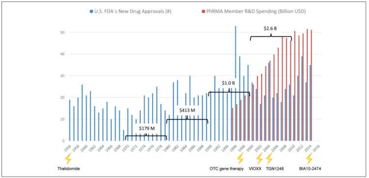
Development of new medicines currently suffers from two major obstacles (Rovida et al., 2015a): The current approach in preclinical therapeutic drug discovery cannot safeguard against high candidate attrition before and even during clinical trials, and constantly increasing regulatory requirements regarding preclinical testing to avoid harm to human individuals reduces R&D productivity. In the last seven decades pharmaceutical drug development costs increased significantly (spending of U.S. PhRMA members) while new drug approval rates by the US Food and Drug Administration (FDA) fluctuated in response to prime drug development disasters (Fig. 1). R&D productivity has declined more than fifteen-fold, as indicated by the respec-tive increase in inflation-adjusted average spending from $179 million per successful drug (including the costs of failures) in the 1970s to $2.6 billion in the twenty-first century (Scannell et al., 2012; Tufts CSDD, 2014) (Fig. 1).
The thalidomide disaster in the late 1950s and early 1960s was the first notorious drug failure resulting in extreme handi-caps in over 10,000 people. This event triggered the introduction of pre-clinical teratogenicity testing. Each subsequent disaster
1 The prediction dilemma of substance testing using laboratory animals
According to the most recent report from the European Com-mission (EC) to the Council and the European Parliament (EC, 2013), 11.5 million animals were used for experimental and other scientific purposes in the Member States of the European Union (EU) in 2011. The report stated that the number of ani-mals used for research and development for human medicine, dentistry and veterinary medicine dropped since the last report in 2008 from 22.8 to 18.8%. The number of animals used for toxi-cological and other safety evaluation, amounting to 8.75% of the total, remained relatively unchanged. However, the percentage of animals used for fundamental biological research increased sharply from 38 to 46%. These three areas use by far the highest number of animals (8.7 million in 2011) for scientific purposes in the EU (Daneshian et al., 2015). Information and results gen-erated from fundamental biological studies using animals lay the groundwork for new medicine development in the pharmaceuti-cal and biotech industries. The EC report highlights this fact by saying that ocular research, bone metabolism, fertility studies, potency testing, immunogenicity testing, studies in the areas of neuroscience and immunology, studies on pathophysiological mechanisms of tumors and research to elucidate mechanisms of action of diseases for therapeutic purposes were the major causes for the increase in fundamental biological research.
However, the last few decades of research and development have shown clearly that data from animal studies are often poorly indicative of the human situation (Hackam and Redelmeier, 2006;
Fig. 1: Changes in drug development over the last seventy years
Number of drugs approved by the US FDA (FDA, 2013; FDA, 2014a) are plotted against the pharmaceutical research and development spending of the members of the Pharmaceutical Research and Manufacturers of America (PhRMA, 2015). Drug or substance failures with detrimental outcome for humans (lightening) (see Tab. 1) and average costs to develop one new drug including costs of failures within the corresponding decade (brackets) are given.
rent human in vitro tests and the human body have not improved attrition rates in clinical trials to a satisfactory level. Although animals represent systemic organisms, they are not human, and the in vitro tests on human cells are neither physiological nor systemic. Despite intense preclinical safety testing in a number of phase I trials, safety issues may arise which can lead to the termination of a program (Cook et al., 2014; Schuster et al., 2005). Failure to predict efficacy and toxicity in the preclini-cal phases leads to serious delays in the development of new drugs, exposure of subjects to inefficient substances and even unwanted side effects, as well as initiating unsuccessful, expen-sive clinical programs, which are the largest investment points in the drug development process (Ledford, 2011).
Next to the pharma industry, other industries, such as chemi-cal and consumer product industries face similar problems re-garding toxicological hazard and risk assessment of substances as they also rely on animal experiments. This situation has led to ethical concerns about the use of large numbers of labora-tory animals.
New approaches are needed to bridge the translational gap and improve productivity of the drug development process. We believe that biology-inspired microphysiological in vitro sys-tems (MPS) will be a cornerstone of this bridge.
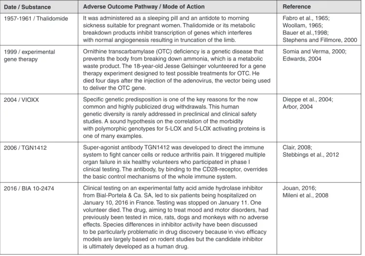
prompted an increase in relevant regulations for substance haz-ard identification, safety testing and efficacy evaluation. Some of these events, including the most recent fatty acid amide hy-droxylase inhibitor failure from Bial-Portela & Ca. SA in 2016, are summarized in Table 1.
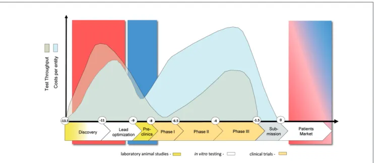
These historical drug failures with severe outcomes led to the establishment of a drug development approach spanning sev-eral years of experimentation and involving animals, in vitro tests and healthy volunteers or patients (Fig. 2). A pool of more than 10,000 entities needs to be fed into the drug development pipeline to finally arrive at one successful product (Kessel and Frank, 2007). A stringent regime of traditionally defined steps guides the process. The main goal of this extensive testing ap-proach is to ensure patient safety. It has been the best apap-proach possible to date. However, it is often inefficient and would today cause blockbuster drugs, such as aspirin or paracetamol, to fail regulatory approval (Hartung, 2009).
The smaller part of the investment during the cycle is spent on discovery and lead optimization (Paul et al., 2010), performing larger numbers of in vitro assays per lead identification and a significant number of laboratory animal tests per target or lead. Despite all these efforts, the phylogenetic distance between lab-oratory animals and humans and the discrepancy between
cur-Date / Substance 1957-1961 / Thalidomide 1999 / experimental gene therapy 2004 / VIOXX 2006 / TGN1412 2016 / BIA 10-2474 Reference Fabro et al., 1965; Woollam, 1965; Bauer et al.,1998;
Stephens and Fillmore, 2000 Somia and Verma, 2000; Edwards, 2004 Dieppe et al., 2004; Arbor, 2004 Clair, 2008; Stebbings et al., 2012 Jouan, 2016; Mileni et al., 2008
Adverse Outcome Pathway / Mode of Action
It was administered as a sleeping pill and an antidote to morning sickness suitable for pregnant women. Thalidomide or its metabolic breakdown products inhibit transcription of genes which interferes with normal angiogenesis resulting in truncation of the limb.
Ornithine transcarbamylase (OTC) deficiency is a genetic disease that prevents the body from breaking down ammonia, which is a metabolic waste product. The 18-year-old Jesse Gelsinger volunteered for a gene therapy experiment designed to test possible treatments for OTC. He died four days after the injection of the adenovirus, the vector being used to deliver the OTC gene.
Specific genetic predisposition is one of the key reasons for the now common and highly publicized drug withdrawals. This human genetic diversity is rarely addressed in preclinical and clinical safety studies. A sound hypothesis on the correlation of the morbidity with polymorphic genotypes for 5-LOX and 5-LOX activating proteins is one of many examples.
Super-agonist antibody TGN1412 was developed to direct the immune system to fight cancer cells or reduce arthritis pain. It triggered multiple organ failure in six healthy volunteers who participated in phase I clinical testing. The antibody, by binding to the CD28-receptor, overrides the basic control mechanisms of the whole immune system.
Clinical testing on an experimental fatty acid amide hydrolase inhibitor from Bial-Portela & Ca. SA, led to six patients being hospitalized on January 10, 2016 in France. Testing was stopped on January 11. One volunteer died. The drug, aiming to treat mood and motor disorders, had previously been tested in mice, rats, dogs and monkeys with no adverse effects. Species differences in inhibitor activity have been discussed to be particularly problematic in drug discovery because in vivo efficacy models are largely based on rodent studies but the candidate inhibitor is ultimately developed as a human drug.
sary features for in vivo-like tissue-specific electro-mechano-bi-ochemical signaling. They support expansion and compression forces especially relevant for lung, bone and cartilage, and mi-croelectrodes for the electrical stimulation and readout of mus-cle tissue (Ahadian et al., 2012; Dvir et al., 2012) or stimulation of cardiac cells or neurons (Bussek et al., 2009; Gramowski et al., 2011; Himmel et al., 2012; Johnstone et al., 2010, Khosh-fetrat-Pakazad 2014). Moreover, such devices could be able to apply other technical means of measurement and control, such as noninvasive optical imaging. Finally, the “system” in an MPS implies a high degree of automation, which makes it robust and scalable. It is necessary to consider all of these in order to en-able reproducible high throughput repeated substance exposure protocols mimicking human drug exposure as closely as possi-ble. Further detailed information on these features is given else-where (Andersson and van den Berg, 2004; Huh et al., 2011; Ingber and Whitesides, 2012; Kim et al., 2007; Park and Shuler, 2003; Wu et al., 2010).
The “physiological” component of MPS stands for the ambi-tion to truly emulate human biology. In the current literature, the “true emulation” of human biology in vitro is described in different ways, depending on the background and prior experi-ence of the author(s). Adherexperi-ence as far as possible to the emula-tion of human organ architecture, including proper cell-to-cell, cell-to-matrix, biochemical and mechanical signaling, is one of the cornerstones of our definition of “physiological.” Glass- and silicone-based devices complemented with polymers, textiles, ceramic or biological matrix entities are individually designed to match the well reviewed requirements regarding shape, sur-face pattern, stiffness, and microarchitecture of each specific
2 Microphysiological systems – an expanding toolbox for hazard, safety, disease
and efficacy prediction of particulate matter, chemicals and drug candidates
2.1 Definition and terminology
Microphysiological systems are microfluidic devices capable of emulating human (or any other animal species’) biology in vitro at the smallest biologically acceptable scale, defined by pur-pose. The application of fluid flow (dynamic) for physiological nutrition of the tissues and for the creation of microenvironmen-tal biomolecular gradients and relevant mechanical cues (e.g., shear stress) is a major aspect of these systems, differentiating them from conventional (static) cell and tissue cultures.
The “system” component of MPS refers to devices which support human-like physiology of tissues and organ equivalents within the devices in vitro. This may include the maintenance of physical factors, such as temperature (e.g., 37°C), relevant pH, and supply and control of oxygen and humidity levels. Further-more, it comprises mechanical coupling of organs by mimick-ing, for example, the flow of blood, urine, air, bile, pancreatic juice or cerebral fluid; shear stress regarding blood and lym-phatic vessels; physical pressure on bone and cartilage; strain on skin, lung and stomach wall; intestinal peristaltic movement and muscle contraction. For more details on how microfluidic tools can be used to study mechanobiology we refer to a com-prehensive review (Polacheck et al., 2013). In addition, the term “system” may incorporate readout of electrical activity of neu-ronal and cardiac tissues. The implementation of miniaturized relevant actuators and sensors into the devices enables neces-Fig. 2: Drug development cycle: test throughput and cost profile
The vertical axis illustrates approximate numbers of tests performed (grey) and related spending (blue). The horizontal axis illustrates the development time in years (Paul et al., 2010). Animal tests are used in early discovery for mechanistic mode of action research and for toxicity and ADME profiling in the preclinical phase, while conventional in vitro assays are largely used in discovery for target validation, target-to-lead translation and lead optimization steps.
Chips of microscope slide size and other formats are operated by an active microfluidic flow (external or on-chip pumps are used). This supports the emulation of shear stress at physiologi-cal intra-capillary or interstitial rates mandatory to maintain sta-ble protein and oxygen gradient-based microenvironments over long time periods. Typically, the single chip format is used when more complex tissue architectures need to be mimicked, while plate-based formats are preferred when a minimal amount of data points per system are needed.
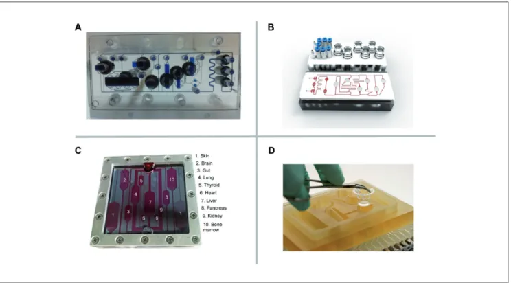
Alternatively, microphysiological systems can be broken down into three distinct types: Single-organ systems, multi-organ systems and more complex systems that are often termed human “body-on-a-chip” systems (see Fig. 3).
Single-organ systems are plates or chips emulating single tis-sue or organ function and are designed to improve early pre-dictivity of human single-organ toxicity of a particulate matter, a chemical or a drug candidate. These tissues or organs can be modeled at a two-dimensional (2D), three dimensional (3D) or organotypic complexity: 2D stands for suspension or monolayer cell cultures; 3D involves multilayer cultures of different ge-ometry, e.g., spheroids, strata and irregular tissue formations; and organotypic differs from 3D by capturing as many desir-able features of the in vivo architecture as necessary to gain the appropriate organ function (Hartung, 2014). Single-organ for-mats range from those employing passive gravity-based flow in plates adapted from standard cell culture plates to a variety of plate-based and microfluidic chip-type approaches using active flow from on-board or external pumps. These approaches allow the generation of various flow profiles, different fluid-to-tissue ratios and other physiologically relevant characteristics. Multi-organ systems emulate systemic interactions of two or more organ models within one system to enable adverse outcome pathway (AOP) and mode of action (MoA) data generation from their crosstalk. In contrast, human-on-a-chip (or body-on-a-chip) systems are envisioned to mimic the physiological interaction of a number of organs capable of emulating entire organismal functionality. A minimum of ten organs has been set as a goal by the Defense Advanced Research Projects Agency (DARPA)/National Institutes of Health (NIH) MPS program (Hartung and Zurlo, 2012), for example, as a means to identify the field of technical challenges for such organ combinations. This number has yet to be defined by the scientific community and regulatory bodies.
The current literature on MPS describes different culture times: short-term cultures last hours or days, supporting single exposure acute substance testing, and long-term cultures last weeks and months, enabling disease modeling and repeated dose substance exposure.
2.2 Human cell supply for MPS
Human beings are the source for human cells and tissues used today in in vitro research and industrial applications (Fig. 4).
Historically, human cell lines derived from adult donors (blue arrow, Fig. 4) have been the source of first choice for industrial
in vitro assays due to their unlimited expansion potential, relative
stability and the amenability to long-term cryostorage. The first human cell line – HeLa – was derived from the cervical adeno-organ microenvironment (Griffith and Swartz, 2006; Nelson
and Gleghorn, 2011; Pexton et al., 2011). Another cornerstone encompasses the maintenance of healthy, long-term organ ho-meostasis. The third cornerstone covers the ability to repair and regenerate organs that have been damaged or diseased. Thus, the latter cornerstone logically introduces one pathophysiologi-cal dimension of MPS. The final crucial cornerstone is to mimic human diseased tissue in the chip, this further introduces the pathophysiological dimension.
The “micro” component of MPS stands for miniaturization to the smallest biologically acceptable scale for each application. This miniaturization is driven by four major factors: mimicking organ function at laminar fluid flow in the cultured tissue, test throughput in the different phases of the drug development cy-cle (fit-for-purpose), minimum use of human cells and tissues, and, along with this, cost consideration. Scaling MPS-based organ equivalents down to an organoid level, as defined below, is one of the possible scaling strategies in the field. Almost all human organs consist of multiple, identical, functionally self-reliant, structural units, realizing the most relevant functions of the respective organ. Multiplication of these structures within a given organ is nature’s risk management tool to prevent a to-tal loss of functionality during partial organ damage or disease for the majority of body organs. During evolution, this concept has allowed the easy adjustment of organ size and shape to the needs of a given species – for example, liver size in mice and men – still using nearly the same master plan to build up single functional organoids. The term “organoid” was used as early as 1876 in different ways in life science (Bristowe, 1876, p. 73) and means “the smallest functional organ or tissue unit.” A selection of organoid histologies, all with a relevant func-tionality and highly variable conglomerate geometry, has been published for 15 key human organs (Marx et al. 2012). Ideally, the reactivity of organoids to drugs is representative of the re-activity of the organ. Therefore, a single organoid should reflect the smallest possible scale that can be used in MPS in order to emulate organ functionality. Selected functions may be emu-lated on a smaller scale, however, the investigation of disease emergence and progression, as well as tissue repair and regen-eration processes requires more identical organoids to represent the organ response in an MPS. With regard to the scaling aspect of MPS, a computational methodology for design and opera-tions for pharmacology applicaopera-tions was developed by Yu et al. (2015). Stokes et al. (2015) discuss on-platform scaling based on physiology and pharmacology and in vitro-in vivo transla-tion of results for complex MPS technologies. These different aspects reflect the current diversity of MPS within the scientific community.
MPS devices exist in two categories: plates and chips, as il-lustrated in Figure 3. Plates are usually based on the microtiter plate dimensions as defined by ANSI/SLAS (SLAS, 2014) and use passive, gravity-based microfluidic flow or active on-board pumping (Ebrahimkhani et al., 2014). An advantage of plate-based systems, whether gravity-plate-based or active flow-plate-based, is that they can be used with standard liquid handling equipment, such as robotic pipettors, and are fully compatible with auto-mated microscopes and plate readers.
Clevers, 2015). Then single cells are isolated from the tissues, expanded and reassembled in vitro. Prime targets for such recon-struction are the human barrier organs of skin, lung and intestine: EpiDerm™ from MatTek, EpiSkin™ from EpiSkin, EpiAir-way™ and MucilAir™ from Epithelix and EpiIntestinal™ from MatTek are prime reconstruction examples, see Gordon et al. (2015) for a comprehensive overview of non-animal models of epithelial skin, lung and intestinal barriers. In order to integrate such models into MPS devices, the latter need to be adapted to the standard architecture of the respective reconstructed model. Optimized manufacturing procedures and a pool of standardized donors at the vendor site support a certain robustness of such reconstructed human tissue models. The drawbacks are limited architectural complexity, short-term stability and costs.
There are three further human cell sources for organoid engi-neering in MPS. All three are stem cell-based – embryonic, fetal and induced pluripotent – but only the induced pluripotent stem cell (iPSC) approach works with primary cells from an adult do-nor (red arrow, Fig. 4). Since the inception of the induced pluri-potent stem cell technology by Takahashi and Yamanaka (2006), induction of pluripotency in primary human cells from patients or healthy donors has become an experimental routine. An im-pressive panel of companies, such as Axiogenesis, Axol, Bio-Talentum, Cellular Dynamics International, Pluriomics, Lonza, Takara Bio Europe AB, Viagene Biotech, iXCells Biotechnolo-gies, Applied StemCell, Applied Biological Materials, Centre for Commercialization of Regenerative Medicine and Cedars-Sinai provide reprogramming and/or differentiation services. While reprogramming has become a professional industrial standard, leading to more than 1,500 cryopreserved and well characterized iPSC lines, differentiation into organ-specific human cells still remains a big challenge, see Bellin et al. (2012) for a compre-hensive review. Within the last three years there has been an un-precedented increase of experimental research into differentia-tion protocols, starting from induced pluripotency. Here we list a number of papers trying to differentiate cell types relevant for MPS-based organoid engineering and subsequent assay devel-opment: hepatocytes (Szkolnicka et al., 2014), cardiomyocytes (Burridge et al., 2014), small intestine (Kauffman et al., 2013), neurons (Hoelting et al., 2013; Lippmann et al., 2014; Lancaster and Knoblich, 2014; Terrasso et al., 2015; Simão et al., 2016), lung (Ghaedi et al., 2013), gallbladder (Kauffman et al., 2013), pancreas (Pagliuca et al., 2014), kidney (Song et al., 2012), skin (Itoh et al., 2013), biliary ducts (Ogawa et al., 2015), bone-mar-row (Hynes et al., 2014), ovaries (Leng et al., 2015) and vascu-lature (Patsch et al., 2015). To the best of our knowledge, none of the protocols or procedures have yet been adopted by phar-maceutical industry for robust reproducible assay establishment, but there are first data indicating that MPS might be the right ap-proach to drive the differentiation of pre-differentiated iPS cells toward a final stage by supplying the physiological environment of the respective organoid (Moreno et al, 2015).
Fetal cells are rarely used in the engineering of human adult tissue, but recently, mesenchymal stem cells derived from fetal tissues could be differentiated into neuronal (Zhang et al., 2011) and osteoblastic lineages (Zhang et al., 2010). The main advantage with fetal cells is that they retain carcinoma of Henrietta Lacks as early as 1951. Today, thousands
of human cell lines more or less representative of different or-gan/tissue cell types are available, for example, patient-derived human cell lines like HepaRG, used in a number of toxicity says, and the CaCo-2 cell line used for intestinal permeation as-says. With increasing knowledge on the genetic engineering of mammalian cells, an ever growing panel of designed human cell lines has been integrated into industrial and research applications (Sandig and Jordan, 2007). Well established human cell lines are a common cell source for MPS-based cell culture. The drawbacks of cell lines are the partial loss of or gain of functionality due to their cancer background in addition to an inability to form com-plex heterogenic tissue architecture. Primary tissue explants and biopsies from healthy donors are the most complex and in vivo-like human tissue sources. These sources are used primarily in basic research and tissue engineering. Regarding use in MPS, the respective microsystem needs to be adapted for direct integra-tion of such human explants and biopsies into the fluid flow. The drawback of tissue explants is their limited availability.
Another common approach to generate human tissue architec-ture in vitro is the de novo assembly or reconstruction of human organoids. All primary tissues and/or their necessary adult tissue-specific stem cells are harvested from a donor or patient (Fig. 4) (Clevers and Bender, 2015; Rookmaaker et al., 2015; Sato and Fig. 3: Types of MPS used for emulation of human biology
in vitro
The MIMETAS OrganoPlate for 3D perfused cell culture in microtiter format (top left), the lung-on-a-chip developed by the Wyss Institute (middle left), the hanging drop microtiter plate with microfluidic channel connecting multiple spheroids developed by ETH Basel (top center), an artist’s impression of the four organ system developed by TissUse (center bottom), and artist’s impressions of a human body-on-a-chip platform (right) (courtesy of MIMETAS, The Netherlands; Wyss Institute, USA; ETH, Switzerland; and TissUse GmbH, Germany, respectively).
hepatocytes in static culture flasks, for example, was described as early as 1968 (McLimans et al., 1968). Microfabrication and microfluidics technologies, initiated as a scientific disci-pline in its own right as early as 1990 (Manz et al., 1990) de-livered commercially available lab-on-a-chip platforms at the end of the 20th century (see El-Ali et al., 2006 and Whitesides,
2006 for reviews), and advances in bioengineering related to materials science enabled the appearance of first plate- and chip-based microfluidic tissue culture systems aiming to solve the two aforementioned limitations of static cultures and to replicate the spatiotemporal, mechanical and biochemical cues that drive the physiological behavior inherent in those tissues. MPS developers approached their goal from two dif-ferent angles, some of them aiming to integrate microfluidic principles of nutrient supply into well-established industrial high throughput compliant static cell culture microtiter well formats and some trying to miniaturize well established in-dustrial tissue culture bioreactor systems.
Plate-based MPS developments accommodated conventional static cell culture microtiter plates with arrays of 96, 384, 1536 and 3,456 wells, which have become the standard of choice in any biological laboratory (SLAS, 2014). These platforms appeared on the grounds that the sheer nature of experiments and inherent variability in biology requires a vast amount of control experiments, repetitions and dilutions. In addition, the trend in molecular and cell biological sciences has progressed to ever higher throughput approaches, testing more conditions in one experimental session. It is for these reasons that manual and automated liquid handling and readout equipment has been designed in accordance with this standard. Moreover, the high number of wells in a plate offers the additional advantage of their proliferative capacity while being committed to an
end-point, and are not teratogenic in nature. However, their source is controversial.
Embryonic stem cells (ESC) are derived early after fertili-zation from the inner cell mass of human blastocysts. A vari-ety of human embryonic stem cell lines has been created over the last few decades (Mummery, 2011). These pluripotent progenitor cells lack the epigenetic modifications that may sometimes be seen in iPSC. Therefore, ESC might provide the broadest background for differentiation into any human organoid. The disadvantages of human embryonal cells or cell lines are the ethical debate surrounding their use and their propensity to give rise to teratomas, as shown in transplanta-tion experiments (Lees et al., 2007; Leist et al., 2008)
Pluripotent stem cells of the origin described above have been used for the de novo assembly of human gastric (Mc-Cracken et al., 2011), cerebral (Lancaster et al., 2013), kidney (Xia et al., 2014) and lung organoids (Dye et al., 2015) in static culture systems. Physiological, mechanical and electri-cal coupling of such human organoids using MPS seems to be an attractive approach for their further differentiation into
in vivo-like functional organ units. See Huch and Koo (2015)
and Li et al. (2014a) for a review on organoid cultures based on pluripotent human stem cells.
2.3 Current state of the art of MPS 2.3.1 Introduction
Technical drawbacks of any static cell culture-based assay in-clude the limitation of oxygen and nutrient supply to a given 3D tissue size or monolayer, and the lack of dynamic protein gradients. Oxygen restriction in cultures of primary human Fig. 4: Human cell sources for in vitro formation of organoids
The expansion potential of different human cell and tissue sources used in MPS are plotted against their appearance in the human life-span. (Grey arrows) – indicate Differentiation potential of the respective stem cell pool (grey arrows); induction of pluripotency using primary cells (red arrow); unlimited expansion potential of immortalized cell lines (blue arrow).
ed incubator-independent systems at relevant throughput. In summary, plate- and chip-based MPS aim to reflect physi-ologically relevant parameters, including proper cell-to-cell, cell-to-matrix, and biochemical and mechanical signaling. These capabilities present unprecedented opportunities to cre-ate MPS with the potential of capturing the dynamics of disease appearance, of repair and regeneration processes and of drug effects in the human body. Recent advances in MPS develop-ment have made it possible to initiate the engineering of cel-lular environments and/or functional units of lung, heart, blood vessels, muscles, bones, liver, reproductive system, nervous system (including eye), gut, skin, intestine and kidney.
In this chapter, we report the status of the development of biology-inspired microphysiological single- and multi-organ systems and their use for substance testing. Furthermore, we introduce achievements in modeling diseases using MPS. Re-cent initiatives to advance multi-organ systems into human body-on-a-chip solutions are outlined in Chapter 4.
2.3.2 State of the art of microphysiological single-organ systems
Early microtiter plate-based microfluidic cell culture formats with passive gravity-based microfluidic flow approaches to
in vitro tissue regeneration with application in human disease
modeling and drug development were developed by CellAsic (Lee et al., 2007a). Inspired by the work of Luke Lee’s group (Lee et al., 2007b; Hung et al., 2005b), they developed a micro-titer plate with eight independent flow units that did not require tubing for perfusion flow, but relied on passive leveling instead. The device consists of a polydimethylsiloxane (PDMS) mold-ed microfluidic layer that is bondmold-ed to a glass bottom and an acrylic user interface. Cells were introduced with the help of a vacuum manifold and positioned by a filter consisting of dense-ly packed micropillars. Perfusion flow was induced by gravity leveling in conjunction with a carefully tuned fluid resistance of the microfluidic channels. HP Medizintechnik GmbH’s (Ober-schleißheim, Germany) intelligent microplate reader is an ad-vanced 24-well-based microphysiological system incorporating an automated microscope, pipetting robot and microsensors for pH, dissolved oxygen and impedance, and is premounted in an incubator (Demmel et al., 2015). It was successfully used to monitor the viability of various cell types and primary tissue (Kleinhans et al., 2012) and to develop new toxicological end-points (Wolf et al., 2011). The relatively high grade of paralleli-zation and the automation of the pipetting robot and the micro-scope allow autonomous, label-free long-term experiments in the field of MPS. The PDMS material used, however, displays high unspecific binding of drug candidates.
Dave Beebe’s group at the University of Wisconsin, Madison, WI, US, developed a series of microtiter plate-based cell culture devices, including a 3D culture plate and a chemotaxis plate (Berthier et al., 2010). A total of 192 single channels on a micro-titer plate were filled with cell-loaded gels (Meyvantsson et al., 2008). An interstitial flow through the gel was applied, making use of differential meniscus pressure between two droplets of different size at the in- and outlet, respectively. A similar pump-ing concept was used for a 49-channel network chemotaxis de-reduced volumes of cells and reagents, leading to a reduction
of cost per assay.
Chip-based microphysiological systems for tissue and organ modeling have usually been designed by downscaling the flu-idic and tissue culture compartments and miniaturizing the sen-sors and actuators of conventional industrial cell-culture biore-actors. Automated dynamic bioreactors, such as hollow-fiber bioreactors, invented in the early-1970s, solved the problem of large scale oxygen and nutrient supply– up to one liter tissue culture – by the introduction of artificial capillaries for medium perfusion (Knazek, 1972). These dynamic perfusion tissue cul-ture systems later were advanced into functional long-term in
vitro organ equivalents introduced into medical practice as
ex-tracorporeal bioartificial liver devices in the late-1990s (Cata-pano and Gerlach, 2007; Gerlach, 1996; Wang et al., 2010). Following from this, numerous dynamic tissue-specific biore-actor devices, such as human artificial lymph nodes for immu-nogenicity testing, were miniaturized for in vitro testing pur-poses towards the lower ml per tissue culture scale (Giese et al., 2006). Figure 5 illustrates a miniaturized incubator-dependent dynamic bioreactor prototype for the simultaneous culture of human immune tissues at 0.5 ml each in operation.
Unfortunately, it became obvious in the first decade of this century that the lower ml scale still required too many cells and was still far too large to enable simultaneous high content test-ing at reasonable costs. This triggered the advent of chip-based MPS, which adopted lab-on-a-chip platforms to decrease tissue culture volumes down from micro- to nanoliter scale per tissue culture compartment. These aim to apply automated pumping systems and electrical, optical and mechanical transduction to monitor and control complex minute tissue cultures in automat-Fig. 5: Single-organ microcassette bioreactor device with hollow-fiber-based perfusion
(A) Integration of electromechanically controlled peristaltic pumps; (B) controller for ten microcassettes; (C) micro-bioreactor in operation in an incubator for temperature, humidity and CO2 control (courtesy of ProBioGen AG, Germany).
hepatocytes (Carraro et al., 2008; Goral et al., 2010; Ho et al., 2006; Leclerc et al., 2004; Lee et al., 2007b; Park et al., 2008; Powers et al., 2002; Toh et al., 2007, 2009), neurons (Rhee et al., 2005), mammary epithelial cells (Grafton et al., 2011), adipose cells (Nakayama et al., 2008), and human embryo cells (Chung et al., 2005; Hung et al., 2005a; Smith et al., 2012; Villa-Diaz et al., 2009).
In a next phase, heterotypic microfluidic single-organ co-cul-ture systems combining crucial cell types of a specific organoid into artificial functional units reflected aspects of the individual organs more realistically.
Scientific activities in the field of organ-on-a-chip models have increased dramatically in the last five years (van de Stolpe et al., 2013). Therefore, we decided to exemplarily highlight the single-organ MPS developments for human liver and lung mod-eling whilst referring readers to the original MPS literature for other organs and systems.
MPS-based liver cultures have matured from dynamic, ran-dom cell culture towards systems of higher tissue complexity. The unique importance of liver for organismal homeostasis (e.g., plasma protein synthesis, glucose biotransformation) and blood detoxification (e.g., urea, xenobiotic drug metabolism) has led to an evolutionary optimization of the human liver architecture at the scale of its smallest functional unit – the liver lobule. A vice consisting of a source and a sink channel that are connected
to a very thin gradient channel.
The OrganoPlate was recently introduced by the company MIMETAS, Leiden, The Netherlands, consisting of 40 to 96 individually addressable microfluidic networks (Jang et al., 2015; Moreno et al., 2015; Trietsch et al., 2013). The Organo-Plate makes use of PhaseGuide technology to stratify extracel-lular matrix (ECM) gels in orderly layers, one next to the other. This enables a co-culture approach that is fully free of artificial membranes, while still enabling the engineering of tissues. The OrganoPlate is fully devoid of high absorbance materials, such as PDMS, does not require any other handling equipment apart from standard pipettes for operation, and provides continuous perfusion flow through passive leveling.
Plate-based microfluidic cell culture formats with active on-board pumping were introduced for liver models by the group of Linda Griffith at MIT, Cambridge, MA, US and commercialized by CN Bio Innovations, London, UK. The Liverchip™ system, such a plate-based system, is described in more detail below.
Many single-organ chip concepts covering a wide range of tissues were proposed during the last decade and were actively introduced into biological research. They began with single cell type cultures of different organ specificity, such as endothelial cells (Young and Simmons, 2010), myoblasts (Gu et al., 2004),
Fig. 6: Increasing complexity of MPS-based liver models
Inspired by the lobulus architecture of human liver lobules, MPS have evolved from bile canaliculi forming cord-like liver cultures (A) through sinusoid-like arrangements supporting functional space Disse structures (B) towards tissue slice cultures maintaining functional organoid liver structures (C).
CN Bio Innovations can be scaled for 10,000 - 1,000,000 cells, and plates are similarly scaled in 12- or 36-well formats. Ac-tive pumping allows flow rates through the tissue to be uncou-pled from the morphology of the tissue. The system provides high-content data for drug metabolism and pharmacokinetics, toxicology and disease biology applications (Sarkar et al., 2015; Vivares et al., 2015). It has been applied in preclinical develop-ment (Dash et al., 2009; Vivares et al., 2015). Furthermore, a model of micrometastasis of cancer cells in liver has been de-veloped on the platform and a demonstration of dormancy of the aggressive MDA-MB-231 cell line has been established in the model (Wheeler et al., 2014). A model of hepatitis B virus infec-tion has also been developed using the same 3D perfused liver model. Infection of cryopreserved primary human hepatocytes is achieved through incubation with patient serum. The full viral life cycle is recapitulated, making the model of utility in explor-ing both the basic biology of hepatitis B virus and evaluatexplor-ing novel treatments (Wai et al., 2014).
The highest level of architecture – the entire liver lobule – has not yet been reached, but a chip-based microfluidic approach to containing a major part of a liver lobule has been made by van Midwoud and colleagues (van Midwoud et al., 2010a), who cul-tivated 3 mg of liver slice tissue in a microfluidic chip (Fig. 6c). The precision-cut liver slice (100 µm thick, 4 mm diameter) was perfused with a flow of 10 µl/min in an incubator chamber with constant pH and dissolved oxygen. Biotransformation activity was shown to be equal in control slices in static culture over three days. The system does not demonstrate any advantage with regard to metabolic functionality over the 3 d measurement time. The authors highlight the advantage of continuous media perfusion at low tissue-to-fluid volume ratios in contrast to steady metabolite accumulation in static culture. Unfortunately, data on the culture performance over times longer than 72 hours are not yet presented (van Midwoud et al., 2011a).
In addition to the examples described above, MPS-based sin-gle-organ liver modeling has led to a large variety of liver-on-a-chip devices (Baudoin et al., 2014a; Hwa et al., 2007; Kane et al., 2006; Khetani and Bhatia, 2008; Leclerc et al., 2014, 2015; Legendre et al., 2013, 2014; Pasirayi et al., 2014; Snouber et al., 2013). Research tools and assays have been developed to investigate liver metabolism and toxicity in single-organ chips (Baudoin et al., 2014b; Leclerc et al., 2014, 2015; Legendre et al., 2013, 2014; Pasirayi et al., 2014; Snouber et al., 2013a). For further details on the development of liver-on-a-chip models readers are referred to respective reviews (Materne et al., 2013; van Midwoud et al., 2011b).
Another complex organ where MPS-based models are already making a difference regarding the understanding of human or-gan functionality, testing safety and disease modeling is the hu-man lung. Huhu-man in vitro models of small lung airways and alveoli were developed historically in static air-liquid interface cultures on cell culture inserts. For a comprehensive overview on such models, readers are referred to Gordon and colleagues (2015). Until recently, only a few MPS trying to emulate func-tional parts of the lung had been developed (Nalayanda et al., 2007, 2010), but in 2010, a microphysiological lung-on-a-chip system developed by Donald Ingber’s group at the Wyss Insti-precise zonal division of labor along a 500 µm long stretch of
about 25 hepatocytes arranged in cords allows the management of blood detoxification at a blood contact time of only a few sec-onds. Not much is known about the fluid dynamics of plasma-hepatocyte contact. The nature of the plasma flow in the space of Disse and the mechanics applied to hepatocytes at the basolat-eral surfaces are under dispute. The high degree of fenestration may result in transmission of fluid shear stress to hepatocytes. Furthermore, the sinusoids in the periportal zone are of a very small diameter, i.e., the size of an erythrocyte. Blood cell pas-sage through these tight sinusoids may additionally modulate shear stress. Four types of MPS-based liver equivalents have been developed so far: random dynamic hepatocyte monocul-tures, cord-like liver equivalents, endothelial sinusoid models and hepatic lobule “equivalents” (Materne et al., 2015). Figure 6 highlights prominent examples for the latter three types.
In contrast to random dynamic hepatocyte cultures, the for-mation of 3D cord-like structures composed of polarized cells that form extended bile canalicular structures was presented by Goral and colleagues (2010). A series of retention pillars formed a microchannel centered between two side-channels (Fig. 6a). However, unlike other perfusion-based microdevices, the bot-tom of the cell culture chamber was patterned with microstruc-tures, which provided additional control of hepatocyte polarity. After two weeks of perfusion culture, the cells remained viable and had formed a cord-like structure. An extended bile canali-cular structure and the formation of gap junctions between the 3D structured cells could be shown. The system thus allowed the arrangement of hepatocytes into an artificial but cord-like assembly, control of hepatocyte polarity and sporadic bile seg-regation. Co-culture of hepatocytes with non-parenchymal cells in a spatially arranged 3D environment under constant perfu-sion might lead to the formation of a next level of architectural resemblance to the in vivo situation. A very interesting approach to modeling sinusoid-like structures in a microfluidic system was developed by Linda Griffith’s group at MIT (Domansky et al., 2010). An array of multiple bioreactors was built into a mul-tiwell plate comprising 12 autonomous microfluidic systems (Fig. 6b), each perfused by an integrated pneumatic micropump circulating a total volume of 3 ml. Each tissue culture scaffold contains 769 multichannels (0.24 mm deep, 0.34 mm diameter) and is seeded with 106 rat hepatocytes and endothelial cells at a 1:1 ratio. A continuous adjustable oxygen gradient can be es-tablished over long operating times. The scaffold supports near physiological tissue densities and the functional zonation of hepatocytes can be stipulated. The large channels of the scaffold support self-assembly of the two cell types in dynamic condi-tions. Liver sinusoid endothelial cells, known to lose their dif-ferentiated phenotype in vitro, maintained the expression of the functional marker SE-1 throughout the culture. The important features of adjustable flow rates on the basis of an oxygen con-sumption model, long-term steady gradient maintenance and the amenability to co-culture of hepatocytes with different types of non-parenchymal cells made the system an interesting ap-proach for toxicity testing. It is one of the still rare cases where a research MPS has been transferred into industrial application. The scaffold size of the LiverChip™ system commercialized by
and colleagues (2012) confirmed that administration of high doses of interleukin-2 (IL-2) caused a state consistent with pul-monary edema in their lung-on-a-chip system, similar to the ef-fects of administering IL-2 to human cancer patients. They also found that angiopoietin-1 and GSK2193874, an inhibitor of the transient receptor potential vanilloid 4 ion channel, attenuated edema in vitro and may prevent this dose-limiting toxicity of IL-2 in clinical settings. The authors concluded that the system could be used to model diseases in other organs and predict the efficacies and toxicities of other drugs in humans. Emulate Inc. (Boston, USA) is commercializing tools based on this platform. The system has also been used to examine the production of intracellular reactive oxygen species (ROS) in response to na-noparticles (Huh, 2015; Huh et al., 2010). In these studies, silica nanoparticles with a diameter of 12 nm induced and amplified the acute toxic responses measured in terms of ROS genera-tion. Cyclic stretching and relaxation of the porous membrane to mimic breathing further enhanced the absorption and trans-location of the silica nanoparticles from the upper air channel to the lower fluid channel. Similar findings were observed in a ventilation-perfusion model using whole mouse lung.
Other research groups have developed lung-on-a-chip sys-tems using human primary pulmonary alveolar epithelial cells from patients who had undergone partial lung resection (Stucki et al., 2015) or primary airway epithelia (Bol et al., 2014; Sell-gren et al., 2014) for selection of candidate drugs to treat pul-monary pathologies.
Finally, a model of chemotherapy-induced pulmonary edema has been developed on the basis of a small-airway-on-a-chip model (Benam et al., 2015b). The main biology-inspired aspect of the systems is the application of physiological cyclic strain placed on the cells, which not only closely mimics normal res-piratory movements, but also influences the permeability of the cell layers. In vivo studies have shown that distention of the lung, as in inhalation, increases the permeability of the lung to hy-drophilic molecules (Marks et al., 1985). This is probably due to the stretching of the intercellular junctions, which increases the size of the pores and allows larger hydrophilic molecules to cross the junctions (Mason et al., 2001). Innovative approaches to develop microfluidic systems for the culture of other types of epithelia could potentially also be adapted to establish new lung-on-a-chip systems. For further information on MPS-based lung model development readers are referred to a recent review (Nichols et al., 2014). Although the consensus is that lung-on-a-chip systems are useful tools for toxicological research, very few toxicology studies have been performed using these systems.
Other organs models in single-organ MPS include the small artery (Günter et al., 2010), the nervous system (Booth and Kim, 2012; Brown et al., 2014; Kerman et al., 2015; Nery et al., 2015; Park et al., 2009; Taylor et al., 2005), the pancreas (Silva et al., 2013; Jun et al., 2013; Lee et al., 2012), the kidney (Snouber et al., 2011, 2012; Jang et al., 2013; Ferrell et al., 2012; Kim and Takayama, 2015; Huang et al., 2013; Mu et al., 2013), the bone-marrow (Cui et al., 2007), the skin and hair (Ataç et al., 2013) and the intestine (Esch et al., 2012; Kim and Ingber, 2013; Kim et al., 2012, 2013a; Kimura et al., 2008; Lahar et al., 2011; Mahler et al., 2009a; McAuliffe et al., 2008; Ootani et al., 2010; tute for Biologically Inspired Engineering at Harvard University
Boston, MA, USA, made it for the first time into Science (Huh et al., 2010). Figure 7a shows the design and principles of this system, which mimics the function of a lung alveolus. The sys-tem is prepared by microfabricating a chamber with two micro-channels separated by a thin (10 μm) porous PDMS membrane coated with an ECM (fibronectin or collagen). Human alveolar epithelial cells are cultivated on top of the membrane (in the up-per channel) and human pulmonary microvascular endothelial cells are cultivated on the lower side of the membrane (in the lower channel). Air is pumped through the top channel and a culture fluid is pumped through the lower channel as a blood substitute. Thus, the system mimics the alveolar-capillary bar-rier. By applying a vacuum to the side channels, it is possible to stretch the culture layer; release of vacuum causes elastic recoil of the membrane, returning it to its natural state, mimicking the stretching of the alveoli during breathing (Fig. 7b). Molecules present in the air cross the culture layer through a combination of passive diffusion and active transport, as in the alveoli in the lung. Huh and colleagues (2012) used their lung-on-a-chip sys-tem to develop a model of pulmonary edema. Pulmonary edema is a life-threatening disease associated with increased hydro-static pressure or microvascular permeability caused by various diseases or dose-limiting drug toxicities (Willett, 2014). Huh Fig. 7: Biology-inspired microfluidic alveolar models applying mechanical stretch
Design of a lung-on-a-chip system. (A) Cross-sectional view of the device in the native and stretched state. Human alveolar epithelial cells are cultivated on the top of the membrane and human pulmonary microvascular endothelial cells are cultivated on the bottom of the membrane. (B) View of the lung showing the stretch and resulting distribution of air during inhalation. Applying a cyclic vacuum through the side chambers causes the cell layer to stretch, mimicking natural stretching during inhalation (reprinted from Huh et al., 2010).
Another important finding of the survey of Hankemeier and colleagues (van Duinen et al., 2015) was the fact that microflu-idic cancer models published between 2012 and early 2015 hold approximately the same numeric share as the aforementioned microfluidic tissue models. Breast and lung cancer models comprise half of the cancer models published and many of the cancer models developed recently include a vascular compo-nent. Here again, MPS technologies provide the only platform to model tumor cell intravasation into a surrogate blood stream or immune cell extravasation into the tumor by combining hu-man microperfused 3D tumor models with huhu-man vasculature. These systems add to our understanding of tumor progression. Migration (Haessler et al., 2012; Hockemeyer et al., 2014), in-travasation (Zervantonakis et al., 2012) exin-travasation (Bersini et al., 2014; Jeon et al., 2013) and metastasis (Griep et al., 2013) have been studied in such MPS-based cancer models.
2.3.3 Microphysiological multi-organ system developments
Combining single-organ models towards integrated multi-organ configurations lifts the degree of complexity to a systemic level of organ interaction. Emulation of human organ-to-organ cross-talk, ADME pathways and systemic regulatory circuits between organs by such multi-organ systems is envisioned. Their devel-opment poses significant challenges both for plate- and chip-based formats. Different organ models have to be handled in the same device, remain fully functional at the same time and inter-act through the same circulating liquid phase. These challenges raise the qualification requirements for tools and approaches using multi-organ MPS to generate results that are reproduc-ible and predictive of humans. Any technological plate- or chip-based MPS approaches chosen for these systemic models are a trade-off between complexity and in vivo resemblance on the one hand, and ease of use, reproducibility and potential for parallelization on the other hand (van Midwoud et al., 2011b). Olivier Frey’s group at the ETH Zurich and InSphero AG, Swit-zerland, and collaborators pioneered the development of multi-organ plate concepts using 3D micro-tissue spheroids as a 3D tissue model (Kim et al., 2015a,b). The microfluidic platform is built in a way that micro-tissue development is completely uncoupled from microfluidic culturing and the loading of the spheroids is realized in a modular way (Fig. 8A).
The plate consists of straight channels connecting up to 10 identical compartments into which spheroids can be loaded using simple pipetting. The medium is perfused between two lateral medium reservoirs using gravity-based flow through tilting the platform periodically. A single plate comprises up to 10 chan-nels so that up to 60 multi-organ conditions can be tested on a single tilting device (Fig. 8B). Human tissue spheroids generally possess inherent organotypic functionality and biomimetic mor-phology and can be manufactured precisely, reliably and flex-ibly in off-line automated systems. Their spherical shape makes them easy to handle and has enabled the development of plate- or chip-based MPS that are simple to handle and robust to oper-ate. Multi-organ arrangements using spheroids can be built up in a very flexible way regarding the large variety of cell types with which spheroids can be formed so as to represent Sato et al., 2009; Sung et al., 2011; Yu et al., 2012) at
differ-ent levels of biological complexity. Gao and colleagues (2013) designed an integrated microfluidic device directly coupled to a mass spectrometer, for instance, to characterize drug perme-ability of the intestinal barrier in a real-time manner. They were able to measure the permeation of curcumin through a Caco-2 cell monolayer in real-time and obtained results consistent with published in vivo data. Another integrated microfluidic platform, called the “NutriChip,” was set up to investigate the potential of the immune-modulatory function of dairy food (Ramadan et al., 2013). Ramadan and colleagues quantified the variation of pro-inflammatory cytokine expression directly online using magnetic beads and an optical detection device in their epithe-lial/immune cell co-culture model. These techniques offer use-ful tools for substance permeability studies or physiological re-sponse measurement. Finally, the lung-on-a-chip platform of the Wyss Institute at Harvard, Boston, USA, has recently been used for a gut-on-a-chip model of inflammatory bowel disease (Kim et al., 2016). This single-organ chip was used to co-culture mul-tiple commensal microbes in contact with living human intesti-nal epithelial cells and to aintesti-nalyze how gut microbiome, inflam-matory cells and peristalsis-associated mechanical deformations contribute independently to intestinal bacterial overgrowth and inflammation. This in vitro model replicated results from past animal and human studies, including a demonstration that pro-biotic and antipro-biotic therapies can suppress villus injury induced by pathogenic bacteria. By ceasing peristalsis-like motions while maintaining luminal flow, lack of epithelial deformation was shown to trigger bacterial overgrowth similar to that observed in patients with ileus and inflammatory bowel disease. Thus, this human gut-on-a-chip can be used to analyze contributions of the microbiome to intestinal pathophysiology and dissect disease mechanisms in a controlled manner that is not possible using existing in vitro systems or animal models.
For further information on single-organ systems modeling tissues of the respiratory, nervous, digestive and excretory systems, we refer readers to a recent systematic review by Pe-restrelo and co-authors (2015), whilst the use of single-organ MPS to model diseases has been reviewed elsewhere (Benam et al., 2015a).
A recent survey of microfluidic and microengineered 3D cell culture systems conducted by Hankemeier and colleagues (van Duinen et al., 2015) at Leiden University, The Netherlands, found that most tissue modeling efforts published between 2012 and early-2015 in the field of MPS were focused on the vascula-ture. Authors explained the striking dominance of efforts in vas-cular modeling by the fact that MPS is the only platform capable of perfusing such vessels, thereby including the vitally important flow and accompanying shear stress. MPS emulating microves-sels to study angiogenesis (Bischel et al., 2013; Zheng et al., 2012), permeability (Lee et al., 2014), pattern diffusive gradients (Baker et al., 2013), micro-vascular environments (Hasenberg et al., 2015; Kim et al., 2013b; Park et al., 2014; Tourovskaia et al., 2014; Wang et al., 2014a) and vascular response to vessel geometries (Ye et al., 2014) have been developed within the last few years. Finally, vascular MPS are used as models for arterial thrombosis (Huh et al., 2007; Westein et al., 2013).
achieved with frequent discrete media exchanges. Suitable trans-fer timing and incubation times that are long enough to obtain the desired metabolic compounds are, however, difficult to estimate and optimize. Furthermore, the comparably large well volumes of conventional setups may result in the active metabolites be-ing diluted too much. Increasbe-ing the cell-to-medium volume ratio by co-culturing spheroids in the same well would lead to uncon-trolled tissue fusion. The findings further demonstrate that the use of well formats with an already labor-intensive pipetting protocol cannot reproduce the results obtained with continuous media ex-change, as has been exemplified for a setup including only two different tissue types. Pipetting approaches are no longer an op-tion for experimental scenarios requiring more than two different tissue types, as the respective protocols become very complex, whereas microfluidic networks offer viable solutions. This plate-based MPS technology is being developed for commercialization by InSphero AG, Switzerland.
Another approach of the same group includes the use of the hanging drop itself – the primary technique to form spheroids – as an on-chip culturing compartment (Frey et al., 2014). Ar-rays of hanging drops are linked to functional hanging-drop networks, in which the medium can be perfused between the drops and interconnects different spheroid types. The platform combines formation and culturing of spheroids of different cell types without any risk of adhesion or functional loss, as sphe-roids are located on the liquid-air interface. The array-based for-mat enables parallel multi-tissue experiments and reproduced the cyclophosphamide bioactivation study described above. The integration of an on-chip pulsatile micropump, with its stroke ent organ models and fluidically interconnected systems of many
spheroid compartments. The spheroid model can, thereby, be improved and further developed continuously without the need to redesign the microfluidic test platform itself. While offering a large amount of flexibility in arranging different organ models in physiological order as well as tuning the different tissue vol-umes and ratios over the number of spheroids introduced into the system, the limitation of this approach lies within the spheroid model itself. Spheroids are frequently considered as the small-est functional tissue unit. However, it is clear that spheroids do not include mechanical cues (dynamic forces such as breathing strains) nor are they vascularized. Multi-organ models based on spheroids will, therefore, focus predominantly on biochemical and metabolic interactions between different tissue types (e.g., adding metabolic liver functionality to bioactivation applica-tions). For proof of concept (Kim et al., 2015a,b) primary rat liver tissues were cultured over eight days in combination with colorectal tumor micro-tissues (HCT-116). Interestingly, albumin secretion of the rat liver micro-tissues increased over the first few days in the fluidic devices compared to static culture conditions, which indicates further tissue maturation under flow conditions in the chip. The importance of interconnecting liver and tumor tissues was demonstrated by applying cyclophosphamide, a pro-drug which requires activation by the liver metabolism (mainly CYP2B6) to become effective. The impact of cyclophosphamide on tumor growth was simultaneously assessed under static cul-ture conditions by discrete liquid transfer using a pipetting pro-tocol and under perfusion conditions on the chip. Remarkably, hardly any effect on tumor growth was observed under static cul-ture conditions, whereas a clear size decrease of the microtumor was detected after treatment with cyclophosphamide in the case of direct and continuous fluidic coupling in the chip. These find-ings illustrate the importance of continuous liquid and metabolite transfer between the different tissues or tissue compartments. In isolated spheroid configurations such transfer could only be Fig. 8: A prime example of a plate-based multi-organ system
(A) A 96-well format multi-tissue interaction testing chip (close-up shows spheroid compartment with loading port). Ten parallel microfluidic channels interconnect six culturing compartments, in which spheroids of different types can be loaded.
(B) Platform operated in a standard incubator tilting the chips back and forth producing a gravity-induced flow between the different culturing compartments (courtesy of ETH, Switzerland).
Fig. 9: Scheme of the flow diagram of a µCCA
The chip is 25 by 25 mm and flow channels are 20-100 µm wide. Flow is laminar and typically more than 10,000 cells populate each tissue culture compartment. The design is based on the Hagen Poiseulle Law, which allows matching human-like fluid velocity in each channel and liquid residence time in each compartment with the respective PKPD model in silico (modified from Marx et al., 2012).