Publisher’s version / Version de l'éditeur:
Diffusion Fundamentals, 10, special issue, pp. 29.1-29.4, 2009-06
READ THESE TERMS AND CONDITIONS CAREFULLY BEFORE USING THIS WEBSITE. https://nrc-publications.canada.ca/eng/copyright
Vous avez des questions? Nous pouvons vous aider. Pour communiquer directement avec un auteur, consultez la première page de la revue dans laquelle son article a été publié afin de trouver ses coordonnées. Si vous n’arrivez pas à les repérer, communiquez avec nous à PublicationsArchive-ArchivesPublications@nrc-cnrc.gc.ca.
Questions? Contact the NRC Publications Archive team at
PublicationsArchive-ArchivesPublications@nrc-cnrc.gc.ca. If you wish to email the authors directly, please see the first page of the publication for their contact information.
NRC Publications Archive
Archives des publications du CNRC
This publication could be one of several versions: author’s original, accepted manuscript or the publisher’s version. / La version de cette publication peut être l’une des suivantes : la version prépublication de l’auteur, la version acceptée du manuscrit ou la version de l’éditeur.
Access and use of this website and the material on it are subject to the Terms and Conditions set forth at
MRI visualisation of moisture ingress into porous tissue of decayed teeth
Weglarz, W. P.; Tanasiewicz, M. M.; Gruwel, M. L. H.; Tomanek, B.
https://publications-cnrc.canada.ca/fra/droits
L’accès à ce site Web et l’utilisation de son contenu sont assujettis aux conditions présentées dans le site LISEZ CES CONDITIONS ATTENTIVEMENT AVANT D’UTILISER CE SITE WEB.
NRC Publications Record / Notice d'Archives des publications de CNRC:
https://nrc-publications.canada.ca/eng/view/object/?id=f24dd56e-2f66-4c32-8176-096f3c2362cd https://publications-cnrc.canada.ca/fra/voir/objet/?id=f24dd56e-2f66-4c32-8176-096f3c2362cd
The Open-Access Journal for the Basic Principles of Diffusion Theory, Experiment and Application
MRI Visualisation of Moisture Ingress into Porous Tissue of
Decayed Teeth
W.P. Węglarz
1, M.M. Tanasiewicz
2, M.L.H. Gruwel
3, B. Tomanek
31
Department of Magnetic Resonance Imaging, H. Niewodniczański Institute of Nuclear Physics, Polish Academy of Sciences, Kraków, Poland
2
Department of Material Science and Propaedeutics in Dentistry, Medical University of Silesia, Bytom, Poland
3
National Research Council of Canada, Institute for Biodiagnostics, Winnipeg, Canada Corresponding author: Władysław P. Węglarz, Department of Magnetic Resonance Imaging,
H. Niewodniczański Institute of Nuclear Physics, Polish Academy of Sciences, Kraków, Poland, E-mail: Wladyslaw.Weglarz@ifj.edu.pl
(received 14 July 2008, accepted 29 May 2009)
Abstract
MR imaging of moisture ingression into porous tissue of decayed teeth in vitro with resolution of 30×120×180 µm3 was obtained using a 4.7 T research MRI scanner and a spin-echo pulse sequence. High resolution images allowed the visualisation of the 3D structure of tooth pores and the estimation of their size and extent. Using a Single Point Imaging method, images of the mineralized tissue of teeth were obtained.
Keywords
Dental MRI, spin-echo, SPI, caries, cavity detection, moisture ingression
1. Introduction
The last decade brought an increasing research interest in the application of MRI to the study of healthy and decayed teeth. The aim of this study has been the visualization of the geometry of the dental surface as well as the differentiation of soft (pulp) and mineralized tissue (enamel, dentine and root cement) in the extracted teeth [1-4]. Although X-ray detection is still the predominant diagnostic method of choice in dentistry, MR imaging has also been considered due to the lack of harmful radiation. This issue is especially important if repetitive examination is required (e.g. for children). Recent developments in MRI technology
presented here was performed in vitro, using extracted teeth, as a first step toward the assessment of the application of the method.
2. Materials and Methods
Five extracted human teeth with different levels of decay (see for example Fig. 1. were used for the study. 3D Spin Echo (SE) MR Imaging of the teeth was carried out at the Department of MRI, Institute of Nuclear Physics (INP), Polish Academy of Sciences (PAN) Teeth were extracted with minimum trauma and stored after cleaning in 0.5 % chloramine for 24 h, before permanent storage in distilled water at 4 oC until needed for MRI. Prior to the MR experiment, teeth were degassed to minimize the magnetic susceptibility artifacts at the air-tooth boundary. A 3D spin echo pulse sequence without slice selection (TE = 20 ms, TR = 1.8 s, NA = 2) was used for the experiments on a 4.7 T, 30 cm bore, MRI system, equipped with Maran DRX console. A dedicated home-built volume radio-frequency (RF) coil of 2 cm diameter was used to obtain three dimensional images (512×128×128 pixels) of teeth with a resolution of 30×120×180 µm3. MR images were analyzed using software developed at the INP PAN using IDL (Research Systems Inc.) and ImageJ (NIH) software. The software allows various 2D and 3D image manipulation as well as structure segmentation and volume calculation.
The studies using Single Point Imaging (SPI) were carried out on a 11.7 T / 72.5 mm vertical bore magnet (Magnex, UK) and a Bruker Avance DRX Console (Bruker Germany) MRI system at the Instititute for Biodiagnostics, Winnipeg, Canada. Images were obtained using a data matrix of 96×96×32. A detection time (Tp) of 125 µs was used in combination with a 10 µs excitation pulse and a repetition time of 15 ms. Gradients of the magnetic field with sinusoidally shaped ramp of 1.0 ms length, were used in order to minimize acoustic noise [7].Image resolution was 315×315×940 µm3.
3. Results
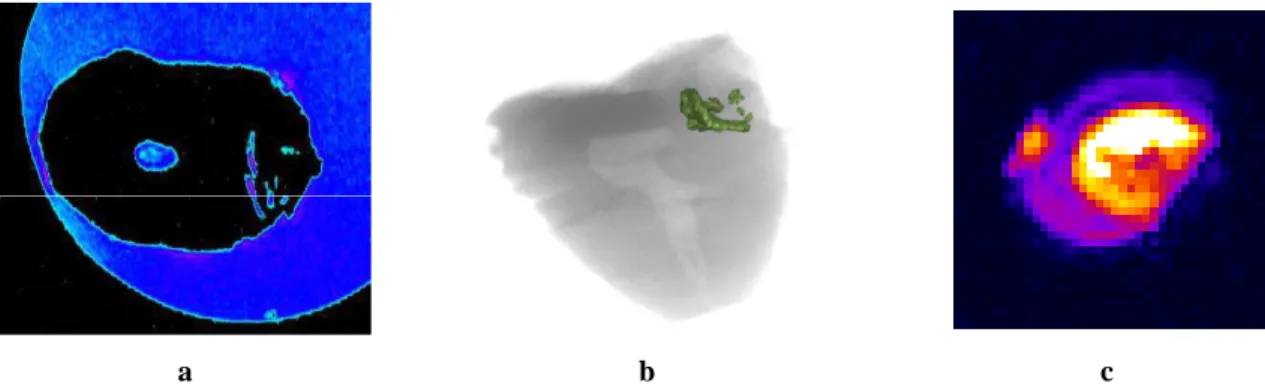
Fig. 2a shows a 2D cross-section of a 3D MR image of a carious tooth. High intensity water signal was observed in the porous, decayed, regions of the tooth due to water penetration into this region. No signal from mineralized tooth tissue could be observed this way. This allowed visualization of the presence of pores only. Pores were shown to extend deep inside the tooth and showed the moisture penetration path into the internal tooth structures. Black areas in the image correspond to mineralized tissue of the tooth, using a 3D SE pulse sequence. Water surrounding the tooth is shown in blue for better visualization. The area of pulp is easily recognized in the centre of tooth (see Fig. 2a). Irregular, blue regions between the pulp and the tooth surface indicate the presence of water in the pores of the decayed tooth.
Fig. 2b shows a pseudo-3D representation of the pores within the decayed region of the tooth, obtained with MRI.
In Fig. 2c cross-section through SPI data of the decayed tooth is shown. Obtained spatial resolution is lower than in the corresponding SE-based image. However, variation of signal intensity in the tissue is visible in contrary to SE image, where no signal is present in this region of tooth.
Fig. 1 Photo image of a decayed tooth.
a b c
Fig. 2. Cross-section of a 3D SE MR image (a) and the pseudo 3D reconstruction (b) of water in the porous tissue of a decayed tooth, obtained from 3D SE MR data. Fig (c) shows a 2D cross-section of a 3D SPI data set of a tooth with decay comparable that shown in figures a and b.
4. Discussion
The 3D SE pulse sequence used for imaging in the presented work is considered a “liquid” MRI measuring method, capable to detect signal from tissues with T2 longer than few ms.
However, this method is severely limited obtaining MR images of solids, such as mineralized tooth tissue. Thus, in the obtained MRI images, water surrounding the teeth and penetrating pores in decayed tissue are seen as bright areas, whereas rigid tissue of the tooth is invisible. High resolution images allow us to precisely visualize the 3D structure of pores and to estimate their size and extent. This type of MRI measurement could be used for the diagnosis of the extend of caries on and below the surface of teeth, as can be seen from Fig 2.
A possible improvement in the visualization of the mineralized tissue using MRI may be achieved by utilizing imaging methods capable of detecting signal with T2 of 1 ms or less (e.g.
SPI). However such methods are yet to be developed due to the current hardware limitations. The results presented here were obtained in vitro. The present MRI hardware and long acquisition time of the experiment necessary to collect data for high resolution images do not allow the same quality results to be obtained in vivo. However, significantly shorter acquisition time in the case of SE imaging can be relatively easily achieved using multi-echo sequences (e.g. RARE). Also, compromising the resolution would allow the experimental time to be acceptable for in vivo imaging. Recent works in the field show that a nominal isotropic resolution of 250 - 300 µm is achievable within 5 - 10 minutes using whole body MRI scanner [5, 6].
5. Conclusions
Spin-echo based MR imaging methods allow for high resolution imaging of the moisture entering into decayed teeth. This method enables the investigation of the extent of pores through which moisture penetrates within decayed tooth tissue. These pores are paths for microorganisms which can cause further damage of the tissue. Thus a method for the non-invasive visualization of pores in teeth is important. SE MRI, unfortunately, does not allow the visualization of any details of mineralized tooth tissue. Especially the gradual decrease in tissue density in the early stages of caries development cannot be followed with SE MRI. However SPI, which allows for the visualization of mineralized tissue may be applied, as shown in our studies, to obtain this information.
[3] W.P. Węglarz, M. Tanasiewicz, T.W. Kupka, T. Skórka, Z. Sułek, A. Jasiński, Solid State NMR, 25 (2004) 84.
[4] M. Tanasiewicz, W.P. Węglarz, T.W. Kupka, A. Jasiński, Pol. J. of Environ. Stud. 16 (2C) (2007) 169-172.
[5] O. Tymofiyeva,K. Rottner, F. Schmid, E.-J. Richter, P.M. Jakob, Proc. ISMRM (Toronto, 2008).
[6] S. Boujraf, C. Hofmann, J. Ulrici, E. Hell, B. Haller, V. Rasche, Proc. Intl. Soc. Mag. Reson. Med. 17 (2009) 4518
[7] P. Latta, M.L.H. Gruwel, E. Edie, M. Sramek, B. Tomanek, J. Magn. Reson. 170 (2004) 177.