Publisher’s version / Version de l'éditeur:
mBio, 7, 2, 2016-03
READ THESE TERMS AND CONDITIONS CAREFULLY BEFORE USING THIS WEBSITE. https://nrc-publications.canada.ca/eng/copyright
Vous avez des questions? Nous pouvons vous aider. Pour communiquer directement avec un auteur, consultez la première page de la revue dans laquelle son article a été publié afin de trouver ses coordonnées. Si vous n’arrivez pas à les repérer, communiquez avec nous à PublicationsArchive-ArchivesPublications@nrc-cnrc.gc.ca.
Questions? Contact the NRC Publications Archive team at
PublicationsArchive-ArchivesPublications@nrc-cnrc.gc.ca. If you wish to email the authors directly, please see the first page of the publication for their contact information.
NRC Publications Archive
Archives des publications du CNRC
This publication could be one of several versions: author’s original, accepted manuscript or the publisher’s version. / La version de cette publication peut être l’une des suivantes : la version prépublication de l’auteur, la version acceptée du manuscrit ou la version de l’éditeur.
For the publisher’s version, please access the DOI link below./ Pour consulter la version de l’éditeur, utilisez le lien DOI ci-dessous.
https://doi.org/10.1128/mBio.00252-16
Access and use of this website and the material on it are subject to the Terms and Conditions set forth at
Deacetylation of fungal exopolysaccharide mediates adhesion and
biofilm formation
Lee, Mark J.; Geller, Alexander M.; Bamford, Natalie C.; Liu, Hong; Gravelat,
Fabrice N.; Snarr, rendan D.; Le Mauff, François; Chabot, Joseé; Ralph,
Benjamin; Ostapska, Hanna; Lehoux, Mélanie; Cerone, Robert P.; Baptista,
Stephanie D.; Vinogradov, Evgeny; Stajich, Jason E.; Filler, Scott G.; Howell,
P. Lynne; Sheppard, Donald C.
https://publications-cnrc.canada.ca/fra/droits
L’accès à ce site Web et l’utilisation de son contenu sont assujettis aux conditions présentées dans le site LISEZ CES CONDITIONS ATTENTIVEMENT AVANT D’UTILISER CE SITE WEB.
NRC Publications Record / Notice d'Archives des publications de CNRC:
https://nrc-publications.canada.ca/eng/view/object/?id=0c2d9ccd-950d-4c89-993f-ca1e594b821c
https://publications-cnrc.canada.ca/fra/voir/objet/?id=0c2d9ccd-950d-4c89-993f-ca1e594b821c
Deacetylation of Fungal Exopolysaccharide Mediates Adhesion and
Biofilm Formation
Mark J. Lee,a* Alexander M. Geller,a* Natalie C. Bamford,c,dHong Liu,bFabrice N. Gravelat,a,gBrendan D. Snarr,a,g
François Le Mauff,a,gJoseé Chabot,a,gBenjamin Ralph,a,gHanna Ostapska,a,gMélanie Lehoux,a,gRobert P. Cerone,a
Stephanie D. Baptista,aEvgeny Vinogradov,e Jason E. Stajich,fScott G. Filler,bP. Lynne Howell,c,dDonald C. Shepparda,g
Departments of Medicine and of Microbiology and Immunology, McGill University, Montréal, Canadaa; Division of Infectious Diseases, Los Angeles Biomedical Medical
Institute at Harbor-University of California, Los Angeles Medical Center, Torrance, California, USAb; Program in Molecular Structure & Function, The Hospital for Sick
Children, Toronto, Canadac; Department of Biochemistry, Faculty of Medicine, University of Toronto, Toronto, Canadad; National Research Council, Ottawa, Canadae;
Department of Plant Pathology and Microbiology, University of California, Riverside, Riverside, California, USAf; Infectious Diseases and Immunity in Global Health
Program, Research Institute of the McGill University Health Centre, Montréal, Canadag
* Present address: Mark J. Lee, Department of Pathology and Laboratory Medicine, David Geffen School of Medicine at UCLA, Los Angeles, California, USA; Alexander M. Geller, Department of Molecular Biology, Princeton University, Princeton, New Jersey, USA.
ABSTRACT
The mold Aspergillus fumigatus causes invasive infection in immunocompromised patients. Recently,
galactosami-nogalactan (GAG), an exopolysaccharide composed of galactose and N-acetylgalactosamine (GalNAc), was identified as a
viru-lence factor required for biofilm formation. The molecular mechanisms underlying GAG biosynthesis and GAG-mediated
bio-film formation were unknown. We identified a cluster of five coregulated genes that were dysregulated in GAG-deficient mutants
and whose gene products share functional similarity with proteins that mediate the synthesis of the bacterial biofilm
exopolysac-charide poly-(1-6)-N-acetyl-
D-glucosamine (PNAG). Bioinformatic analyses suggested that the GAG cluster gene agd3 encodes
a protein containing a deacetylase domain. Because deacetylation of N-acetylglucosamine residues is critical for the function of
PNAG, we investigated the role of GAG deacetylation in fungal biofilm formation. Agd3 was found to mediate deacetylation of
GalNAc residues within GAG and render the polysaccharide polycationic. As with PNAG, deacetylation is required for the
adher-ence of GAG to hyphae and for biofilm formation. Growth of the ⌬agd3 mutant in the presadher-ence of culture supernatants of the
GAG-deficient ⌬uge3 mutant rescued the biofilm defect of the ⌬agd3 mutant and restored the adhesive properties of GAG,
sug-gesting that deacetylation is an extracellular process. The GAG biosynthetic gene cluster is present in the genomes of members of
the Pezizomycotina subphylum of the Ascomycota including a number of plant-pathogenic fungi and a single basidiomycete
spe-cies, Trichosporon asahii, likely a result of recent horizontal gene transfer. The current study demonstrates that the production
of cationic, deacetylated exopolysaccharides is a strategy used by both fungi and bacteria for biofilm formation.
IMPORTANCE
This study sheds light on the biosynthetic pathways governing the synthesis of galactosaminogalactan (GAG),
which plays a key role in A. fumigatus virulence and biofilm formation. We find that bacteria and fungi use similar strategies to
synthesize adhesive biofilm exopolysaccharides. The presence of orthologs of the GAG biosynthetic gene clusters in multiple
fungi suggests that this exopolysaccharide may also be important in the virulence of other fungal pathogens. Further, these
stud-ies establish a molecular mechanism of adhesion in which GAG interacts via charge-charge interactions to bind to both fungal
hyphae and other substrates. Finally, the importance of deacetylation in the synthesis of functional GAG and the extracellular
localization of this process suggest that inhibition of deacetylation may be an attractive target for the development of novel
anti-fungal therapies.
Received 19 February 2016 Accepted 25 February 2016 Published 5 April 2016
Citation Lee MJ, Geller AM, Bamford NC, Liu H, Gravelat FN, Snarr BD, Le Mauff F, Chabot J, Ralph B, Ostapska H, Lehoux M, Cerone RP, Baptista SD, Vinogradov E, Stajich JE, Filler SG, Howell PL, Sheppard DC. 2016. Deacetylation of fungal exopolysaccharide mediates adhesion and biofilm formation. mBio 7(2):e00252-16. doi:10.1128/mBio.00252-16.
Editor Joseph Heitman, Duke University
Copyright © 2016 Lee et al. This is an open-access article distributed under the terms of theCreative Commons Attribution 4.0 International license. Address correspondence to Donald C. Sheppard, don.sheppard@mcgill.ca.
This article is a direct contribution from a Fellow of the American Academy of Microbiology.
A
spergillus fumigatus
is an opportunistic mold that causes
inva-sive infections in immunosuppressed patients. Despite
anti-fungal treatment with the currently available antianti-fungal agents,
the mortality of invasive aspergillosis (IA) remains between 50
and 95% (1), highlighting the urgent need for new, effective
ther-apeutic agents for this disease. Identifying and targeting virulence
factors unique to this fungus is one approach to the development
of novel treatments for invasive aspergillosis.
Galactosaminogalactan (GAG) is an exopolysaccharide
re-cently identified in A. fumigatus that plays a number of roles in the
pathogenesis of IA (2, 3). GAG is a linear heteropolymer
com-posed of ␣-1,4-linked galactose and N-acetylgalactosamine
(GalNAc) that is bound to the outer cell wall and found within the
extracellular matrix of biofilms of Aspergillus species (4, 5). Cell
wall-associated GAG mediates a number of virulence-associated
traits, including adherence to host cells and other substrates,
bio-crossmark
mbio.asm.org
on April 21, 2016 - Published by
film formation, masking of -1,3-glucans from immune
recogni-tion, and resistance to neutrophil extracellular traps (2, 6).
How-ever, the biosynthetic pathways underlying GAG synthesis and the
molecular mechanisms by which GAG mediates these virulence
traits remain poorly understood.
A number of pathogenic bacteria also produce exopolysaccharide
composed of N-acetylhexosamines, most commonly -1,6-linked
N
-acetylglucosamine (GlcNAc). This exopolysaccharide, known as
polysaccharide intercellular adhesion (PIA) in Staphylococcus
au-reus
(7) or poly-(1-6)-N-acetyl-
D-glucosamine (PNAG) in
Esch-erichia coli
(8), mediates many of the same virulence properties as
GAG does, including adherence of the organism to both biotic and
abiotic surfaces, resistance to neutrophil killing, and masking of
pathogen-associated molecular patterns (PAMPs) (8–11). The
mechanisms governing the syntheses of these bacterial
exopoly-saccharides have been well characterized and are mediated by the
gene products of the ica or pga operons, respectively (Fig. 1A and
B and Fig. 2). Briefly, PIA/PNAG is synthesized and transported
FIG 1 Bacterial polysaccharide biosynthetic operons and the putative GAG biosynthetic gene cluster. (A to C) Schematic of the ica operon responsible for the
synthesis of polysaccharide intercellular adhesion (PIA) (A), pga operon responsible for the synthesis of PNAG [poly-(1-6)-N-acetyl-D-glucosamine] (B), and
the putative GAG gene cluster (C). (D) Heatmap showing differential gene expression of the A. fumigatus ⌬medA and ⌬stuA regulatory mutants compared to wild-type A. fumigatus, highlighting the coregulation of the genes in the GAG biosynthesis gene cluster. Fold induction is shown in red (upregulation), green (downregulation), black (no change), and gray (missing data point). Locus ID, locus identification.
Lee et al.
mbio.asm.org
on April 21, 2016 - Published by
mbio.asm.org
across the membrane and extruded extracellularly by the action of
a glycosyltransferase complex composed of either IcaA/D or
PgaC/D, respectively. The nascent polysaccharide undergoes
par-tial deacetylation of GlcNAc residues by IcaB/PgaB, resulting in
interspersed glucosamine (GlcN) residues (11–13). In
Gram-negative bacteria, the partially deacetylated polymer is then
trans-ported across the outer membrane by PgaA (14). Under acidic
conditions, these GlcN residues are protonated, conferring a
pos-itive charge on the mature polysaccharide, and thereby mediating
adherence to negatively charged surfaces including host cells and
the organism itself, as well as enhancing resistance to cationic
mol-ecules such as aminoglycoside antibiotics and antimicrobial peptides
(15). Loss of deacetylase activity results in the formation of a fully
acetylated polysaccharide that cannot adhere to the organism or
other substrates, and which is shed into the culture supernatant,
ren-dering it nonfunctional (11, 16).
Although the type of hexosamine and linkages differ between
PIA/PNAG and GAG, we hypothesized that the biosynthetic
path-ways for these glycans are similar and that partial deacetylation of
GAG is also required for its function. Comparative
transcrip-tome studies identified a cluster of genes in A. fumigatus that is
similar in composition to the bacterial exopolysaccharide
operons. This cluster encompasses the uge3 gene, encoding a
glucose 4-epimerase that is required for GAG biosynthesis (2,
17). Molecular and biochemical studies revealed that, as with
bac-terial exopolysaccharide, GAG is partially deacetylated. This
deacetylation is mediated by Agd3, which is encoded by another
gene within the GAG biosynthetic gene cluster. Extracellular Agd3
activity was required for binding of GAG to the hyphal cell wall,
biofilm formation, masking of -1,3-glucan, and full virulence.
Bioinformatic analysis of fungal genomes revealed the presence of
the GAG biosynthetic gene cluster within the genome of a number
of other filamentous ascomycetes. The complete cluster and
evi-dence for GAG production was also found within the
basidiomy-cete Trichosporon asahii, likely as a consequence of a recent
hori-zontal gene transfer event.
RESULTS
Identification of a gene cluster encoding putative GAG
biosyn-thetic proteins.
Previously, comparative transcriptomic analysis
of two transcription factor mutants, ⌬stuA and ⌬medA mutants,
identified uge3, a gene encoding a bifunctional epimerase, as
re-quired for GAG synthesis (2, 17). A more detailed analysis of this
data set revealed that the uge3 gene is flanked by four other
coregulated genes (Fig. 1C and D). These genes include genes
predicted to encode a glycosyltransferase (gtb3),
polysaccha-ride deacetylase (agd3), polysacchapolysaccha-ride hydrolase (ega3), and
spherulin-4-like protein (sph3). The predicted functions of these
proteins suggest a model for exopolysaccharide synthesis
analo-gous to the bacterial ICA/PGA systems (Fig. 2A). In this model,
UDP-galactose and UDP-GalNAc are synthesized by the activity
of the epimerase Uge3 and then polymerized and extruded from
the cell by the putative transmembrane glycosyltransferase Gtb3.
As with bacterial exopolysaccharide systems, the function of the
putative glycosyl hydrolase within the fungal system is unclear,
but a recent study from our group suggests that it may be required
for polymer modification and remodeling (18). Importantly, the
final step in this putative pathway is predicted to be the partial
deacetylation of N-acetylgalactosamine residues within the
nas-cent polymer by the deacetylase Agd3 to generate a polycationic
glycan that adheres to the hyphal and other surfaces. To begin to
validate this model, we therefore sought to determine the role of
Agd3 in GAG deacetylation and function.
Agd3 is predicted to contain a polysaccharide deacetylase
do-main.
Bioinformatic analysis of Agd3 suggests that it is a
multido-main extracellular protein including a predicted signal peptide
with a cleavage site between Cys18 and Thr19 (19, 20), suggesting
that Agd3 is secreted as an extracellular protein (Fig. 2B).
Struc-ture prediction server Phyre
2analysis of Agd3 predicted several
distinct domains and extended regions of low complexity (21).
The N-terminal region of the protein from residues 38 to 129
contains a serine-rich region that is predicted to be structurally
disordered. When Agd3 lacking the signal peptide was analyzed
using the structure prediction server Phyre
2, the top hits (⬎95%
confidence) for residues 509 to 726 were deacetylase domains of
members of carbohydrate esterase family 4 (CE 4) (Fig. 3A). The
CE 4 family contains a large number of metal-dependent
polysac-charide deacetylases, which have been shown to remove N- or
O
-linked acetate groups from chitin, peptidoglycan, acetylxylan,
and poly--1,6-N-acetylglucosamine (12, 22–24). CE 4 domains
adopt a (/␣)
7fold with five canonical amino acid motifs that are
important for active site formation and activity (12, 23). The
structural prediction for Agd3 contains only a partial CE 4 fold
starting at the second active site motif, which usually occurs at the
end of the third -strand of the barrel (Fig. 2B).
Using the whole protein, Phyre
2was unable to predict any
function-associated motifs for residues 140 to 509 (25). However,
when submitted to Phyre
2in isolation, residues 147 to 234 were
predicted (86.2% confidence) to be similar to the flavodoxin-like
fold present in Thermotoga maritima class I glutamine
amido-transferase (GATase) (PDB code
1O1Y
). Class I GATases are
de-fined by a conserved Cys-His-Glu catalytic triad that is important
for the reductase activity (25). These residues could not be
iden-tified in the Agd3 model, which contained only 88 of the 140 to
180 residues typically found in this fold. This domain is followed
by a region of low structural complexity, as residues 243 to 300
were predicted to be largely disordered by both Phyre
2and
Glob-Plot. When residues 200 to 500 were submitted, a partial (/␣)
barrel was identified between residues 392 and 468 with 75.3%
confidence. This region may represent the first /␣ repeats of the
CE 4 domain.
The metal coordinating triad (Asp-His-His) is conserved in
CE 4 enzymes and is localized to two of the five canonical linear
motifs (MTs). The MT1, DD and DXD motifs contain the
coor-dinating aspartic acid, as well as the catalytic base (Fig. 2C). In
Agd3, MT1 is most likely D377 and D378, which are highly
con-served in Agd3 orthologs. These residues are located outside the
predicted CE 4 domain and slightly upstream of the partial (/␣)
barrel motif. MT2 contains the metal coordinating histidines. In
Agd3, H510 and H514, which are highly conserved and align with
active site residues of known polysaccharide deacetylases using
ClustalW, probably constitute MT2 (Fig. 2C). The linear distance
between MT1 and MT2 is larger than that found in most CE 4
family members and is predicted to be a region of low structural
complexity. Similar insertions between motifs are present in
Hel-icobacter pylori
PgdA and Staphylococcus epidermidis IcaB, where
they are important for homomultimerization and membrane
as-sociation, respectively (26, 27). Circular permutations of the CE 4
motifs have also been reported in multiple members (23, 26).
Based on these findings, Agd3 has a unique CE 4 domain
compo-mbio.asm.org
on April 21, 2016 - Published by
sition that may have prevented accurate modeling of the entire
domain by Phyre
2, making the exact domain boundaries unclear.
Collectively, these in silico analyses suggest that Agd3 contains
functional domains similar to known bacterial polysaccharide
deacetylases, and therefore Agd3 may mediate the
deacetyla-tion of GAG.
Deletion of agd3 blocks deacetylation of GAG.
To determine
whether GAG is partially deacetylated, the presence of primary
amines in purified GAG was quantified by a chemical approach
using trinitrobenzene sulfonate (TNBS) (28). This analysis
iden-tified that the presence of primary amines consistent with the
presence of non-acetylated galactosamine (GalN) in GAG purified
from wild-type A. fumigatus (Fig. 3A). The presence of GalN
res-idues within purified GAG was also confirmed by nuclear
mag-netic resonance (NMR) analysis (Fig. 3B). Taken together, these
data suggest that GAG undergoes partial deacetylation.
To determine whether Agd3 activity is required for GAG
deacetylation, a ⌬agd3 deletion mutant strain was constructed.
Deletion of agd3 resulted in the production of GAG that was
de-void of primary amines and lacked GalN as detected by NMR
analysis (Fig. 3A and B). Deletion of agd3 did not significantly
affect the production of total secreted GAG (Fig. 3C) or affect the
expression of other genes in the GAG biosynthetic gene cluster
under all growth conditions tested (Fig. 3D). As previously
re-ported for the deletion of uge3, loss of Agd3 did not affect
suscep-tibility to antifungal agents or cell wall stressors, including
caspo-fungin and nikkomycin (see Fig. S1 in the supplemental material).
Complementation of the ⌬agd3 mutant with a wild-type allele of
FIG 2 Comparative models and in silico analysis. (A) Comparative models of exopolysaccharide synthesis in bacteria and fungi. The numbered steps are as
follows: (step 1) polymerization of sugar residues by the glycosyltransferases indicated in green (Gtb3, IcaA, and PgaC), (step 2) extrusion of the elongating polysaccharide from the cytosolic side to the extracellular side (or periplasm in Gram-negative bacteria) by the dual action of the glycosyltransferase (Gtb3, IcaA, and PgaC) and associated protein for the bacterial species (IcaD and PgaD), and (step 3) deacetylation of the N-acetylhexosamine unit of the nascent polysac-charide by the de-N-acetylase indicated in dark red (Agd3, IcaB, and PgaB). The extracellular matrix (ECM), cell wall (CW), plasma membrane (M), pepti-doglycan (PG), outer membrane (OM), and inner membrane (IM) are shown. CoA, coenzyme A. (B) Predicted domains and conserved regions in the Agd3 protein. From the N terminus, these domains include signal peptide (SS), a serine-rich region, a glutamine amidotransferase domain (reductase), metal-coordinating linear motifs (MT1/MT2), /␣ barrel, a carbohydrate esterase-4 like domain (CE 4), and a -strand-rich region. (C) Multiple-sequence alignment showing conserved DXD/DD motif located within the MT1 and MT2 conserved sites. Sequences include S. epidermidis IcaB, E. coli PgaB, P. aeruginosa PelA, and Sinorhizobium melilotiNodB. Highly conserved and similar residues are highlighted in dark and light gray, respectively. Gaps introduced to maximize alignment are indicated by hyphens.
Lee et al.
mbio.asm.org
on April 21, 2016 - Published by
mbio.asm.org
agd3
restored deacetylation to wild-type levels. These data suggest
that Agd3 is required for the deacetylation of GAG.
Deletion of agd3 is associated with the loss of adherence and
changes in the cell wall morphology.
To examine the role of
deacetylation and significance of the resulting cationic charge on
the functional properties of GAG, the ⌬agd3 mutant was
com-pared with the wild-type parent and agd3 complemented strains
for a variety of GAG-dependent phenotypes. Deletion of agd3 was
associated with a loss of adherence to negatively charged tissue
culture-treated surfaces (Fig. 4A). However, consistent with the
hypothesis that GAG-mediated adherence is dependent on
charge-charge interactions, the ⌬agd3 mutant retained the ability
to form wild-type biofilms on positively charged poly-
D-lysine-coated plates (Fig. 4A). Thus, deacetylation of GAG mediates
ad-herence to negatively charged surfaces, but not to positively
charged surfaces.
The loss of Agd3 resulted in the loss of detectable cell
wall-associated GAG as measured by direct immunofluorescence with
the GalNAc-specific soybean agglutinin (SBA) lectin (Fig. 4B).
These findings were also confirmed by scanning electron
micros-copy, which demonstrated a loss of the GAG-dependent cell wall
decorations (Fig. 4C). Consistent with this loss of cell
wall-associated GAG, hyphae of the ⌬agd3 mutant displayed increased
exposure of -1,3-glucan as detected by enhanced binding of
re-FIG 3 Deletion of agd3 blocks deacetylation of GAG. (A) Detection of primary amine of purified GAG from the strains indicated in the figure as measured by
evolution of colorimetric byproduct of the TNBS reaction. O.D., optical density. (B)1H NMR analysis of purified GAG from the indicated strains. Black arrows
indicate the detection of the hydrogen resonance peak originating from galactosamine. (C) Total secreted GAG production by the indicated strains. (D) Comparison of relative expression of GAG cluster genes between wild-type Af293 and ⌬agd3 mutant as measured against reference gene tef1 under various growth conditions (RPMI 1640 supplemented with MOPS [RPMI-MOPS], Brian medium, Aspergillus minimal medium [AspMM]). For anaerobic growth, additional reference genes, actin 1, and -tubulin were also used. For all graphs, data are represented as means plus standard errors of the means (SEM) (error bars). The values of the wild-type A. fumigatus Af293 and the ⌬agd3 mutant strain were significantly different (P ⬍ 0.05 by analysis of variance [ANOVA] with Tukey’s test for pairwise comparison) as indicated by the asterisk.
mbio.asm.org
on April 21, 2016 - Published by
combinant Fc-dectin-1 by fluorescence microscopy (Fig. 4D; see
Fig. S2 in the supplemental material). The phenotype of the ⌬agd3
mutant observed in these assays was indistinguishable from the
GAG-deficient ⌬uge3 mutant (2). Collectively, these data suggest
that Agd3-mediated deacetylation of GAG is required for
adher-ence of the polysaccharide to the fungal cell wall and mediates
adherence of hyphae to positively charged surfaces.
De-N-acetylated GAG confers a positive charge on the
hy-phal surface.
To confirm that the presence of deacetylated GAG
alters the surface charge of A. fumigatus hyphae, the ability of
hyphae to bind negatively charged Sephadex beads was quantified
(29). When coincubated with anionic Sephadex beads, there was a
greater binding of negatively charged beads by wild-type A.
fu-migatus
or by the agd3 complemented strain than by the ⌬agd3
mutant (Fig. 5). Binding of anionic beads by hyphae of the ⌬agd3
mutant was similar to the binding of these beads by the completely
GAG-deficient ⌬uge3 mutant. Thus, deacetylation is required to
render GAG cationic and mediates adhesion to both the hyphal
cell wall and other surfaces.
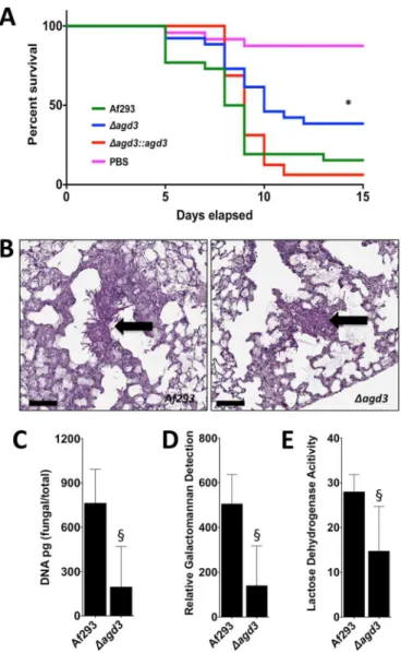
Deletion of agd3 attenuates virulence.
To confirm that
deacetylation of GAG plays a role in pathogenesis, the virulence of
the ⌬agd3 mutant was compared to the wild-type Af293 strain and
the agd3 complemented strain in a leukopenic murine model of
invasive aspergillosis (Fig. 6A) (2, 30). Consistent with the in vitro
findings of impaired GAG function, the ⌬agd3 mutant was
hypo-virulent compared to wild-type A. fumigatus or the agd3
comple-mented strain. Histopathologic examination of the lungs of mice
infected with the ⌬agd3 mutant revealed no difference in the
ap-pearance of fungal lesions in mice infected with the wild-type or
⌬agd3 mutant strains (Fig. 6B). However, determination of fungal
burden by quantitative PCR (Fig. 6C) or pulmonary
galactoman-nan content (Fig. 6D) revealed a lower fungal burden in the lungs
of mice infected with the ⌬agd3 mutant than in mice infected with
the wild-type parent strain, as was previously reported with the
⌬uge3 mutant strain. Consistent with the lower fungal burden
observed in mice infected with the ⌬agd3 mutant, lower
pulmo-nary injury was observed in these animals as measured by lactose
dehydrogenase activity in bronchoalveolar lavage fluid (Fig. 6E).
Thus, deacetylation of GAG is required for full virulence of A.
fu-migatus
.
FIG 4 Deletion of agd3 is associated with loss of adherence and changes in the
cell wall. (A) Formation of adherent biofilms by the strains indicated in the figure on either positively charged (poly-D-lysine-treated [PDL]) or negatively charged (tissue culture-treated polystyrene [TC]) surfaces. Biofilms were washed and visualized by staining with crystal violet (gray). (B) Confocal mi-croscopy images of hyphae stained with FITC-tagged soybean agglutinin lectin (top) and corresponding differential interference contrast (DIC) (bottom). (C) Scanning electron micrographs of hyphae grown for 24 h. The white ar-rows point to hyphal surface decorations associated with GAG production. (D) Confocal microscopy images of hyphae stained with Fc-dectin-1 detected by FITC-tagged Fc-receptor antibody (top) and corresponding DIC (bottom).
FIG 5 Agd3 activity augments the positive charge on the surfaces of hyphae.
The graph shows the percentage of negatively charged Sephadex beads bound by hyphae of the indicated strains. Data are presented as means plus standard errors of the means (SEM) (error bars). The values for the indicated mutant strains were significantly different (P ⬍ 0.05 by ANOVA with Tukey’s test for pairwise comparison) from the value for wild-type A. fumigatus Af293 strain as indicated by the asterisk. The value for the ⌬agd3::agd3 strain was not signifi-cantly different (n.s.) from the value for the wild-type A. fumigatus Af293 strain. Lee et al. mbio.asm.org on April 21, 2016 - Published by mbio.asm.org Downloaded from
Restoration of biofilm formation by coculture of
biofilm-deficient strains.
Since the ⌬agd3 mutant produces fully
acety-lated GAG but not Agd3, and the ⌬uge3 mutant produces Agd3
but not GAG, we hypothesized that these strains could
comple-ment one another to produce deacetylated GAG and form
bio-films. As predicted, coculture of the ⌬uge3 and ⌬agd3 mutants
resulted in the formation of biofilms that were indistinguishable
from those produced by wild-type A. fumigatus (Fig. 7A). Further,
growth of the ⌬agd3 mutant in the presence of culture filtrates
from the ⌬uge3 mutant resulted in the formation of adherent
biofilms (Fig. 7B) and restored the presence of GAG-associated
cell wall decorations as detected by scanning electron microscopy
(Fig. 7C). Growth of the ⌬agd3 or ⌬uge3 mutant in the presence of
their own respective culture filtrates had no effects on adherence
(Fig. 7B) or hyphal morphology (Fig. 7C).
To extend these findings, we also examined the ability of
cell-free culture filtrates from both ⌬agd3 and ⌬uge3 mutants to
gen-erate deacetylated GAG in vitro in the absence of A. fumigatus
hyphae. Culture supernatants of each mutant or a 1:1 mixture of
both culture supernatants were incubated in anionic polystyrene
enzyme immunoassay (EIA) plates to capture adherent,
deacety-lated GAG. Adherent, deacetydeacety-lated GAG was then quantified
us-ing an anti-GAG antibody (3). A 1:1 mixture of culture
superna-tants of the ⌬agd3 and ⌬uge3 musuperna-tants resulted in detection of
adherent GAG at a level similar to that recovered from culture
supernatants of wild-type A. fumigatus (see Fig. S3 in the
supple-mental material), while no adherent GAG was detected in culture
filtrates of the ⌬agd3 mutant or ⌬uge3 mutant alone. Collectively,
these results support the proposed model of GAG biosynthesis in
which GAG undergoes deacetylation by extracellular Agd3.
FIG 6 Agd3 is required for full virulence in a mouse model of invasive
aspergillosis. (A) Survival of BALB/c mice treated with cortisone and cyclo-phosphamide and then infected with the indicated conidial strains. Graphs are the combined results of two independent experiments with 26 mice per group for all groups of mice infected with fungal strains and 24 mice in the PBS sham infection group. There was a significant difference in the survival of mice infected with wild-type Af293 or ⌬agd3::agd3 strain compared to those in-fected with the ⌬agd3 strain as determined by the Mantel-Cox log rank test with pairwise comparison applying Bonferroni’s correction as indicated by the asterisk. (B) Pulmonary histopathology sections from BALB/c mice infected with indicated strains and stained with PAS for visualization of fungal ele-ments. Black arrows indicate fungal elements found within pulmonary lesions. Bars, 100 m. (C) Pulmonary fungal burden of mice infected with the indi-cated strains, as measured by quantitative PCR. There were 8 mice in each group. (D) Pulmonary fungal burden of mice infected with the indicated strains, as measured by determination of pulmonary galactomannan content. There were 8 mice in each group. (E) Pulmonary injury as measured by lactose dehydrogenase activity in the bronchoalveolar lavage fluid of mice infected with the indicated strains. There were 8 mice in each group. Values are medi-ans plus interquartile ranged (error bars). There was a significant difference in either the fungal burden or lung injury in mice infected with wild-type Af293 strain and those infected with the ⌬agd3 mutant strain as determined by the Mann-Whitney test as indicated by the § symbol.
FIG 7 Culture filtrates from the ⌬uge3 mutant complement the defects in
adherence and cell wall morphology of ⌬agd3 mutant. (A) Biofilm formation by the indicated strains grown alone or in coculture. After the biofilm was washed, adherent biofilm was visualized by crystal violet staining. (B) Biofilm formation by the indicated strains grown in the presence of culture filtrates (C/F) from the ⌬uge3 or ⌬agd3 mutant. After the biofilm was washed, adher-ent biofilm was visualized by crystal violet staining. (C) Scanning electron microscopy visualization of cell wall morphology of the indicated strains grown in the presence of culture filtrates from the ⌬uge3 mutant. The white arrows indicate cell wall decorations associated with cell wall-bound GAG.
mbio.asm.org
on April 21, 2016 - Published by
Agd3 is localized on the hyphal surface.
The results of our
biochemical and complementation studies suggest that
Agd3-mediated deacetylation is an extracellular process. Consistent
with these findings, a recent study identified Agd3 within the
se-cretome of A. fumigatus (31). To determine whether Agd3 is also
present within the cell wall, a red fluorescent protein
(RFP)-tagged agd3 allele was expressed under the native agd3 promoter
in A. fumigatus Af293. In this strain, while low levels of red
fluo-rescence in the cell wall were observed, the signal was obscured by
significant red autofluorescence of hyphae. To enhance
visualiza-tion of RFP-tagged Agd3 (Agd3-RFP), indirect
immunofluores-cence using a fluorescein isothiocyanate (FITC)-tagged anti-RFP
antibody was performed. Using this technique, low levels of
Agd3-RFP were observed on the surfaces of hyphae (Fig. 8). Western
blot analysis of culture supernatants and fungal biomass
con-firmed that although Agd3-RFP was found both in the culture
supernatant and the fungal biomass, the majority of Agd3-RFP
was associated with hyphae (see Fig. S4 in the supplemental
ma-terial). Interestingly, from the fungal biomass, four discrete bands
were detected using anti-RFP antibodies, while in the secreted
fraction, only a single band was observed. The same pattern of
bands was detected in multiple independent experiments. Liquid
chromatography-mass spectrometry (LC-MS) analysis of all five
of these bands confirmed that these bands contained Agd3-RFP.
Further, the relative abundance of individual peptides did not vary
significantly between these bands, suggesting that the differences
in band migration are more likely due to differences in
glycosyla-tion rather than proteolytic cleavage.
The GAG biosynthesis gene cluster is widespread in
Ascomy-cetes but present only in Trichosporon asahii among
Basidiomy-cetes.
Using an in silico approach, the GAG gene cluster sequences
from A. fumigatus were queried against more than 250 publically
available fungal genomes at NCBI Genomes and FungiDB (32).
Orthologous genes of the GAG gene cluster present at the same
locus were found in 28 fungal species. In the majority of these
species, synteny of the cluster genes was observed, suggesting a
common origin for this cluster in these fungal species (see Fig. S5
in the supplemental material). All of the species containing the
GAG gene cluster belong to the Pezizomycotina subphylum of the
Ascomycota
with the exception of one member of the phylum
Ba-sidiomycota
, Trichosporon asahii (Fig. 9). More than half of the
fungal species that possess the GAG gene cluster have been
de-scribed as pathogenic species (see Table S1 in the supplemental
material). Of these species, 13 are plant pathogens, while only 3 are
known pathogens to insects and animals, suggesting a possible
role for GAG in the pathogenesis of plant fungal infections.
To elucidate the evolutionary relationship among these gene
cluster-containing species, and specifically of Agd3 orthologs, we
constructed a gene tree by aligning the amino acid sequences of
Agd3 orthologs from all 28 gene cluster-containing species (Fig. 9)
and constructed a maximum likelihood tree rooted by the copies
from the early branching ascomycete and nematode-trapping
fungus Arthrobotrys oligospora. While the relationship among the
orthologs of Agd3 largely mirrors the evolutionary relationship of
the respective species as established by the fungal tree of life (33,
34), we found that the T. asahii Agd3 ortholog was nested in the
tree within members of the class Sordariomycetes (Fig. 9),
suggest-ing that a recent horizontal gene transfer event may have occurred.
Consistent with this hypothesis, a predicted mutator-like element
(MULE) domain-containing protein was found within the GAG
gene cluster of T. asahii (see Fig. S5 in the supplemental material).
The MULE domain-containing proteins are class II DNA
trans-posases (35) and are found in fungal species (36) and are
com-monly found in eukaryotes (37). Analysis of the sequences
flank-ing the cluster did not reveal an identifiable tandem inverted
repeat at the borders of the GAG synthetic cluster in T. asahii or
any other mobile element signatures. It therefore seems most
likely that the transposable element is no longer active and that
only the transposase domain remains, while other features of the
mobile element have degraded.
To ascertain whether the GAG cluster was functional in T.
asa-hii
, mature hyphae of this organism were stained with
fluorescein-tagged SBA lectin to detect the production of GAG. Lectin staining
suggests that T. asahii secretes GAG into the extracellular matrix
(Fig. 10). GAG produced by T. asahii was less uniformly bound to
the cell wall of hyphae compared with A. fumigatus and seemed to
form a matrix between hyphae.
DISCUSSION
In the present study, we report a cluster of coregulated genes on
chromosome 3 of A. fumigatus with composition and functions
similar to those of bacterial operons encoding proteins required
for exopolysaccharide synthesis. Characterization of one of the
enzymes in this cluster demonstrates that, as with bacterial
exopo-lysaccharide, deacetylation of GAG is required for GAG to adhere
both to substrates and to the organism itself, and for full virulence.
Further, these studies suggest that GAG-mediated adherence is
largely a consequence of charge-charge interactions between the
polycationic polysaccharide and negatively charged surfaces.
The results of this study illustrate important similarities
be-tween the Agd3 deacetylase and other multidomain CE 4 family
members from bacteria. In S. epidermis, deacetylation of PIA by
the CE 4 domain-containing deacetylase IcaB is required to confer
a net positive charge to PIA and retain the polysaccharide on the
bacterial cell wall (38). Similarly, PelA, a deacetylase in the
Pseu-domonas aeruginosa
Pel exopolysaccharide biosynthetic operon,
has also been reported to be required for the production of
adhe-sive polysaccharide and biofilm formation in this organism (26).
As with Agd3, PelA contains a small reductase fold and CE 4
FIG 8 Agd3 localizes to the surfaces of hyphae. Hyphae expressing an
Agd3-RFP fusion protein are visualized by confocal microscopy and indirect immu-nofluorescence. Lee et al. mbio.asm.org on April 21, 2016 - Published by mbio.asm.org Downloaded from
deacetylase domain separated by a region of predicted low
com-plexity, as well as a -sheet-rich region at the C terminus. While
the role of the predicted reductase and C-terminal domains in
PelA are unknown, the similarities between the domain
arrange-ments within PelA and Agd3 suggest they likely mediate similar
functions in the synthesis of Pel and GAG, respectively. PgaB,
from E. coli, requires an adjacent domain, located C terminally to
the CE 4 domain, for the deacetylation of substrate (20). While
there are domains other than CE 4 predicted in Agd3, their specific
role in deacetylation is currently unknown. Finally, a protein
con-taining the CE 4 domain, PssB, is required for the production of
cell surface-bound extracellular polysaccharide in Listeria
mono-cytogenes
(39). Loss of PssB results in impaired bacterial cell
aggregation and biofilm formation. Thus, postsynthesis
modi-fication of extracellular polysaccharides by deacetylation of
hexosamine sugars seems to be a common theme used by
di-verse microorganisms in the formation of biofilms (40).
Although these polysaccharide systems display many
similari-ties, several lines of evidence suggest that these biosynthetic
path-ways are the product of convergent, rather than divergent,
evolu-tion from a common ancestor of both fungi and bacteria. First, the
polysaccharide composition and linkages of the
exopolysaccha-ride product of these systems differ markedly between organisms.
Although the composition of Pel is unknown, PIA/PNAG is
composed of -1,6-linked GlcNAc residues; the
exopolysac-charide of L. monocytogenes is composed of a -1,4-linked
N
-acetylmannosamine chain decorated with a terminal
␣-1,6-FIG 9 Agd3 orthologs are found within a wide range of Ascomyces species. A gene tree is shown categorized by taxonomic class of Agd3 orthologs found in the
28 fungal species that possess the GAG gene cluster. The gene tree was built by aligning and trimming Agd3 ortholog sequences, followed by maximum likelihood phylogenetic analysis with 100 bootstrap replicates. The outgroup was rooted with sequences of Arthrobotrys species. The scale bar is the genetic distance representing amino acid substitutions per site.
FIG 10 Trichosporon asahii produces a GAG-like exopolysaccharide. Hyphae
of T. asahii were stained with FITC-tagged soybean agglutinin (SBA) lectin binding for the detection of GalNAc-rich exopolysaccharide.
mbio.asm.org
on April 21, 2016 - Published by
linked galactose; and GAG is a heteropolymer of ␣-1,4-linked
ga-lactose and GalNAc. Further, there is little sequence similarity
between these bacterial and fungal enzymes beyond the conserved
enzymatic domains themselves.
While it is possible that the GAG gene cluster arose de novo in
fungi, it is also possible that these genes were acquired by
horizon-tal gene transfer by a common ancestor of the Pezizomycotina that
may have originated in bacteria or another organism. The
clus-tered consecutive organization of the GAG biosynthetic genes is
reminiscent of a bacterial operon and is relatively unusual in
fungi. Fungal gene clusters have been best described as encoding
enzymes required for the biosynthesis of secondary metabolites.
Horizontal gene transfer of these secondary metabolite clusters
has been suggested as a mechanism for their acquisition (41). The
observations that T. asahii is the only basidiomycete found to
contain the GAG gene cluster and that the T. asahii Agd3 ortholog
nests with strong bootstrap support within the Soridariomycetes
sequences in the gene tree are very strong evidence for a horizontal
gene transfer event from an ascomycete species belonging to the
Sordariomycetes
class into T. asahii or its ancestor. Furthermore,
the homology of T. asahii Agd3 and overall synteny of the cluster
genes with other ascomycetes strongly suggest that the entire
clus-ter was acquired in T. asahii or its parental linages as a single
horizontal transfer event. Although sequence analysis did not
re-veal any clear evidence of a specific mechanism and integration of
the gene transfer, it is possible that it was mediated by a MULE
domain-containing transposase, which is found near the
bound-ary of the cluster in T. asahii. As genomes of more fungal species
are made available and queried, a better picture of the timing of
this exchange and overall evolution of this gene cluster will likely
become clearer.
Interestingly, many of the species containing the GAG gene
cluster are known plant pathogens, while no primary
animal-associated fungi, such as the dermatophytes or Onygenales fungi,
contain a homolog of the gene. Whether the ability to produce
GAG confers an advantage to colonize and invade plant hosts and
whether this selection pressure resulted in the convergent
evolu-tion of caevolu-tionic polysaccharides in microorganisms remains to be
determined. The observation that bacterial and fungal pathogens
have developed similar strategies to produce cationic
exopolysac-charide might suggest that these polymers facilitate pathogenicity.
However, as humans are dead-end hosts for Aspergillus species, it
is likely that the pressures responsible for the development of GAG
were found in the soil or other natural environment. A shared
requirement for adherence to substrates and resistance to
envi-ronmental stresses through the production of an adherent layer of
exopolysaccharide may underlie the development of
exopolysac-charide synthesis by all of these microorganisms. Future studies
examining the role of exopolysaccharide in fungal and bacterial
growth in nonhuman environments may shed light on the factors
leading to the evolution of these glycans in these varied
microor-ganisms.
Deacetylation of GalNAc by Agd3 is required for A. fumigatus
to produce cell wall-bound GAG and for mediating the
pheno-types that have been associated with cell wall-associated GAG.
Further, the mixed mutant biofilm studies reported here would
suggest that, as with Gram-positive bacterial exopolysaccharide,
deacetylation occurs in the extracellular space. This observation
suggests that inhibition of GAG deacetylation may represent an
attractive antifungal target since intracellular penetration of a
can-didate deacetylase inhibitor would not be required for activity.
Deacetylation of GalNAc within human cells has not been
re-ported, suggesting that antifungal specificity may be achievable.
As loss of GAG deacetylation has no direct effect on fungal
viabil-ity, such a strategy would be best paired with a conventional
anti-fungal agent.
Although a previous report did not identify the presence of
deacetylated GalNAc within secreted GAG from A. fumigatus (3),
compositional analysis of GAG from Aspergillus niger and
Asper-gillus parasiticus
has reported the presence of GalN in preparations
from these species (42, 43). Because deacetylated GAG adheres
avidly to glass and plastics, it is possible that during the
purifica-tion of A. fumigatus GAG, the preparapurifica-tion was enriched for fully
acetylated, nonadherent polysaccharide. Alternately, it is possible
that deacetylation varies under different growth conditions as
re-ported in other species (44–46). Further experiments are required
to test these possibilities.
In conclusion, we report that deacetylation of GAG by Agd3 is
essential for the function of this exopolysaccharide, including
ad-herence of GAG to the fungal cell wall and other substrates. These
experiments shed light on the molecular mechanisms underlying
GAG-mediated adhesion and draw important parallels between
this glycan and bacterial exopolysaccharides. Finally, these data
suggest the exciting possibility that targeting deacetylation of
GAG may represent a promising antifungal strategy.
MATERIALS AND METHODS
Fungal strains and growth conditions. Aspergillus fumigatusstrain Af293
(a generous gift from P. Magee, University of Minnesota, St. Paul, MN) was used as the parent strain for all genetic manipulations and experimen-tal controls. Unless otherwise noted, all A. fumigatus strains were main-tained on yeast extract-peptone-dextrose (YPD) agar (Fisher Scientific) at 37°C. For growth in liquid culture, Brian medium (3) or phenol-free RPMI 1640 (Wisent) was used as indicated in the figures at 37°C and 5% CO2incubation, unless otherwise noted. A clinical isolate of Trichosporon asahiiwas obtained from the McGill University Health Centre, Montreal, Québec, Canada, and maintained on potato-dextrose medium at 30°C.
Molecular and genetic manipulations.A split marker, double
homol-ogous recombination approach was used to generate the ⌬agd3 mutant (47). Sequences flanking the open reading frame (ORF) of the agd3 gene were amplified from the genomic DNA using PCR (see Table S2 in the supplemental material). The resulting PCR fragments were then cloned into the entry vector pENTR-D-TOPO (Gateway, Inc.) and then recom-bined with previously described destination plasmids, pHY and pYG, containing the hygromycin split marker resistance cassettes (2). Target DNA was amplified by PCR from the recombined plasmids and then used for transformation of A. fumigatus as previously described (47).
To generate the ⌬agd3::agd3 complemented strain, the agd3 open reading frame and flanking sequences (680 bp downstream and 560 bp upstream) were amplified by PCR using the Sgf-compl-fow (compl stands for complemented, and fow stands for forward) and Asc-compl-rev (rev stands for reverse) primers, respectively (see Table S2 in the supplemental material). The resulting PCR fragment was cloned into pTAPA, a newly designed destination plasmid containing the phleomycin selection marker. The resulting pTAPA::agd3 plasmid contained the following se-quence: Pagd3::ORFagd3::Tcyc1::ORFble::PthiA::TtrpC. Two fragments for transformation were amplified by PCR from the pTAPA::agd3 plasmid using Sgf-compl-fow and LE4 primers and BL4 and Asc-compl-rev prim-ers. Phleomycin-resistant transformants were selected, and correct inte-gration at the native locus was verified by genomic PCR. Restoration of agd3mRNA expression was confirmed by real-time reverse transcription-PCR (RT-transcription-PCR) (Table S2).
To generate the agd3::rfp mutant, the ORF of agd3 was amplified using
Lee et al.
mbio.asm.org
on April 21, 2016 - Published by
mbio.asm.org
primers AgeI-agd-end-fow and EcoRV-agd-end-rev (see Table S2 in the supplemental material), cloned upstream of the rfp ORF in the pRFP␥ plasmid (17), and digested by AgeI and EcoRV, which excised the gpdA promoter region on the plasmid. Amplification of the linearized plasmid produced the (agd3-end-ORF)::(RFP)::(TtrpC)::(3=-half of HYG cas-sette) PCR fragment. From the agd3 terminator on the pRFP␥ plasmid, a 735-bp PCR product was made, using BsrGI-agd-Tr-fow and SacI-agd-TR-rev. This product was cloned upstream of the hygromycin (HYG) cassette in the pAN7.1 plasmid (2), digested by BsrGI and SacI, resulting in an amplified PCR fragment (5=-half of HYG cassette)::(agd3-Tr). A. fu-migatusAf293 was then transformed with this construct (47). All integra-tions were of agd3, and integration of the hygromycin resistance was verified by genomic PCR confirmed using real-time RT-PCR (see Ta-ble S2 for relevant primers) (2).
Gene expression studies.Expression of the galactosaminogalactan
(GAG) cluster genes AFUA_3G07910 (uge3), AFUA_3G07900 (sph3), AFUA_3G07890 (ega3), and AFUA_3G07860 (gtb3) in the ⌬agd3 mutant was compared to the wild-type Af293. The fungal strains indicated in the figures were grown for 18 h at 37°C under various growth conditions including phenol-free RPMI 1640 (Wisent) buffered with morpho-linepropanesulfonic acid (MOPS) (Bioshop, Inc.) at pH 5.4 and pH 7.0, Brian medium (3), or Aspergillus medium (2) with iron supplementation of 0, 2, or 30 mM. For anaerobic (AnaeroPack; Mitsubishi Gas Chemical, Inc.) or microaerophilic (MicroAeroPack; Mitsubishi Gas Chemical, Inc.) conditions, fungi were grown in phenol-free RPMI 1640 (Wisent) buff-ered with MOPS (Bioshop, Inc.) at pH 7.0 in anaerobic chambers with the appropriate gas packs. Mycelia were collected, RNA was extracted, and quantitative PCR (qPCR) was performed and analyzed as previously de-scribed (see Table S2 in the supplemental material) (2).
Bioinformatic analyses.For homology structural analysis, the amino
acid sequence of Agd3 from Aspergillus fumigatus was obtained from the Aspergillus Genome Database (19) and analyzed using a number of dif-ferent web-based servers, including Phyre2, SignalP v3.0, GlobPlot, NetOGlyc 4.0, and SMART (20, 21, 48). The full-length sequence of Agd3 was initially used for the bioinformatic analysis. As analysis of full-length Agd3 using Phyre2failed to predict a structural model for residues 140 to 500, these residues were submitted to the server with different boundaries including residues 140 to 250, 141 to 400, 200 to 500, 300 to 806, and 350 to 733. Alignments were performed using ClustalW2 and Phyre2(20, 22). To assess the prevalence of the GAG gene cluster across the fungal kingdom, amino acid sequences of each gene in the cluster in A. fumigatus was queried with FastA to search the predicted proteomes of publically available taxa in the Ascomycetes and Basidiomycetes (49). Species having a cluster containing a predicted glycosyltransferase, epimerase, deacetylase, and either the hydrolase or spherulin-like protein were selected. To deter-mine the phylogenetic relationship of Agd3 among these species, a phy-logenetic tree was constructed by first performing multiple alignment using T-Coffee (50), trimming the alignment with trimAl (51), followed by phylogenetic analysis using RAxML (v 8.1.1) with 100 bootstrap repli-cates and PROTGAMMA matrix with automatic model selection (52). The resultant tree was visualized with FigTree, and colors were added by using Adobe Illustrator (Adobe Systems Inc., Palo Alto, CA).
Polysaccharide analysis.GAG was extracted by ethanol precipitation
and purified as previously described (17). For chemical analysis to quan-tify the presence of primary amines in GAG, colorimetric detection using trinitrobenzene sulfonate (TNBS) (Fisher Scientific, Inc.) was performed following the manufacturer’s instructions. For nuclear magnetic reso-nance (NMR) detection of the primary amine group, dried GAG samples were reconstituted in deuterated water, and cold deuterium chloride was added to dissolve the samples.1H heteronuclear multiple quantum cor-relation (HMQC) NMR experiments were conducted on a Varian INOVA 500-MHz spectrometer with a 3-mm gradient probe. Assignment of tra was performed using Bruker Topspin version 3.1 program for spec-trum visualization.
Hyphal surfa ce characterization.For biofilm adherence assays, fungi
were grown for 24 h in Brian medium on tissue culture-treated six-well polystyrene plates. Nonadherent fungi were removed by vigorous agita-tion and washing. Adherent biofilms were visualized by crystal violet staining (53). Cell wall topography was examined using scanning electron microscopy as previously described (2). For soybean agglutinin (SBA) or recombinant Fc-dectin-1 binding to the surface of hyphae, strains were grown for 7 to 9 h on poly-D-lysine-coated coverslips (BD Biosciences, Inc.), then fixed, and stained with either fluorescein-tagged SBA or re-combinant Fc-dectin-1 (2). For all microscopy experimentation, strains were grown in phenol red-free RPMI 1640. The susceptibility of strains to cell wall-active antifungals was determined as previously described (2). Bright-field images of mycelia were captured at a magnification of ⫻200 (Infinity 2 camera; Lumenera Inc.).
Difference in hyphal surface charge was determined by hyphal binding to negatively charged CM Sephadex beads (Sigma Aldrich, Inc.). The fun-gal strains indicated in the figures were grown in RPMI 1640 in 50-ml centrifuge tubes at 37°C and 5% CO2for 18 to 24 h. Reconstituted sterile beads were added to the tubes containing pregrown fungi, andthe tubes were then shaken for 10 min at 200 rpm. Supernatant samples were taken, and the number of unbound beads was counted by using a light micro-scope.
Agd3 localization studies.To determine the localization of Agd3 by
fluorescence microscopy, the Af293 agd3-rfp mutant was grown on glass coverslips in RPMI 1640 for 9 to 12 h at 37°C and 5% CO2, fixed with 4% paraformaldehyde, stained with anti-red fluorescent protein (anti-RFP) IgG antibody (Abcam, Inc.), and detected with fluorescein-tagged anti-IgG antibody (Abcam, Inc.). The cells were then mounted on microscope slides and imaged by confocal microscopy as previously described (2). For RFP-tagged Agd3 (Agd3-RFP) immunoblotting, mycelia of the wild-type and Agd3-RFP strains were grown at 37°C for 22 to 24 h under shaking conditions, and culture supernatants were recovered by filtration. The biomass was crushed under liquid nitrogen, homogenized, and incubated for 1 h at 4°C in the presence of protease inhibitors (Bioshop, Inc.) and Triton X-100 at a final concentration of 2% (Bioshop, Inc.) prior to ly-ophilization. Protease inhibitors (Bioshop, Inc.) were added to the culture supernatant, lyophilized, and stored at ⫺20°C. Detection of Agd3-RFP was performed by Western blotting using rat anti-RFP antibody (Chro-motek, Inc.) and horseradish peroxidase (HRP)-conjugated donkey anti-rat IgG (Chromotek, Inc.).
For liquid chromatography-mass spectrometry (LC-MS) analysis, bands were excised from sodium dodecyl sulfate-polyacrylamide gels (SDS-PAGs), washed in 0.1 M ammonium bicarbonate, and incubated first in 10 mM dithiothreitol for 45 min at 56°C and then in 0.1 M ammo-nium bicarbonate containing 55 mM iodoacetamide for 20 min at room temperature. The gel was washed in 0.1 M ammonium bicarbonate⫺ace-tonitrile (1:1, vol/vol) for 15 min and incubated in sequencing grade mod-ified trypsin (Promega) for 1 h at 4°C and then overnight at 37°C. Peptides were extracted from the gel by successive incubations of 10 min in aceto-nitrile, 0.1 M ammonium bicarbonate, acetoaceto-nitrile, and 5% formic acid. Pooled supernatants were lyophilized, resolubilized in 0.1% aqueous for-mic acid, and then loaded onto a Thermo Acclaim PepMap precolumn (75-m inside diameter [i.d.] by 2 cm with 3-m C18 beads) and a Thermo PepMap EASY-spray analytical column (75 m by 15 cm with 2-m C18beads) for separation using a Dionex NLC 3000 liquid chro-matograph (LC) at 200 nl/min with a gradient of 2 to 35% organic 0.1% formic acid in acetonitrile over 2 h. Peptides were analyzed using a Thermo Orbitrap Fusion mass spectrometer operating at 120,000 resolu-tion (full width at half maximum [FWHM] in the first stage of mass analysis [MS1] and 15,000 for tandem mass spectrometry [MS-MS]) with higher-energy collision-induced dissociation (HCD) sequencing of all peptides with a charge of 2⫹ or greater. Amino acid sequence was queried using the X! Tandem (Beavis Informatics) search engine VENGEANCE (accessed on 15 December 2015) and analyzed and visualized using Scaf-fold Q⫹ ScafScaf-fold_4.4.8 (Proteome Sciences). Confirmatory analysis was
mbio.asm.org
on April 21, 2016 - Published by
performed using Pinnacle v1.0.30.0 (Optys Technologies) and the inte-grated counts from MS1 extracted ion currents specific to peptides in RFP and Agd3 verified by MS-MS.
GAG enzyme immunoassay (EIA).Culture filtrates from wild-type,
⌬uge3 mutant, or ⌬agd3 mutant strains alone or in combination were incubated for 1 h in a high-binding enzyme-linked immunosorbent assay (ELISA) plate (Nunclon Inc.). The wells were then washed with washing buffer, incubated for 1 h on a rotator with anti-GAG antibody (a kind gift from Jean-Paul Latgé, Institut Pasteur, Paris, France), then washed, incu-bated with anti-IgG HRP-tagged secondary antibody (Jackson Laborato-ries, Inc.), and developed by adding HRP substrate solution (Clontech, Inc.).
Virulence studies.Male BALB/c mice, 5 to 6 weeks old, were infected
with the fungal strain indicated in the figures or sham infected with phosphate-buffered saline (PBS) using an aerosol chamber as previously described (30). Mice were immunosuppressed with 250 mg of cortisone acetate (Sigma-Aldrich) per kg of body weight by subcutaneous injection on days ⫺2 and ⫹3 and with 250 mg of cyclophosphamide (Western Medical Supply, Inc.) per kg intraperitoneally on day ⫺2 and 200 mg/kg on day ⫹3 relative to conidial challenge (30). To prevent bacterial infec-tion, enrofloxacin was added to the drinking water (Baytril, Inc.). Mice were monitored for a period of 2 weeks for signs of illness, and moribund animals were euthanized.
For pulmonary fungal burden determination, eight 5- to 6-week-old immunosuppressed male BALB/c mice were infected endotracheally with the fungal strains indicated in the figures as previously described (54). At day 4 postinfection, the lungs were harvested and either homogenized for fungal burden determination or fixed in formalin for histopathology. Fungal burden in lung homogenates was quantified by PCR, or the rela-tive content of galactomannan was determined as previously described (2). To assess the extent of lung injury, bronchoalveolar lavage fluid sam-ples from infected animals were collected prior to harvesting the lungs, and the activity of lactose dehydrogenase was determined per the manu-facturer’s instructions (CytoTox96 nonradioactive cytotoxicity assay kit; Promega). For pulmonary histopathology examination, sections of lung were stained with periodic acid-Schiff (PAS) and digitally scanned at the Institute for Research in Immunology and Cancer (Université de Montréal). Scanned images were viewed, analyzed, and captured using ObjectiView (Objective Pathology Services). All procedures involving mice were approved by the Los Angeles Biomedical Research Institute Animal Use and Care Committee and the McGill University Animal Use and Care Committee.
SUPPLEMENTAL MATERIAL
Supplemental material for this article may be found athttp://mbio.asm.org/ lookup/suppl/doi:10.1128/mBio.00252-16/-/DCSupplemental.
Figure S1, TIF file, 2 MB. Figure S2, TIF file, 2 MB. Figure S3, TIF file, 1.5 MB. Figure S4, TIF file, 2 MB. Figure S5, TIF file, 2.4 MB. Table S1, XLSX file, 0.04 MB. Table S2, XLSX file, 0.04 MB.
ACKNOWLEDGMENTS
This work was supported by the following grants: Canadian Institutes of Health Research (CIHR) grant 13337 to P.L.H. and D.C.S. and grant 236182 to D.C.S., Cystic Fibrosis Canada grant 3019 to P.L.H. and D.C.S., and the National Institutes of Health grant R01AI073829 to D.C.S. and S.G.F. D.C.S. is the recipient of a Chercheur-Boursier award from the Fonds de Recherche Quebec en Sante. P.L.H. is the recipient of a Canada Research Chair. N.C.B. has been supported by graduate scholarships from Natural Sciences and Engineering Research Council (NSERC) Canada Graduate Scholarships, Cystic Fibrosis Canada, and The Hospital for Sick Children Foundation Student Scholarship Program.
FUNDING INFORMATION
This work, including the efforts of Scott G. Filler and Donald C. Sheppard, was funded by HHS | National Institutes of Health (NIH) (R01AI073829). This work, including the efforts of Patricia Lynne Howell, was funded by Canada Research Chairs (Chaires de recherche du Canada). This work, including the efforts of Patricia Lynne Howell and Donald C. Sheppard, was funded by Cystic Fibrosis Canada (Fibrose kystique Canada) (3019). This work, including the efforts of Natalie C. Bamford, was funded by Cystic Fibrosis Canada (Fibrose kystique Canada). This work, including the efforts of Natalie C. Bamford, was funded by Gouvernement du Can-ada | Natural Sciences and Engineering Research Council of CanCan-ada (NSERC). This work, including the efforts of Patricia Lynne Howell and Donald C. Sheppard, was funded by Gouvernement du Canada | Cana-dian Institutes of Health Research (CIHR) (81361). This work, including the efforts of Donald C. Sheppard, was funded by Gouvernement du Can-ada | Canadian Institutes of Health Research (CIHR) (236182). This work, including the efforts of Donald C. Sheppard, was funded by Fonds de Recherche du Québec - Santé (FRQS). This work, including the efforts of Natalie C. Bamford, was funded by The Hospital for Sick Children Foun-dation Student Scholarship Program. This work, including the efforts of Natalie C. Bamford, was funded by Mary H. Beatty, and Dr. James A. and Connie P. Dickson Scholarships, University of Toronto.
REFERENCES
1. Abad A, Fernández-Molina JV, Bikandi J, Ramírez A, Margareto J,
Sendino J, Hernando FL, Pontón J, Garaizar J, Rementeria A. 2010.
What makes Aspergillus fumigatus a successful pathogen? Genes and mol-ecules involved in invasive aspergillosis. Rev Iberoam Micol 27:155–182.
http://dx.doi.org/10.1016/j.riam.2010.10.003.
2. Gravelat FN, Beauvais A, Liu H, Lee MJ, Snarr BD, Chen D, Xu W,
Kravtsov I, Hoareau CMQ, Vanier G, Urb M, Campoli P, Al Abdallah Q, Lehoux M, Chabot JC, Ouimet M-C, Baptista SD, Fritz JH, Nierman
WC, Latgé JP, Mitchell AP, Filler SG, Fontaine T, Sheppard DC. 2013.
Aspergillus galactosaminogalactan mediates adherence to host constitu-ents and conceals hyphal -glucan from the immune system. PLoS Pathog
9:e1003575.http://dx.doi.org/10.1371/journal.ppat.1003575.
3. Fontaine T, Delangle A, Simenel C, Coddeville B, van Vliet SJ, van
Kooyk Y, Bozza S, Moretti S, Schwarz F, Trichot C, Aebi M, Delepierre
M, Elbim C, Romani L, Latgé JP. 2011. Galactosaminogalactan, a new
immunosuppressive polysaccharide of Aspergillus fumigatus. PLoS Pat-hog 7:e1002372.http://dx.doi.org/10.1371/journal.ppat.1002372. 4. Gorin PAJ, Eveleigh DE. 1970. Extracellular 2-acetamido-2-deoxy-D
-galacto-D-galactan from Aspergillus nidulans. Biochemistry 9:5023–5027.
http://dx.doi.org/10.1021/bi00827a029.
5. Takada H, Araki Y, Ito E. 1981. Structure of polygalactosamine produced by Aspergillus parasiticus. J Biochem 89:1265–1274.
6. Sheppard DC. 2011. Molecular mechanism of Aspergillus fumigatus ad-herence to host constituents. Curr Opin Microbiol 14:375–379.http:// dx.doi.org/10.1016/j.mib.2011.07.006.
7. Cramton SE, Gerke C, Schnell NF, Nichols WW, Götz F. 1999. The intercellular adhesion (ica) locus is present in Staphylococcus aureus and is required for biofilm formation. Infect Immun 67:5427–5433.
8. Wang X, Preston JF, III, Romeo T. 2004. The pgaABCD locus of Esche-richia colipromotes the synthesis of a polysaccharide adhesin required for biofilm formation. J Bacteriol 186:2724 –2734.http://dx.doi.org/10.1128/ JB.186.9.2724-2734.2004.
9. O’Gara JP. 2007. ica and beyond: biofilm mechanisms and regulation in Staphylococcus epidermidis and Staphylococcus aureus. FEMS Microbiol Lett 270:179 –188.http://dx.doi.org/10.1111/j.1574-6968.2007.00688.x. 10. Izano EA, Sadovskaya I, Vinogradov E, Mulks MH, Velliyagounder K,
Ragunath C, Kher WB, Ramasubbu N, Jabbouri S, Perry MB, Kaplan JB. 2007. Poly-N-acetylglucosamine mediates biofilm formation and an-tibiotic resistance in Actinobacillus pleuropneumoniae. Microb Pathog
43:1–9.http://dx.doi.org/10.1016/j.micpath.2007.02.004.
11. Vuong C, Kocianova S, Voyich JM, Yao Y, Fischer ER, DeLeo FR, Otto
M. 2004. A crucial role for exopolysaccharide modification in bacterial biofilm formation, immune evasion, and virulence. J Biol Chem 279: 54881–54886.http://dx.doi.org/10.1074/jbc.M411374200.
12. Little DJ, Poloczek J, Whitney JC, Robinson H, Nitz M, Howell PL. 2012. The structure- and metal-dependent activity of Escherichia coli PgaB
Lee et al.
mbio.asm.org
on April 21, 2016 - Published by
mbio.asm.org