HAL Id: hal-01613323
https://hal.sorbonne-universite.fr/hal-01613323
Submitted on 9 Oct 2017
HAL is a multi-disciplinary open access archive for the deposit and dissemination of sci-entific research documents, whether they are pub-lished or not. The documents may come from teaching and research institutions in France or abroad, or from public or private research centers.
L’archive ouverte pluridisciplinaire HAL, est destinée au dépôt et à la diffusion de documents scientifiques de niveau recherche, publiés ou non, émanant des établissements d’enseignement et de recherche français ou étrangers, des laboratoires publics ou privés.
Tyroscherin and tyroscherin analogs from
Pseudallescheria boydii SNB-CN85 isolated from
termite Termes cf. hispaniolae
Jonathan Sorres, Charlotte Nirma, Morgane Barthélemy, Véronique Eparvier,
Didier Stien
To cite this version:
Jonathan Sorres, Charlotte Nirma, Morgane Barthélemy, Véronique Eparvier, Didier Stien. Ty-roscherin and tyTy-roscherin analogs from Pseudallescheria boydii SNB-CN85 isolated from ter-mite Termes cf. hispaniolae. Phytochemistry Letters, Elsevier, 2017, 22, pp.142-144. �10.1016/j.phytol.2017.09.013�. �hal-01613323�
Tyroscherin and tyroscherin analogs from
Pseudallescheria boydii SNB-CN85 isolated from
termite Termes cf. hispaniolae
Jonathan Sorres a, Charlotte Nirma a, Morgane Barthélemy a,Véronique Eparvier a,*,Didier
Stien b,*
a CNRS, Institut de Chimie des Substances Naturelles, UPR 2301, 1 avenue de la Terrasse,
91198 Gif-sur-Yvette, France
b Sorbonne Universités, UPMC Univ. Paris 06, CNRS, Laboratoire de Biodiversité et
Biotechnologies Microbiennes (LBBM), Observatoire Océanologique, 66650 Banyuls/Mer, France
Corresponding Authors
* Tel: +33 1 69 82 36 79; Fax: +33 1 69 82 37 84; E-mail: veronique.eparvier@cnrs.fr (V.
ABSTRACT
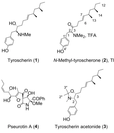
Two new tyroscherin derivatives: N-methyl-tyroscherone (2) and tyroscherin N,O-acetonide
(3), as well as two known secondary metabolites tyroscherin (1) and pseurotin A (4), were
isolated from a solid culture of the termite-borne fungus Pseudallescheria boydii SNB-CN85.
The structures were elucidated by spectroscopic analysis and chemical modification.
Compound 3 was synthesized from tyroscherin (1) in the free amine form by heating in
deuterated acetone, followed by deuterium-proton exchange in MeOH. All compounds were
tested for their antimicrobial activities on C. albicans and S. aureus, and 2 and 3 exhibited
minimal inhibitory concentrations between 128 and 8 g.mL-1.
Keywords:
The exploration of secondary metabolites involved in microbial-host interactions has
emerged as a successful strategy to identify novel chemical entities and to shed light on the
ecology and evolution of defensive association (Beemelmanns et al., 2016; Cantley and
Clardy, 2015).These associations are well described in social insects which include ants,
bees, termites and wasps, although termites are somewhat less studied than others (Carr et al.,
2012; Matsui et al., 2012; Nirma et al., 2015a, 2015b, 2013; Nowak et al., 2010; Sorres et al.,
2017; Yan et al., 2011). Indeed, termites microorganisms interactions have been explored
mainly to study trophobioses (Carr et al., 2012; Nowak et al., 2010; Yan et al., 2011). Our
previous work on Pseudallescheria boydii SNB-CN73 strain had demonstrated that this
fungus produced two antimicrobial compounds tyroscherin and N-methyl-tyroscherin along
with ovalicin and several analogs of these metabolites (Nirma et al., 2013). Interestingly, the
random isolation work of entomogenous microbes that was conducted in 2011 yielded six P.
boydii strains from three different termite nests located in somewhat distant places in French
Guiana. After our work on SNB-CN73, it was relevant to investigate other strains see
whether or not these strains also produce tyroscherin and analogs.
SNB-CN85 is one of these P. boydii strains. It was isolated from host Termes cf. hispaniolae,
whereas SNB-CN73 was isolated from Nasutitermes corniger. In addition to being from
different termite species, both nests were located 3 km apart from each other as the crow
flies. Technically, SNB-CN85 was isolated from a surface-sterilized worker placed in a Petri
dish containing a solid potato dextrose agar medium. It was identified by amplification and
sequencing of the nuclear ribosomal internal transcribed spacer region ITS4 and comparison
with NCBI database of sequences. The EtOAc extract of a solid culture of P. boydii
investigation of this extract led to the isolation of tyroscherin (1) (Hayakawa Y, Yamashita T,
Mori T, Nagai K, Shin-Ya K, 2004; Katsuta et al., 2008; Nirma et al., 2013) and pseurotin A
(4) (Bloch and Tamm, 1981; Hayashi et al., 2003) along with two new tyroscherin analogs
N-methyl-tyroscherone (2) and tyroscherin N,O-acetonide (3).
Fig. 1. Structures of compounds 1-4 with configurations presented as absolute
Compound 2 TFA salt was obtained as white amorphous powder. HRESIMS analysis
indicated a molecular formula of C22H35NO2 (m/z for 346.2749 for [M+H]+), implying 6
degrees of unsaturation, i.e., one more than for N-methyl-tyroscherin with the same number
of carbons. Preliminary inspection of the 1H and 13C NMR spectra, along with the analysis of
the HSQC correlations confirmed that compound 2 was a tyroscherin analog (Table 1). The
main difference between these compounds was for 1H and 13C chemical shifts in the C-2–C-4
in 2. Also, 13C NMR spectrum of 2 displayed a carbonyl signal at C 207.7 ppm, and we also
observed the lack of the carbinol at C 68.7 ppm in tyroscherin. In HMBC, methylene protons
in position 1 at H 3.37 and 2.96 ppm, methylene protons in position 4 at H 2.37 and 2.00
ppm, and the methine proton at H 4.45 ppm all correlated with this carbonyl. Consequently,
it was clear that compound 2 had a carbonyl in position 3. The complete proton assignment of
compound 2 was deduced from the careful examination of COSY, HSQC and HMBC
correlations. However, although the overall integration of protons was correct, it was noticed
that many signals in the 1H and 13C NMR experiments were split. Attempts to separate two
different compounds by HPLC failed. Since all NMR data were almost identical, we assumed
that the position 2 in compound 2 may in fact epimerize due to the presence of the carbonyl
group at C-3, yielding a mixture of epimers. Treatment of 2 with NaOD in CD3OD at room
temperature provided N-methyl-tyroscherone-2-d (2-d) with 69% conversion (as measured by
1H NMR by integration of the residual H-2 proton signal), confirming that carbon C-2 is most
prone to epimerization. We concluded then that compound 2 was isolated in the form of a
mixture of 2R and 2S epimers. Biosynthetic considerations would provide the configurations
of other asymmetric centres as being identical to those observed for tyroscherin (8R and
10R). Compound 2 was named N-methyl-tyroscherone from a putative oxo analog of
tyroscherin.
The molecular formula of 3 was determined to be C24H39NO2 by HRESIMS (m/z 374.3046
for [M+H]+), indicating the presence of three additional carbons compared to the molecular
formula of 1. Compound 3 was originally isolated as TFA salt, and was converted to the free
base for 1D and 2D NMR analyses. The 1H NMR spectra of compounds 1 and 3 presented
compound 3 had para-substituted aromatic systems characteristic signals at H 7.06 (d, J =
8.6 Hz, 2H) and H 6.71 (d, J = 8.6 Hz, 2H). It also had the tyroscherin series methyl signals
H 0.90 (d, J = 6.7 Hz, 3H), 0.86 (t, J = 7.3 Hz, 3H), and 0.81 (d, J = 6.5 Hz, 3H), ethylenic
protons at H 5.31 and H 5.10, a carbinol at H 3.92, and a proton to a nitrogen at H 3.23.
On the other hand, the remarkable difference between compounds 1 and 3 in 1H NMR was
the presence of 2 additional singlet signals integrating for 3H at H 1.33 and 1.14 in
Table 1
NMR spectroscopic data for N-methyl-tyroscherone (2) TFA salt (two diastereoisomers 1:1) and tyroscherin acetonide (3) at 500 MHz in CD3OD.
N-Methyl-tyroscherone (2) Tyroscherin N,O-acetonide (3)
position C, type H (J in Hz) COSY HMBCa C H (J in Hz) COSY HMBCa
1 34.33/34.28, CH2 a: 3.361/3.365, dd (13.7, 5.8) 1b, 2 2, 3, 1’, 2’ 34.4, CH2 a: 2.83, dd (14.3, 5.6) 1b, 2 2, 3, 1’, 2’ b: 2.956/2.964, dd (13.7, 9.7) 1a, 2 2, 3, 1’, 2’ b: 2.57, dd (14.3, 9.1) 1a, 2 2, 3, 1’, 2’ 2 74.2, CH 4.445/4.452, dd (9.7, 5.8) 1a, 1b 1, 3, 1’ 66.8, CH 3.23, ddd (9.1, 7.0, 5.6) 1a, 1b, 3 1 3 207.75/207.68, C 77.6, CH 3.92, ddd (10.3, 7.0, 3.1) 2, 4 4 44.9/44.8, CH2 a: 2.37, m 4b, 5a, 5b 3, 5, 6 32.0, CH2 1.55, m 3, 5a 3, 6 b: 2.00, m 4a, 5a, 5b 3, 5, 6 5 26.7, CH2 a: 2.12, m 4a, 4b, 5b, 6 3, 4, 6, 7 29.5, CH2 a: 2.12, m 4, 5b 4, 6, 7
b: 1.99, m 4a, 4b, 5a, 6 3, 4, 6, 7 b: 1.93, m 4, 5a, 6 4, 6, 7
6 127.3, CH 5.17, m 5a, 5b, 7 4, 5, 7, 8 137.7, CH 5.31, m 5a, 5b, 7 5, 8 7 139.21/139.18, CH 5.14, m 6, 8 5, 6, 8, 9, 13 129.0, CH 5.10, ddt (15.3, 8.4, 1.4) 6, 8 5 8 35.82/35.79, CH 2.11, m 7, 9a, 9b, 13 6, 7 35.7, CH 2.10, m 9 45.59/45.58, CH2 a: 1.21, m 8, 9b, 10 7, 8, 10, 11, 13, 14 45.5, CH2 a: 1.21, m 9b b: 0.97, ddd (13.5 8.6 5.0) 8, 9a, 10 7, 8, 10, 11, 13, 14 b: 0.97, m 9a, 10 7, 8, 10, 11 10 33.3, CH 1.26, m 32.9, CH 1.30, m 14 11 31.3, CH2 a: 1.29, m 11b, 12 31.2, CH2 a: 1.27, m 12 b: 1.13, m 12, 11a 9, 10, 12, 14 b: 1.14, m 12 12 11.81/11.79, CH3 0.85/0.86, t (7.3) 11a, 11b 10, 11 11.6, CH3 0.86, t (7.3) 11a, 11b 10, 11 13 22.32/22.29, CH3 0.898/0.899, d (6.7) 8 7, 8, 9 22.4, CH3 0.90, d (6.7) 8 7, 8, 9 14 19.44/19.41, CH3 0.804/0.806, d (6.5) 10 9, 10, 11 19.3, CH3 0.81, d (6.5) 10 9, 10, 11 1’ 125.43/125.40, C 130.6, C 2’/6’ 131.7, CH 7.10, bd (8.7) 3’/5’ 1, 4’, 2’/6’ 130.7, CH 7.06, d (8.6) 3’/5’ 1, 4’, 2’/6’ 3’/5’ 117.3, CH 6.80, bd 2’/6’ 1’, 4’, 3’/5’ 116.0, CH 6.71, d (8.6) 2’/6’ 1’, 4’, 3’/5’ 4’ 158.7, C 156.5, C NMe 42.6 (broad) , CH3 2.90, bs 2 34.5, CH3 2.20, s 2, 1’’ 1’’ 95.6, C 2’’ 26.8, CH3 1.33, s 1’’ 3’’ 20.0, CH3 1.14, s 1’’, 2’’
These two additional methyls were in geminal position, both linked to C1” as witnessed
by HMBC. The correlation between the NMe and C1” as well as the C1” chemical shift
at C 95.6 confirmed the existence of an oxazolidine ring including carbons C2, C3 and
C1”. Compound 3 was named tyroscherin N,O-acetonide. Its presence was confirmed in
the HPLC profile of the crude extract, and we therefore assumed that this compound did
not originate from an unwanted addition of tyroscherin onto acetone after extraction (no
acetone was used in the process).
NMR spectral data of 3 were recorded in CD3OD for 12 hours. Interestingly, after this
period, we observed that both methyl signals at H 1.33 (H2”) and 1.14 ppm (H3”) had
disappeared in 1H NMR. Deuteration had occurred in these positions yielding 3-d6, most
likely via a transient acyclic enamine resulting from the opening of the oxazolidine ring
(Scheme 1). Besides, deuteriums were exchanged back to protons in anhydrous
methanol. Also, after a long time in solution (~1 year), it was found that compound 3 had
been converted into tyroscherin, therefore confirming the absolute configuration of 3 as
2S, 3R, 8R, 10R. Further confirmation was obtained by reacting tyroscherin with
acetone-d6 under reflux for 50 hours (Scheme 1). 3-d6 was obtained and was converted in 3 in
methanol. The overall yield was 88%. The NMR data for 3-d6 and 3 obtained with this
process were identical to those of natural 3 and of 3-d6 obtained by deuteration of natural
3. Note that acetonides are rather uncommon in Nature (Shao et al., 2011; Yu et al.,
2016). Compound 3 was isolated without using acetone, neither for extraction, nor for
purification. Also, condensation of tyroscherin with acetone-d6 did not occur at room
temperature. Fungal cultures can produce acetone (Scotter et al., 2005), which we
should therefore be considered as a genuine natural product although the condensation
may not have been enzyme-catalyzed.
Scheme 1
The antimicrobial potential of compounds 1-4 was evaluated against human pathogens
Staphylococcus aureus and Candida albicans (Table 2). In accordance with the literature,
tyroscherin (1) was strongly active on both pathogens, and pseurotin A (4) was inactive
(Lu et al., 2014; Nirma et al., 2013). Interestingly, both tyroscherin analogs were
significantly active, with minimal inhibitory concentrations of 8 µg.mL-1 for 2 on S.
aureus, and 16 and 8 µg.mL-1 for 3 on C. albicans and S. aureus, respectively. In
addition, compounds 1-3 showed a moderate cytotoxicity towards MRC5 cells with IC50
in the 20-50 µg.mL-1 range.
Table 2. Antimicrobial activity of compounds 1-4
MIC (g.mL-1)
Molecule C. albicansa S. aureusb
Tyroscherin (1) 16 8 N-Methyl-tyroscherone (2) 128 8 Tyroscherin N,O-acetonide (3) 16 8 Pseurotin A (4) > 128 > 128 Oxacillin - 4 Fluconazole 4 -
aCandida albicans ATCC10213
b Staphylococcus aureus ATCC 29213
for the first time. These compounds were isolated along with pseurotin A and tyroscherin
from the termite-borne fungus Pseudallescheria boydii SNB-CN85. Their relative and
absolute configurations were determined by chemical modifications and comparison with
tyroscherin. Along with our previous report,(Nirma et al., 2013) this article further
demonstrates that tyroscherin-producing Pseudallescheria fungal strains can be found in
several termite nests in French Guiana. The exact occurrence of Pseudallescheria sp. and
the ability of this strain to deliver tyroscherin to its host termites are currently being
investigated.
Acknowledgment
This work has benefited from an "Investissement d’Avenir" grant of the Agence
Nationale de la Recherche (CEBA, ref. ANR-10-LABX-0025, France).
Appendix A. Supplementary data
Supplementary data associated with this article include experimental information, full
spectroscopic data, and NMR spectra of compounds 1-4. It can be found at http://
References
Beemelmanns, C., Guo, H., Rischer, M., Poulsen, M., 2016. Natural products from
microbes associated with insects. Beilstein J. Org. Chem. 12, 314–327.
Bloch, P., Tamm, C., 1981. Isolation and structure of pseurotin A, a microbial metabolite
Chim. Acta 64, 304–315.
Cantley, A.M., Clardy, J., 2015. Animals in a bacterial world: opportunities for chemical
ecology. Nat. Prod. Rep. 32, 888–892.
Carr, G., Poulsen, M., Klassen, J.L., Hou, Y., Wyche, T.P., Bugni, T.S., Currie, C.R.,
Clardy, J., 2012. Microtermolides A and B from termite-associated streptomyces sp.
and structural revision of vinylamycin. Org. Lett. 14, 2822–2825.
Hayakawa Y, Yamashita T, Mori T, Nagai K, Shin-Ya K, W.H., 2004. Structure of
tyroscherin, an antitumor antibiotic against IGF-1-dependent cells from
Pseudallescheria sp. J. Antibiot. 57, 634–638.
Hayashi, Y., Shoji, M., Yamaguchi, S., Mukaiyama, T., Yamaguchi, J., Kakeya, H.,
Osada, H., 2003. Asymmetric total synthesis of pseurotin A. Org. Lett. 5, 2287–
2290.
Katsuta, R., Shibata, C., Ishigami, K., Watanabe, H., Kitahara, T., 2008. Synthesis and
structure revision of tyroscherin, a growth inhibitor of IGF-1-dependent tumor cells.
Tetrahedron Lett. 49, 7042–7045.
Lu, Q.-Q., Tian, J.-M., Wei, J., Gao, J.-M., 2014. Bioactive metabolites from the mycelia
of the basidiomycete Hericium erinaceum. Nat. Prod. Res. 28, 1288–1292.
Matsui, T., Tanaka, J., Namihira, T., Shinzato, N., 2012. Antibiotics production by an
actinomycete isolated from the termite gut. J. Basic Microbiol. 52, 731–735.
Nirma, C., Eparvier, V., Stien, D., 2015a. Antibacterial ilicicolinic Acids C and D and
ilicicolinal from Neonectria discophora SNB-CN63 isolated from a termite nest. J.
Nat. Prod. 78, 159–162.
Nirma, C., Eparvier, V., Stien, D., 2015b. Reactivation of antibiosis in the entomogenous
fungus Chrysoporthe sp. SNB-CN74. J. Antibiot. 68, 586–590.
SNB-CN73 isolated from a Nasutitermes sp. termite. J. Nat. Prod. 76, 988–991.
Nowak, M.A., Tarnita, C.E., Wilson, E.O., 2010. The evolution of eusociality. Nature
466, 1057–1062.
Scotter, J.M., Langford, V.S., Wilson, P.F., McEwan, M.J., Chambers, S.T., 2005.
Real-time detection of common microbial volatile organic compounds from medically
important fungi by Selected Ion Flow Tube-Mass Spectrometry (SIFT-MS). J.
Microbiol. Methods 63, 127–134.
Shao, C.L., Wu, H.X., Wang, C.Y., Liu, Q.A., Xu, Y., Wei, M.Y., Qian, P.Y., Gu, Y.C.,
Zheng, C.J., She, Z.G., Lin, Y.C., 2011. Potent antifouling resorcylic acid lactones
from the gorgonian-derived fungus Cochliobolus lunatus. J. Nat. Prod. 74, 629–633.
Sorres, J., Nirma, C., Eparvier, V., Stien, D., 2017. Pseudallicins A-D, Four Complex
Ovalicin Derivatives from Pseudallescheria boydii SNB-CN85. Org. Lett. 19,
3978–3981.
Yan, S., Li, S., Wu, W., Zhao, F., Bao, L., Ding, R., Gao, H., Wen, H.A., Song, F., Liu,
H.W., 2011. Terpenoid and phenolic metabolites from the fungus Xylaria sp.
associated with termite nests. Chem. Biodivers. 8, 1689–1700.
Yu, J.S., Moon, E., Choi, S.U., Kim, K.H., 2016. Asarotonide, a new phenylpropanoid
with a rare natural acetonide group from the rhizomes of Acorus gramineus.