Assessment of Magic Angle Spinning Spectroscopy for Studying Migration in Solid Milk Chocolate
by
Dwight McCoy Chambers
SUBMITTED TO THE DEPARTMENT OF NUCLEAR SCIENCE AND ENGINEERING IN PARTIAL FULFULLMENT OF THE REQUIREMENTS FOR THE DEGREE OF
BACHELORS OF SCIENCE IN NUCLEAR SCIENCE AND ENGINEERING AT THE
MASSACHUSETTS INSTITUE OF TECHNOLOGY JUNE 2007
© Dwight McCoy Chambers. All rights reserved. The author hereby grants to MIT permission to reproduce
and to distribute publicly paper and electronic copies of this thesis document in whole or in part
in any medium now known or hereafter created.
Signature of Author:
Signature of Author: rtm t of Nuclear Science and Engineering
/"-7/
May 21, 2007
Certified by:
David Cory Professor of Nuclear Science an ngineering Theh ' Supervisor
Accepted by:
David Cory ef f Nuclear Science and Engineering Chairman, NSE Committee for Undergraduate Students MASAcHuSS I NSTITU
OF
TECHNOLOGy
OCT 12 2007
ARCHIVES
Assessment of Magic Angle Spinning Spectroscopy for Studying Migration in Solid Milk Chocolate
by
Dwight McCoy Chambers
Submitted to the Department of Nuclear Science and Engineering on May 21, 2007 In Partial Fulfillment of the Requirements for the Degree of
Bachelor of Science in Nuclear Science and Engineering
Abstract
In the confectionery industry, there is considerable interest in understanding the mechanisms of liquid lipid migration inside chocolates as it relates to combating fat-bloom defects in confectionery products. [1] This thesis investigates the ability of MAS NMR spectroscopy to adequately resolve chocolate spectra and deliver diffusion data specific to individual chemical species. Using a Bruker 300 MHz spectrometer equipped with liquid state and hr-MAS probes various spectroscopic characteristics of a milk chocolate sample were recorded. The MAS spectrometer resolved the chocolate spectrum into individual chemical signals and demonstrated multi-compartment diffusion behavior.
Thesis Supervisor: David Cory
Table of Contents
Abstract 2
List of Figures 4
List of Tables 5
1. Introduction 6
1-1. NMR Spectroscopy of Chocolates and the Fat Blooming Phenomena 6 1-2. Magic Angle Spinning (MAS) Spectroscopy of Solids in NMR 7 1-3. Stimulated Echo and its Application to Diffusion Measurements 8
2. Methodology 10
2-1. Sample Preparation 10
2-2. T1, T2, and Diffusion NMR Experiments 10
2-3. MAS NMR Experiments 12
3. Data Analysis and Results 14
3-1. Gross Sample Relaxation Characteristics and Diffusion Behavior 14
3-2. MAS NMR Spectrum and Diffusion Measurements 16
4. Discussion 19
5. Conclusions 20
References 21
List of Figures
Figure 1: Cartoon of chocolate 6
Figure 2: Visual comparison of normal milk chocolate and milk chocolate with fat bloom 6
Figure 3: Illustration of sample in MAS configuration 8
Figure 4: Generalized Stimulated Echo with Continuous Gradient 9
Figure 5: Inversion recovery sequence 10
Figure 6: CPMG sequence 11
Figure 7: Stimulated echo sequence 12
Figure 8: MAS stimulated echo sequence 13
Figure 9: Plot of Z(t) versus the D2 delay from the inversion recovery sequence 15 Figure 10: Plot of Max Observed Signal versus Total Relaxation Time 15 Figure 11: Observed Signal versus Gradient Strength for Gross Diffusion Behavior 16
Figure 12: NMR Spectrum of the Stationary Sample 17
Figure 13: Chocolate MAS Spectrum at 3 kHz 17
Figure 14: MAS Diffusion Spectrum with 10% MA Gradient 18
Figure 15: MAS Diffusion Spectrum with 20% MA Gradient 18
List of Tables
Table 1:Inversion Recovery Experiment Parameters 11
Table 2: CPMG Experiment Parameters 11
Table 3:Stimulated Echo Experiment Parameters 12
Table 4:MAS Stimulated Echo Experiment Parameters 13
1. Introduction
1-1. NMR Spectroscopy of Chocolates and the Fat Blooming Phenomena
Chocolate is a complex composite food composed of a matrix of cocoa, sugar and milk awash in a sea of liquid fats. [1] The behavior of this composite (depicted in the cartoon in Figure 1) is extremely sensitive to many factors including processing techniques, temperature, and the proximity of foreign liquid fats in composite foods. [1, 2]
Surface bloom Crack
,f
Q Interparticle pore
Cocoa solids
LII
Crystalline sugar C> Fat crystal[77 Cocoa butter [. ] Milk powder - Lecithin
Figure 1: Cartoon of chocolate [1]
Many food scientists and companies have studied chocolate using NMR spectroscopy, and a significant portion of this research has been dedicated to studying the migrations of lipids inside chocolate. [1-3] This research is often motivated by a desire to understand the underlying mechanisms causing "fat-blooms" -a chocolate blight in which lipids migrate to the surface of the chocolate,
causing a white discoloration and poor taste. [4] An example of fat bloom can be seen on the surface of the right chocolate bar in Figure 2.
Figure 2: Visual comparison of normal milk chocolate and milk chocolate with fat bloom [4]
~an 1CI. ~: ~d~B ,,~&f~B~I~ '' '"` '""' i ~ag~i~ ~~-s
Initially, researchers tried to explain lipid migratory behavior solely in terms of molecular diffusion. Now, there is an emerging view of the mechanistic foundations of fat-blooming in which the phenomena is a combination of molecular diffusion and capillary action. [1, 3] In order to further clarify the matter, it may be helpful to scientists to be able to more accurately track the migration of individual chemical species through chocolates. This thesis will examine the suitability of using MAS NMR spectroscopy in order to study the migratory behavior of individual chemical species in
chocolates. Specifically, this study will assess whether MAS NMR techniques can accurately resolve a chocolate spectrum with enough sensitivity so as to pick out individual species migratory behavior.
1-2. Magic Angle Spinning (MAS) Spectroscopy of Solids in NMR
MAS spectroscopy is a technique used in NMR to acquire high resolution spectra in solid state systems. [5-6] In all media, there are dipole interactions between nuclei. In a general magnetic field, the dipole interaction between any two nuclei, i and j, at a distance R from each other, and where the vector R is at an angle 0 to the magnetic field, is proportional to:
(1)(3*cos(O)2-1) (Equ. 1) [6]
In many solutions, the random motion and rotational freedom on the nuclei cancel out any net dipole interactions for the solution. However, in solid state systems, the nuclei are often much more constrained both translationally and rotationally. The presence of dipole interactions generally leads to broad line widths in NMR spectroscopy. [5,6]
Many of the interactions averaged out by MAS in composite systems such as chocolate arise from the inherent heterogeneity of bulk magnetic susceptibility in the sample. This is a reflection of the fundamental compositional heterogeneity and the differing magnetic susceptibilities of each
individual component of the sample composite. [9] The effective magnetic field is thus spatially dependent, receiving contributions from the magnet's static field, Bo, and the local field variations as shown in Equation 2:
Bef=Bo+B10o (Equ. 2) [9]
MAS spectroscopy exploits the angular dependence of this phenomena in order to address the limited motion of solid state nuclei by rapidly rotating the sample about an axis which is 54.74' with respect to the orientation of the magnetic field as shown in Figure 3. [5,6]
BO
0
RotorNMR coil
Figure 3: Illustration of sample in MAS configuration [5]
Through sufficiently rapid rotation of the sample, the average NMR frequency suppresses the dipole interactions and varying magnetic susceptibilities of the constituent parts, resulting in a solution like spectrum. [5,6] The extent of the narrowing achieved in an NMR spectrum by MAS spectroscopy is dependent on the rate of spinning of the sample. [6]
1-3. Stimulated Echo and Its Application to Diffusion Measurements
Given the desire to study diffusion behavior in chocolate, it is important to choose a pulse sequence which can adequately highlight its affect on the signal. In NMR, the attenuation of a signal by molecular diffusion is a function of essentially two properties of a pulse sequence: the amount of time the nuclei are left to diffuse (storage time) and the "steepness" of the phase encoding done by the gradient (which is to say the product of the gradient strength and the gradient's application time). [6] The stimulated echo is a simple pulse sequence which gives a researcher broad flexibility in adjusting all three of these features so as to maximize their ability to measure diffusion.
The sequence is depicted schematically in Figure 4 (Figure 4 represents a generic version of the stimulated echo sequence but all discussion related to Figure 4 will apply generically to those other
possible versions as well). The three x/2 pulses are separated by two delays, t and T. These delays represent the gradient application time and the spin storage time respectively. Also shown in Figure 4 are the 5 echoes that form as a result of this pulse sequence - 1,3,4 and 5 are simple spin echoes while 2 is the stimulated echo which occurs 2t+T after the initial pulse. [7-8]
RF
G
Figure 4: Generalized Stimulated Echo with Continuous Gradient /7]
The stimulated echo is different from the 4 other echoes because it spends the T delay along the Bo axis, and accordingly only undergoes T1 and not T2 relaxation during that period. [7-8] Since the T1
is necessarily larger than T2, these spins are substantially less attenuated upon refocusing into an echo. [6-8] This helpful feature of the sequence frees the researcher from T2 constrains which might
otherwise severely limit the possible storage time. [7-8] It is also worth noting that the research maintains broad control over the gradient strength and gradient application time in the stimulated echo sequence.
2. Methodology:
2-1. Sample Preparation
A sample of Flyer Swiss milk chocolate was acquire from a local food store. For the early NMR experiments, a portion of this sample was cut off and loaded into an NMR sample tube (Wilmad-Labglass product 504-PP-9) which was then sealed with a rubber cap. For the later MAS experiments,
a second sample was prepared from the remaining chocolate originally purchased. This sample was tightly packed into a ceramic MAS rotor (Bruker hr-MAS product B200147) with the use of a plunger so as to remove any possible voids in the chocolate. This rotor was then sealed as well.
2-2. T1, T2, and Diffusion NMR Experiments
These experiments were conducted using a 300 MHz Bruker NMR Spectrometer using a SEI 300 MHz probe in Prof. David Cory's laboratory at the Massachusetts Institute of Technology. The '
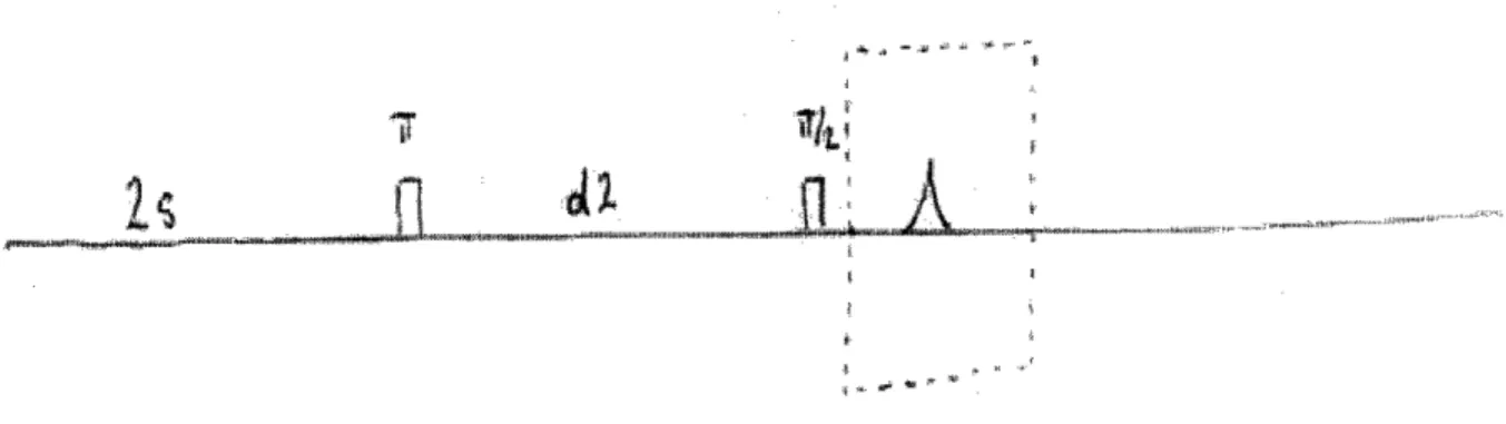
and 7T/2 pulses were determined to be 22.85 us and 11.42 us respectively. In order to measure the spin-lattice (Ti) relaxation time of the sample, an inversion recovery sequence, shown in Figure 5, was used.
Figure 5: Inversion recovery sequence
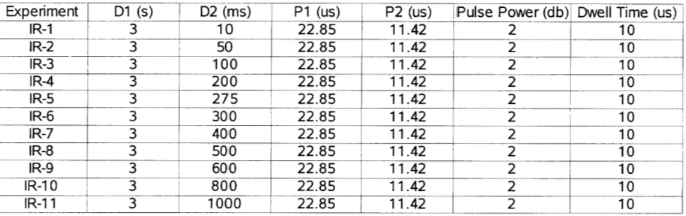
The experimental parameters for the 11 inversion recovery experiments are summarized in Table 1.
D2 (ms) P1 (us) j P2 (us) IR-1 3 IR-2 3 IR-3 3 IR-4 3 IR-5 3 IR-6 3 IR-7 3 IR-8 3 IDA i 3 10 22.85 11.42 50 22.85 11.42 100 22.85 11.42 200 22.85 11.42 275 22.85 11.42 300 22.85 11.42 400 22.85 11.42 mnn ') A 1 1 An cAAr 11OC~ A7
Pulse Power (db) Dwell Time (us)
2 10 2 10 2 10 2 10 2 10 2 10 2 "9 I3 JUV LL.03 I 1.4L L IR-10 3 800 22.85 11.42 2 IR-1 1 3 1000 22.85 11.42 2 10 10 10 10 10
Table 1: Inversion Recovery Experiment Parameters
In order to characterize the spin-spin relaxation time (T2), a CPMG sequence, which is shown generically in Figure 6, was used. The experimental parameters for the 8 CPMG experiments are summarized in Table 2. 4jL i ; Figure 6: CPMG sequence VpMG-. L Uu CPMG-1 2 100 CPMG-2 2 200 CPMG-3 2 300 CPMG-4 2 600 CPMG-5 2 900 CPMG-6 2 1000 CPMG-7 2 1500 CPMG-8 2 2000 \u1 / 11.42 11.42 11.42 11.42 11.42 11.42 11.42 11.42 P2 (us) 22.85 22.85 22.85 22.85 22.85 22.85 22.85 22.85 Pulse Power (db) 2 2 2 2 2 2 2
Table 2: CPMG Experiment Parameters
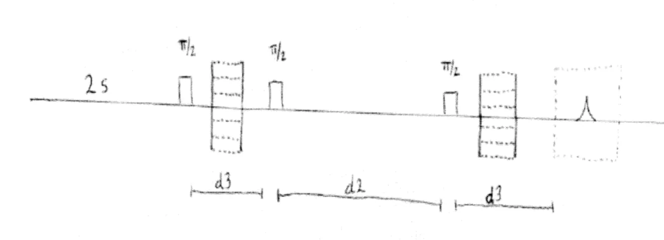
In order to assess the bulk diffusion characteristics of the sample, a stimulated echo sequence, which is shown in Figure 7, was used. The experimental parameters for the stimulated echo
experiments are reported in Table 3.
xE eriment D1 (s) D2 ( ) ~ ---- ~---Experiment D1 (s) ~a i i d2, ii
· i_..a~
--I-Figure 7: Stimulated echo sequence
Experiment D1 (s)
D-1 2
D-2 2
D-3 2
D-4 2
Table 3: Stimulated echo
D2 (us) D3 (us) 437.9 700 437.9 700 437.9 700 437.9 700 experiment parameters 2-3. MAS NMR Experiments
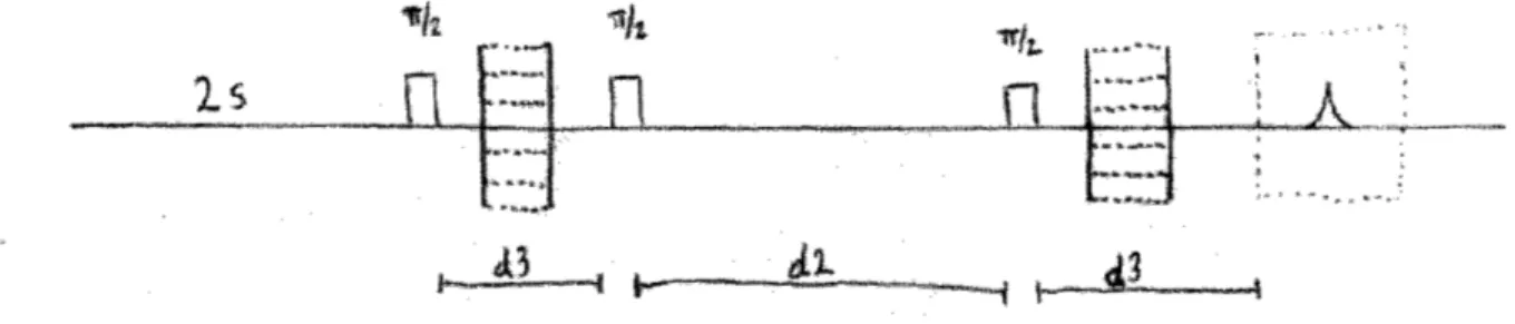
All MAS experiments were conducted using a hr-MAS Bruker probe in the same 300 MHz system as the relaxation experiments. After loading the MAS sample rotor, the sample was spun in the probe at 3 kHz. The 71 and 7/2 pulses were 9 us and 18 us, respectively. A simple spectrum of the sample was acquired by using a hard 7z/2 pulse. The MAS diffusion spectra were acquired using a modified version of the earlier stimulated echo sequence and is shown in Figure 8. The experimental parameters are reported in Table 4.
i. ,·:.··-ihr
i "
'" ~::*i')-l"* "*-re *X;;i-~· SGradient Strength (%) 20 25 35 50 20 25 ~ 35 50 ---_ ti!n;,; ·-···:-i-:;-·;I-~ :~~_2, i;
A id^j
dn
-··-1 i·-~I-~
I
Is
__Th.k tL~b
i
LA
A3
A3
Figure 8: MAS stimulated echo sequence
ExperimentA D (s) D2 (ms) D3 (ms) Gradient Strength (%)
MASD- 2 10 400 10
MASD-2 2 2 -400 10 20
Table 4: MAS stimulated echo experiment parameters '"l""nr~*~e~r~-~9-mrax(l~a~b*bsr~~arc~,.
· i
;.,,;a*,~~r,,,,,,
3. Data Analysis and Results
3-1. Gross Sample Relaxation Characteristics and Diffusion Behavior
In an inversion recovery experiment, the amplitude of the FID signal, MT1, is:
MT, (t)=Mo* (1-2+ e-ITi') (Equ. 3) [6]
where t is the delay between the initial 7c pulse and the subsequent 'T/2 pulse, and Mo is the initial magnetization at t = 0. It is possible to rewrite Equation 3 so that:
Z(t)= 1 (1-MM (t)/Mo)=et/- , (Equ. 4)
2
After fitting the experimental results, shown in Figure 9, to a simple exponential curve, the TI value can be measured directly from the fit. The measured TI value for this chocolate sample is 456
ms.
In a CPMGr sequence, amplitude of the FID signal, MT2, is a function of the total time, t, spent in plane transverse to the Bo field and the initial magnetization of the sample Mo. In the simplest case, the relationship is a simple exponential decay as shown in Equation 5:
MT2(t)= M0* e- t/T2 (Equ. 5) [6]
Using a best fit line for the experimental data shown in Figure 10, the measured T2 value for the
chocolate sample is 6.12 ms.
Finally, the signal amplitude in the pulsed gradient diffusion experiment is a function of the gradient strength, the gradient application time (D3 from Table 3) and the storage time (D2 from Table 3):
-D*G 2*D22*y2* (D3+(2)*D2)
M (G, D2, D3)= Mo *(e D 3+ D2 ) (Equ. 6) [6]
where D is the molecular diffusion coefficient of the mobile species. By taking the natural log of the observed signal's maximum, the linear best fit in Figure 11 estimates the diffusion coefficient of the mobile species in the chocolate to be 2.87 ýIm 2/ms.
Z Value versus D2 delay
0.1 0.2 0.3 0.4 0.5 0.6 0.7 0.8 0.9 1
D2 delay in seconds
Figure 9: Plot of Z(t) versus the D2 delay from the inversion recovery sequence
Max Observed Signal versus Total Relaxation Time using CPMG sequence 12000 11000 10000 9000 6000 7000 6000 5000 4000 3000 1000 2000 3000 4000 5000 6000 7000 8000 9000 10000
Total Relaxtion Time (us)
I
I
Grait 8I6W th (% of ma x mt)
Figure 11: Maximum Observed Signal versus Gradient Strength for Molecular Diffusion Behavior
3-2. MAS NMR Spectrum and Diffusion Measurements
Using the liquid state probe, the chocolate spectrum was dominated by a single peak with a full-width-half-maximum (FWHM) of 2.196 ppm. The peak, shown in Figure 12, has a generally
Lorentzian line-shape with the obvious exception of the subsumed peak on the left. At 3 kHz in the hr-MAS probe, the chocolate's spectrum breaks into 9 distinguishable peaks as can be seen in Figure 13 The FWHM of peak of the 9 peaks are listed in Table 5 with Peak 1 on the right.
Peak 1 2 3 4 5 6 7 8 9
Resonant Frequency (ppm) 0.641 1.047 1.324 1.763 1.991 2.536 3.821 4.016 5.066
Spectrum FWHM (ppm) 0.105 0.114 0.146 0.130 0.114 0.097 0.122 0.114 0.114
Diffusion (14) Spectrum FWHM (ppm) 0.181 0.249 N/A N/A N/A N/A N/A N/A 0.206
Table 5: Peak Information for MAS Spectrum and MAS Diffusion Spectrum
Figures 14 and 15 show the resulting chocolate MAS spectra using the pulse sequence from Figure 8 and the experimental parameters listed in Table 4. Despite the two-fold increase in gradient strength between trials, there is no significant change to the spectrum from additional diffusion.
Most, notably, the signal in Figure 14 is weaker, more noisy and has worse resolution. There are also prominent spinning sidebands present in Figure 13 at 11.020 ppm and -9.007 ppm which are absent in Figure 14 and 15.
x 10 Stationary Chocolate Spectrum
4.5 4 3.5 3 2.5 2 . 1.5 1 0.5 0 15 10 5 0 -5 -10 ppm
Figure 12: NMR Spectrum of the Stationary Sample
I
s15 10 5 0 -5 -10
ppm
Figure 13: Chocolate MAS Spectrum at 3 kFIz
MAS Diffusion Spectrum with 10% Magic Angle Gradient
Z
rg ;P 15 10 5 ppmFigure 14: AMAS Diffusion Spectrum with
0 -5
10% MA Gradient
MAS Diffusion pectrunm using 20% Magic Angle Gradient
15 10 5 0 -s
ppm
Figure 15: MAS Diffusion Spectrum using 20% Magic Angle
Gradient
4. Discussion
The two spectra in the Results section from the MAS and liquid state probes quite clearly illustrate the ability of MAS NMR spectroscopy to return a high resolution spectrum of a solid-state sample. MAS, even at a modest spinning rate, quite clearly reduced the chocolate spectrum from a single lumped, broad signal to its fundamental component peaks. Using the chemical shift data stored in Figure 13, it is possible to assign each peak to particular chemical species present in chocolate
-thus, fulfilling the first expectation of the MAS assembly.
In order to address the needs of researchers further exploring lipid migration in chocolate, the MAS assembly also needed to be able to study the migration behavior of individual species. The MAS spectra in Figure 13, 14 and 15 clearly report on diffusion behavior of two distinct groups within the chocolate: a liquid-like group and a solid state group. The solid state group is restricted in its mobility, while the liquid state is much less constrained. Evidence for the mobility restriction of the solid state comes from the comparison of Figures 14 and 15 in which a significant increase in gradient strength did not attenuate signal strength, indicating a population unable to diffuse.
The apparent mobility of the liquid like spins can be seen both in the presence of spinning sidebands in Figure 13 [9], as well as the contrast between the smooth sharp peaks in Figure 13 and the broader, nosier peaks in Figure 14. Referring to Table 5, the FWHM about doubled in all cases
between Figures 13 and 14. This is most likely a reflection of the diversity of the chemical
environments seen by the stationary sample in Figure 14. Each differing environment should shift (minutely in this case) the peaks , resulting in broader, nosier peaks. While the solid state behavior is also present in Figure 13, it is masked by the presence of the liquid state population (which had been attenuated by the diffusion experiments in Figure 14). The mobility of the liquid phase spins allows them to sample many different chemical environments - averaging out this shifting and resulting in
5. Conclusions
MAS spectroscopy is capable of resolving the chocolate spectrum into its component peaks, and during investigation of diffusion behavior in the chocolate, MAS spectroscopy was able to clearly identify two unique populations within the sample. Future work in the area should focus on identifying potential species involved in both a diffusion and capillary action model of fat blooming. Having developed theoretical models, scientists could use the species specific migration data from potential future MAS NMR spectroscopy experiments to evaluate the validity of such models. Beyond
information on the behavior of individual species, the liquid state population, in particular, may also be a source of valuable information about the characteristic spaces inside the chocolate. Information about the geometry of the chocolate matrix may also help researchers refine their models.
References
[1] Aguilera, J.M., Michel, M., and G. Mayor, "Fat Migration in Chocolate: Diffusion or Capillary Flow in a Particulate Solid?- A Hypothesis Paper," Journal of Food Science, Vol. 69, Nr. 7, pp.
167-174, 2004.
[2] Walter, Peggy and Paul Cornillon, "Lipid migration in two-phase chocolate systems investigated by NMR and DSC," Food Research International, Volume 35, pp 761-767, 2002.
[3] Guiheneuf, T.M., Couzens, P. J, Wille, H. J., and L. Hall, "Visualization of Liquid Triacylglycerol Migration in Chocolate by Magnetic Resonance Imaging," Journal of the Science of Food and
Agriculture, Vol. 73, Issue 3, pp. 265-273, 1999.
[4] R. Peschar, M.M. Pop, D.J.A. De Ridder, J.B. Van Mechelen, R.A.J. Driessen, H. Schenk,
"Structure of Chocolate Clarified with Synchrotron Powder Diffraction Data," J. Phys. Chem. B., Vol. 108, Nr. 40, pp. 15450-15453, 2004.
[5] Jeschke, G. "High-Resolution Solid-State NMR: Magic Angle Sample Spinning and Cross Polarization," Lecture Notes: www.mpip-mainz.mpg.de/-~jeschke/lect9.pdf, 2004.
[6] Fukushima, Elichi and Stephen B.W. Roeder, Experimental Pulse NMR: a Nuts and Bolts Approach, Addison-Wesley Publishing Company: Reading, MA, 1993.
[7] Sodickson, A and David Cory, "A generalized k-space formalism for treating spatial aspects of a variety of NMR experiments," Progress in Nuclear Magnetic Resonance Spectroscopy, Vol. 33, pp. 77-108, 1998.
[8] Cho, Z.-H., Jones, J. P., and Manbir Singh, Foundations of Medical Imaging, John Wiley & Sons, Inc.: New York, 1993.
[9] Liu, Y., Gabriela, L., Singer, S., Cory, D., and Pabitra Sen, "Manipulation of phase and amplitude modulation of spin magnetization in magic angle spinning nuclear magnetic resonance in the presence of molecular diffusion," Journal of Chemical Physics, Vol 114, Nr. 13, 2001.
Acknowledgments
The author would like to thank the following people: Prof. Cory time and effort spent advising me during this project and for the use of your lab equipment; Sekhar for the sound advice, the technical assistance with the probes and the Nuts and Bolts; Jonathan, for his help with all things Athena and printing, Troy for a through introduction to experimental NMR, and Michael for patent advice,
catching typos in the first line of my abstract and laughing at my numerous chocolate jokes which sadly never made it into this thesis.
![Figure 2: Visual comparison of normal milk chocolate and milk chocolate with fat bloom [4]](https://thumb-eu.123doks.com/thumbv2/123doknet/14107553.466235/6.918.338.569.879.1041/figure-visual-comparison-normal-milk-chocolate-chocolate-bloom.webp)
![Figure 4: Generalized Stimulated Echo with Continuous Gradient /7]](https://thumb-eu.123doks.com/thumbv2/123doknet/14107553.466235/9.918.170.757.282.446/figure-generalized-stimulated-echo-continuous-gradient.webp)