HAL Id: tel-01228252
https://tel.archives-ouvertes.fr/tel-01228252
Submitted on 12 Nov 2015
HAL is a multi-disciplinary open access
archive for the deposit and dissemination of sci-entific research documents, whether they are pub-lished or not. The documents may come from teaching and research institutions in France or abroad, or from public or private research centers.
L’archive ouverte pluridisciplinaire HAL, est destinée au dépôt et à la diffusion de documents scientifiques de niveau recherche, publiés ou non, émanant des établissements d’enseignement et de recherche français ou étrangers, des laboratoires publics ou privés.
The endothelial dysfunction in portal hypertension : role
of the oxidative stress and angiotensin system
Sherzad Khorsheed Rashid
To cite this version:
Sherzad Khorsheed Rashid. The endothelial dysfunction in portal hypertension : role of the oxidative stress and angiotensin system. Cardiology and cardiovascular system. Université de Strasbourg, 2014. English. �NNT : 2014STRAJ096�. �tel-01228252�
UNIVERSITE DE STRASBOURG
Ecole Doctorale des sciences de la Vie et de la Santé
Faculté de Pharmacie – Equipe Pharmarmacologie et Physiopathologie Cardiovasculaires
UMR CNRS 7213- Laboratoire de Biophotonique et Pharmacologie
THESE
Présentée pour l’obtention du grade de
DOCTEUR DE L’UNIVERSITE DE STRASBOURG
Discipline : Sciences pharmaceutiques
Spécialité : Pharmacologie
THE ENDOTHELIAL DYSFUNCTION IN PORTAL HYPERTENSION:
ROLE OF THE OXIDATIVE STRESS AND ANGIOTENSIN SYSTEM
Par Sherzad Khorsheed RASHID
Soutenue publiquement le 18 Juin 2014 devant le jury compose de:
Professor Josiane Cillard Rapporteur externe
Professor Dinh-Xuan Rapporteur externe
Professor Frédéric de Blay Rapporteur interne Professor Valérie B.SCHINI-KERTH Directeur de thése Dr. Monique Oswald-Mammosser Co-directeur de thése
TO ASLAN, YASSER, TAQWA AND MY BEST BELOVER; My WIFE
I DEDICATE THIS WORK
!"# $ % & % ' & ' ( $ ( & ( ' ( ) * + , * && ( ( & - ( ( & ' ' ' + % & ( ( & * ( . $ % ' % ( % (
& / .") #0 ' ' $ & 1 "023 &
( &
) * & 04 , & %
( % ( (
5 & ( ' & &% %6
) 0.7!" - 8' % !& && 4 7 9 9 0 0& ' ' ' 8 " % * !# ! ! !44,.* )" ,):0 !$ 40 , "!##! ", !" ' ,44!# : , 4!! & &% % .*" ;<=>
( & & & 6 & & ' &
& & % ' & 6 & %
% ' 0 <?=@ ?A =A
Publications issues de ce travail
Publications
1. Sherzad K. Rashid, Noureddine Idris Khodja, Cyril Auger, Mahmoud Alhosin, Nelly Boehm, Monique Oswald-Mammosser and Valérie B Schini-Kerth. Blackcurrant juice prevents endothelial dysfunction and vascular oxidative stress in the mesenteric artery of rats with portal hypertension, soumis pour publication.
2. Sherzad K. Rashid, Noureddine Idris-Khodja, Cyril Auger, Mahmoud Alhosin, Nelly Boehm, Monique Oswald-Mammosser and Valérie B. Schini-Kerth. Probiotics (VSL#3) prevent endothelial dysfunction in rats with portal hypertension: role of the angiotensin system. En press PLOS ONE.
3. Oswald-Mammosser Monique, Rashid Sherzad K., Boehm Nelly, Agin Arnaud,
Geny Bernard, Schini-Kerth Valérie, Charloux Anne. Effect of Fulvestrant,an estrogen receptor antagonist, on the cirrhotic rat lung, soumis pour publication
4. Amissi Said, Boisramé-Helms Julie, Burban Mélanie, Rashid Sherzad K., Cyril Auger, Florence Toti, Ferhat Meziani, Valérie B. Schini-Kerth. Lipid emulsions used in parenteral nutrition induce endothelial dysfunction in porcine coronary artery rings: Role of oxidative stress and cyclooxygenase-derived vasoconstrictors, En preparation 5. Mahmoud Alhosin, Eric Anselm, Sherzad K. Rashid, Jong Hun Kim, Socorro
Vanesca Frota Madeira, Christian Bronner, and Valérie B. Schini-Kerth. Redox-Sensitive Up-Regulation of eNOS by Purple Grape Juice in Endothelial Cells: Role of PI3-Kinase/Akt, p38MAPK, JNK, FoxO1 and FoxO3a. 2013;8(3):e57883. doi: 10.1371/journal.pone.0057883. Epub 2013 Mar 22.
6. Faraj Zgheel, Mahmoud Alhosin, Sherzad K. Rashid, Cyril Auger and Valérie B.Schini-Kerth. The highly purified EPA:DHA 6:1 product-evoked endothelium-dependent NO-mediated relaxation in the coronary artery involves a copper-endothelium-dependent event triggering the redox-sensitive PI3-kinase/Akt pathway to activate eNOS by phosphorylation at Ser 1177, soumis pour publication.
7. Mahmoud Alhosin, Israa Dandache, Agnès Lelay, Sherzad K. Rashid, Luc-Matthieu Fornecker, Laurent Mauvieux, Raoul Herbrecht and Valérie B. Schini-Kerth. Bilberry
extract (Antho 50) selectively induces a redox sensitive caspase 3-related apoptosis in chronic lymphocytic leukemia cells through targeting Bcl-2, soumis pour publication
Présentations
-Posters-
1. Sherzad K. Rashid, Noureddine Idris-Khodja, Cyril Auger, Mahmoud Alhosin, Nelly Boehm, Monique Oswald-Mammosser and Valérie B. Schini-Kerth. The probiotic VSL#3 prevents endothelial dysfunction in the mesenteric artery of cirrhotic rats with hepatopulmonary syndrome.5th International Symposium Nutrition, Oxygen Biology and Medicine, P40, 5 - 7 June 2013, Paris, France.
2. Sherzad K. Rashid, Noureddine Idris-Khodja, Cyril Auger, Mahmoud Alhosin, Nelly Boehm, Monique Oswald-Mammosser and Valérie B. Schini-Kerth. Blackcurrant juice prevents endothelial dysfunction and vascular oxidative stress in the mesenteric artery of cirrhotic rats with hepatopulmonary syndrome.5th International Symposium Nutrition, Oxygen Biology and Medicine, P28, 5 - 7 June 2013, Paris, France.
3. Sherzad K. Rashid, Noureddine Idris-Khodja, Cyril Auger , Mahmoud Alhosin, Nelly Boehm, Monique Oswald-Mammosser and Valérie B. Schini-Kerth. Oral intake of blackcurrant juice prevents endothelial dysfunction in the mesenteric artery of cirrhotic rats with portal hypertension.MOVD 2013, 11th International Symposium on Mechanisms of Vasodilatation, P2/2, 4-6 October 2013 University hospital Zurich.
4. Faraj Zgheel, Mahmoud Alhosin, Sherzad K. Rashid, Cyril Auger and Valérie B. Schini-Kerth. The NO-mediated relaxation induced by highly purified EPA: DHA 6:1 product involves a copper-dependent redox-sensitive activation of the PI3-Kinase/Akt pathway leading to eNOS activation.11th International Symposium on Mechanisms of Vasodilatation, P1/2, 4-6 October 2013 University hospital Zurich.
5. Sherzad K. Rashid, Noureddine Idris-Khodja, Cyril Auger , Mahmoud Alhosin, Nelly Boehm, Monique Oswald-Mammosser and Valérie B. Schini-Kerth. Blackcurrant juice prevents endothelial dysfunction and vascular oxidative stress in the mesenteric
artery of cirrhotic rats with hepatopulmonary syndrome. P23, Doctoral school day 2014, Strasbourg.
6. Amissi Said, Boisramé-Helms Julie, Burban Mélanie, Sherzad K. Rashid, Cyril Auger, Florence Toti, Ferhat Meziani, Valérie B. Schini-Kerth. Lipid emulsions used in parenteral nutrition induce endothelial dysfunction in porcine coronary artery rings: Role of oxidative stress and cyclooxygenase-derived vasoconstrictors. P2, Doctoral school day 2014, Strasbourg.
7. Faraj Zgheel, Mahmoud Alhosin, Sherzad K. Rashid, Cyril Auger and Valérie B. Schini-Kerth. The EPA:DHA 6:1-evoked endothelium-dependent NO-mediated relaxation in the coronary artery involves a copper-dependent pro-oxidant response triggering the PI3-kinase/Akt-mediated activation of eNOS, P29, Doctoral school day 2014, Strasbourg.
-Communications orales –
1. Sherzad K. Rashid, Noureddine Idris Khodja, Cyril Auger, Mahmoud Alhosin, Nelly Boehm, Monique Oswald-Mammosser and Valérie B Schini-Kerth. Blackcurrant juice prevents endothelial dysfunction and vascular oxidative stress in the mesenteric artery of cirrhotic rats with hepatopulmonary syndrome, Journees Campus d'Illkirch 2012.
2. Monique Oswald-Mammosser, Sherzad K. Rashid, Nelly Boehm, Arnaud Agin, Bernard Geny, Valérie B. Schini-Kerth, Anne Charloux. Effect of fulvestrant in an experimental model of hepato-pulmonary syndrome (HPS) in rats. European Respiratory Society, Barcelone 2013.
Awards “prix”
The young investigator award, 2013 obtained from the “THE LINUS PAULING INSTITUTE” 5th International of Symposium Nutrition, Oxygen Biology and Medicine, P40, 5 - 7 June 2013, Paris, France.
ABBRAVATIONS
AA : arachidonic acids
AC : adenylyl cyclise
ACE : angiotensin converting enzyme
ACh : acetylcholine
ACE : angiotensin-converting enzyme
ADP : adenosine diphosphate
ATI : angiotensin I
AT II : angiotensin II
ATP : adenosine triphosphate
BH4 : tetrahydrobiopterin
cAMP : cyclic adenosine-3’,5’-monophosphate
CBDL : common bile duct ligation
CO : carbon monooxide
COX : cyclooxygenase
EC : endothelial cell
EDCF : endothelium-derived contracting factor
EDH : endothelial derived hyperpolarizing
EDHF : endothelium-derived hyperpolarizing factor
EDRF : endothelium-derived relaxing factor
EETs : epoxyeicosatrienoic acids
ER : estrogen receptor
ET-1 : endothelin-1
GPx : glutathione peroxidase
H2O2 : hydrogen peroxide
HO-1 : hemeoxygense-1
HPS : hepatopulmonary syndrome
IKca channels : potassium-dependant channels of intermediate-conductance iNOS : inducible NO synthase
Kca : Ca2+- sensitive K+ channels L-NA : N-nitro-L-arginine
L-NAME : L-nitro-L-arginine methylester
LPS : lipopolysaccaride
MCP-1 : monocyte chemoattractant protein-1
MGJ : myoendothelial gap junction
MnTMPyP : Mn(III) tetrakis(1-methyl-4pyridyl)porphyrin, superoxyde dismutase mimetic
MMPs : matrix metalloproteinases
NO : nitric oxide
NOS : nitric oxide synthase
nNOS : neuronal nitric oxide synthase
O2- : superoxide anions OH.- : hydroxyl groups
PDGF : platelet-derived growth factor
PEG-SOD : polyethylene glycol-superoxide dismutase
PGE2 : prostaglandins E2 PGH2 : prostaglandins H2 PGI2 : prostacyclin
PI3K : Phosphoinositide 3-kinase
PIMs : pulmonary intravascular macrophage
PKA : protein kinase A
PKC : protein kinase C
RAAS : rennin-angiotensin-aldosterone-system
ROS : reactive oxygen species
SID : selective intestinal decontamination
SKca channels : Kca channels of small-conductance sGC : soluble guanylyl cyclase
SMC : smooth muscle cell
SOD : superoxide dismutase
TGF-B1 : transforming growth factor-B1
TNF- : tumor necrosis factor-alpha
TXA2 : thrombxane A2
VEGF-A : vacular endothelial growth factor A
LIST OF THE FIGURES AND THE TABLES
FIGURESFigure 1: The three layers of normal artery.
Figure 2: NO generation from L-Arginine and its functional properties.
Figure 3: NO synthesis pathway through eNOS phosphorylation in calcium dependent and independent pathway.
Figure 4: Hypothesis describing the nature of EDHF pathway.
Figure 5: Arachidonic acid metabolism pathway.
Figure 6: The pathways of endothelial-derived relaxation.
Figure 7: The renin angiotensin system.
Figure 8: Pathways of endothelial derived contraction.
Figure 9: Overview of how portal hypertension developed.
Figure 10: Overview of factors contributing to PH initiation and it is worsening.
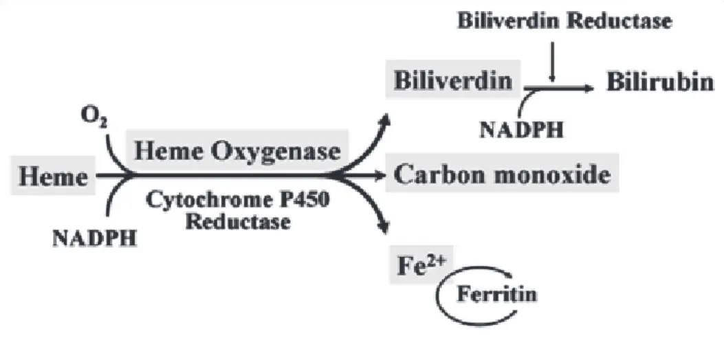
Figure 11: Production of CO by the enzymatic activity of Heme-oxygenase.
Figure 12: Potential mechanisms and target therapeutic options in hepatopulmonary syndrome (HPS) and HCS.
Figure 13: Role of angiogenesis in the pathology of HPS.
Figure 14: Strauctures of some polyphenols.
Figure 15: Schematic of pharmacokinetically relevant flavonol features pertaining to the composition in foods, abdorption, metabolism, distribution and excretion.
Figure 16: Various photographs of blackcurrants (Ribes nigrum) showing a commercial blackcurrant plantation (A), shrub with fruits (B and C), and isolated fruits (D).
Figure 17: Chemical structures of the major anthocyans present in blackcurrants.
Figure 19: Histological study of liver section using heamatoxylin and eosin staining, sample taken at the end of study; A, sham; B, CBDL
Figure 20: CBDL operation; consist of identification of the common bile duct and then put double ligature and cut between the ligature with scissor.
Figure 21: Study of the vascular reactivity.
Figure 22: Bacterial translocation and the hyperdynamic circulatory state in cirrhosis.
Figure 23: Beneficial effects of commensal and probiotic bacteria.
Figure 24: Schema presenting the effects of CBDL in rats and the therapeutic preventive effects of VSL#3 in the mesenteric arteries and both liver and spleen size.
Figure 25: Schema presenting the effects of CBDL in rats and the therapeutic preventive effects of PRBJ in the mesenteric arteries and /or aorta and both liver and spleen size
Figure 26: Schema presenting the effects of CBDL in rats’ lung and the therapeutic preventive effects of fulvestrant.
Figure 27:Structure of NADPH oxidase.
Figure 28: Schematic representation of different biological events depending on the pathway that angiotensinogen pass through.
Figure 29:Role of TLR in liver pathology. Treg: T regulatory cell.
TABLES
Table 1: Endothelium-derived vasoactive factors.
Table 2: Three isoforms of NO synthase.
Table 3: Physiological and pathophysiological stimuli shown to regulate eNOS expression and their mode of regulation.
TABLE OF CONTENTS
ACKNOWLEDGEMENTS……… 2
LIST OF PUBLICATIONS………. 3
ABBREVIATIONS………... 7
LIST OF THE FIGURES AND THE TABLES……….... 9
TABLE OF CONTENTS……….. 11
ABSTRACT……… 15
INTRODUCTION……… 44
1.1. Vascular endothelium ………. 45
1.2. The major endothelial functions 1.2. Endothelium-derived vasorelaxing factors……… 45
1.2.1. Nitric oxide (NO)……….... 46
1.2.2. Endothelium-derived hyperpolarizing factor (EDHF)……… … 51
1.2.3. Prostacyclin (PGI2)………. 52
1.2.4. Summary of endothelium-derived relaxation………... 53
1.3. Endothelium-derived contracting factors (EDCF)……… 55
1.3.1. Reactive oxygen species (ROS)………... 55
1.3.2. Thromboxane A2 & prostaglandin H2 (TXA2 & PGH2)……….. ... 56
1.3.3. Angiotensin II (AngII)………. 56
1.3.4. Endothelin-1 (ET-1)………... 57
2. Relationship between endothelial dysfunction and portal hypertension………... 60
2.1. Introduction………... 61
2.2. Mechanisms of endothelial dysfunction in portal hypertension………... 63
2.2.2. Nitric oxide ……….. 63
2.2.3. Prostacyclin... 64
2.2.4. Carbon monoxide (CO)……… 65
2.2.5. EDHF………... 66
2.2.6. Intestinal endotoxemia and role of monocyte /macrophage activation……… 67
2.2.7. Angiogenesis in portal hypertension……… 68
2.2.8. Oxidative stress ………... 70
2.2.9. Renin-angiotensin system in portal hypertension……… 71
Chapter three 3.1. Polyphenols………. 73
3.2. Classification of polyphenols……….. 73
3.3. Absorption, metabolism and excretion of polyphenols………... 75
3.4. Beneficial effect of polyphenols in vascular function………... 77
3.5. The blackcurrant and its health benefit……….. .... 78
3.6. Phytochemical constituents of blackcurrant………... 79
3.7. Antioxidant properties………. 82
Chapter four 4.1. AIM OF THE STUDY……… 83
4.2. EXPERIMENTAL PROTOCOLS………... 84
4.3. CBDL model………... 86
4.4. Animal scarification………. 86
4.5. Vascular reactivity study in isolated organ chambers ………... 87
RESULTS ……… 89
PART I……….. 91
Article 1 Probiotics (VSL#3) prevent endothelial dysfunction in rats with portal hypertension: role of the angiotensin system PART II...110
Article 2
Polyphenol-rich blackcurrant juice prevents endothelial dysfunction in the mesenteric artery of cirrhotic rats with portal hypertension: Role of oxidative stress and the angiotensin system
PART III...143
Article 3
Effect of Fulvestrant, an estrogen receptor antagonist, on the cirrhotic rat lung
Chapter 5
GENERAL DISCUSSION……… 173
Conclusion
ABSTRACT
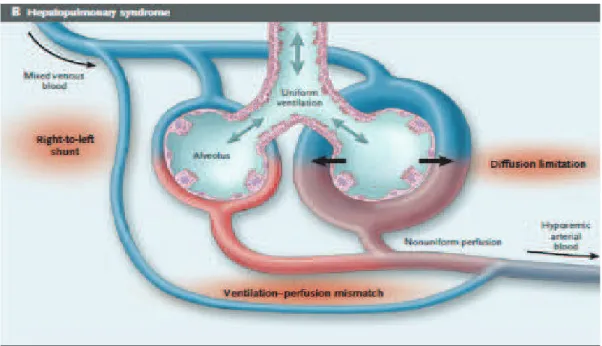
Chronic liver diseases are characterized by a progressive vasodilation that particularly affects splanchnic and pulmonary vascular beds. The vasodilation is due to portal hypertension with or without cirrhosis.
Vasodilation in the lungs can cause a shunt which may lead to hypoxemia. These abnormalities are known as the hepatopulmonary syndrome (HPS): arterial de-oxygenation due to pulmonary vascular dilatation in the context of liver disease. It is important to diagnose HPS because it is an independent prognostic factor of survival and because it also affects postoperative mortality in liver transplantation. Till now the only available treatment for HPS is liver transplantation which leads to a regression and often a disappearance of the HPS. Nevertheless, having an efficient medical treatment is necessary for two reasons. First, given that there is no close correlation between the occurrence of hypoxemia and the severity of liver disease, there are cases where severe hypoxemia requires liver transplantation although liver disease is only mild and would otherwise not be an indication for transplantation. Improving hypoxemia would therefore allow delaying transplantation in such cases. Second, since HPS is associated with an increased transplantation morbidity and mortality, improving hypoxemia could ameliorate survival in patients with a HPS during and after transplantation.
The mechanisms leading to vasodilation due to portal hypertension are not fully understood. Till now, nitric oxide (NO) has been thought to play a major role. It is a well known vasodilator the production of which has been shown to be increased in the rat model of biliary cirrhosis. Moreover, in animal studies chronic treatment with NG-nitro-L-arginine methyl ester (L-NAME), a non-specific NO synthase inhibitor attenuated or prevented the occurrence of HPS. However clinical trials of treatment with inhaled L-NAME led to contradictory results. This can be explained by the fact that other factors play a role. It has been suggested that the renin-angiotensin-aldosterone system associated to oxidative stress may be important. Oxidative stress is linked in part to bacterial translocation that is present in a significant proportion of patients with advanced cirrhosis. Finally, other factors have been implicated: the carbon monoxide which is a vasodilator produced by the heme-oxygenase and it has also been suggested that estrogens (high levels in some patients) could lead to pulmonary vascular dilatation.
The aims of our work were thus to estimate the effects of a probiotic which could act on bacterial translocation, the effects of polyphenols on oxidative stress and the possible improvement of HPS by the estrogen receptor antagonist fulvestrant in a rat model of biliary cirrhosis. The first two studies have been devoted to the effects of a probiotic and a polyphenol rich compound on the splanchnic vessels and the last study to the effects of fulvestrant on the lung. Biliary cirrhosis with portal hypertension has been induced by bile duct ligation in wistar rats under general anesthesia. This leads to a hyperdynamic circulatory state and a hepatopulmonary syndrome. The rats were examined 4 weeks after surgery.
It was shown in an experimental model of partial portal vein ligation in the rat that the initial event because of portal hypertension is an up-regulation in the expression of VEGF (vascular endothelial growth factor) and eNOS (endothelial nitric oxide synthase) in the intestinal microcirculation. Thereafter, a systemic vasodilation occurs, and in particular in the lung leading to a HPS.
The effects of the probiotic and polyphenols were mainly studied on the mesenteric artery and the aorta:
- Vascular reactivity studies were performed in isolated organ chambers in the presence of indomethacin to prevent the formation of vasoactive prostanoids. The NO component of relaxation was studied in the presence of charybdotoxin and apamin (inhibitors of the EDHF response). The EDHF (endothelial derived hyperpolarizing factor) component was studied in the presence of NG-nitro-L-arginine.
- The formation of reactive oxygen species (ROS) was estimated using the fluorescent probe dihydroethidine before the determination of different sources of ROS.
- Immunofluorescence and western blot analyses were used to quantify the expression of different components of the vascular reactivity and of the renin-angiotensin-aldosterone system (RAAS).
- Inflammatory cytokines levels were measured in the plasma
The effects of fulvestrant were studied on the lungs:
- Immunohistochemistry on lung sections allowed to quantify the intravascular macrophages and the small vessels diameter.
- Western blot analyses was used to quantify the expression of eNOS, iNOS, nitrotyrosine and p-VASP, and VEGF and p-Akt.
- Nitrites and hormones levels were measured in the plasma and COHb was measured in arterial blood samples.
The first study showed that treatment with probiotics improved the NO- and EDHF-dependent components of relaxation in the mesenteric vascular bed. This was accompanied by a decreased production of ROS and expression of RAAS factors and a decrease in pro-inflammatory cytokines in the plasma. Bacterial translocation and oxidative stress leading to the production of pro-inflammatory cytokines and to the stimulation of local RAAS is probably an important factor in the development of endothelial dysfunction in cirrhotic rats. The administration of probiotics has improved these abnormalities.
The second study showed that the administration of polyphenols improves endothelial dysfunction in cirrhotic rats: improved EDHF-mediated relaxations, decreased expression of eNOS, ROS production, and some components of the RAAS and decreased levels of proinflammatory cytokines. Oxidative stress, part of which may be due to bacterial translocation, but other factors are also involved, is probably a key factor in the endothelial dysfunction in cirrhotic rats. The relation between the RAAS and oxidative stress is again highlighted in this study which confirms our previous studies where treatment with losartan (angiotensin type 1 receptor antagonist) improved endothelial dysfunction in rats with biliary cirrhosis.
In the third study, treatment with fulvestrant (an estrogen receptor antagonist) decreased the expression of eNOS and nitrotyrosines production in the lung. However, the expression of p-VASP which is a marker of the action of NO was reduced in cirrhotic rat lungs and fulvestrant had no effect on this marker. The hypothesis is that despite the increase in eNOS expression, the produced NO is captured by ROS which would prevent its activity. This could also explain the impaired NO component of relaxation in the two earlier studies. Fulvestrant had no effect neither on the expression of VEGF, nor on the number of macrophages in the pulmonary vessels, nor on the diameter of small pulmonary vessels. In conclusion, either fuvestrant is not effective in cirrhotic rats or the relative increase in estradiol in male rat has no role in the occurrence of a HPS
The important role of oxidative stress in the development of endothelial dysfunction in rats with biliary cirrhosis is emphasized in our studies. Oxidative stress is associated with the stimulation of the RAAS and bacterial translocation. Treatment with antioxidants (polyphenols) and probiotics have clearly demonstrated their beneficial effects on the endothelial dysfunction in our cirrhotic rats. The role of NO as a pure vasodilator is perhaps not essential which may explain the disappointing clinical results published by some authors.
Résumé
Les atteintes hépatiques chroniques se caractérisent par une vasodilatation progressive qui touche en particulier les lits vasculaires splanchnique et pulmonaire. La vasodilatation est due à l’hypertension portale que celle-ci soit la conséquence d’une cirrhose hépatique ou qu’elle soit primitive.
Au niveau pulmonaire la vasodilatation peut entraîner un shunt pouvant conduire à une hypoxémie. On parle alors de syndrome hépato-pulmonaire (SHP) dont la définition est une triade : défaut d’oxygénation artériel dû à des dilatations vasculaires pulmonaires dans un contexte de maladie hépatique. Il est important d’en faire le diagnostic car c’est un facteur pronostique indépendant de survie qui conditionne aussi la mortalité post-opératoire au cours de la greffe hépatique. Pour le moment seule la transplantation hépatique permet une régression ou une disparition du SHP mais un traitement médical efficace est nécessaire pour deux raisons. D’une part étant donné qu’il n’existe pas de corrélation étroite entre la survenue d’une hypoxémie et la sévérité de l’atteinte hépatique, il y a des cas où l’hypoxémie profonde nécessite la transplantation hépatique alors que d’un point de vue hépatique elle n’est pas encore justifiée. Améliorer l’hypoxémie pourrait donc surseoir à la greffe. D’autre part la survenue d’un SHP sévère entraîne un risque opératoire important et un traitement médical atténuant la complication pulmonaire devrait pouvoir faciliter la prise en charge pré- et post-opératoire de ces patients.
Les mécanismes conduisant à la vasodilatation due à l’hypertension portale ne sont pas complètement élucidés. La molécule qui a été incriminé le plus souvent est le NO. C’est un vasodilatateur bien connu et dont on a montré la surproduction dans un modèle de rat de cirrhose biliaire accompagnée d’un SHP. En expérimentation animale un traitement chronique par le NG-nitro-L-arginine methyl ester (L-NAME), inhibiteur non spécifique des NO
synthases atténuait ou empêchait la survenue du SHP. Néanmoins des essais cliniques de traitement par inhalation de L-NAME donnent des résultats contradictoires. Ceci peut s’expliquer par le fait que d’autres facteurs jouent un rôle. Il en est ainsi du système-rénine-angiotensine-aldostérone probablement par le biais d’un stress oxydant. Ce dernier a aussi été lié à la translocation bactérienne qui est présente chez une proportion importante de patients présentant une cirrhose évoluée. Enfin d’autres facteurs ont été incriminés : le monoxide de carbone qui est un vasodilatateur produit par la hème-oxygénase et il a aussi été suggéré que les estrogènes (taux élevés chez certains patients) pouvaient entraîner une dilatation vasculaire pulmonaire.
Le but des différents travaux était donc d’estimer l’action de probiotiques agissant sur la translocation bactérienne, de polyphénols agissant sur la composante stress oxydant et d’un antagoniste des récepteurs de l’estrogène dans un modèle de rat ayant une cirrhose biliaire. L’analyse des effets a été essentiellement réalisée dans la circulation splanchnique et dans une moindre mesure (étude fulvestrant) au niveau pulmonaire.
Premier chapitre
Un rappel général sur les cellules endothéliales et notamment leur action sur le tonus vasculaire par le biais des différents facteurs vasoconstricteurs et vasodilatateurs produits par l’endothélium est fait dans un premier chapitre. Ainsi ont été revus comme vasodilatateurs le monoxyde d’azote (NO), le facteur hyperpolarisant dérivé de l’endothelium (endothelium-derived-hyperpolarization-factor, EDHF), et la prostacycline (PGI2). Parmi les facteurs vasoconstricteurs ont été présentés : le thromboxane A2 (TXA2), et la prostaglandine H2 (PGH2), les espèces réactives de l’oxygène (ROS), l’endotheline 1 (ET1) and l’angiotensine II.
Le deuxième chapitre est dédié à la dysfonction endothéliale dans l’hypertension portale.
D’après des études antérieures réalisées sur un modèle de ligature partielle de la veine porte chez le rat, les premières anomalies survenant au niveau de la microcirculation intestinale est une augmentation de l’expression du facteur de croissance vasculaire (vascular–endothelial-growth factor, VEGF) et de la NO synthase endothéliale (eNOS). Survient par la suite une vasodilatation généralisée avec syndrome hyperdynamique imputable à des mécanismes encore non complètement élucidés. Plusieurs facteurs sont très probablement impliqués : NO, le monoxyde de carbone (CO, possiblement en relation étroite avec le système NO), VEGF et peut-être la prostacycline (peu étudiée à vrai dire). Il est aussi insisté dans ce chapitre sur le rôle très probable de la translocation bactérienne (BT) par le biais de cytokines et des ROS en partie étroitement liés à l’activation du système rénine-angiotensine (SRA). Enfin, ces dernières années a été souligné le rôle très probable d’une angiogénèse anormale qui survient tant au niveau de la circulation splanchnique qu’au niveau pulmonaire.
Troisième chapitre
Les résultats observés au cours d’études antérieures justifient les choix faits en termes d’expérimentations réalisées pour cette thèse : action sur la translocation bactérienne par l’utilisation de probiotiques, action sur le stress oxydant en évaluant un traitement par polyphénols. La troisième étude a permis d’estimer la possibilité d’agir sur la composante NO par le biais de l’administration de fulvestrant. Ce dernier est un antagoniste des récepteurs de l’estrogène dont le taux est souvent augmenté dans la cirrhose et qui stimule l’expression de eNOS.
Le modèle de cirrhose biliaire a été bien validé et consiste à ligaturer la voie biliaire du rat sous anesthésie générale. Ceci induit une cirrhose biliaire avec hypertension portale, un état circulatoire hyperkinétique et un syndrome hépato-pulmonaire. Les rats ont été étudiés 4 semaines après l’intervention.
La figure ci-dessous montre à gauche un foie chez un rat normal et à droite le foie d’un rat présentant une cirrhose biliaire. Dans ce dernier cas il existe une nette prolifération des canaux biliaires.
Foie normal Cirhhose biliaire
Dans les études concernant l’action des probiotiques et polyphénols ont été essentiellement étudiés les effets au niveau de l’artère mésentérique et de l’aorte :
réactivité vasculaire dans des cuves à organe isolé en présence d’indométacine pour prévenir la formation de prostanoïdes vasoactifs. La composante NO de la relaxation est étudiée en présence de charybdotoxine et d’apamine (inhibiteurs de la réponse EDHF-dépendante). La composante EDHF (endothelial derived hyperpolarizing factor) est étudiée en présence de NG-nitro-L-arginine, inhibiteur de la réponse NO-dépendante.
formation d’espèces réactives de l’oxygène (ROS) déterminée à l’aide d’une sonde fluorescente, la dihydroéthidine et détermination des différentes sources de ROS
quantifications de l’expression de différents composants de la réactivité vasculaire et du système rénine-angiotensine-aldostérone (SRAA) par immunofluorescence et en western blot
Quant à l’action du fulvestrant (antagoniste des récepteurs de l’estrogène) il a été étudié au niveau du poumon :
étude des macrophages intravasculaires pulmonaires et des petits vaisseaux (immunohistochimie sur des coupes du poumon)
western blot sur tissu pulmonaire pour la quantification de l’expression de eNOS, iNOS, nitrotyrosine, VEGF et des marqueurs de l’action de NO (p-VASP) et de VEGF (p-Akt), récepteurs- des estrogènes
dosages plasmatiques de nitrites, HbCO et du taux d’hormones.
Etude de la réactivité vasculaire
La première étude a montré qu’un traitement par le probiotique VSL#3 améliorait la composante EDHF- dépendante de la relaxation au niveau mésentérique. Ceci était accompagné de la diminution de production des ROS et des expressions facteurs du SRA ainsi qu’une diminution des cytokines pro-inflammatoires dans le plasma. Le stress oxydant dû à la translocation bactérienne et conduisant à la production de cytokines pro-inflammatoires et à une stimulation du SRA local est probablement un facteur important dans la survenue de la dysfonction endothéliale chez le rat cirrhotique. L’administration de probiotiques a amélioré ces anomalies.
Le probiotique VSL#3 améliore la fonction EDHF-dépendante de la relaxation de l’artère mésentérique chez le rat cirrhotique ; A) composante NO en présence d’indométacine (10 µM) et d’apamine plus charybdotoxine (100 nM chacun), B) composante EDH en présence d’indométacine et de NG-nitro-L-arginine (300 µM) C) réponse au nitroprussiate de sodium (donneur exogène de NO), D) réponse au levcromakalim (ouvreur de canaux potassiques ) ; CBDL : rats cirrhotiques ; *P<0.05 CBDL vs sham, et #P<0.05 CBDL+VSL#3 vs CBDL.
Immmuno-fluorescence au niveau de l’artère mésentérique : VSL#3 diminue l’expression de eNOS et augmente celle des canaux potassiques SKCa et IKCa (A) et des connexines (B) impliquées dans la réponse EDHF-dépendante. Panneau du haut : exemple d’immunofluorescence, panneau du bas : données cumulées pour 4-5 rats ; *P<0.05 CBDL vs sham, et #P<0.05 CBDL+VSL#3 vs CBDL.
Le traitement par VSL#3 diminue le stress oxydant et l’expression de eNOS dans la paroi de l’aorte des rats cirrhotiques (A) ; rôle d’anions superoxydes intracellulaires et du superoxyde d’hydrogène, anneaux aortiques exposés au MnTMPyP (membrane permeant superoxide dismutase mimetic), à la superoxyde dismutase (SOD), PEG-catalase (membrane permeant catalase) et à la catalase. (B) ; sources cellulaires des ROS, anneaux aortiques exposés à l’antioxydant et inhibiteur de la NADPH oxydase apocynine, au L-NA inhibiteur de la NOS, au sulfaphenazol inhibiteur du cytochrome P450, à l’indométacine inhibiteur de la cyclooxygénase et à la combinaison KCN, myxothiazol et roténone (KCN+MY+Rot), inhibitrice de la respiration mitochondriale, 30 minutes avant le marquage DHE (C) ; A) *P<0.05 CBDL vs sham, et #P<0.05 CBDL+VSL#3 vs CBDL; B et C) *P<0.05 pour CBDL avec et sans inhibiteurs.
Le traitement par VSL#3 prévient la surexpression des sous-unités p22phox et p47phox de la NADPH oxydase et de COX-2 mais pas de COX-1 dans la paroi aortique. Panneau de gauche : exemple d’immunofluorescence, panneau de droite : données cumulées pour 4-5 rats ; *P<0.05 CBDL vs sham, et #P<0.05 CBDL+VSL# 3 vs CBDL.
Action du VSL#3 sur les composantes du système RAS (Ang II : angiotensine II, AT1R : récepteur 1 de l‘angiotensine, ACE : enzyme de conversion de l’angiotensine) au niveau de la paroi aortique; Panneau de gauche : exemple d’immunofluorescence, panneau de droite : données cumulées pour 4 rats différents; *P<0.05 CBDL vs sham, et #P<0.05 CBDL+VSL#3 vs CBDL.
Diminution des cytokines pro-inflammatoires IL-1 , MCP-1 et TNF- et augmentation de la cytokine anti-inflammatoire IL-4 par le VSL#3 ; *P<0.05 CBDL vs sham, et #P< 0.05 CBDL+VSL#3 vs CBDL.
La deuxième étude a montré que l’administration de polyphénols (jus de cassis riche en polyphénols, PRBJ) améliorait la dysfonction endothéliale chez le rat cirrhotique : amélioration de la relaxation EDHF-dépendante, diminution de l’expression de eNOS, des ROS, du SRA et des cytokines pro-inflammatoires. Le stress oxydant, dont une part peut être due à la translocation bactérienne mais d’autres facteurs sont également en jeu, est probablement un facteur clé dans la dysfonction endothéliale chez le rat cirrhotique. Le rôle du SRA lié au stress oxydant est à nouveau mis en exergue dans cette étude ce qui confirme une de nos études antérieures où un traitement par losartan (antagoniste des récepteurs 1 de l’angiotensine) améliorait la dysfonction endothéliale chez le rat présentant une cirrhose biliaire.
Le traitement par PRBJ améliore la fonction EDHF mésentérique chez le rat cirrhotique
µM) et d’apamine plus charybdotoxine (100 nM chacun d’indométacine et de NG-nitro
(donneur exogène de NO), D) répons
CBDL : rats cirrhotiques ; *P<0.05 CBDL vs sham, et
Le traitement par PRBJ améliore la fonction EDHF-dépendante de la relaxation de l’artère mésentérique chez le rat cirrhotique ; A) composante NO en présence d’indométacine (10 µM) et d’apamine plus charybdotoxine (100 nM chacun), B) composante EDHF en présence
nitro-L-arginine (300 µM) C) réponse au nitroprussiate de s (donneur exogène de NO), D) réponse au levcromakalim (ouvreur de canaux potassiques )
<0.05 CBDL vs sham, et #P<0.05 CBDL+PRBJ vs CBDL
dépendante de la relaxation de l’artère composante NO en présence d’indométacine (10 ), B) composante EDHF en présence arginine (300 µM) C) réponse au nitroprussiate de sodium e au levcromakalim (ouvreur de canaux potassiques ) ;
Immmuno-fluorescence au niveau de l’artère mésentérique eNOS et augmente celle des canaux potassiques SK
la réponse EDHF-dépendante.
fluorescence au niveau de l’artère mésentérique : PRBJ diminue l’expression de et augmente celle des canaux potassiques SKCa et de la connexine 37 impliqués dans
dépendante. Panneau du haut : exemple d’immunofluorescence, pan
: PRBJ diminue l’expression de et de la connexine 37 impliqués dans Panneau du haut : exemple d’immunofluorescence, panneau du
bas : données cumulées pour 4 vs CBDL
Le traitement par PRBJ diminue le stress oxydant et l’aorte des rats cirrhotiques (A)
d’hydrogène, anneaux aortiques exposés au
dismutase mimetic), à la superoxyde dismutase (SOD) catalase) et à la catalase (B)
Le traitement par PRBJ diminue le stress oxydant et l’expression de eNOS dans la paroi de l’aorte des rats cirrhotiques (A) ; rôle d’anions superoxydes intracellulaires et du
d’hydrogène, anneaux aortiques exposés au MnTMPyP (membrane permeant superoxide dismutase mimetic), à la superoxyde dismutase (SOD), PEG-catalase (membrane permeant
(B) ; sources cellulaires des ROS, anneaux aortiques ex
l’expression de eNOS dans la paroi de ; rôle d’anions superoxydes intracellulaires et du superoxyde MnTMPyP (membrane permeant superoxide catalase (membrane permeant ; sources cellulaires des ROS, anneaux aortiques exposés à
l’antioxydant et inhibiteur de la NADPH oxydase apocynine, au L-NA inhibiteur de la NOS, au sulfaphenazol inhibiteur du cytochrome P450, à l’indométacine inhibiteur de la cyclooxygénase et à la combinaison KCN, myxothiazol et roténone (KCN+MY+Rot), inhibiteur de la respiration mitochondriale, avant le marquage DHE (C). Panneaux du haut : exemple d’immunofluorescence, panneaux du bas : données cumulées pour 4 rats A) *P<0.05 CBDL vs sham, et #P<0.05 CBDL+VSL#3 vs CBDL; B and C) *P<0.05 for CBDL avec et sans inhibiteurs.
Le traitement par PRBJ prévient la surexpression des sous
NADPH oxydase, de COX-2 et de iNOS dans la paroi aortique. Panneau de ga d’immunofluorescence, panneau de droite
CBDL vs sham, et #P<0.05 CBDL+PRBJ vs CBDL.
prévient la surexpression des sous-unités p22phox et p47phox de la 2 et de iNOS dans la paroi aortique. Panneau de ga
d’immunofluorescence, panneau de droite : données cumulées pour 4 rats différents 0.05 CBDL+PRBJ vs CBDL.
p22phox et p47phox de la 2 et de iNOS dans la paroi aortique. Panneau de gauche : exemple : données cumulées pour 4 rats différents ;*P<0.05
Action du PRBJ sur les composantes du système RAS ( récepteur 1 de l‘angiotensine, ACE
paroi aortique; Panneau du haut : exemple d’immunofluorescence, pan
Action du PRBJ sur les composantes du système RAS (Ang II : angiotensine II, AT1R récepteur 1 de l‘angiotensine, ACE : enzyme de conversion de l’angiotensine) au niveau
Panneau du haut : exemple d’immunofluorescence, panneau du bas : données : angiotensine II, AT1R : : enzyme de conversion de l’angiotensine) au niveau de la neau du bas : données
cumulées pour 4 rats différents CBDL.
Diminution des cytokines pro-inflammatoires IL-1 , MCP-1 et TNF- et augmentation de la cytokine anti-inflammatoire IL-4 par le PRBJ ; *P<0.05 CBDL vs sham, et #P< 0.05 CBDL+PRBJ vs CBDL.
Dans la troisième étude, un traitement par fulvestrant (antagoniste des récepteurs de l’estrogène) diminuait l’expression de eNOS et des nitrotyrosines au niveau du poumon. Néanmoins le taux de p-VASP qui est un marqueur de l’action de NO était diminué dans le poumon du rat cirrhotique et le fulvestrant n’avait aucun effet sur ce marqueur. L’hypothèse est que malgré l’augmentation de l’expression de eNOS, le NO produit est capté par les ROS ce qui empêcherait son activité. Ceci pourrait d’ailleurs aussi expliquer la diminution de la composante NO de la relaxation dans les 2 études précédentes. Le fulvestrant n’a aucun effet non plus sur l’expression de VEGF ni sur la quantification des macrophages dans les vaisseaux pulmonaires, ni sur le diamètre des petits vaisseaux pulmonaires. En conclusion, soit le fuvestrant n’est pas efficace, soit l’hyperoestrogénie relative chez nos rats cirrhotiques mâles n’a pas de rôle à jouer dans le survenue du SHP.
Résultats des western blots pour eNOS (NO synthase
inductiblel), nitrotyrosine, p-VASP (phosphorylated vasodilator dans les poumons.
Le fulvestrant diminue l’expression de eNOS et de l
p-VASP qui est un marqueur de l’action de NO, est diminué cirrhotique et le fulvestrant n’a aucun effet sur c
* : P<0.05 pour CBDL vs sham rats; ** : P<0.01 pour vs CBDL+F
Résultats des western blots pour eNOS (NO synthase endothéliale), iNOS
VASP (phosphorylated vasodilator-stimulated phosphoprotein)
Le fulvestrant diminue l’expression de eNOS et de la nitrotyrosine ; de manière surprenante, qui est un marqueur de l’action de NO, est diminué dans le poumon du rat cirrhotique et le fulvestrant n’a aucun effet sur ce marqueur
* : P<0.05 pour CBDL vs sham rats; ** : P<0.01 pour CDL vs sham; # : P<0.01 pour CBDL endothéliale), iNOS (NO synthase stimulated phosphoprotein)
; de manière surprenante, dans le poumon du rat
Résultats des western blots pour VEGF
–alpha des estrogènes), p-Akt (phosphorylated serine/threonine kinase Akt) da poumons. Le fulvestrant augmente l’expression de ER
Akt.* : P<0.05 pour CBDL vs sham; ** : P<0.01 pour CBDL vs
tern blots pour VEGF (vascular endothelial growth factor), ER Akt (phosphorylated serine/threonine kinase Akt) da Le fulvestrant augmente l’expression de ER- mais n’a aucun effet sur VEGF ni p P<0.05 pour CBDL vs sham; ** : P<0.01 pour CBDL vs CBDL+F
(vascular endothelial growth factor), ER- (récepteur Akt (phosphorylated serine/threonine kinase Akt) dans les mais n’a aucun effet sur VEGF ni
Marquage en immunofluorescence pour VEGF (vascular
hème-oxygénase-1 dans les poumons. Les deux facteurs sont élevés mai diminuée avec le traitement par fulvestrant (possib
stimuler HO-1, par le fulvestrant).
** : P<0.05 pour CBDL vs sham; # : P<0.05 pour CBDL
Marquage en immunofluorescence pour VEGF (vascular endothelial growth factor) et la dans les poumons. Les deux facteurs sont élevés mai
diminuée avec le traitement par fulvestrant (possible effet de la diminution de NO, qui peut 1, par le fulvestrant).
** : P<0.05 pour CBDL vs sham; # : P<0.05 pour CBDL vs CBDL+F
endothelial growth factor) et la dans les poumons. Les deux facteurs sont élevés mais seule HO-1 est le effet de la diminution de NO, qui peut
Immunohistochimie d’échantillons pulmonaires. On ob intravasculaires (étoiles) et une augmentation du d
poumon de rats cirrhotiques (panneaux du bas, 2 rat normaux (panneaux du haut).
Conclusions
L’ensemble de nos études soulignent le rôle importa stimulation du SRA dans la survenue de la dysfoncti cirrhose biliaire. L’action sur des facteurs pouvan
Immunohistochimie d’échantillons pulmonaires. On observe de nombreux macrophages intravasculaires (étoiles) et une augmentation du diamètre de petits vaisseaux (flèches) dans le poumon de rats cirrhotiques (panneaux du bas, 2 rats différents) en comparaison avec des rats
L’ensemble de nos études soulignent le rôle important du stress oxydant qui est lié à la stimulation du SRA dans la survenue de la dysfonction endothéliale chez le rat présentant une cirrhose biliaire. L’action sur des facteurs pouvant induire le stress ox
serve de nombreux macrophages iamètre de petits vaisseaux (flèches) dans le comparaison avec des rats
nt du stress oxydant qui est lié à la on endothéliale chez le rat présentant une t induire le stress oxydant comme la
translocation bactérienne et le traitement par des antioxydants (polyphénols) a clairement montré des actions bénéfiques chez nos rats cirrhotiques. Le rôle du NO, en tous cas en tant que vasodilatateur pur, n’est peut-être pas primordial ce qui peut expliquer les résultats décevants en clinique publiés par certains auteurs et ce qui peut également expliquer en partie les résultats de notre troisième étude.
CHAPTER I
INTRODUCTION
Chapter 1
Introduction:
1.1. Vascular endothelium
The vascular endothelium plays a major role in controlling the vascular tone through different mechanisms. The vessel’s wall consists of three individualized layers (Figure 1) from the lumen to the periphery: the tunica intima (comprising the endothelium with a basement membrane and the internal elastic lamina), the media (smooth muscle cells and collagen fibers limited by the external elastic lamina) and the adventitia (where are nerve endings, vasa vasorum, fibroblast and macrophages in connective tissue) (Mulvany 1990). The importance of each layer depends on the size of the arteries: more elastic fibers in the media in large conducting arteries, more smooth muscles in muscular arteries, less smooth muscle cells with only an internal elastic lamina in arterioles and only endothelium and basement membrane and connective tissue in capillaries). The endothelium covers the whole entire wall of all vessels from the heart to small vessels. Hence the endothelium has many function, this enables it to be called the chief regulator of body homeostasis. Due to their position and their large surface area, endothelial cells (ECs) assume a large variety of functions: synthesis and secretion of various molecules, including vasodilator and vasoconstrictor factors, control of smooth muscle cell (SMC) proliferation, exchanges of molecules between the plasma and the interstitial fluid; they have also a role in the balance between pro- and anticoagulant factors and in immunity.
1.2. The major endothelial functions
Endothelial cells respond to physical and chemical stimuli such as pressure, shear stress, and pH (Feletou and Vanhoutte 2006; Moncada and Higgs 2006) and respond also to other stimuli like microparticles and pro-inflammatory mediators. In response to stimuli the endothelium has the capacity to regulate local vascular homeostasis by maintaining the balance between vasodilation and vasoconstriction, by modulating the proliferation and migration of smooth muscle cells and by acting on thrombosis and fibrinolysis by the release of various factors (Davignon and Ganz 2004). In case of endothelial dysfunction, imbalance of these different mechanisms occurs and may lead to serious cardiovascular diseases. One of the endothelial dysfunction may be due to imbalance between vasodilators and vasoconstrictors. The principal vasoconstrictors are: thromboxane A2, prostaglandin H2
(PGH2), endothelin-1, angiotensin II and the superoxide anions, (Furchgott and Vanhoutte 1989; Touyz, Yao et al. 2004). Vasodilator factors are : nitric oxide (NO), the prostacyclin or prostaglandin I2 (PGI2) and the endothelium derived hyperpolarizing (EDH) (Feletou and Vanhoutte 2006; Mombouli and Vanhoutte 1999; Moncada and Higgs 2006) (Table1).
Figure 1. The three layers of normal artery
Vasodilation Vasocontraction
Nitric oxide (NO Reactive oxygen species (ROS) Hyperpolarizing factor Angiotensin II (Ang II)
Prostacyclin (PGI2) Thromboxane A2 (TXA2) Prostaglandin H2 (PGH2) Endothelin
Table 1. Endothelium-derived vasoactive factors. (Calles-Escandon and Cipolla, 2001)
1.2.1. Endothelium-derived vasorelaxing factors
1.2.2. Nitric oxide (NO)
NO is an important cellular signaling molecule involved in different physiological and pathological processes. NO was first known as a ligand capable of activating the soluble guanylyl cyclase and responsible for vascular smooth muscle cells relaxation (Katsuki, Arnold et al. 1977). Soon after, Furchgott and Zawadzki (1980) discovered that the endothelium is responsible for vascular smooth muscle relaxation through production of an endothelium-derived relaxing factor, EDRF. Finally, NO was identified as being the EDRF
described earlier (Palmer, Ferrige et al. 1987; Palmer, Ashton et al. 1988; Palmer and Moncada 1989; Moncada 1997). The endothelium constitutively expresses a NO synthase (endothelial NO synthase, eNOS) which is one of the three isoforms of NOS (Mombouli and Vanhoutte 1999; Stuehr and Griffith 1992) (Table 2). Under normal conditions, inactive eNOS is bound to the protein caveolin and is located in microdomains in the cell membrane called caveolae (Bucci, Gratton et al. 2000). When intracellular Ca2+ levels increase, (in calcium-dependent activation of eNOS) (Figure 3) calmodulin detaches eNOS from caveolin permitting the enzyme to become active. Apart from the increases in intracellular [Ca2+] , other mechanisms (calcium-independent activation of eNOS), leading to the phosphorylation of eNOS (especially phosphorylation of Ser1177) and hence to the activation of eNOS occur via protein kinases pathways, such as protein kinase A (PKA) and cGMP dependent protein kinase II (PKG). The relative contributions of different kinase pathways remain under active investigation, but it is clear that extra-cellular stimuli (shear stress, estrogen, ET-1 and VEGF) activate distinct kinase pathways leading to eNOS phosphorylation and regulating the eNOS state at transcriptional level and stability (Dimmeler et al. 1999, Haynes et al. 2000; Sandoo et al. 2010) (Table 3). eNOS generates NO upon the conversion of L-arginine to L-citrulline in the endothelium (Figure 2). NO has the ability to diffuse towards the underlying vascular smooth muscle to reduce vascular tone through activation of soluble guanylyl cyclase (sGC), and to prevent smooth muscle cell proliferation and migration thereby, maintaining the arterial wall in a quiescent state (Murad et al, 1978). Apart from its action on the smooth muscles, NO has another effect: NO has also been shown to prevent the expression of numerous pro-inflammatory and pro-atherothrombotic mediators such as monocyte chemoattractant protein-1 (MCP-1), tissue factors and adhesion molecules. Moreover, NO helps maintaining blood fluidity by preventing the adhesion and aggregation of platelets and the adhesion of monocytes (Lee et al, 2011). Diminished NO production or bioavailability has been implicated in the pathogenesis of essential and pulmonary hypertension and in multiple other vascular disorders including atherosclerosis (Voetsch et al. 2004). In portal hypertension: NO is diminished in the liver and increased in the splanchnic and systemic vascular beds. Many factors contribute to intra-hepatic reduced eNOS activity including increased oxidative stress, increased binding ability of caveolin-1 to eNOS and increased activity of asymmetric dimethylarginine (ADMA), an endogenous inhibitor of NOS causing un-coupling of NOS and leading to the generation of peroxynitrate (Shah et al. 1999; Laleman et al. 2005). Other factors play a role like decreased Akt activity and tetra-hydrobiopterin (BH4) level (BH4 acts as important catalytic agent in the oxidation of L-arginine by NO
synthases) (Alp and Channon 2004) (Figure 2), and impairment of the antioxidant system (Hu et al, 2013). In contrast to hypoactive hepatic endothelial cells, endothelial cells in the splanchnic and systemic circulation have been shown to produce NO which could modulate the vascular changes observed in cirrhosis. Increased activity of vascular Akt signaling (Fernandez-Varo et al, 2010), VEGF-induced NO production have been also implicated (Abraldes et al, 2006). Likewise, in portal hypertensive rats, NO production is increased in response to shear stress (Tazi et al, 2002) and finally the role of bacterial lipopolysaccharide (LPS) induced overproduction of pro-inflammatory cytokines like TNF- and subsequent induction of inducible NOS (iNOS) in extrahepatic vasculature have been reported (Kajita et al, 2011; Moreau et al, 2002; Huang et al, 2009). It is worthy to mention, that the iNOS induced in the endothelial and other inflammatory cells following immunological activation (Geller et al. 1993), is calcium independent, mostly transcriptionally regulated and is not normally produced in most cell (Forstermann et al. 1994), iNOS generates NO in 100-1000 fold more than eNOS and the NO activity persists for many hours (Morris and Billiar 1994; Nathan and Xie 1994).
Name location main function Neuronal NOS nervous tissue
(nNOSorNOS1) skeletal muscle type II Cell communication Inducible NOS Immune system Immune defense against (iNOS or NOS2) Cardiovascular system Endothelial NOS Endothelium
(eNOS or NOS3) Vasodilatation
Table 2. Three isoforms of NO synthase. (Stuehr and Griffith 1992)
Figure 3. NO synthesis pathway through eNOS phosphorylation in calcium dependent and independent pathway. PI3K, phosphatidylinositol-3- kinase; PDK1, phosphoinositide-dependent
kinase 1; eNOS, endothelial NO synthase; NO, nitric oxide; ER, estrogen receptor; CaM, calmodulin; sGC, soluble guanylyl cyclase modified. (Schini-Kerth et al. 2010)
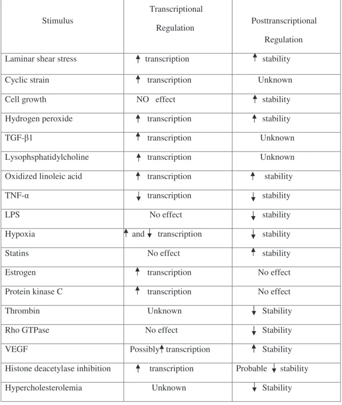
Stimulus Transcriptional Regulation Posttranscriptional Regulation
Laminar shear stress transcription stability
Cyclic strain transcription Unknown
Cell growth NO effect stability
Hydrogen peroxide transcription stability
TGF- 1 transcription Unknown
Lysophsphatidylcholine transcription Unknown
Oxidized linoleic acid transcription stability
TNF- transcription stability
LPS No effect stability
Hypoxia and transcription stability
Statins No effect stability
Estrogen transcription No effect
Protein kinase C transcription No effect
Thrombin Unknown Stability
Rho GTPase No effect Stability
VEGF Possibly transcription Stability
Histone deacetylase inhibition transcription Probable stability
Hypercholesterolemia Unknown Stability
Table 3. Physiological and pathophysiological stimuli shown to regulate eNOS expression and their mode of regulation: (increased : , decreased: ) (Searles 2006).
1.2.3. Endothelium-derived hyperpolarizing factor (EDHF)
Beside NO and prostacyclin, EDHF plays an important role in endothelium-dependent relaxation in most medium-to small sized arteries (Feletou and Vanhoutte 1996). EDHF is an important relaxing factor in the coronary artery as well as in small arteries and arterioles such as second and third-order mesenteric arteries (Shimokawa et al, 1996). After its discovery in 1987, the identity of EDHF is still in the center of controversy because it differs depending on the animal species and the type of blood vessel. In 1996, Shimokawa et al. reported that EDHF is more important in resistance vessels than NO and prostacyclin (Shimokawa et al, 1996). Also, in human arteries, endothelium-dependent vasodilation involves EDHF (Nakashima et al, 1993). EDHF is defined as a hyperpolarization of endothelial origin that is transmitted to the vascular smooth muscle leading to its relaxation. The EDHF component of the relaxation is evaluated in the presence of the combination of inhibitors of eNOS like L-NAME and COXs like indomethacin. To block also the EDHF mediated relaxation, we need to add the inhibitors, apamin and charybdotoxin for both calcium-dependent potassium channels of intermediate (IKCa) and small conductance (SKCa). In the EDHF-mediated response, IKCa and SKCa are activated so that potassium ions move from the intracellular compartment to the extracellular space of endothelial cells, which leads to their hyperpolarization (Figure 4). Thereafter, low concentrations of potassium ions in the extracellular space can activate inwardly rectifying K+ (KIR) channels and Na+/K+-ATPase to cause hyperpolarization of smooth muscle cells through the movement of potassium ions out of the smooth muscle cells (Edwards et al, 1998; Feletou and Vanhoutte 2006; Feletou et al, 2003). Another pathway leading to a direct transfer of the hyperpolarization from endothelial cells to smooth muscle cells occurs via myoendothelial gap junctions (meGJ) as reported previously by Edwards et al., 1998 (Edwards et al, 1998). Gap junctions are intracellular channels which can transfer signals from the endothelial cells to the underling smooth muscle cells (Sandoo et al, 2010) In addition, in some tissues, the hyperpolarization of the endothelial cells might be regulated by hydrogen peroxide (H2O2) (Matoba et al, 2002) or the activation of cytochrome P450 and the resulting generation of epoxyeicosatrienoic acids (EET), which are metabolites of arachidonic acid (Quilley and McGiff 2000). The EET results in increasing K+ efflux from the smooth muscle cells resulting in hyperpolarization and relaxation (Figure 4). Hyperpolarization of smooth muscle cells leads to a decrease in cytosolic calcium concentration with subsequent relaxation.
Figure 4. Hypothesis describing the nature of EDHF pathway (Grgic et al, 2009).
AA, arachidonic acid; ACh, acetylcholine, [Ca2+]i, intracellular calcium concentration; CYP, cytochrome P450 epoxygenase; EC, endothelial cell; EDHF, endothelium-derived hyperpolarizing factor; EETs, epoxyeicosatrienoic acids; ER, endoplasmic reticulum; GPCR, G protein-coupled receptor; BKCa, large conductance Ca
2+
-activated K+ channel; SKCa, small-conductance Ca 2+
-activated K+ channel subtype 3; IKCa, intermediate-conductance Ca
2+
-activated K+ channel; Kir, inwardly rectifying K+ channel; meGJ, myo-endothelial gap-junction; RyR, ryanodine receptor; SR, sarcoplasmic reticulum; VDCC, voltage dependent Ca2+ channel; VSMC, vascular smooth muscle cell.
1.2.4. Prostacyclin (PGI2)
Another important factor of endothelium-dependant relaxation is PGI2. PGI2 is a member of prostanoids (together with other prostaglandins and thromboxanes) which are products from the arachidonic acid metabolism (Figure 5). PGI2 is a potent vasodilator, and effective endogenous inhibitor of platelet aggregation (Coleman et al, 1994; Moncada and Vane 1979). PGI2 is produced in the endothelium from prostaglandin H2 (PGH2) by the action of the enzyme prostacyclin synthase. PGI2, PGG2 and PGH2 are major products of vascular cycloxygenase (COX). There are two isoforms of COX encoded by two separate genes. 1 is constitutively expressed and is present in many tissues, including endothelial cells. COX-2 is not constitutively expressed, but can be induced rapidly and transiently in many cells, including vascular endothelial cells and smooth muscle cells, under the effect of physical stimuli and pro-inflammatory agents (Marnett et al, 1999; Hong and Deykin 1982; Topper et al, 1996). PGI2 elicits smooth muscle relaxation by stimulating adenylyl cyclase and formation of cyclic adenosine -3', 5'- monophosphate. Its vasodilator activity is determined by the expression of specific receptors which are prostaglandin I2 receptors of the G-protein
coupled receptor family in vascular smooth muscle cells (Coleman et al, 1994). The binding of PGI2 to its receptors causes a change of conformation of the receptors, which will lead to an increase of cyclic adenosine-3', 5'-monophosphate (cAMP) levels in the vascular smooth muscle. cAMP then activates protein kinase A, which reduces intracellular Ca2+ by decreasing Ca2+ release from the endoplasmic reticulum and by stimulating its uptake by it (Kukovetz et al, 1979). Furthermore, PGI2 is a potent inhibitor of platelet adhesion, aggregation, and degranulation (Mustard et al, 1980). In addition, PGI2 facilitates the release of NO by endothelial cells (Shimokawa et al, 1988) and in turn, the action of PGI2 in vascular smooth muscle cells and platelets is potentiated by NO (Delpy et al, 1996).
Figure 5. Arachidonic acid metabolism pathway. (Tang and Vanhoutte, 2009)
1.2.5. Summary of endothelium-derived relaxation
EDHF causes hyperpolarization of the endothelium via the activation of calcium dependant potassium channels of intermediate conductance (IKCa) and small conductance (SKCa). Furthermore, vascular smooth muscle relaxation is induced subsequently to the transmission of hyperpolarization through the myoendothelial junction (MGJ). NO is produced by eNOS which is activated by Src/PI3 kinase/Akt pathway or by the calcium calmodulin signaling and relaxes vascular smooth muscle via activation of soluble guanylyl cyclase (sGC). Moreover, PGI2 bind to prostacyclin receptor (IP1) receptor and induce
relaxation via adenylyl cyclase. Endothelin-1(ET-1) binds to ET-B1 receptor on the endothelial receptor to activate eNOS (Figure 6).
Figure 6. The pathways of endothelial-derived relaxation. AA, arachidonic acid; AC,
adenylyl cyclase; ATP, Adenosine triphosphate ; Big ET-1, precursor of endothelin-1; cAMP, cyclic adenosine monophosphate; cGMP, cyclic guanosine monophosphate ; eNOS, endothelium NO synthase; ECE, endothelin converting enzyme; ET-1, endothelin-1, ET-B1 endothelin receptor; IP1,
prostacyclin receptor; GTP, guanosine triphosphate; NO , nitric oxide; PI3K, phosphatidylinositol 3-kinse; Src, Src family. Dash line represents inhibition.
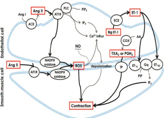
1.3. Endothelium-derived contracting factors (EDCF)
There is large heterogeneity in the formation of EDCF, depending on the stimuli, vascular bed and the age of the experimental animal used. Among the different contracting factors that are produced by the endothelial cells, we retain: thromboxane A2 (TXA2), prostaglandin H2 (PGH2) and also reactive oxygen species, endothelin-1 (ET1) and angiotensin II. (Figure 8) summarizes the different EDCF factors.
1.3.1. Reactive oxygen species (ROS)
Under normal conditions ROS are produced and released from endothelial cells for vascular homeostasis. However, in different pathological conditions like hypertension, diabetes mellitus, atherosclerosis and in acute and chronic inflammatory diseases there is over-production of ROS causing vascular oxidative stress (Eisenberg and Ghigliotti 1999); (Mathis et al, 2012; Sedeek et al, 2012; Xi et al, 2007). ROS in the vasculature are produced by enzymes including cytochrome P450, cyclooxygenases (COXs including COX-1 and COX-2), lipoxygenases, uncoupled-eNOS, xanthine oxidase, NADPH oxidase, and also by the mitochondrial respiratory chain (Griendling and Ushio-Fukai 1997). Superoxide anions (O2 -), hydroxyl radical (OH -) and H2O2 are the major ROS. ROS may have direct vasoconstracting effects by facilitating the mobilization of cytosolic Ca2+ or promoting Ca2+ sensitization of the contractile elements (Jin et al, 1991); (Suzuki and Ford 1992). In addition, ROS in particular O2 - can also potentiate the contractile responses by reducing the bioavailability of NO (Rubanyi and Vanhoutte 1986) or by activating COXs in vascular smooth muscle cells (Hibino, Okumura et al. 1999) or associated with impairment of endothelium-dependent relaxation (Aubin et al, 2006; Liu et al, 2007). The effect of oxidative stress on EDHF pathway occur through reducing the activity of calcium dependent potassium channels (SKCa and IKCa ) (Kusama et al, 2005) and also via modifying the passage of hyperpolarization from endothelial cells to underling smooth muscle cells through myoendothelial gap junctions (Griffith et al, 2005). O2 - can be catalyzed to H2O2 by superoxide dismutase (SOD). Thereafter, H2O2 is degraded to water (H2O) and molecular oxygen (O2) by catalase. Glutathione peroxidase (GPx) also degrades H2O2 to H2O (Thorin-Trescases et al, 2010). This has been supported by earlier works where it has been shown that the antioxidants are able to improve the deleterious effect of oxidant stress on vascular endothelial function in vitro and in vivo in animals (Aubin et al, 2006; Liu et al, 2007) and as