Publisher’s version / Version de l'éditeur: World Neurosurgery, 2012-11-22
READ THESE TERMS AND CONDITIONS CAREFULLY BEFORE USING THIS WEBSITE.
https://nrc-publications.canada.ca/eng/copyright
Vous avez des questions? Nous pouvons vous aider. Pour communiquer directement avec un auteur, consultez la
première page de la revue dans laquelle son article a été publié afin de trouver ses coordonnées. Si vous n’arrivez pas à les repérer, communiquez avec nous à PublicationsArchive-ArchivesPublications@nrc-cnrc.gc.ca.
Questions? Contact the NRC Publications Archive team at
PublicationsArchive-ArchivesPublications@nrc-cnrc.gc.ca. If you wish to email the authors directly, please see the first page of the publication for their contact information.
This publication could be one of several versions: author’s original, accepted manuscript or the publisher’s version. / La version de cette publication peut être l’une des suivantes : la version prépublication de l’auteur, la version acceptée du manuscrit ou la version de l’éditeur.
For the publisher’s version, please access the DOI link below./ Pour consulter la version de l’éditeur, utilisez le lien DOI ci-dessous.
https://doi.org/10.1016/j.wneu.2012.08.022
Access and use of this website and the material on it are subject to the Terms and Conditions set forth at
Fundamentals of Neurosurgery : virtual reality tasks for training and
evaluation of technical skills
Choudhury, Nusrat; Gélinas-Phaneuf, Nicholas; Delorme, Sébastien; Del
Maestro, Rolando
https://publications-cnrc.canada.ca/fra/droits
L’accès à ce site Web et l’utilisation de son contenu sont assujettis aux conditions présentées dans le site LISEZ CES CONDITIONS ATTENTIVEMENT AVANT D’UTILISER CE SITE WEB.
NRC Publications Record / Notice d'Archives des publications de CNRC: https://nrc-publications.canada.ca/eng/view/object/?id=482c4848-9237-41e1-bb5d-d4de3928c82a https://publications-cnrc.canada.ca/fra/voir/objet/?id=482c4848-9237-41e1-bb5d-d4de3928c82a
Fundamentals of Neurosurgery: virtual reality tasks for training and evaluation of technical skills
Nusrat Choudhury, MEng Nicholas Gélinas-Phaneuf, MD Sébastien Delorme, PhD Rolando Del Maestro, MD, PhD, FRCS(C)
PII: S1878-8750(12)01359-9 DOI: 10.1016/j.wneu.2012.08.022 Reference: WNEU 1574
To appear in: World Neurosurgery Received Date: 27 September 2011 Revised Date: 25 April 2012 Accepted Date: 30 August 2012
Please cite this article as: Choudhury N, Gélinas-Phaneuf N, Delorme S, Del Maestro R, Fundamentals of Neurosurgery: virtual reality tasks for training and evaluation of technical skills, World Neurosurgery (2012), doi: 10.1016/j.wneu.2012.08.022.
This is a PDF file of an unedited manuscript that has been accepted for publication. As a service to our customers we are providing this early version of the manuscript. The manuscript will undergo copyediting, typesetting, and review of the resulting proof before it is published in its final form. Please note that during the production process errors may be discovered which could affect the content, and all legal disclaimers that apply to the journal pertain.
M
A
NUS
C
R
IP
T
A
C
C
E
P
TE
D
1TITLE:
Fundamentals of Neurosurgery: virtual reality tasks for training and evaluation of technical skillsAuthors:
Nusrat Choudhury, MEng, Simulation of Deformable Materials, Industrial Materials Institute, National Research Council Canada, Boucherville, Canada
Nicholas Gélinas-Phaneuf, MD, Department of Neurology and Neurosurgery, Montreal Neurological Institute and Hospital, Brain Tumour Research Centre, McGill University, Montréal, Quebec, Canada
Sébastien Delorme, PhD, Simulation of Deformable Materials, Industrial Materials Institute, National Research Council Canada, Boucherville, Canada
Rolando Del Maestro, MD, PhD, FRCS(C), Department of Neurology and Neurosurgery, Montreal Neurological Institute and Hospital, Brain Tumour Research Centre, McGill University, Montréal, Quebec, Canada
Contact information:
Nusrat Choudhury
nusrat.choudhury@nrc.gc.ca (450) 641-5208
M
A
NUS
C
R
IP
T
A
C
C
E
P
TE
D
2ABSTRACT
Background: Technical skills training in neurosurgery is mostly done in the operating room. New
educational paradigms are encouraging the development of novel training methods for surgical skills. Simulation could answer some of these needs. This paper presents the development of a conceptual training framework for use on a virtual reality (VR) neurosurgical simulator.
Methods: Appropriate tasks were identified by reviewing neurosurgical oncology curricula
requirements and performing cognitive task analyses of basic techniques and representative surgeries. The tasks were then elaborated into training modules by including learning objectives, instructions, levels of difficulty and performance metrics. Surveys and interviews were iteratively conducted with subject matter experts (SMEs) to delimitate, review, discuss and approve each of the development stages.
Results: Five tasks were selected as representative of basic and advanced neurosurgical skill. These tasks
were: 1) ventriculostomy, 2) endoscopic nasal navigation, 3) tumour debulking, 4) hemostasis and 5) microdissection. The complete training modules were structured into easy, intermediate and advanced settings. Performance metrics were also integrated to provide feedback on outcome, efficiency and errors. The SMEs deemed the proposed modules as pertinent and useful for neurosurgical skills training.
Conclusion: The conceptual framework presented here, the Fundamentals of Neurosurgery (FNS),
represents a first attempt to develop standardized training modules for technical skills acquisition in neurosurgical oncology. The National Research Council Canada is currently developing NeuroTouch, a VR simulator for cranial microneurosurgery. The simulator presently includes the five FNS modules at varying stages of completion. A first pilot study has shown that neurosurgical residents obtained higher
M
A
NUS
C
R
IP
T
A
C
C
E
P
TE
D
3performance scores on the simulator than medical students. Further work will validate its components and use in a training curriculum.
M
A
NUS
C
R
IP
T
A
C
C
E
P
TE
D
4INTRODUCTION
Technical skills proficiency is an essential component of a surgeon’s competency. An important aspect in cranial neurosurgery is microsurgical skill. Typically, a neurosurgeon must execute precise and delicate manipulations through small openings on magnified structures, performed under the operating microscope (25). Injury to critical areas could lead to major post-operative deficits or fatal outcomes (33). As is the case for many surgical disciplines, neurosurgery is becoming less invasive. The range of procedures that can be done endoscopically is steadily expanding, introducing new and increasingly sophisticated tools to surgeons and ultimately widening the already considerable scope of skills that a trainee must master.
Concerns for patient safety and reduced resident duty hours have limited the time available to train in the operating room (OR) (28). This restriction is at odds with learning new and added techniques; encouraging the development of novel training methods for surgical skills outside of the OR. Current curricula include laboratory sessions with hands-on components such as cadaveric dissection and animal surgeries (40). Cadavers are useful for learning surgical anatomy, however dynamic properties such as bleeding or pulsing organs are missing. In live animal surgeries, the anatomy might differ. None of these alternatives are able to incorporate the anatomic variability and pathology seen during live training in the OR. Surgical simulation is emerging as a potential answer. These systems can consist of task box trainers, mannequins, virtual reality (VR) and hybrid systems (5, 24). A benefit of VR simulation, is that in addition to complementing training in the OR, it can also serve as an assessment tool by providing immediate objective feedback to the trainee through automated performance scores (47). VR simulation can allow autonomous skills training. It can also incorporate the different techniques, anatomies and pathologies required for a variety of surgical specialties. Using advanced graphics and haptics, simulation is striving towards realistic, dynamic tissue behaviour.
M
A
NUS
C
R
IP
T
A
C
C
E
P
TE
D
5Systems are being developed for neurosurgery by research teams, including simulators for
ventriculostomy (35), brain tissue manipulation and dissection (21, 49), endoscopic surgery (38, 39) and cranial bone drilling (1, 31, 50). To our knowledge, there is no commercially available VR simulator specific to neurosurgery. The end goal for a given VR simulator is acceptance by the medical community. A lesson that has been learned during first generation development of these simulators is that the technology should not be constructed prior to determining the needs of the end user (11, 23). Proper simulator design facilitates its integration into surgical training curricula. The first step is the
identification of the educational requirements. Next, face and content validity are subjective measures used to respectively establish that the simulator is realistic and targets training the skills that are required to be trained (9, 18). The scores obtained in simulation should correlate with actual operative technical skill by discriminating novices from experts, demonstrated through construct validation studies (9, 18). Finally, concurrent validation is required to establish that the skills acquired from training on the simulator are transferable to the OR (9, 18, 22, 32, 43).
The National Research Council Canada (NRC) is currently developing NeuroTouch, a VR surgical
simulator for cranial neurosurgery, Figure 1. NeuroTouch is an integrated platform simulating both the stereovision and ergonomics of an OR microscope as well as the 2D indirect view of an endoscopic procedure. The system is equipped with two haptic devices, providing tactile feedback for each hand and permitting interaction with virtual soft tissue. An array of interchangeable physical handles is available (suction tool, ultrasonic aspirator, bipolar forceps, microscissors and endoscope). The developed software allows physics–based simulation of tissue-tool interaction and bleeding. Further details on the system extend beyond the scope of this paper. For a comprehensive description of NeuroTouch, the reader is referred to a work that introduces the technology (13).
M
A
NUS
C
R
IP
T
A
C
C
E
P
TE
D
6A conceptual framework for training was defined prior to developing NeuroTouch. This paper describes the efforts undertaken to define the content for simulation with the input of surgeons. For basic skills training, we took inspiration from the Fundamentals of Laparoscopic Surgery (FLS) manual skills exercises (14, 16) to draft the Fundamentals of Neurosurgery (FNS) tasks targeting neurosurgical oncology. The objective of the FNS is to facilitate the acquisition of psychomotor skills. Consensus was reached to define only five main tasks as a starting point for fundamental skills training.
METHODS
Identification of core technical skills
We first identified the skills that a resident is required to master in order to graduate in neurosurgery. Of the sub-specialties, we focused on neurosurgical oncology as a preliminary effort. Canadian and
American neurosurgical oncology curricula detailing basic requirements were consulted (Royal College of Physicians and Surgeons of Canada, Congress of Neurological Surgeons as well as the McGill
University and Yale School of Medicine neurosurgery training programs). Performance objectives involving hands-on techniques and procedures were sought out. The identified skills were grouped into broad categories.
Selection of appropriate training tasks
Meningiomas, gliomas and pituitary adenomas account for approximately 80% of primary brain and central nervous system tumours in the United States (10). Procedures involving these most commonly occurring brain tumours were investigated as a start to discerning appropriate training tasks. Three generalized surgeries were chosen: removal of a convexity meningioma, low grade frontal lobe glioma resection and endoscopic resection of a pituitary adenoma. We executed cognitive task analyses (CTAs), breaking each of these procedures down into elemental subtasks, including surgical cues and decision
M
A
NUS
C
R
IP
T
A
C
C
E
P
TE
D
7loops (7, 23, 45). The CTAs revealed simplified tasks that could be tailored for training a large number of the skill requirements for graduation. Technical tasks that could be used in many types of procedures were preferentially considered, such as performing a ventriculostomy to relax brain swelling. Of these, the tasks that would most benefit from advanced technology for training were selected. Our intention was to address areas where implementing tasks as modules in NeuroTouch could compliment current curricula for technical skills training.
Development of training modules
The identified tasks were then expanded into structured training modules including learning objectives, instructions, levels of difficulty and performance metrics. The FNS modules were designed with
incremental difficulty (that the trainee must master sequentially) to favour optimal learning (17). Each FNS was organized to first allow the user to become familiar with a given surgical tool proceeding to more advanced levels to practise technique. Some of the exercises target the development of bimanual coordination, others the familiarization with commonly used neurosurgical instruments or learning basic surgical techniques in neurosurgical oncology.
Appropriate performance metrics were identified and integrated to each of the training modules to score performance and provide feedback to the user. Currently, little work has been done for the development of metrics in neurosurgery. However, the performance objective of any given surgery is to attain a favourable patient outcome, providing optimal, efficient treatment while minimizing permanent damage and OR time. As such, the metrics that were defined for each task were derived from the main neurosurgical oncology performance objectives of minimizing tumour cells remaining after surgery, permanent damage to critical areas, blood loss and the duration of the surgery. Also, positive performance measures such as noting a successful outcome of the task were included.
M
A
NUS
C
R
IP
T
A
C
C
E
P
TE
D
8Validation
A key element to the NRC’s VR surgical simulation initiative is the presence of an advisory network of subject matter experts (SMEs) that meet as a collective at semi-annual program meetings. Specifically, the SMEs consist of neurosurgeons and surgeons involved in medical education research. The network is pan-Canadian, including surgeons from 23 teaching hospitals. The participants act as consultants to assure clinical pertinence and realism to the program, indicating their priorities for simulation, providing feedback, clinical guidance, medical images and OR access to our development team.
The SMEs were consulted for performing the CTAs and guided the identification of the five basic tasks. The analyses were then further detailed through expert interviews to determine which features to include in the tasks to maximize the educational value. Questionnaires were sent out to the program SMEs to categorize the identified features as essential or optional. Different questionnaires and SMEs were used for each task. The results from the surveys were used to set the levels of difficulty, define appropriate performance metrics and prioritize the features to be developed in simulation.
The identification of the FNS tasks first began in April 2008 at the start of the program. The tasks were further detailed concurrent to the development of the NeuroTouch simulator. As the learning modules were refined, we assured that they remained useful and pertinent with iterative validation. This was achieved through surveys, discussions and interviews using select SMEs with an interest in the given topic. Feedback has been ongoing for the last four years.
RESULTS
The technical skill requirements for graduation in neurosurgical oncology are shown in Table 1. The list is extensive, demonstrating that many different types of skills are required. The items are listed in
M
A
NUS
C
R
IP
T
A
C
C
E
P
TE
D
9progression of postgraduate year, ranging from performing basic techniques for cerebrospinal fluid management to complete tumour resection procedures.
The cognitive task analyses (CTAs) proved informative in revealing appropriate tasks for training the skills required for graduation. An example of a CTA that was performed is represented in Figure 2. The deconstruction of the approach and resection of a convexity meningioma into subtasks is shown. In the selection of tasks, we opted for the subtasks most associated with decisions and surgical cues. Such tasks would most likely benefit from advanced technology for training. In this example, the most demanding steps in the resection of a convexity meningioma are dissection and debulking, executed to fully expose the tumour. These tasks can be related to #7, 8, 10 and 11 of the graduation requirements in Table 1.
The aim was to address as many of the identified skill requirements as possible while also imposing a limit on the number of tasks to be developed. The selected tasks were: 1) ventriculostomy, 2) endoscopic nasal navigation, 3) tumour debulking, 4) hemostasis and 5) microdissection. Further decomposition of the CTAs exposed surgical cues and appropriate performance metrics that could be used as features to expand the five selected tasks into complete training modules. These five tasks touched upon 9 out of 15 of the skill requirements (scalp incisions, patient positioning, performing craniotomies, skull lesion resections and image-guided biopsies as well as handling unexpected complications were not addressed).
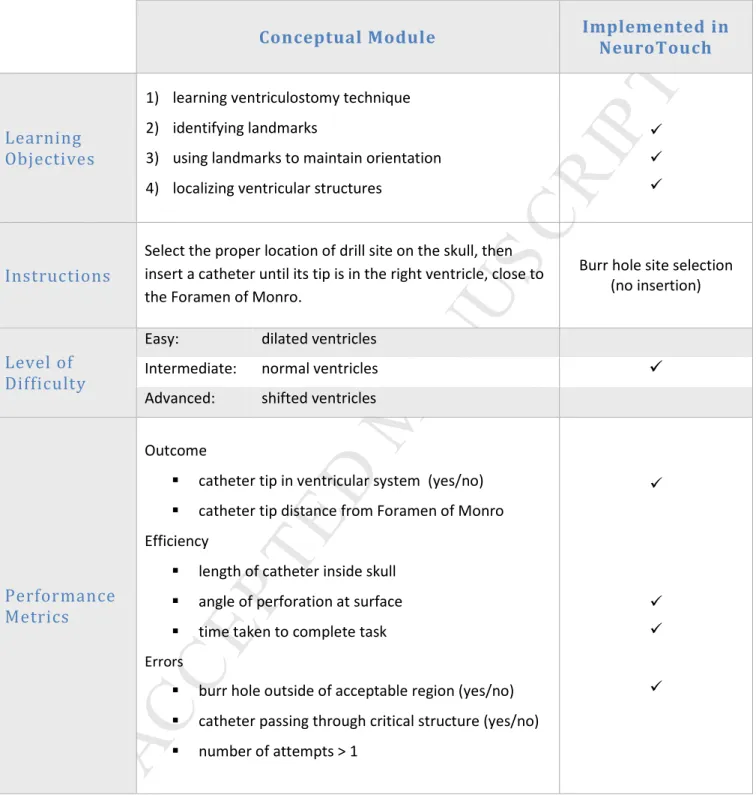
FNS Ventriculostomy
The first training task is to practise the correct insertion of a ventricular catheter. This is a frequently performed procedure and one of the first that a neurosurgical resident encounters (as indicated in Table 1). The challenge for this basic technique, identified from the CTAs, is to be able to properly guide the drain by referring to the anatomical landmarks. Correct use of the landmarks circumvents functional
M
A
NUS
C
R
IP
T
A
C
C
E
P
TE
D
10areas of the cortex and facilitates placement of the catheter tip close to the target. Performing the procedure in a single pass minimizes the risk of complications.
The conceptual FNS training module for ventriculostomy is summarized in Table 2. To test knowledge of the landmarks, the exercise is to select the location of the burr hole using the eyes, nose and ears of the head, Figure 3 (2, 15). The goal is to advance the drain into the brain until the ventricular lining is pierced and placed anterior to the Foramen of Monro, Figure 3. Interactive anatomical models of the skull, brain and ventricles are required to recreate the surgical cues used in performing the procedure, such as feeling a haptic pop as the catheter perforates the ventricle lining. Realistic representation of the catheter is required, including providing the demarcations on the tool to indicate when the perforation is likely to occur (~6 cm from the scalp (2)). The level of difficulty of the module can vary with the anatomy. An easy case being enlarged ventricles due to hydrocephalus and a more difficult one with shifted ventricles due to the presence of a tumour. Potential errors that can occur include
breaching no-go zones which may cause permanent damage, such as crossing the midline or inserting the catheter too deep and into the brain stem.
The work for taking the ventriculostomy training module from concept to VR simulation has begun with the current features implemented in simulation indicated in the last column of Table 2. The simulation is interactive, using haptics to track the selected entry site and angle of insertion on a mannequin head. Currently only the selection of entry site location and angle can be performed (without the actual insertion of the catheter). The resulting trajectory is projected onto the virtual ventricular model for immediate feedback on performance, Figure 3.
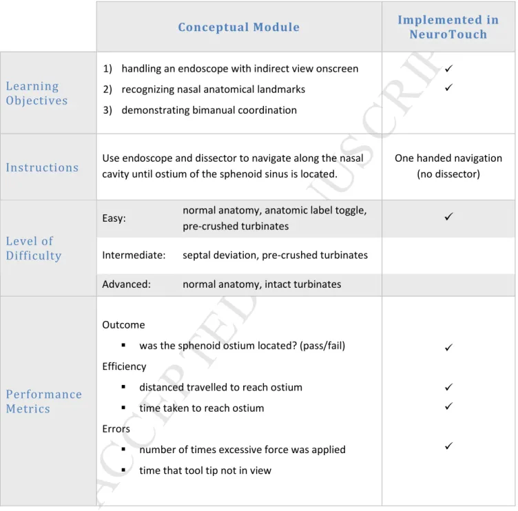
FNS Endoscopic Navigation
This exercise is included such that the trainee can practise skills unique to endoscopic procedures. The main challenges identified from the CTAs were learning the unfamiliar anatomy (44), maintaining spatial
M
A
NUS
C
R
IP
T
A
C
C
E
P
TE
D
11orientation to recognize the anatomy and location of the tools (3) as well as properly navigating with both hands. The goal of the exercise is to locate and identify the sphenoid ostium. This task was selected because it integrates the identified challenges and is a major step in the endoscopic transnasal approach to resecting a pituitary adenoma. The endoscope is inserted and advanced along the nasal cavity. The dissector is then inserted and the tool tip is visualized. The anatomical landmarks are used for guidance to locate the ostium of the sphenoid sinus.
The training module for this task is summarized in Table 3. To practise proper scope handling, the user is required to manoeuvre through the narrow surgical corridors of the nose. Interactive models of the nasal cavities are used to gain familiarity with the anatomy and to recreate the cues identified by the CTAs, such as using the turbinates, choana and spheno-ethmoid recess to guide the trajectory (8, 29). The level of difficulty for this task is related to the anatomy of the patient. The most challenging case is when the middle turbinate is blocking access. The turbinate must first be crushed with the dissector so that the endoscope can be advanced to visualize the ostium. Errors can include improper tool handling or using too much force which can cause the mucosa to bleed or septum perforation. Optimal
performance is indicated when the ostium is located with efficient handling of the endoscope without error.
FNS endoscopic navigation as a VR training module in NeuroTouch has been implemented, with the current features summarized in the last column of Table 3. The simulation is presently one handed permitting only the navigation of an endoscope. The nose is a physical replica of the exterior with a virtual model for the nasal cavity anatomy, Figure 4. Navigation can take place in either nostril to locate each ostium. The simulated anatomy is complete with the required landmarks with performance
feedback through automated measures of force, distance travelled to target as well as the time taken to complete the task.
M
A
NUS
C
R
IP
T
A
C
C
E
P
TE
D
12FNS Tumour Debulking
Tumour debulking is a task that permits the trainee to gain familiarity with surgical aspirators, one of the most widely used neurosurgery tools (51), as well as to practise bimanual dexterity under the operating microscope. The basic task consists of using an ultrasonic aspirator to core out a convexity meningioma, leaving only the outer capsule.
This FNS training module is described in Table 4, consisting of an ultrasonic aspirator and suction tool. Realistic tool handles and accessories permitting adjustment of the tool settings as in the OR are
required. As a training exercise, the user is instructed to debulk the tumour until a small margin from the capsule is reached. As tumour tissue is removed, suction is used to clear the operating field of blood. The level of difficulty is adjusted by modifying the tumour shape, consistency and colour. The easy setting involves a geometric shape rather than an anatomically realistic tumour. This allows the user to first focus on becoming comfortable using each tool and coordinating them bimanually. Errors include injury to or removal of healthy brain which can occur by debulking straight through the capsule, using improper settings on the ultrasonic aspirator or using too much force when retracting. A proficient level in skill is achieved when sufficient tumour is removed efficiently for proper capsule retraction without any damage to healthy brain.
Currently, the intermediate and advanced virtual scenarios are available in NeuroTouch, with the features indicated in the last column of Table 4. The simulation requires the use of both hands with suction and the ultrasonic aspirator available, Figure 5 (30, 37). The module currently includes performance metrics on the quality of the resection as well as efficiency and error measures.
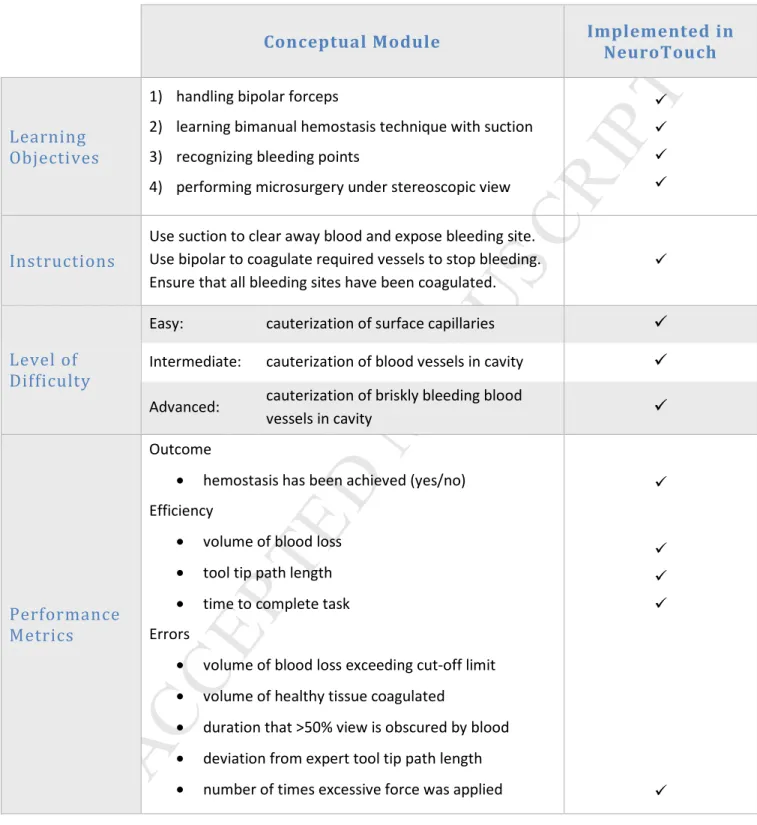
FNS Hemostasis
This task was selected to allow practice with the bipolar forceps and to use it in performing hemostasis, an essential technique to any surgical procedure. Some of the identified challenges from the CTAs
M
A
NUS
C
R
IP
T
A
C
C
E
P
TE
D
13included locating the bleeding site, being able to keep the view clear of blood and managing bleeding vessels. Mastery of proper technique is required when coagulating. The forceps must be gently applied over the bleeding site rather than pinching forcefully on the blood vessel.
The full training module concept is detailed in Table 5. It was designed to practise the cauterisation of blood vessels using bipolar forceps as well as to train the bimanual coordination required. The exercise first requires the use of suction to aspirate blood to reveal the bleeding site and to maintain a clear field of view. A bleeding vessel can be grasped with the forceps and is sealed only if proper technique is used. This must be repeated until all of the bleeding sites have been dealt with for a successful outcome. The level of difficulty is related to the bleeding rate and working depth or angle. Brisk bleeding can occur from the feeding arteries near tumour bed which will cause the operating cavity to quickly fill up with blood. Potential errors include cauterisation of healthy tissue, using an inappropriate technique leading to tissue sticking to bipolar tips and excessive blood loss occurring when too much time is taken to successfully stop the bleeding.
This training module is currently under development in NeuroTouch with different levels of difficulty permitting cauterisation of surface capillaries as well as vessels in a cavity. The work in progress is summarized in the last column of Table 5 and displayed in Figure 6. The simulation is one handed with the bipolar forceps or bimanual with suction in the other hand. Performance metrics have been implemented to detect whether hemostasis has been achieved and include measures of efficiency and error.
FNS Microdissection
This task allows the trainee to gain familiarity with microscissors and to practise an important
M
A
NUS
C
R
IP
T
A
C
C
E
P
TE
D
14tremors when squeezing the tool shaft. Another aspect is being able to correctly identify the natural cutting plane to preserve the normal anatomy in the brain.
The complete FNS module is detailed in Table 6. The training exercise makes use of a classic situation requiring microdissection, which is arachnoid dissection for the removal of convexity meningiomas, Figure 7. This task involves first identifying the tumour-tissue interface to be used as the surgical plane. The tumour is grasped using forceps and retracted to expose the interface. Microscissors are used to cut arachnoid bands within the plane to separate the tumour. The goal is to achieve complete separation of the tissues without injury to healthy tissue. The level of difficulty of the task is related to the level of retraction possible, as well as the depth and angles at which the bands must be cut. The first exercise consists of a debulked meningioma where only the tumour capsule remains. Here sufficient tissue retraction can be achieved to make cutting easily accessible. The more difficult case is a meningioma that has not been debulked, where awkward cutting angles can be encountered. The major error is breaching the surgical plane and cutting into healthy brain. As well, if too much force is used to retract the tumour, some of the bands may be torn rather than cut causing unnecessary bleeding or damage to healthy structures.
The VR simulation work in progress is summarized in the last column of Table 6 and can be seen in Figure 7. The simulation is bimanual, with microscissors in one hand and a grasper in the other used to retract tissue. Currently, the difficult scenario is available for the en bloc removal of a small meningioma. The tissue-tumour interface contains only the adhesions without any blood vessels present.
Validation of the conceptual FNS
We have had the content of the FNS evaluated by the program subject matter experts (SMEs). The proposed tasks were deemed as appropriate and pertinent through iterative discussions and surveys
M
A
NUS
C
R
IP
T
A
C
C
E
P
TE
D
15with our advisory network of surgeons. These findings have established face and content validation that the FNS modules are suitable and target training the skills required in neurosurgical oncology.
DISCUSSION
The Fundamentals of Neurosurgery (FNS) represent the first set of training modules developed to teach basic and advanced neurosurgical technical skills. The FNS modules were designed according to the skill requirements of graduating residents in neurosurgical oncology. As a starting point, curricula
requirements were reviewed and cognitive task analyses were performed to identify five tasks as representative of required skills, including aspects of operating either under the microscope or with an endoscope. They were: 1) ventriculostomy, 2) endoscopic nasal navigation, 3) tumour debulking, 4) hemostasis and 5) microdissection. These tasks were elaborated into complete training modules by integrating learning objectives, instructions, levels of difficulty and performance metrics. NeuroTouch, an interactive bimanual cranial neurosurgery simulator, is being developed by the National Research Council Canada (NRC) to bring the FNS into application (13, 21).
Current neurosurgical training curricula focus primarily on developing microsurgical skill, the basis for neurosurgery (42). The hands-on components require many hours of practice in skills labs, using exercises such as training with synthetic materials (6), manoeuvring through restrictive corridors (46), dissection of animal vessels and nerves (26, 27) or separating of fruit layers (41) under the microscope. The FNS were developed in the same vein as the standardized tasks eventually used for the
Fundamentals of Laparoscopic Surgery (FLS) program (14). Incorporating such a set of tasks to current curricula would allow practice of a diversity of skills, familiarization with multiple surgical tools, training on tasks with incremental difficulty and assessment with objective measures of performance. Validation studies of the Fundamentals of Laparoscopic Surgery (FLS) set of manual skills exercises showed high
M
A
NUS
C
R
IP
T
A
C
C
E
P
TE
D
16levels of construct validity of the tasks as well as high reliability, making it suitable for certification examinations (16).
Feedback on NeuroTouch
Validation of the actual simulator (FNS concepts integrated into NeuroTouch) was done throughout the development of the technology and is ongoing. Formal and informal feedback was obtained from the program SMEs. This feedback has indicated that the look and sense of touch in simulation has reached an acceptable level of realism. Validation outside of the program was achieved during demonstrations at major neurosurgical annual conferences, including the earlier version of NeuroTouch (13, 21) being featured in the sixth Top Gun skills competition held during the 2011 American Association of
Neurological Surgeons meeting. Prototypes of this version of NeuroTouch have been deployed to seven teaching hospitals across Canada. The feedback generated from local staff and residents trialling the system was used to guide the ongoing development of the simulator. Currently all five tasks, at varying stages of completion, have been implemented into the system.
Future Challenges
A limitation of the present work is the lack of sufficient objective data to demonstrate the efficacy of the proposed training modules in neurosurgical education. It will be possible to obtain objective data once the proposed training modules have been completely integrated into NeuroTouch. Many developments are still required in order to completely recreate the conceptual training modules in simulation. The challenge in virtual surgery is to be able to reproduce what a surgeon sees and feels in the operating room instantaneously. This implies that the simulation must run in real-time with high enough
resolution to reproduce the sensory feedback that the surgeon perceives, for example while under the microscope with instrument in hand.
M
A
NUS
C
R
IP
T
A
C
C
E
P
TE
D
17Software developments in NeuroTouch have focused on the integration of computationally effective simulation techniques (4, 12, 13, 30, 37). Today, we have achieved real-time simulation of both the touch and visual feedback as long as the virtual soft tissue surgical corridor is small enough to permit it. Presently, the simulation scenarios in NeuroTouch involve the outer region of the brain. A skull-base procedure or deep tumour resection simulation at an acceptable level of resolution is currently not possible. We are also currently unable to include smaller structures such as the pial membrane, tumour feeders and the meningioma capsule. These structures are crucial in modelling the resection of a meningioma. Future developments will be oriented towards being able to simulate virtual tissue models with greater detail. Finally, based on the latest recommendations arising from the prototype
deployments at our participating collaborator sites, we will prioritize the development of dynamic tool change (currently the surgical instrument selected must be used for the duration of the simulation) and sharp dissection of tissue because presently only fibres can be cut in this manner.
Regarding the use of the simulator as a training tool, future work will focus on providing proficiency goals for the trainee. To ascertain the level, neurosurgical staff (experts) will be asked to perform the VR tasks and their performance recorded. Future validation studies will include medical students,
residents, fellows and staff performance on the simulator. We will investigate its construct validity, specifically whether the simulator metrics can distinguish between varying levels of experience. We have started with a pilot study at the Top Gun skills competition in 2011 (19). We found that neurosurgery residents obtained higher performance scores compared to medical students in the tumour debulking simulation exercise.
Finally, concurrent validation demonstrating that the skills learned on the simulator are transferrable to the operating room is required. Training programs are more inclined to accept expensive technology, such as VR trainers, if objective data can justify the costs. Concurrent validation has been shown in other
M
A
NUS
C
R
IP
T
A
C
C
E
P
TE
D
18surgical disciplines (22, 32, 34, 43). In a particular study involving skills training for laparoscopic surgery, it was concluded that novice residents with little to no surgical experience performed at the level of an intermediately experienced resident after undergoing a training program on a simulator (32). Not only was the learning curve shortened but the time to complete the procedure was halved. Such studies are currently lacking in neurosurgery. A reason for this may be the lack of an objective assessment tool in neurosurgery such as the Objective Structured Assessment of Technical Skill (OSATS) (36) and Global Operative Assessment of Laparoscopic Skills (GOALS) (48) scales used respectively in general and laparoscopic surgery. Current studies are underway to develop a global rating scale to measure neurosurgical performance in the operating room called the Global Assessment of Intraoperative Neurosurgical Skills (GAINS) (20). Using this rating tool, it will then be possible to assess the value of neurosurgical skills training both inside and outside of the operating room permitting the investigation of skill transfer from VR to OR.
The ultimate goal is to establish a VR training curriculum for neurosurgery residents. As in the FLS program, the FNS psychomotor skills training modules should eventually be combined with didactic content to represent a complete training framework. Trainees could use this program to acquire a baseline level of competency before performing neurosurgical interventions on live patients, potentially increasing patient safety.
CONCLUSION
The conceptual framework of the FNS is a first attempt to develop standardized training modules for technical skills acquisition in neurosurgical oncology. The FNS were designed to provide access to skills training in a structured format to allow residents graduating in neurosurgery to sequentially acquire the required skills. The next step is to fully incorporate the modules into a simulated environment. This work has already begun, with implementation of the five FNS into the NRC cranial microneurosurgery VR
M
A
NUS
C
R
IP
T
A
C
C
E
P
TE
D
19simulator. Our first pilot study demonstrated that neurosurgical residents obtained higher performance scores on the simulator compared to medical students. The results indicate that NeuroTouch is a promising tool for neurosurgical technical skills training. Further work will validate its components and integrate them in a complete simulation training curriculum.
ACKNOWLEDGEMENTS
The authors wish to acknowledge the contributions of the NeuroTouch team, consisting of over 50 researchers from the National Research Council Canada (NRC) Industrial Materials Institute, the Institute for Information Technology and the Institute for Biodiagnostics. This VR surgical simulation program has been funded by the NRC Genomics and Health Initiative. This work has also been supported by the Franco Di Giovanni Foundation along with brain tumour research funds from B-Strong, Alex Pavanel Family, Raymonde and Tony Boeckh. The authors also thank the Montreal English School Board, the Brainstorm Foundation, the Colannini Foundation and the Brain Tumour Foundation of Canada for their financial support. Dr. R. F. Del Maestro holds the William Feindel Chair in Neuro-Oncology at McGill University. Dr. Gélinas-Phaneuf would like to thank the Harold and Audrey Delphine Fisher Brain Tumour Research Award for their support. The authors have no personal or institutional financial interest in the devices described in the manuscript.
M
A
NUS
C
R
IP
T
A
C
C
E
P
TE
D
20 Reference List1. Acosta E, Liu A, Armonda R, Fiorill M, Haluck R, Lake C, Muniz G, Bowyer M: Burrhole simulation for an intracranial hematoma simulator. Stud Health Technol Inform 125:1-6, 2007.
2. Aitken AR: Neuroanatomical and cranial geometry of the frontal horn of the lateral ventricle. J Clin Neurosci 2:329-332, 1995.
3. Bakker NH, Fokkens WJ, Grimbergen CA: Investigation of training needs for functional endoscopic sinus surgery (FESS). Rhinology 43:104-108, 2005.
4. Borgeat L, Massicotte P, Poirier G, Godin G: Layered surface fluid simulation for surgical training. Med Image Comput Comput Assist Interv 14:323-330, 2011.
5. Botden SM, Jakimowicz JJ: What is going on in augmented reality simulation in laparoscopic surgery? Surg Endosc 23:1693-1700, 2009.
6. Buis DR, Buis CR, Feller RE, Mandl ES, Peerdeman SM: A basic model for practice of intracranial microsurgery. Surg Neurol 71:254-256, 2009.
7. Cao CG, MacKenzie CL, Ibbotson JA, Turner LJ, Blair NP, Nagy AG: Hierarchical decomposition of laparoscopic procedures. Stud Health Technol Inform 62:83-89, 1999.
8. Cappabianca P, Cavallo LM, Esposito F, de Divitiis E: Endoscopic endonasal transsphenoidal surgery: procedure, endoscopic equipment and instrumentation. Childs Nerv Syst 20:796-801, 2004.
9. Carter FJ, Schijven MP, Aggarwal R, Grantcharov T, Francis NK, Hanna GB, Jakimowicz JJ: Consensus guidelines for validation of virtual reality surgical simulators. Surg Endosc 19:1523-1532, 2005.
10. Central Brain Tumor Registry of the United States (CBTRUS) Statistical Report: NCPR and SEER. Distribution and Incidence Rates of Primary (Malignant and Non-Malignant) Brain and Central Nervous System Tumors by Major Histology Groupings and Histology, Age-Adjusted to the 2000 U.S. Standard Population. 2006.
11. Dawson S: Procedural simulation: a primer. Radiology 241:17-25, 2006.
12. Delorme S, Cabral A, Ayres F, Jiang D: Modeling the thermal effect of the bipolar electrocautery for neurosurgery simulation. Stud Health Technol Inform 163:166-172, 2011.
13. Delorme S, Laroche D, Diraddo R, Del MR: NeuroTouch: A Physics-Based Virtual Simulator for Cranial Microneurosurgery Training. Neurosurgery 2012.
M
A
NUS
C
R
IP
T
A
C
C
E
P
TE
D
2114. Derossis AM, Fried GM, Abrahamowicz M, Sigman HH, Barkun JS, Meakins JL: Development of a model for training and evaluation of laparoscopic skills. Am J Surg 175:482-487, 1998.
15. Epstein ML: Surgical Management of Hydrocephalus, in Schmidek HH, Sweet WH (eds): Operative
Neurosurgical Techniques: indications, methods, and results. Philadelphia, W.B Saunders
Company, 1988, pp 141-150.
16. Fried GM: FLS assessment of competency using simulated laparoscopic tasks. J Gastrointest Surg 12:210-212, 2008.
17. Gallagher AG, Ritter EM, Champion H, Higgins G, Fried MP, Moses G, Smith CD, Satava RM: Virtual reality simulation for the operating room: proficiency-based training as a paradigm shift in surgical skills training. Ann Surg 241:364-372, 2005.
18. Gallagher AG, Ritter EM, Satava RM: Fundamental principles of validation, and reliability: rigorous science for the assessment of surgical education and training. Surg Endosc 17:1525-1529, 2003. 19. Gélinas-Phaneuf N, Choudhury N, Al-Habib A, Cabral A, Nadeau E, Mora V, Pazos V, Debergue P,
DiRaddo R, Del Maestro R: Assessing performance in brain tumor debulking simulation using a novel virtual reality simulator. Neuro Oncol 13 (suppl 3):ST-25, 2011 (abstr).
20. Gélinas-Phaneuf N, Del Maestro R, Okrainec A, Fried G, Choudhury N: The assessment of technical skills in neurosurgery: the development of the global assessment of intraoperative neurosurgical skills (GAINS) scale. Canadian Conference on Medical Education 2010 (abstr).
21. Gélinas-Phaneuf N, DiRaddo R, Del Maestro RF: A novel way to train and assess neurosurgeons: NeuroTouch, a virtual reality simulator. Presented at the Proceedings from the 14th Biennial Canadian Neuro-Oncology Meeting.
22. Grantcharov TP, Kristiansen VB, Bendix J, Bardram L, Rosenberg J, Funch-Jensen P: Randomized clinical trial of virtual reality simulation for laparoscopic skills training. Br J Surg 91:146-150, 2004. 23. Grunwald T, Clark D, Fisher SS, McLaughlin M, Narayanan S, Piepol D: Using cognitive task analysis
to facilitate collaboration in development of simulator to accelerate surgical training. Stud Health Technol Inform 98:114-120, 2004.
24. Halvorsen FH, Elle OJ, Fosse E: Simulators in surgery. Minim Invasive Ther Allied Technol 14:214-223, 2005.
25. Hernesniemi J, Niemela M, Karatas A, Kivipelto L, Ishii K, Rinne J, Ronkainen A, Koivisto T, Kivisaari R, Shen H, Lehecka M, Frosen J, Piippo A, Jaaskelainen JE: Some collected principles of
microneurosurgery: simple and fast, while preserving normal anatomy: a review. Surg Neurol 64:195-200, 2005.
26. Hicdonmez T, Hamamcioglu MK, Tiryaki M, Cukur Z, Cobanoglu S: Microneurosurgical training model in fresh cadaveric cow brain: a laboratory study simulating the approach to the circle of Willis. Surg Neurol 66:100-104, 2006.
M
A
NUS
C
R
IP
T
A
C
C
E
P
TE
D
2227. Hino A: Training in microvascular surgery using a chicken wing artery. Neurosurgery 52:1495-1497, 2003.
28. Jagannathan J, Vates GE, Pouratian N, Sheehan JP, Patrie J, Grady MS, Jane JA: Impact of the Accreditation Council for Graduate Medical Education work-hour regulations on neurosurgical resident education and productivity. J Neurosurg 110:820-827, 2009.
29. Jho HD: Endoscopic transsphenoidal surgery. J Neurooncol 54:187-195, 2001.
30. Jiang D, Choudhury N, Mora V, Delorme S: Characterization of suction and CUSA interaction with brain tissue. Presented at the LNCS.
31. Kockro RA, Hwang PY: Virtual temporal bone: an interactive 3-dimensional learning aid for cranial base surgery. Neurosurgery 64:216-229, 2009.
32. Larsen CR, Soerensen JL, Grantcharov TP, Dalsgaard T, Schouenborg L, Ottosen C, Schroeder TV, Ottesen BS: Effect of virtual reality training on laparoscopic surgery: randomised controlled trial. BMJ 338:b1802, 2009.
33. Lassen B, Helseth E, Ronning P, Scheie D, Johannesen TB, Maehlen J, Langmoen IA, Meling TR: Surgical mortality at 30 days and complications leading to recraniotomy in 2630 consecutive craniotomies for intracranial tumors. Neurosurgery 68:1259-1268, 2011.
34. Lee JY, Mucksavage P, Kerbl DC, Huynh VB, Etafy M, McDougall EM: Validation study of a virtual reality robotic simulator--role as an assessment tool? J Urol 187:998-1002, 2012.
35. Lemole GM, Jr., Banerjee PP, Luciano C, Neckrysh S, Charbel FT: Virtual reality in neurosurgical education: part-task ventriculostomy simulation with dynamic visual and haptic feedback. Neurosurgery 61:142-148, 2007.
36. Martin JA, Regehr G, Reznick R, MacRae H, Murnaghan J, Hutchison C, Brown M: Objective structured assessment of technical skill (OSATS) for surgical residents. Br J Surg 84:273-278, 1997. 37. Mora V, Jiang D, Brooks R, Delorme S: A computer model of soft tissue interaction with a surgical
aspirator.
38. Neubauer A, Wolfsberger S, Forster MT, Mroz L, Wegenkittl R, Buhler K: Advanced virtual endoscopic pituitary surgery. IEEE Trans Vis Comput Graph 11:497-507, 2005.
39. Parikh SS, Chan S, Agrawal SK, Hwang PH, Salisbury CM, Rafii BY, Varma G, Salisbury KJ, Blevins NH: Integration of patient-specific paranasal sinus computed tomographic data into a virtual surgical environment. Am J Rhinol Allergy 23:442-447, 2009.
40. Reznick RK, MacRae H: Teaching surgical skills--changes in the wind. N Engl J Med 355:2664-2669, 2006.
41. Scholz M, Dick S, Fricke B, Schmieder K, Engelhardt M, Tombrock S, Pechlivanis I, Harders A, Konen W: Consideration of ergonomic aspects in the development of a new endoscopic navigation system. Br J Neurosurg 19:402-408, 2005.
M
A
NUS
C
R
IP
T
A
C
C
E
P
TE
D
2342. Scholz M, Mucke T, Holzle F, Schmieder K, Engelhardt M, Pechlivanis I, Harders AG: A program of microsurgical training for young medical students: are younger students better? Microsurgery 26:450-455, 2006.
43. Seymour NE, Gallagher AG, Roman SA, O'Brien MK, Bansal VK, Andersen DK, Satava RM: Virtual reality training improves operating room performance: results of a randomized, double-blinded study. Ann Surg 236:458-463, 2002.
44. Snyderman C, Kassam A, Carrau R, Mintz A, Gardner P, Prevedello DM: Acquisition of surgical skills for endonasal skull base surgery: a training program. Laryngoscope 117:699-705, 2007.
45. Sullivan ME, Ortega A, Wasserberg N, Kaufman H, Nyquist J, Clark R: Assessing the teaching of procedural skills: can cognitive task analysis add to our traditional teaching methods? Am J Surg 195:20-23, 2008.
46. Takeuchi M, Hayashi N, Hamada H, Matsumura N, Nishijo H, Endo S: A new training method to improve deep microsurgical skills using a mannequin head. Microsurgery 28:168-170, 2008. 47. Tsuda S, Scott D, Doyle J, Jones DB: Surgical skills training and simulation. Curr Probl Surg
46:271-370, 2009.
48. Vassiliou MC, Feldman LS, Andrew CG, Bergman S, Leffondre K, Stanbridge D, Fried GM: A global assessment tool for evaluation of intraoperative laparoscopic skills. Am J Surg 190:107-113, 2005. 49. Wang P, Becker AA, Jones IA, Glover AT, Benford SD, Greenhalgh CM, Vloeberghs M: A virtual
reality surgery simulation of cutting and retraction in neurosurgery with force-feedback. Comput Methods Programs Biomed 84:11-18, 2006.
50. Wiet GJ, Stredney D, Sessanna D, Bryan JA, Welling DB, Schmalbrock P: Virtual temporal bone dissection: an interactive surgical simulator. Otolaryngol Head Neck Surg 127:79-83, 2002. 51. Yasargil M: Instrumentation and Equipment, Microneurosurgery of CNS tumors IVB. Stuttgart,
M
A
NUS
C
R
IP
T
A
C
C
E
P
TE
D
24Figure 1: NeuroTouch, the National Research Council’s cranial microneurosurgery simulator equipped
with i) stereoscopic view, bimanual force feedback handles and mannequin head, and an endoscopic view with a force feedback handle for ii) insertion into a physical replica of the nose or iii) use with a mannequin head.
Figure 2: Cognitive task analysis deconstructing the resection of a convexity meningioma. The top level
subtasks are shown. In this example, the approach to the tumour, its exposure and removal phases were selected as possible training tasks due to the decision-making involved. These subtasks were further broken down to expose features and cues that were potentially important to simulate for training (not detailed here).
Figure 3: Conceptual FNS Ventriculostomy with i) burr hole localization and proper catheter insertion
angle determined using the eyes, nose and ears of the head to reach the target: ii) anterior to the Foramen of Monro. The work for taking the ventriculostomy training module from concept to VR simulation has begun. The selection of entry site location and angle is performed on iii) a mannequin head. iv) Ending the simulation gives qualitative feedback of performance by indicating whether the trainee’s projected path falls into the acceptable region defined by experts.
Figure 4: The FNS Endoscopic Navigation requires use of nasal anatomical landmarks and proper
navigation to locate sphenoid ostium. The implementation of this training module into NeuroTouch consists of the nose as ii) a physical replica with a virtual nasal cavity anatomy. ii) The simulated
anatomy is complete with the required landmarks including anatomic labels for the inferior, middle and superior turbinates as well as the nasal septum. iii) The simulator detects a successful outcome if the user is able to locate the ostium and hold it in the centre of the virtual endoscopic view. iv) At simulation end, the user is provided with measures of force, distance travelled to target as well as the time taken to complete the task as feedback on performance.
M
A
NUS
C
R
IP
T
A
C
C
E
P
TE
D
25Figure 5: The FNS Tumour Debulking training module conceptualization including i) a meningioma with
its capsule, suction and an ultrasonic aspirator. NeuroTouch training module is under development with ii) different colours for tumour core and healthy brain. iii) The simulator is equipped with a haptic handle in one hand allowing adjustment of suction pressure at the fingertips. The ultrasonic aspiration in the other hand is activated via a foot pedal and the settings are varied through a console. iv) The module includes performance metrics of the percentage of tumour resected, the time taken to complete the task with penalty if any healthy tissue is removed or if any excessive force was applied on the tissue.
Figure 6: The FNS Hemostasis was designed to practise cauterisation of blood vessels. This training
module is currently under development in NeuroTouch with i) easy level of difficulty involving the removal of capillaries from the surface of the brain and ii) advanced level requiring suction to first clear the view, locating the blood source and cauterisation of bleeding site in a cavity. The simulated bipolar forceps are activated using a foot pedal. The bleeding rate depends on the type of blood vessel. Major ones bleed rapidly, smaller ones bleed at slower rates and capillaries do not bleed, simply disappearing under cauterisation. iii) Tissue in contact with active bipolar tips changes in colour from a whitish to a burnt hue as a result of proximal heating, which depends on the power, distance between the bipolar tips and duration of cauterisation (12). iv) Performance metrics that have been implemented include outcome assessment, tool tip displacement and time taken to complete the task as well as tracking the volume of blood loss. A penalty is allotted for the use of excessive force or damage to healthy brain.
Figure 7: Microdissection training i) conceptualization including meningioma with tissue interface and ii)
NeuroTouch module under development. The VR simulation is bimanual, with microscissors in the right hand and a grasper in the left. Currently, only the difficult scenario is available. The simulation requires the user to locate the tumour-tissue interface by retracting the tumour with a grasper. The microscissors are used to cut any bands that can be seen. It includes performance metrics of the percentage of the
M
A
NUS
C
R
IP
T
A
C
C
E
P
TE
D
26bands cut and the time taken to complete the task. Penalties are assigned if bands are torn from excessive retraction and if any healthy tissue is damaged.
M
A
NUS
C
R
IP
T
A
C
C
E
P
TE
D
Keywords: neurosurgery, clinical skills, computer simulation, training, virtual systems
List of abbreviations: cognitive task analyses (CTAs), Fundamentals of Laparoscopy (FLS), Fundamentals of Neurosurgery (FNS), National Research Council (NRC), operating room (OR), subject matter experts (SMEs), virtual reality (VR).
M
A
NUS
C
R
IP
T
A
C
C
E
P
TE
D
Table 1: Technical skill requirements for graduation in neurosurgical oncology
1. Open and close scalp incisions
2. Perform ventriculostomies, place lumbar drains and intracranial monitors
3. Position patients for craniotomy
4. Perform the opening and closing of craniotomies
5. Resect skull lesions
6. Perform image-guided biopsies
7. Demonstrate facility with the use of surgical instruments including operating microscope and endoscope
8. Identify interface between tumour and brain and use as operating plane for tumour resection
9. Identify anatomic landmarks, functional regions and major structures
10. Show how to minimize and control intraoperative bleeding
11. Perform resection of extra-axial and intra-axial brain tumours
12. Perform resection of supra- and infratentorial brain tumours
13. Perform resection of pituitary lesions
14. Perform basic skull base procedures
M
A
NUS
C
R
IP
T
A
C
C
E
P
TE
D
Table 2: FNS Ventriculostomy training module with task to correctly place a ventricular drain
Conceptual Module
Implemented in
NeuroTouch
Learning
Objectives
1) learning ventriculostomy technique 2) identifying landmarks
3) using landmarks to maintain orientation 4) localizing ventricular structures
Instructions
Select the proper location of drill site on the skull, then insert a catheter until its tip is in the right ventricle, close to the Foramen of Monro.
Burr hole site selection (no insertion)
Easy: dilated ventricles Intermediate: normal ventricles
Level of
Difficulty
Advanced: shifted ventricles
Performance
Metrics
Outcome
catheter tip in ventricular system (yes/no) catheter tip distance from Foramen of Monro Efficiency
length of catheter inside skull angle of perforation at surface time taken to complete task
Errors
burr hole outside of acceptable region (yes/no) catheter passing through critical structure (yes/no) number of attempts > 1
M
A
NUS
C
R
IP
T
A
C
C
E
P
TE
D
Table 3: FNS Endoscopic Nasal Navigation training scenario with task to locate the ostium of the
sphenoid sinus
Conceptual Module
Implemented in
NeuroTouch
Learning
Objectives
1) handling an endoscope with indirect view onscreen 2) recognizing nasal anatomical landmarks
3) demonstrating bimanual coordination
Instructions
Use endoscope and dissector to navigate along the nasal cavity until ostium of the sphenoid sinus is located. One handed navigation (no dissector)Easy: normal anatomy, anatomic label toggle, pre-crushed turbinates
Intermediate: septal deviation, pre-crushed turbinates
Level of
Difficulty
Advanced: normal anatomy, intact turbinates
Performance
Metrics
Outcome
was the sphenoid ostium located? (pass/fail) Efficiency
distanced travelled to reach ostium time taken to reach ostium
Errors
number of times excessive force was applied time that tool tip not in view
M
A
NUS
C
R
IP
T
A
C
C
E
P
TE
D
Table 4: FNS Tumour Debulking training scenario with task to debulk a meningioma
Conceptual Module
Implemented in
NeuroTouch
Learning
Objectives
1) handling the ultrasonic aspirator and suction
2) discriminating tumour from healthy brain using visual and tactile cues
3) learning the bimanual debulking technique 4) performing microsurgery under stereoscopic view
Instructions
Remove as much of the tumour as possible with ultrasonic aspiration. Use suction to aspirate blood.Easy: removal of hemispheric “tumour”
embedded in a cube
Intermediate: removal of complex shape “tumour” embedded in a cube
Level of
Difficulty
Advanced: removal of meningioma embedded in brain
Performance
Metrics
Outcome
percentage tumour resected Efficiency
path length
time taken to complete task Errors
percentage healthy tissue removed
number of times no-go zones have been breached deviation from expert tool tip path length
M
A
NUS
C
R
IP
T
A
C
C
E
P
TE
D
Table 5: FNS Hemostasis training scenario with task to coagulate blood vessels
Conceptual Module
Implemented in
NeuroTouch
Learning
Objectives
1) handling bipolar forceps
2) learning bimanual hemostasis technique with suction 3) recognizing bleeding points
4) performing microsurgery under stereoscopic view
Instructions
Use suction to clear away blood and expose bleeding site. Use bipolar to coagulate required vessels to stop bleeding. Ensure that all bleeding sites have been coagulated. Easy: cauterization of surface capillaries Intermediate: cauterization of blood vessels in cavity
Level of
Difficulty
Advanced: cauterization of briskly bleeding blood vessels in cavity
Performance
Metrics
Outcome
• hemostasis has been achieved (yes/no) Efficiency
• volume of blood loss
• tool tip path length
• time to complete task Errors
• volume of blood loss exceeding cut-off limit
• volume of healthy tissue coagulated
• duration that >50% view is obscured by blood
• deviation from expert tool tip path length
M
A
NUS
C
R
IP
T
A
C
C
E
P
TE
D
Table 6: FNS-5 Microdissection training scenario with arachnoid dissection task
Conceptual Module
Implemented in
NeuroTouch
Learning
Objectives
1) handling microscissors and forceps
2) learning bimanual microdissection technique 3) localizing tumour-tissue interface
4) performing microsurgery under stereoscopic view
Instructions
Dissect a convexity meningioma from the arachnoid using forceps to grasp tumour and microscissors to cut connecting bands. Use arachnoid as the dissection plane and free the tumour with proper retraction and sharp cutting.
Easy: 50 adhesions near surface, debulked tumour
Intermediate: 100 adhesions along entire plane, debulked tumour
Level of
Difficulty
Advanced: 100 adhesions along entire plane, en bloc removal of a small meningioma
Performance
Metrics
Outcome
percentage of bands cut Efficiency
tool tip path length
time taken to complete task
Errors
number of times no-go zones breached number of bands torn
deviation from expert tool tip path length volume of healthy tissue damaged