S80 CANADIAN JOURNALOF ANESTHESIA
Purpose: To describe the drugs used to treat venous throm-boembolism (VTE) and to review particular aspects of the management (elastic stockings, thrombolysis, thrombectomy, vena cava filter).
Source: Our review of the literature is focused on consensus documents and recent large randomized trials.
Principal findings: Subcutaneous low molecular weight hepa-rins (LMWH) have been shown to be both safe and effective for the initial treatment of VTE and have largely replaced unfractionated heparin, unless there is a contraindication to LMWH such as severe renal insufficiency. Low molecular weight heparins or unfractionated heparin are usually administered for five to seven days. Treatment is gradually switched from heparin to oral vitamin K antagonists (VKA) which are usually started the same day as heparin. The duration of oral anticoagulation must be tailored to the individual patient according to the pres-ence of reversible or continuing risk factors. In patients with active cancer, long-term treatment of VTE with LMWH has been shown to be more effective than oral anticoagulation and is recommended for the first three to six months of long-term anticoagulant therapy as an alternative approach to VKA. Elastic stockings are recommended because they have been shown to prevent postthrombotic syndrome. Thrombolysis is, usually, not justified for the treatment of deep venous thrombosis, but is used in cases of massive pulmonary embolism with arterial hypotension and/or shock. Vena cava filter placement is mainly indicated in patients with a proximal deep venous thrombosis and an absolute contraindication to anticoagulation.
Conclusions: The initial management of patients with acute VTE has largely been simplified due to the use of LMWH. Early conversion to VKA is recommended for the great majority of patients. New agents, such as anti-Xa or oral thrombin inhibi-tors, are promising alternatives to heparins or VKA.
Objectif : Présenter les médicaments utilisés pour traiter la
mala-die thromboembolique veineuse (MTEV) et revoir des aspects par-ticuliers de la thérapie comme les bas élastiques, la thrombolyse, la thrombectomie et le filtre cave.
Source : Revue de documents de consensus et de grandes études
récentes.
Constatations principales : Les héparines de bas poids
molécu-laire (HBPM) sont sûres et efficaces comme traitement initial de la MTEV et remplacent largement l’héparine non fractionnée, à moins d’une contre-indication à l’HBPM comme l’insuffisance rénale sévère. Les HBPM ou l’héparine non fractionnée sont habi-tuellement administrées pendant cinq à sept jours. Puis, on passe graduellement de l’héparine à la prise orale d’antagonistes de la vitamine K (AVK), débutés en général le même jour que l’héparine. La durée de l’anticoagulation orale doit être adaptée au patient en fonction de facteurs de risque réversible ou continu. Dans les cas de cancer actif, le traitement de la MTEV avec l’HBPM s’est mon-tré plus efficace que l’anticoagulation orale et il est recommandé pour les trois à six premiers mois de traitement. Les bas élastiques sont recommandés pour prévenir le syndrome post-thrombotique. La thrombolyse n’est pas habituellement justifiée pour traiter la thrombose veineuse profonde, mais est utilisée en cas d’embolie pulmonaire massive avec hypotension et/ou choc artériels. La mise en place d’un filtre cave est principalement indiquée chez les patients souffrant de thrombose veineuse profonde proximale chez qui l’anticoagulation est une contre-indication absolue.
Conclusion : Le traitement initial des patients atteints de MTEV
a été grandement simplifié avec l’usage de l’HBPM. Le passage précoce aux AVK est recommandé pour la grande majorité des patients. De nouveaux médicaments comme les anti-Xa ou les inhibiteurs de la thrombine oraux, sont des équivalents pro- metteurs des héparines ou des AVK.
S80 SUPPLEMENT
CAN J ANESTH 2006 / 53: 6 / pp S80–S88
Management of venous thromboembolism
[Traitement de la maladie thromboembolique veineuse]
Philippe de Moerloose MD,* Charles Marc Samama MDPhD,† Serge Motte MDPhD‡
From the Service d’Angiologie et d’Hémostase,* Hôpitaux Universitaires de Genève, Genève, Suisse; the Department of Anesthésie-Réanimation,† Assistance Publique-Hôpitaux de Paris; the Centre Hospitalier Universitaire Avicenne, Bobigny, France; and the Service de Pathologie Vasculaire,‡ Cliniques Universitaires de Bruxelles, Hôpital Erasme, Université Libre de Bruxelles, Belgique.
Address correspondence to: Pr. Philippe de Moerloose, Unité d’Hémostase, Hôpitaux Universitaires de Genève, 1211 Geneva 14,
de Moerloose et al.: TREATMENTOFVENOUSTHROMBOEMBOLISM S81
I
N most cases, pulmonary embolism (PE) is secondary to deep venous thrombosis (DVT); moreover, asymptomatic PE is observed in 40% of patients presenting with proximal DVT when systematic lung scintigraphy is performed.1 Therefore DVT and PE may be considered as the same disease and will be treated together under the common term of venous thromboembolism (VTE). The treatment (anticoagulation) is also essentially the same.2 In this review we will first discuss the drugs (old and new) and then some particular aspects of the manage-ment of VTE (early ambulation and elastic stockings, thrombolysis, thrombectomy, embolectomy, vena cava filter). The management of anticoagulated patients who require surgery and for over-anticoagulated patients, as well as the problem of heparin-induced thrombocytopenia are dealt with elsewhere.A. Anticoagulant treatment
Venous thromboembolism is a medical emergency and the first step, in most patients, is to administer heparin immediately with an early switch to vitamin K antagonists (VKA). Three options are available for the initial treatment of VTE: low-molecular weight heparins (LMWH), iv unfractionated heparin (UFH) or sc UFH.
1. Unfractionated heparin and LMWH
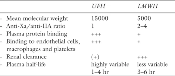
Treatment of VTE starts with heparins. The advan-tages of LMWH over UFH are summarized in Table I. Although LMWH have more favourable pharma-cological properties than UFH, clinical studies have not shown important differences concerning their efficacy or safety, although it is possible that the risk of mortality is decreased with LMWH.2,3 Due to their simplicity of administration, LMWH have become the drug of choice in the initial treatment of VTE. Indeed, LMWH provide a higher level of comfort for the patient, their use is less time consuming for nurses and laboratory technicians since no laboratory controls are required. They carry a lower risk of heparin-induced thrombocytopenia and osteopenia, two adverse events that may be observed if the treatment lasts more than the usual five days. Moreover, LWMH are associated with cost savings since they allow early hospital dis-charge and home therapy.
2. Is there still a place for unfractionated heparin? The difficulties of attaining therapeutic concentrations with UFH are well known. Unfractionated heparin has an unpredictable dose response and a narrow therapeutic window. For example, an audit conducted in three North American hospitals has shown that 60%
of patients did not have a therapeutic activated par-tial thromboplastin time (aPTT) in the first 24 hr of treatment and that 30 to 40% of patients did not have therapeutic anticoagulation during the first four days.4 Most often the doses of UFH are subtherapeutic, phy-sicians being afraid of hemorrhagic complications.
If LMWH are generally given in cases of DVT, their use is not generalized in cases of PE, despite the results of clinical studies.5,6 This is particularly true for severe PE where UFH is still preferred. The main contraindication to LMWH is renal insufficiency (a particular consideration in elderly patients). For very young children and for pregnant women, some physicians still prefer to give UFH. In unstable situa-tions and/or when an invasive procedure is planned, UFH is preferred due to its shorter half-life (approxi-mately one hour as compared to about four hours for LMWH). Finally, when VTE occurs soon after a sur-gical procedure and/or when a bleeding risk persists, UFH is also favoured.
3. Unfractionated heparin: administration and control Once a diagnosis of VTE is suspected, an initial iv bolus of 80 UI·kg–1 of UFH should be administered. An infusion of UFH is initiated (18 UI·kg–1·hr–1) and a control of the aPTT should be obtained four hours after beginning the infusion. The aim is to obtain a prolongation of the aPTT two to three times the control value. It is important to realize that the aPTT depends on reagents and devices used. Therefore therapeutic intervals should be defined by each labo-ratory. When the target level is obtained, a daily con-trol is necessary. Changes can be made according to a nomogram.7 Controls are mandatory; indeed the incidence of early recurrent VTE is correlated with failure to obtain an effective anticoagulation rapidly.2,7 Moreover, the majority of bleeding complications occur at the early stage of the treatment.2,3,8
de Moerloose et al.: TREATMENTOFVENOUSTHROMBOEMBOLISM S81
TABLE I Main characteristics of UFH and LMWH
UFH LMWH
− Mean molecular weight 15000 5000
− Anti-Xa/anti-IIA ratio 1 2–4
− Plasma protein binding +++ +
− Binding to endothelial cells, +++ + macrophages and platelets
− Renal clearance (+) +++
− Plasma half-life highly variable less variable
1–4 hr 3–6 hr
UFH = unfractionated heparin; LMWH = low molecular weight heparins.
S82 CANADIAN JOURNALOF ANESTHESIA It is possible to check UFH activity by tests other
than the aPTT. One of the most frequently performed assays is the measure of anti-Xa activity. For example, some patients require very high doses of UFH to reach a therapeutic aPTT, often because of an impor-tant inflammatory syndrome. For these patients it is preferable to measure anti-Xa levels which, probably, are a better reflection of the degree of anticoagulation and thus avoid the administration of excessive doses of UFH.9 A patient is considered to be well anticoagu-lated when the anti-Xa activity ranges between 0.3 and 0.7 IU·mL–1 by the amidolytic assay.
4. Subcutaneous unfractionated heparin: administra-tion and control
Unfractionated heparin can also be given by the sc route which is as efficacious as its iv administration. Doses to be given are comparable or slightly higher than when UFH is given iv. Usually, controls are performed four to six hours after the sc injection, which corresponds to the peak activity of UFH in the blood.10 Target values are in the range of those expected in the case of iv administration. Another possibility is to check the aPTT just prior to the next injection, in order to evaluate residual UFH activity. In this case, the aPTT should be approximately 1.5 × the control, which corresponds to an anti-Xa activity close to 0.15 IU·mL–1.
5. Low molecular weight heparin: administration and control
Because of their favourable pharmacological charac-teristics, LWMH are administered according to the patient’s weight, without need for a bolus or laborato-ry control.11 The simplicity of treatment with LMWH avoids hospitalization for patients with uncomplicated lower limb DVT.12,13 However it is important to note that one third of patients eligible to home treatment in one study12 and two thirds in a second study13 were excluded. Outpatient treatment is possible if ambula-tory care is well organized.
Low-molecular weight heparin can be adminis-tered in one or two daily injections.14 Due to their renal clearance, LMWH are usually contraindicated in patients having a creatinine clearance below 30 mL·min–1. In some situations (hemorrhagic or throm-botic complications under LMWH, extremes of age or weight, pregnant women), laboratory controls may be performed. Anti-Xa activity is measured, usually three to five hours after the injection.15 If the LMWH is given twice daily, the target concentration will be 0.5– 1.0 IU·mL–1 anti-Xa activity while, if it is administered once a day, the target concentration will be 0.8–1.6
IU·mL–1 anti-Xa activity. As the aim is to detect an overdose due to possible accumulation of the LMWH, measurement of anti-Xa activity is recommended after three injections.15
Various LMWH exist and the doses and the target interval may be different for each LMWH (Table II). For example, for tinzaparin, the peak anti-Xa activity is 0.8 IU·mL–1 whereas for nadroparin, another LMWH also administered once a day, the peak anti-Xa activity is 1.3 IU·mL–1.15 As for UFH, platelet counts should be monitored if LMWH are given for more than five days.
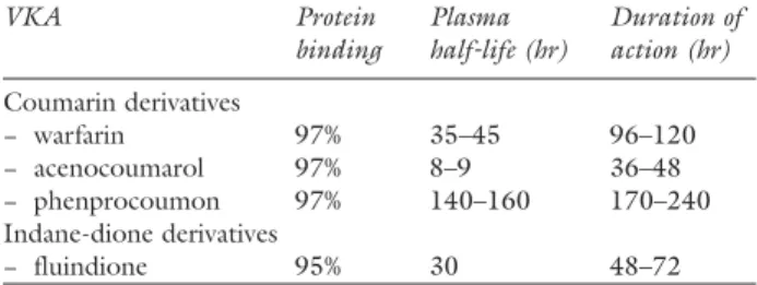
6. Vitamin K antagonists
Various VKA are available and some of their character-istics are presented in Table III.
Treatment is usually started the same day as hepa-rin, without a loading dose. Four to six days are usually necessary to obtain two international normalized ratio (INR) of the prothrombin time within the therapeutic range (2.0–3.0) and therefore there is an overlap of the two drugs for a few days. The minimal duration of heparin therapy should be five days. When the tar-get INR is obtained at least twice at an interval of 24 hr, INR controls may be less frequent. It is generally advised to control the INR once a week during the first month of treatment, then monthly thereafter.
If the patient is receiving other medications, pos-sible drug interactions should be considered. It should be kept in mind that any modification of alternate treatments (change of drug or any change of dosage of a drug already given) may modify the interaction with VKA and therefore change the INR.16 Other factors will influence anticoagulation, mainly liver and renal diseases, fever, hyperthyroidism as well as food. The so-called “VKA resistance” is often due to a poor understanding of anticoagulation by the patient. Bleeding risk depends on numerous fac-tors such as patient compliance, concomitant intake of other drugs, intensity and variability of the INR, comorbidities, age more than 80 yr and length of treatment.16,17
S82 CANADIAN JOURNALOF ANESTHESIA
TABLE II Doses of LMWH for the treatment of venous thromboembolism
LMWH Recommended doses
Enoxaparin 100 U (1 mg·kg–1) twice daily
Nadroparin 85 U·kg–1 twice daily or 170 U·kg–1 once daily Dalteparin 100 U·kg–1 twice daily
Tinzaparin 175 U·kg–1 once daily LMWH = low molecular weight heparins.
de Moerloose et al.: TREATMENTOFVENOUSTHROMBOEMBOLISM S83
The optimal duration of anticoagulation has been the object of several studies and debate.18–26 For distal DVT, six weeks may be enough although the seventh American College of Chest Physicians Conference on Antithrombotic and Thrombolytic therapy pub-lished recently in Chest2 proposes three months of anticoagulation for a symptomatic DVT confined to the calf veins. For a proximal DVT or a PE with an identifiable cause (for example after hip arthroplasty), anticoagulation is required for three months after a first episode. When VTE is considered idiopathic (no obvious triggering factor), anticoagulation should last at least six months, or longer if the risk of bleeding is low. A longer duration of anticoagulation should be contemplated when the risk factor is permanent (cancer for example), when a severe thrombophilia (e.g., antiphospholipid antibodies, antithrombin defi-ciency, combined factor V Leiden and prothrombin G20210A mutations) is detected or when there is recurrence of VTE. In these situations the treatment should be individualized. Two studies have evaluated specifically whether, after a minimum of three months’ treatment with a target INR ranging between 2.0 and 3.0, an INR maintained subsequently between 1.5 and 2.0 retains the antithrombotic benefit while decreas-ing the risk of bleeddecreas-ing complications.25,26 Conflicting results were reported. For some patients indefinite anticoagulant therapy should be considered; however the risk-benefit of continuing such a treatment should be reassessed periodically.
Recent studies suggest that D-dimer levels and ultrasonography after withdrawal of anticoagulation may be useful in estimating the risk of recurrence of VTE.27–29 Increased D-dimer levels were associated with a higher risk of subsequent recurrence. However, it is not yet known whether D-dimer levels or residual venous thrombosis have a role in adjusting the dura-tion of oral anticoagulant therapy. Clinical trials are ongoing to evaluate this possibility.
7. Low molecular weight heparin for the long-term treatment of patients with cancer
Long-term treatment with adjusted doses of a VKA is highly effective for preventing recurrent VTE in patients free of malignancy. This approach is less effec-tive in patients with aceffec-tive cancer and is associated with a higher risk of bleeding than in patients without malignancy.30 Two recent randomized clinical trials have assessed the potential benefit of long-term treat-ment of acute VTE with sc LMWH as an alternative approach to VKA in patients with cancer and acute DVT or PE.31,32 In a study including 676 patients, sc dalteparin alone for six months (200 IU·kg–1 once daily for one month, followed by 150 IU·kg–1 once daily for five months) was compared with dalteparin 200 IU·kg–1 body weight sc once daily for five to seven days, followed by oral treatment with VKA for six months (target INR ranging from 2.0 to 3.0).31 During the six-month study period, recurrent VTE occurred in 15.7% of patients who received the VKA treatment, compared with 8.0% of those who received a LMWH (dalteparin) alone (P = 0.002). Rates of majorbleeding were similar between the two groups. In the second study, Meyer et al. compared enoxapa-rin sodium (1.5 mg·kg–1 once daily) with VKA given for three months in 146 patients.32 The rate of major outcome event (defined as major bleeding or recur-rent VTE within three months) was 21.1% in patients assigned to receive VKA compared with 10.5% in patients assigned to receive enoxaparin (P = 0.09). The trend in favour of enoxaparin was accounted for, mainly, by a higher rate of major bleeding in the VKA group (16% vs 7.0%, P = 0.09). These studies suggest that prolonged LMWH therapy is more effective and possibly safer than oral anticoagulation. Recently, the American College of Chest Physicians consensus con-ference on antithrombotic therapy strongly recom-mended LMWH for the first three to six months of long-term anticoagulant therapy in cancer patients.2 8. New anticoagulants
Several new antithrombotic agents have been devel-oped in recent years. Until now, only the efficacy and safety of fondaparinux and ximelagatran in the treat-ment of established VTE have been evaluated in large phase III trials.
FONDAPARINUX
Fondaparinux is a synthetic analogue of the natu-rally occurring pentasaccharide sequence and, like UFH and LMWH, acts as an anticoagulant by binding antithrombin. Fondaparinux catalyses fac-tor Xa inhibition by antithrombin, but has no effect
TABLE III Main characteristics of vitamin K antagonists
VKA Protein Plasma Duration of binding half-life (hr) action (hr)
Coumarin derivatives − warfarin 97% 35–45 96–120 − acenocoumarol 97% 8–9 36–48 − phenprocoumon 97% 140–160 170–240 Indane-dione derivatives − fluindione 95% 30 48–72
S84 CANADIAN JOURNALOF ANESTHESIA on the rate of thrombin inactivation. Fondaparinux
has an excellent bioavailability after sc injection and is administered once a day. In an open label study, 2,213 patients with symptomatic PE were randomly assigned to receive either fondaparinux (5.0, 7.5 or 10 mg in patients weighing less than 50, 50 to 100 or more than 100 kg respectively) sc once daily or a continuous infusion of UFH for at least five days fol-lowed by VKA for six months.33 The study showed that the incidence of VTE recurrence at three months was 3.8% and 5.0% in the fondaparinux and the UFH group respectively, demonstrating non-inferiority of fondaparinux/VKA vs UFH/VKA. The same regi-men of fondaparinux was compared with enoxaparin 1 mg·kg–1 twice daily for at least five days followed by VKA for three months in a randomized double-blind trial including 2,305 patients with acute DVT.34 The study showed that during a three-month follow up, 3.9% of the fondaparinux-treated patients had symp-tomatic recurrent VTE compared with 4.1% of the enoxaparin-treated patients, demonstrating that once-daily fixed-dose fondaparinux was at least as effective as twice-daily body-weight adjusted enoxaparin. In both studies, the rates of major bleeding did not differ significantly between groups. Fondaparinux is particu-larly interesting because of its synthetic origin and the lack of heparin-induced thrombocytopenia reported until now.
XIMELAGATRAN
Ximelagatran, which can be given orally, is the prodrug of melagatran. Melagatran is a synthetic low molecu-lar weight competitive thrombin inhibitor. This new agent produces a more predictable anticoagulant response than heparin so that no coagulation moni-toring is required. Moreover, in comparison with oral VKA, food or alcohol do not interfere with this new agent and very few drug interactions with drugs have been demonstrated.35 In a randomized double-blind study, 1,233 patients with VTE who had completed six months of standard anticoagulation therapy were randomly assigned to extended secondary prophy-laxis with ximelagatran 24 mg twice a day vs placebo for 18 months without monitoring of coagulation.36 The study showed that, compared with placebo, ximelagatran significantly reduced the cumulative risk of recurrence from 12.6% to 2.8% over 18 months. Bleeding rates did not differ significantly between the two groups. Ximelagatran-treated patients were more likely to have increases in serum alanine ami-notransferase greater than three times the upper limit of normal compared to placebo (6.2% vs 1.2%). Elevations in liver enzymes mostly occurred between
two and six months of treatment and most of them resolved either spontaneously or after discontinuation of treatment. The efficacy and safety of ximelagatran in the treatment of acute DVT has been evaluated in another randomized double-blind study including 2,491 patients.37 Patients were randomly assigned to receive either ximelagatran 36 mg twice a day or enoxaparin 1 mg·kg–1 for at least five days followed by warfarin for six months. The study showed that the cumulative risk of VTE recurrence at six months was 2.1% and 2.0% in the ximelagatran and the enoxaparin group respectively, demonstrating non-inferiority of ximelagatran vs enoxaparin/warfarin. The incidence of major bleeding did not differ significantly between the two groups. The increase of liver enzymes was, again, a matter of very serious concern, which finally leads to withdrawal of the drug.
In conclusion, these two drugs have been shown to be at least as effective and safe as standard therapy in the initial treatment of VTE Ximelagatran has been withdrawn from the market but several new anti-Xa or anti-thrombin inhibitors are under evaluation. Cost-effectiveness analyses will be required to determine the role of these new agents in this clinical setting.
B. Other treatments
1. Compression elastic stockings
Within three years after an episode of DVT of the lower extremities, more than 30% of patients develop pain, swelling, skin pigmentation and venous dilata-tion (and, eventually, leg ulceradilata-tion), all complicadilata-tions usually referred to as postthrombotic syndrome.38
Elastic compression stockings with a pressure of 30 to 40 mmHg at the ankle have been shown to be cru-cial to prevent postthrombotic syndrome. In two ran-domized studies, patients with acute proximal DVT were assignedto wear or not wear compression elastic stockings.39,40 In both studies, compression stock-ings reduced the rate of postthrombotic syndrome by approximately 50%.
Elastic bandages should be used during the first days in combination with leg elevation and ambula-tion in order to prevent stasis. When edema stabilizes, compression stockings are prescribed. The stockings will have to be worn as long as the edema persists. Ideally stockings should be worn at least two years.39 2. Thrombolysis
Thombolysis (as well as thrombectomy) has been recommended for the initial treatment of DVT, based on the assumption that earlier vein patency will preserve venous function and therefore decrease the risk of postthrombotic syndrome. However, the rate
de Moerloose et al.: TREATMENTOFVENOUSTHROMBOEMBOLISM S85 of clinically relevant postthrombotic sequelae does
not appear to differ from that of patients receiving conventional anticoagulation. Therefore, in case of DVT, systemic thrombolysis is usually not justified. Indeed, this treatment carries a risk of major bleeding which is not justified in the case of a non-fatal disease. However, in very rare cases such as patients presenting with phlegmasia cerulea dolens, thrombolysis can be considered. Local thrombolysis may, possibly, provide some benefit but the place of this labour-intensive approach in the management of VTE remains to be established.41
For most patients with PE, systemic thrombo-lytic therapy is not recommended. However in selected patients, thrombolysis can be beneficial.42,43 Thrombolysis is used for massive PE with arte-rial hypotension (< 100 mmHg) and/or a shock. Hemodynamically stable patients with acute PE and echocardiographic abnormalities (right ventricular dysfunction) and/or significant arterial hypertension might also benefit from thrombolysis.44 However, the use of thrombolytic agents in this setting remains controversial.2 In a randomized study, 256 patients with acute PE and pulmonary hypertension or right ventricular dysfunction (but without arterial hypoten-sion or shock) were assigned to receive either heparin plus 100 mg of alteplase or heparin plus placebo over a periodof two hours.45 The study showed that alteplase given in conjunction with heparin could prevent clinical deterioration requiring escalation of treatment. However, no difference between both treatment groups was observed in clinically relevant outcomes such as recurrent non-fatal or fatal PE. Further studies are required to document a clinically relevant improvement with thrombolytic therapy in these patients.
The local administration of thrombolytic therapy via a catheter is not recommended.
Although often relative, contraindications to throm-bolysis should be observed. Contraindications should be balanced against the patient’s clinical status as well as the patient’s prognosis. For example, if the patient is treated with VKA, it is possible to give a preparation of prothrombin complex concentrate which allows normalization of the INR within a few minutes.
Thrombolysis is initiated via a peripheral vein, since there is no clear advantage to administration of a thrombolytic agent in the pulmonary artery. Several agents are available: streptokinase (loading dose of 250 000 UI in 20 min, followed by a continuous infusion of 100 000 UI·hr–1 during 12 to 24 hr), urokinase (loading dose of 4400 UI·kg–1 in ten minutes, fol-lowed by a continuous infusion of 4400 UI·kg–1·hr–1
during 12 to 24 hr) or alteplase (50 mg·hr–1 during two hours, for a total of 100 mg). Various other pro-tocols exist. At the end of the infusion of alteplase, heparin (1000 UI·hr–1) is started and anticoagulation is adjusted according to the aPTT.45 For streptoki-nase or urokistreptoki-nase, heparin is usually begun when the fibrinogen level is above 1.0 g·L–1 and the aPTT less than twice the control value.
3. Thrombectomy and embolectomy
Both are seldom indicated. A surgical thrombectomy may be considered in case of suspended iliac DVT, and an embolectomy can be performed in case of car-diorespiratory arrest or pulmonary hypertension with fresh emboli. The decision to perform such a proce-dure in a severely ill patient is extremely delicate and should be individualized. The decision will rely, espe-cially, on the evolution of the patient under medical treatment, on the contraindications to thrombolysis, on the localization of clots as well as on the surgical team’s experience.46 Various iv catheter systems have been developed over the years to allow the division of clots into fragments and their aspiration; such non-surgical embolectomies might represent an interesting option in some patients.
4. Vena cava filters
A study has shown the limited utility of definitive vena cava filters in case of DVT.47 The main indication is the presence of a proximal DVT with an absolute contra-indication to anticoagulants (or a bleeding complica-tion secondary to anticoagulacomplica-tion). Other indicacomplica-tions may be considered such as well documented recurrent episodes of VTE under therapeutic anticoagulation. Finally, patients with postembolic chronic cor pulmo-nale (any recurrence might be lethal), patients having undergone a recent surgical embolectomy for acute PE as well as those having had a thrombo-endarter-ectomy should be considered candidates for this form of treatment.
Conclusion
Anticoagulant treatment has demonstrated its efficacy to prevent recurrent episodes of VTE. New agents, such as anti-Xa or oral thrombin inhibitors, are prom-ising alternatives for heparins or VKA. As far as other treatments are concerned, the most important are the systematic use of elastic stockings in case of DVT and, possibly, the administration of thrombolytic agents in case of massive PE. Vena cava filters are mainly indi-cated for patients with a proximal DVT and a contra-indication to anticoagulation.
S86 CANADIAN JOURNALOF ANESTHESIA
References
1 Moser KM, Fedullo PF, LitteJohn JK, Crawford R. Frequent asymptomatic pulmonary embolism in patients with deep vein thrombosis. JAMA 1994; 271: 223–5.
2 Büller HR, Agnelli G, Hull RD, Hyers TM, Prins MH, Raskob GE. Antithrombotic therapy for venous throm-boembolic disease: the Seventh ACCP Conference on Antithrombotic and Thrombolytic therapy. Chest 2004; 126 (suppl 3): 401S-8S.
3 Gould MK, Dembitzer AD, Doyle RL, Hastie TJ, Garber AM. Low-molecular-weight heparins compared with unfractionated heparin for treatment of acute deep venous thrombosis. A meta-analysis of randomized, controlled trials. Ann Intern Med 1999; 130: 800–9. 4 Wheeler AP, Jaquiss RD, Newman JH. Physician
prac-tices in the treatment of pulmonary embolism and deep venous thrombosis. Arch Intern Med 1998; 148: 1321–5.
5 Simonneau G, Sors H, Charbonnier B, et al. A com-parison of low-molecular-weight heparin with unfrac-tionated heparin for acute pulmonary embolism. The THESEE Study Group. Tinzaparine ou Heparine Standard: Evaluations dans l’Embolie Pulmonaire. N Engl J Med 1997; 337: 663–9.
6 Anonymous. Low-molecular-weight heparin in the treatment of patients with venous thromboembolism. The Columbus Investigators. N Engl J Med 1997; 337: 657–62.
7 Hirsh J, Raschke R. Heparin and low-molecular-weight heparin: the Seventh ACCP Conference on Antithrombotic and Thrombolytic Therapy. Chest 2004; 126: 188S–203.
8 Zidane M, Schram MT, Planken EW, et al. Frequency of major hemorrhage in patients treated with unfrac-tionated intravenous heparin for deep venous thrombo-sis or pulmonary embolism: a study in routine clinical practice. Arch Intern Med 2000; 160: 2369–73. 9 Levine MN, Hirsh J, Gent M, et al. A randomized trial
comparing activated thromboplastin time with heparin assay in patients with acute venous thromboembolism requiring large daily doses of heparin. Arch Intern Med 1994; 154: 49–56.
10 Prandoni P, Carnovali M, Marchiori A; Galilei Investigators. Subcutaneous adjusted-dose unfractionat-ed heparin vs fixunfractionat-ed-dose low-molecular-weight heparin in the initial treatment of venous thromboembolism. Arch Intern Med 2004; 164: 1077–83.
11 Bounameaux H, de Moerloose P. Is laboratory monitor-ing of low-molecular-weight heparin therapy necessary? No. J Thromb Haemost 2004; 2: 551–4.
12 Levine M, Gent M, Hirsh J, et al. A comparison of low-molecular-weight heparin administered primarily at
home with unfractionated heparin administered in the hospital for proximal deep-vein thrombosis. N Engl J Med 1996; 334: 677–81.
13 Koopman MM, Prandoni P, Piovella F, et al. Treatment of venous thrombosis with intravenous unfractionated heparin administered in the hospital as compared with subcutaneous low-molecular-weight heparin adminis-tered at home. The Tasman Study Group. N Engl J Med 1996; 334: 682–7.
14 Charbonnier BA, Fiessinger JN, Banga JD, Wenzel E, d’Azemar P, Sagnard L; on behalf of the FRAXODI group. Comparison of a once daily with a twice daily subcutaneous low molecular weight heparin regimen in the treatment of deep vein thrombosis. Thromb Haemost 1998; 79: 897–901.
15 Boneu B, de Moerloose P. How and when to monitor a patient treated with low molecular weight heparin. Semin Thromb Hemost 2001; 27: 519–22.
16 Ansell J, Hirsh J, Poller L, Bussey H, Jacobson A, Hylek E. The pharmacology and management of the vita-min K antagonists: the Seventh ACCP Conference on Antithrombotic and Thrombolytic Therapy. Chest 2004; 126 (suppl 3): 204S–33.
17 Levine MN, Raskob G, Beyth RJ, Kearon C, Schulman S. Hemorrhagic complications of anticoagulant treatment: the Seventh ACCP Conference on Antithrombotic and Thrombolytic Therapy. Chest 2004; 126(suppl 3): 287S–310.
18 Schulman S, Rhedin AS, Lindmarker P, et al. A comparison of six weeks with six months of oral anticoagulant therapy after a first episode of venous thromboembolism. Duration of Anticoagulation Trial Study Group. N Engl J Med 1995; 332: 1661–5. 19 Bounameaux H, de Moerloose P, Sarasin FP. Optimal
duration of oral anticoagulant therapy following deep vein thrombosis of lower limbs. Blood Coagul Fibrinolysis 1996; 7: 507–14.
20 Schulman S, Granqvist S, Holmström M, et al. The duration of oral anticoagulant therapy after a second episode of venous thromboembolism. The Duration of Anticoagulation Trial Study Group. N Engl J Med 1997; 336: 393–8.
21 Kearon C, Gent M, Hirsh J, et al. A comparison of three months of anticoagulation with extended anti-coagulation for a first episode of idiopathic venous thromboembolism. N Engl J Med 1999; 340: 901–7. 22 Agnelli G, Prandoni P, Santamaria MG, et al.; for the Warfarin Optimal Duration Italian Trial Investigators. Three months versus one year of oral anticoagu-lant therapy for idiopathic deep venous thrombosis. Warfarin Optimal Duration Italian Trial Investigators. N Engl J Med 2001; 345: 165–9.
de Moerloose et al.: TREATMENTOFVENOUSTHROMBOEMBOLISM S87 Warfarin Optimal Duration Italian Trial Investigators.
Extended oral anticoagulant therapy after a first epi-sode of pulmonary embolism. Ann Intern Med 2003; 139: 19–25.
24 Pinede L, Ninet J, Duhaut P, et al.; for the Investigators of the “Durée optimale du Traitement AntiVitamines K” (DOTAVK). Comparison of 3 and 6 months of oral anticoagulant therapy after a first episode of proximal deep vein thrombosis or pulmonary embolism and comparison of 6 and 12 weeks of therapy after isolated calf deep vein thrombosis. Circulation 2001; 103: 2453–60.
25 Ridker, PM, Goldhaber SZ, Danielson E, et a.l; for the PREVENT Investigators. Long-term, low-intensity warfarin therapy for the prevention of recurrent venous thromboembolism. N Engl J Med 2003; 348: 1425– 34.
26 Kearon C, Ginsberg JS, Kovacs MJ, et al.; for the Extended Low-Intensity Anticoagulation for Thrombo-Embolism (ELATE) Investigators. Comparison of low-intensity warfarin therapy with conventional-low-intensity warfarin therapy for long-term prevention of recurrent venous thromboembolism. N Engl J Med 2003; 349: 631–9.
27 Palareti G, Legnani C, Cosmi B, et al. Predictive value of D-dimers test for recurrent venous thromboembo-lism after anticoagulation withdrawal in subjects with a previous idiopathic event and in carriers of congenital thrombophilia. Circulation 2003; 108: 313–8. 28 Eichinger S, Minar E, Bialonczyk C, et al. D-dimer
levels and risk of recurrent venous thromboembolism. JAMA 2003; 290: 1071–4.
29 Prandoni P, Lensing AW, Prins MH, et al. Residual venous thrombosis as a predictive factor of recurrent venous thromboembolism. Ann Intern Med 2002; 137: 955–60.
30 Prandoni P, Lensing AW, Piccioli A, et al. Recurrent venous thromboembolism and bleeding complications during anticoagulant treatment in patients with cancer and venous thrombosis. Blood 2002; 100: 3484–8. 31 Lee AY, Levine MN, Baker RI, et al.
Low-molecular-weight heparin versus a coumarin for the prevention of recurrent venous thromboembolism in patients with cancer. N Engl J Med 2003; 349: 146–53.
32 Meyer G, Marjanovic Z, Valcke J, et al. Comparison of low-molecular-weight heparin and warfarin for the secondary prevention of venous thromboembolism in patients with cancer: a randomized controlled study. Arch Intern Med 2002; 162: 1729–35.
33 Buller HR, Davidson BL, Decousus H, et al.; for the Matisse Investigators. Subcutaneous fondaparinux ver-sus intravenous unfractionated heparin in the initial treatment of pulmonary embolism. N Engl J Med
2003; 349: 1695–702.
34 Buller HR, Davidson BL, Decousus H, et al.; for the Matisse Investigators. Fondaparinux or enoxaparin for the initial treatment of symptomatic deep venous thrombosis: a randomized trial. Ann Intern Med 2004; 140: 867–73.
35 Bredberg E, Andersson TB, Frison L, et al. Ximelagatran, an oral direct thrombin inhibitor, has a low potential for cytochrome P450-mediated drug-drug interactions. Clin Pharmacokinet 2003; 42: 765–77.
36 Schulman S, Wahlander K, Lundstrom T, Clason SB, Eriksson H; THRIVE III Investigators. Secondary prevention of venous thromboembolism with the oral direct thrombin inhibitor ximelagatran. N Engl J Med 2003; 349: 1713–21.
37 Fiessinger JN, Huisman MV, Davidson BL, et al.; THRIVE Treatment Study Investigators. Ximelagatran vs low-molecular-weight heparin and warfarin for the treatment of deep vein thrombosis: a randomized trial. JAMA 2005; 293: 681–9.
38 Bernardi E, Bagatella P, Frulla M, Simioni P, Prandoni P. Postthrombotic syndrome: incidence, prevention, and management. Semin Vasc Med 2001; 1: 71–80. 39 Brandjes DP, Büller HR, Heijboer H, et al. Randomised
trial of effect of compression stockings in patients with symptomatic proximal-vein thrombosis. Lancet 1997; 349: 759–62.
40 Prandoni P, Lensing AW, Prins MH, et al. Below-knee elastic compression stockings to prevent the post-thrombotic syndrome: a randomized, controlled trial. Ann Intern Med 2004; 141: 249–56.
41 Verhaeghe R, Maleux G. Endovascular local throm-bolytic therapy of iliofemoral and inferior caval vein thrombosis. Sem Vasc Med 2001; 1: 123–8. 42 Goldhaber SZ. Pulmonary embolism
thromboly-sis: broadening the paradigm for its administration. Circulation 1997; 96: 716–8.
43 Dalen JE. The uncertain role of thrombolytic therapy in the treatment of pulmonary embolism. Arch Intern Med 2002; 162: 2521–3.
44 Konstantinides S, Geibel A, Olschewski M, et al. Association between thrombolytic treatment and the prognosis of hemodynamically stable patients with major pulmonary embolism: results of a multicenter registry. Circulation 1997; 96: 882–8.
45 Konstantinides S, Geibel A, Heusel G, Heinrich F, Kasper W; Management Strategies and Prognosis of Pulmonary Embolism-3 Trial Investigators. Heparin plus alteplase compared with heparin alone in patients with submassive pulmonary embolism. N Engl J Med 2002; 347: 1143–50.
46 Aklog L, Williams CS, Byrne JG, Goldhaber SZ. Acute pulmonary embolectomy: a contemporary approach.
S88 CANADIAN JOURNALOF ANESTHESIA Circulation 2002; 105: 1416–9.
47 Decousus H, Leizorovicz A, Parent F, et al. A clinical trial of vena cava filters in the prevention of pulmonary embolism in patients with proximal deep-vein throm-bosis. Prevention du Risque d’Embolie Pulmonaire par Interruption Cave Study Group. N Engl J Med 1998; 338: 409–15.