Acta Neurochir (Wien) (1997) 139:267-270 Acta Neurochirurgica 9 Springer-Verlag 1997 Printed in Ausma
Letter to the Editor: Classification of Pituitary Adenomas
T. M i n d e r m a n nDepartment of Neurosurgery, University Hospitals Basel, Basel, Switzerland
In their article, Sanno
et al.
[15] address an impor- tant issue concerning the classification of pituitary adenomas. Their proposed classification is based on the tumour's histological data. Yet, pituitary adeno- mas are treated by a number of specialists, not all of whom have histological data available. Therefore, I would like to present an alternative classification scheme which can be shared by endocrinologists, gynaecologists, ophthalmologists, neurosurgeons, pathologists, and researchers alike.Advances in the understanding of an adenoma's hormone synthesis (genotype), hormone storage (immunophenotype), and hormone release into the blood stream (clinical phenotype) have lead to sever- al parallel and sometimes confusing classifications of pituitary adenomas. The present confusion on what exactly is, e.g., a prolactinoma or a corticotroph cell adenoma (does it produce, store, or release A C T H ? ) has lead to the occasional use o f such terms as "PRL- oma", " G H - o m a " , " A C T H - o m a " , and " T S H - o m a " [2, 16] or "somatotropinoma" and "corticotropinoma" [3]. Unfortunately these terms do not clarify whether in the case of " P R L - o m a s " the adenomatous cells pro- duce, store, or release PRL or if the excessive release of PRL is caused by the stalk effect or raised intrasel- lar pressure which would mean that the adenomatous cells do not even derive from the mammotroph cell line. Cunently, due to these difficulties and to overlap created by the various terminologies, tumours of the same cell line may be classified differently depending on whether one refers to their genotype, immuno- phenotype, or clinical phenotype. Vice versa, tumours deriving from different cell lines may incorrectly be classified as identical tumours. In an attempt to over- come the difficulties in making a correct diagnosis
based on clinical data, some authors have developed regression equations in order to extract most informa- tion from the clinical data [5]. The introduction of mathematical tools to improve the accuracy of the clinical diagnosis demonstrates the great difficulties encountered when trying to determine an adenoma's true nature in the absence of histological data. On the other hand, classifications based on a tumour's histo- logical examination can often not be shared with cli- nicians who treat their patients before or without sur- gery.
Therefore, I would like to emphasize an unequivo- cal classification of the various adenoma types by crossreferencing an adenoma's cell line of origin, its immunoreactivity, and its clinical phenotype. At the same time, existing anatomical classifications should be used to complement this classification. The aim is to come to a diagnosis that can be shared by research- ers, pathologists, and clinicians.
Proposed Classification of Pituitary Adenomas
The system is based on the work by Kovacs [8] and Landolt [9] whose contributions replaced the classical histological designation of pituitary adenomas as acidophilic, basophilic, and chromophobic with the more meaningful designation as adenomas o f a cer- tain anterior lobe cell line. It takes into account new findings that link somatotroph, mixed mammotroph and somatotroph, and mammosomatotroph adenomas to one basic cell line committed to the production of growth hormone [12].
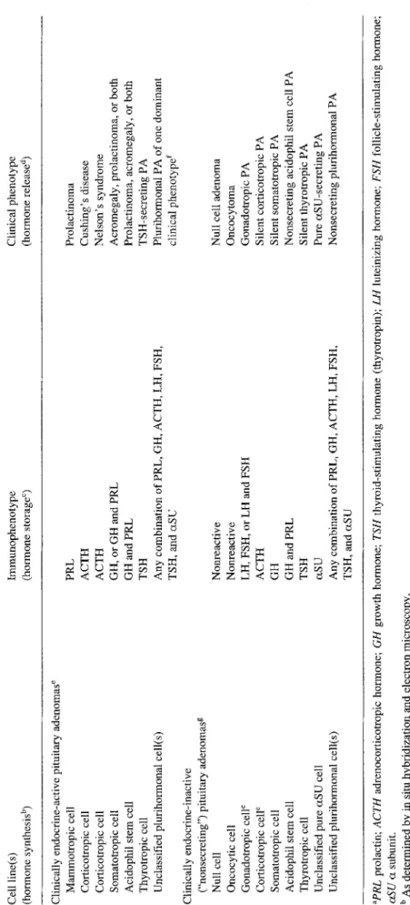
The classification presented in Table 1 has been published elsewhere [18]. It takes into account an adenoma's direct and indirect (stalk effect, raised
Table 1. Classification of Pituitary Adenomas According to ~ Cell line(s) lmmunophenotype Clinical phenotype (hormone synthesis b) (hormone storage ~) (hormone release a) bO Go Clinically endocrine-active pituitary adenomas e Mammotropic cell Corticotropic cell Corticotropic cell Somatotropic cell Acidophil stem cell Thyrotropic cell Unclassified plurihormonal cell(s) Clinically endocrine-inactive ("nonsecreting") pituitary adenomasg Null cell Oncocytic cell Gonadotropic cell ~ Corticotropic celY Somatotropic cell Acidophil stem cell Thyrotropic cell Unclassified pure ctSU cell Unclassified plurihormonal cell(s) PRL ACTH ACTH GH, or GH and PRL GH and PRL TSH Any combination of PRL, GH, ACTH, LH, FSH, TSH, and c~SU Nonreactive Nonreactive LH, FSH, or LH and FSH ACTH GH GH and PRL TSH ctSU Any combination of PRL, GH, ACTH, LH, FSH, TSH, and c~SU Prolactinoma Cushing's disease Nelson's syndrome Acromegaly, prolactinoma, or both Prolactinoma, acromegaly, or both TSH-secreting PA Plurihormonal PA of one dominant clinical phenotypd Null cell adenoma Oncocytoma Gonadotropic PA Silent corticotropic PA Silent somatotropic PA Nonsecreting acidophil stern cell PA Silent thyrotropic PA Pure ctSU-secreting PA Nonsecreting plurihormonal PA aPRL prolactin; ACTH adrenocorticotropic hormone; GH growth hormone; TSH thyroid-stimulating ho~xnone (thyrotropin); LH luteinizing hormone; FSH follicle-stimulating hormone; aSU ct subunit. b As determined by in situ hybridization and electron microscopy. As determined by electron microscopy and immunostaining. As determined clinically by symptoms, signs, and serum hormone levels. Cosynthesis of the clinically inactive c~SU and its release into the bloodstream is possible. e LH and FSH are clinically inactive; their release into the bloodstream can be measured. g May present clinically as prolactinoma when stalk effect or raised intrasellar pressure leads to an increased release of prolactin by non-adenomatous cells or when PRL is cosynthesized by tumorous cells. .=. ;> O
T. Mindermann: Classification of Pituitary Adenomas
intrasellar pressure) endocrine activities. The direct endocrine activities are: hormone synthesis (geno- type), hormone storage (immunophenotype), and hor- mone release (clinical phenotype). The indirect endo- crine activities are: compression of non-adenomatous anterior lobe tissue resulting in partial pituitary insuf- ficiency and excessive release of PRL by non-ade- nomatous cells caused by the stalk effect or raised intrasellar pressure.
The genotype is determined by molecular genetic techniques like in-situ hybridization [13] allowing one to identify both DNA and mRNA in adenomatous cells revealing their genetic fingerprint. It is the most accurate information obtainable concerning the cell line of origin and is mostly used in research.
The immunophenotype is determined by the tumour's immunostaining properties when adenoma- tous tissue is stained with antibodies to the various anterior lobe hormones or hormonal compounds such as the alpha-subunit. This technique reveals which hormone(s) or hormonal compound(s) are stored by tumorous cells. It is a less accurate way of determin- ing an adenoma's cell line of origin since hormonal products may be released without the delay of tissue storage. Immunostaining for the various anterior lobe hormones should be part of today's routine diagnostic procedures.
The clinical phenotype is the least accurate infor- mation concerning the nature of a pituitary adenoma because: (a) hormone excess may be insufficient to cause any clinical symptoms or signs, (b) release of hormonal products may be clinically silent, (c) due to raised intrasellar pressure or the stalk effect prolactin may be released in sufficient amounts to cause ame- norrhea and galactorrhea even though the tumour might not produce prolactin [1, 10, 11], (d) the clini- cal phenotype may be determined by only one of mul- tiple co-existing tumours of differing immunopheno- types [7], (e) tumours may change clinical and immu- nophenotype [14], and (f) in addition to hormone excess, tumours may cause partial pituitary insuffi- ciency.
The proposed classification scheme keeps the doc- tor aware of the degree of certainty concerning the true nature of a pituitary adenoma. For obvious rea- sons, a classification of a patient's adenoma based on clinical data only leads to the least reliable diagnosis. One might grade the three groups represented in the three columns to which a pituitary adenoma may be designated as fair evidence (clinical phenotype), good
269 evidence (immunophenotype), or best evidence (cell line) for the correct diagnosis.
The anatomical classification schemes developed by Hardy [4] and modified by Wilson [17] grade the tumour's extent of sellar destruction and its extrasel- lar extension. A newly developed classification by Knosp et al. [6] based on magnetic resonance imaging (MRI) grades an adenoma's invasion into the cavern- ous sinus space. At surgery, tumour extending beyond the intercarotid line in coronal MRI proved very like- ly to be associated with invasion [6]. These purely anatomical classifications should be used in addition since the data contained in these schemes have prog- nostic value and are helpful in choosing the best treat- ment.
The proposed classification in combination with the various grading schemes should facilitate interdis- ciplinary and interinstitutional communication on what tumour exactly clinicians, pathologists, differ- ent institutions and articles in scientific publications are referring to. It is designed to highten the aware- ness that there are several aspects characterizing a pituitary adenoma ranging from its cell line of origin to its immunophenotype and to its clinical presenta- tion. A hightened awareness of such aspects may help in improving study designs and in the understanding of the tumour biology and the clinical course of the various adenoma types.
References
1. Anonymous (1987) Hype~rolactinaemia. When is a prolactin- oma not a prolactinoma? Lancet 2:1002-1004
2. Beck-Peccoz P (1993) Unusual presentation of a TSH-secret- ing pituitary adenoma. Acta Endocrinol 129:283
3. Faglia G (1993) Epidemiology and pathogenesis of pituitary adenomas. Acta Endocrinol 129 [Suppl 1J: 1-5
4. Hardy J, Wigser SM (1965) Trans-sphenoidal surgery of pitui- tary fossa tumors with televised radiofluoroscopic control. J Neurosurg 23:612-619
5. Hermzo-Cabrera I, Herruzo-Cabrera R, Errazquin-Saez de Tejada L, Garcia-Fernandez JL, Vidarte-Zahala M, Mayer F, Zaragoza-Rubira JR (1992) A multivariant study of pituitary adenoma, obtainment of two logistic regression equations as an auxiliary support in the diagnosis of these tumors. Neoplasma 39:255-260
6. Knosp E, Steiner E, Kitz K, Matula C (1993) Pituitary adeno- mas with invasion of the cavernous sinus space: a magnetic resonance imaging classification compared with surgical find- ings. Neurosurgery 33:610-618
7. Kontogeorgos G, Scheithauer BW, Horvath E, Kovacs K, Lloyd RV, Smyth HS, Rologis D (1992) Double adenomas of the pituitary: a clinicopathologicai study of 11 tumors. Neuro- surgery 31: 840-849
270
Hartmann WH, Sobin LH (eds) Atlas of tumor pathology. Armed Forces Institute of Pathology, Washington DC, pp
1-252
9. Landolt AM (1975) Ultrastructure of human sella tumors. Cor- relation of clinical findings and morphology. Acta Neurochir (Wien) [Suppl] 22:1-167
10. Lees PD (1990) Intrasellar pressure. Acta Neurochir (Wien) [Suppl] 47:68-70
11. Lees PD, Pickard JD (1987) Hyperprolactinemia, intrasellar pituitary pressure, and the stalk compression syndrome. J Neu- rosurg 67:192-196
12. Li J, Stefaneanu L, Kovacs K, Horvath E, Smyth HS (1993) Growth hormone (GH) and prolactin (PRL) gene expression and immunoreactivity in GH- and PRL-producing human pitui- tary adenomas. Virchows Archly A Pathol Anat 422:193-201 13. Lloyd RV, Jin L, Chandler WF (1991) In situ hybridization in
the study of pituitary tissues. Path Res Pract 187:552-555 14. Mindermann T, Kovacs K, Wilson CB (1994) Changes in
T. Mindermann: Classification of Pituitary Adenomas immunophenotype of recurrent pituitary adenomas. Neurosur- gery 35:39-44
15. Sanno N, Teramoto A, Osamura RY (1996) Clinical and cyto- functional classification of pituitary adenomas: proposal of a new classification. Acta Neurochir (Wien) 138:1186-1192 16. Spada A, Vallar L, Faglia G (1994) Cellular alterations in pitui-
tary tumors. Eur J Endocrinol 130:43-52
17. Wilson CB (1983) Surgical management of endocrine-active pituitary adenomas. In: Walker MD (ed) Oncology of the ner- vous system. Martinus Nijhoff, Boston, pp 117-150
18. Wilson CB, Mindermann T (1997) Pituitary neoplasms. In: Holland JF, et al (eds) Cancer medicine, 4th Ed. Williams and Wilkins, Baltimore, pp 1539-1550
Correspondence: Dr. Thomas Mindermann, Department of Neurosurgery, University Hospitals Basel, CH-4031 Basel, Swit- zerland.