Florian M. Buck Bernhard Jost Juerg Hodler Received: 13 January 2008 Revised: 17 May 2008 Accepted: 7 June 2008 Published online: 11 July 2008
# European Society of Radiology 2008
Shoulder arthroplasty
Abstract Shoulder prostheses are now commonly used. Clinical results and patient satisfaction are usually good. The most commonly used types are humeral hemiarthroplasty, uncon-strained total shoulder arthroplasty, and semiconstrained inversed shoul-der prosthesis. Complications of shoulder arthroplasty depend on the prosthesis type used. The most com-mon complications are prosthetic loosening, glenohumeral instability, periprosthetic fracture, rotator cuff tears, nerve injury, infection, and del-toid muscle dysfunction. Standard radiographs are the basis of both pre-and postoperative imaging. Skeletal scintigraphy has a rather limited role
because there is overlap between postoperative changes which may persist for up to 1 year and early loosening and infection. Sonography is most commonly used postopera-tively in order to demonstrate com-plications (hematoma and abscess formation) but may also be useful for the demonstration of rotator cuff tears occurring during follow-up. CT is useful for the demonstration of bone details both pre- and postoperatively. MR imaging is mainly used preoper-atively, for instance for demonstration of rotator cuff tears.
Keywords Shoulder . Prosthesis . Arthroplasty . Review
Introduction
In 1893 Péan performed the first shoulder joint replacement in a patient with tuberculous arthritis [1]. The total shoulder prosthesis consisted of a hardened rubber ball with two deep grooves arranged at right angles each containing a metal loop—one terminating in the shaft of the proximal humerus, the other fixed in the glenoid. Based on records at hand postoperative results were excellent [2]. Nevertheless the prosthesis had to be removed 2 years later because of recurrent tuberculous arthritis and fistulation.
In the 1950s Neer developed surgical techniques and designed prostheses in order to improve treatment of complex fractures of the proximal humerus. Currently, shoulder arthroplasty is well established and increasingly used. Modern prostheses allow the patients to follow an active lifestyle.
This article reviews the current role of imaging in shoulder arthroplasty. The reader will learn about the different prosthesis types, their indications, contraindica-tions, and complications with special regard to the imaging modalities used.
Indications and contraindications for shoulder arthroplasty
Osteoarthritis, rheumatoid arthritis, complex fractures of the proximal humerus, osteonecrosis of the humeral head, irreparable tears of the rotator cuff with or without arthropathy (“cuff tear arthropathy”), and revisions of failed prosthesis are the most common reasons to perform shoulder arthroplasty [3]. This method is contemplated when pain and loss of function (mobility, strength) cannot F. M. Buck (*) . J. Hodler
Department of Radiology,
Orthopedic University Hospital Balgrist, Forchstrasse 340,
8008 Zurich, Switzerland e-mail: [email protected] Tel.: +41-4438-63308
B. Jost
Department of Orthopedic Surgery, Orthopedic University Hospital Balgrist,
be improved with conservative treatment including analge-tics and physiotherapy.
Septic arthritis of the glenohumeral joints and infections in other parts of the body represent absolute contra-indications for shoulder replacement surgery. Even suc-cessfully treated infections increase the probability of prosthetic infection. Severe bone loss at the glenoid may prevent fixation of the glenoid component.
Prostheses
Historical development
Historically, constrained shoulder prostheses were used with the aim of replacing and stabilizing the degenerated gleno-humeral joint, based on the assumption that rotator cuff insufficiency or tear had to be compensated by stabilization. This type of prosthesis is no longer in use. Complications included neurovascular injury, loosening, component disso-ciation, periprosthetic fracture, and ankylosis.
The newer, unconstrained or semiconstrained designs provide far better clinical results. Complications are less common than with constrained implants. They include loosening (mainly of the glenoid component), instability, periprosthetic fracture, rotator cuff tears, and infection.
The outcome after arthroplasty depends on the type of prosthesis, on patient activity, and on the presence of complications. Humeral hemiprostheses implanted before the age of 50 survive more than 10 years in 82% and more than 20 years in 75% of patients. Total shoulder arthro-plasty appears to be even more successful. After 10 years more than 90% of patients are satisfied with the result.
Humeral hemiarthroplasty
Humeral hemiarthroplasty is indicated in avascular necrosis of the humeral head provided the glenoid cartilage is intact (Fig.1). Hemiarthroplasty can also be used in osteoarthritis when there is a bone-deficient glenoid preventing stable implantation of the glenoid component. Because there is no better alternative humeral hemiarthroplasty is also the treatment of choice in advanced rheumatoid arthritis and other abnormalities with advanced glenoid destruction. In proximal humeral fractures with more than two fragments osteosynthesis may not be successful and there is a risk of avascular necrosis. Therefore, some orthopedic surgeons favor primary hemiarthroplasty over osteosynthesis espe-cially in the elderly with osteoporotic bone. For the success of hemiarthroplasty in humeral fracture stable attachment and healing of the greater and lesser tubercle on the hemiprosthesis is crucial. This requires special prosthetic designs. For a successful outcome humeral length, glenoid retroversion, and the center of rotation of the joint must be restored to the original status.
Resurfacing
For resurfacing of the humeral head a shell-like implant replaces the humeral head surface. Anchoring is performed with a short stem. Only minimal resection of the humeral head is performed. There is no need for reaming of the humeral shaft. This type of prosthesis can be combined with a glenoid component. It is mostly employed in young patients with abnormalities limited to the humeral head.
Total shoulder arthroplasty
Primary and secondary osteoarthritis, as well as early rheumatoid arthritis are the main indications for total shoulder arthroplasty (Figs. 1and2). A sufficient rotator cuff and a maintained glenoid (sufficient for successful anchoring of a glenoid component) are required.
Inversed total shoulder prosthesis
This semiconstrained type of prosthesis is used for irreparable rotator cuff tears with painful loss of shoulder function with or without abnormalities of the glenohumeral joint. Inversed prostheses may also be used for revision surgery. Little is known about the long-term outcome. Currently, they are mainly used in elderly patients. The Fig. 1 Examples of the different prosthesis designs most commonly used at the authors’ institution (Courtesy Zimmer, Winterthur, Switzerland). Top Unconstrained fracture prosthesis (Anatomical® fracture prosthesis) with a thicker metaphysis with holes for facilitation of refixation of the tuberosities. Middle Unconstrained humeral hemiprosthesis (Anatomical® prosthesis) which can be combined with different glenoid components for total shoulder arthroplasty (Fig.2). Bottom Semiconstrained inversed total shoul-der prosthesis (Anatomical® inversed prosthesis)
design leads to a medialization of the glenohumeral center of rotation. This improves the moment arm for the deltoid muscle allowing active elevation of the arm independent of the rotator cuff. There is a high mechanical stress to bone in inversed prostheses. Prosthetic loosening and fatigue fractures may occur.
Imaging of shoulder prosthesis
Standard radiography, fluoroscopic examinations
Standard radiographs represent the basis of imaging before and after shoulder arthroplasty. At the authors’ institution the standard protocol includes four projections (Fig.3): (a) An anteroposterior view with the patient rotated approxi-mately 45° towards the abnormal side (beam parallel to the glenoid surface) and with the tube tilted craniocaudally by 15°. The elbow is flexed 90° and the hand is pointing towards the x-ray tube (corresponding to slight internal rotation of the glenohumeral joint); (b) an anteroposterior view comparable to (a), but with the forearm in neutral rotation (sagittal plane with reference to the patient’s body); (c) a cross-table view; and (d) a Neer’s (Y) view with the radiographic beam parallel to the scapula and tilted craniocaudally by 15°.
Standard radiographs do not demonstrate early cartilage damage, but are useful in advanced osteoarthritis of the glenohumeral joints (osteophytes, typically at the inferior humeral head, joint space narrowing, subchondral sclero-sis, and cysts in advanced stages). Radiographs also demonstrate cranial migration of the humeral head which is found in advanced rotator cuff tears. Osteoarthritis of the acromioclavicular joint is another radiographic diagnosis. Postoperatively, standard radiographs demonstrate the position of the prosthesis and intraoperative periprosthetic fractures. Later prosthetic loosening, subluxation or dislo-cation of the prosthesis, and fractures (acute or stress-related) are of interest.
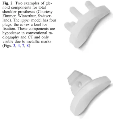
Fluoroscopy is mainly used for image-guided joint aspirations. Arthrography has a limited role after shoulder Fig. 2 Two examples of
gle-noid components for total shoulder prostheses (Courtesy Zimmer, Winterthur, Switzer-land). The upper model has four plugs, the lower a keel for fixation. These components are hypodense in conventional ra-diography and CT and only visible due to metallic marks (Figs.3,4,7,8)
Fig. 3 The four standard projections for standard radiographic evaluation as performed at the author’s institution. Example of a patient with anatomical total shoulder arthroplasty. a Anteroposte-rior view with the patient rotated approximately 45° towards the abnormal side (beam parallel to the glenoid surface) with the tube tilted craniocaudally by 15°. The elbow is flexed 90° and the hand is
pointing towards the x-ray tube (corresponding to slight internal rotation of the glenohumeral joint). b Anteroposterior view comparable to a, but with the forearm in neutral position (sagittal plane with reference to the patient’s body). c Cross-table view. d Neer’s (Y) view with the radiographic beam parallel to the scapula and tilted craniocaudally by 15°
arthroplasty. It may rarely be used for the demonstration of loosening of the glenoid component, with or without CT (Figs.4and5).
Sonography
Sonography is mainly used postoperatively for diagnosing hematoma and infection. Sonography is also suitable for image-guided aspiration and drainage and may demon-strate rotator cuff tears and other soft tissue abnormalities during follow-up of the prosthesis.
Skeletal scintigraphy
Owing to its high negative predictive value, radionuclide imaging is useful for exclusion of prosthetic complications, but has a limited positive predictive value.
Technetium-99m methylene diphosphonate uptake oc-curs wherever new bone formation takes place [4]. Periprosthetic activity can be identified in most patients for more than 1 year after uncomplicated surgery. The use of gallium-67 scintigraphy and labeled leukocyte scintig-raphy improves specificity for infected joint replacement considerably. Publications are rare and do not specifically relate to shoulder arthroplasty [5,6].
Computed tomography
Computed tomography complements standard radiographs in preoperative planning of shoulder arthroplasty. CT is useful for demonstration of the extent of osteoarthritis, the amount of bone available for fixation, and for measuring glenoid version, for which cross-table standard radiographs
are not reliable [7]. Glenoid version is typically measured on a thick reconstructed section at the middle of the glenoid. Normally the glenoid articular surface has a minimal retroversion with regard to a line drawn through the middle of the glenoid and the medial scapular margin [8] (mean 3° retroversion, range 7° anteversion–16° retroversion). Humeral head retroversion may be measured using reference lines drawn through the anterior and posterior limits of the humeral head cartilage and through both humeral epicondyles. Humeral head retroversion varies from 9 to 25°, mean 17° [9]. Fatty degeneration of the rotator cuff muscles is commonly assessed by a grading system published by Goutallier et al. [10] (Table1). CT is also useful in the detection of periprosthetic fracture of the humerus, stress fractures of the acromion, and the coracoid as well as prosthetic loosening [11] (Fig.5).
Beam hardening artifacts depend on the diameter, density, and geometry of the metallic implants. Artifacts are proportional to material density. Therefore, cobalt– chrome alloy causes more pronounced artifacts than titanium [12]. Artifacts are least pronounced in the direction of shortest diameter of the implant [13, 14]. Beam hardening artifacts can be reduced with increasing
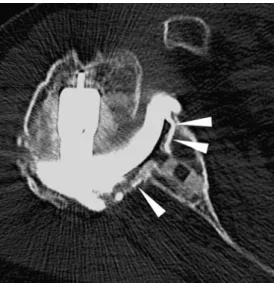
Fig. 4 Arthrography with iodinated contrast between glenoid component and bone (long arrowheads). Normal bone–cement interface at the keel of the glenoid component (short arrowheads)
Fig. 5 Computed tomography of the same patient as in Fig.4after arthrography. Identical spread of intra-articular injected contrast media with glenoid component loosening (arrowheads)
Table 1 Classification of fatty degeneration of rotator cuff muscles (Goutallier et al.) [10] Grade of fatty degeneration Definition 0 No fat 1 Streaks of fat
2 Less intramuscular fat than muscle tissue
3 Equal amount of intramuscular fat and muscle tissue 4 More intramuscular fat than muscle tissue
tube voltage [15]. Increasing tube current improves the signal-to-noise ratio. Decreasing pitch and slice thickness have a similar effect by increasing effective mAs [13,16]. Increased slice thickness of reformatted images, smooth reconstruction filters, and increased window width im-prove image quality [13]. However, smooth reconstruction filters reduce spatial resolution [15]. Tables2and3present an imaging protocol optimized for arthroplasty and a checklist for optimization of CT protocols for patients with orthopedic implants.
Intra-articular contrast in the presence of implants is rarely employed. There is some additional information such as joint capsule width and possibly contrast leakage into bone–cement or bone–implant interfaces in loosening. MR imaging
MR imaging is mainly used preoperatively for imaging of the rotator cuff tendons (Fig.6) and muscles (Fig.7).
Postoperatively, susceptibility artifacts interfere with imaging, but it may still be useful for the assessment of the rotator cuff tendons and muscles, soft tissue hematoma and abscess, or radiographically occult fractures. Different imaging protocols are in use [17–19]. Table4provides the standard protocol for postoperative shoulder imaging used at the authors’ institution.
Intra-articular contrast is rarely helpful after shoulder arthroplasty. Susceptibility artifacts prevent demonstration of at least part of the contrasted joint. In addition, after arthroplasty, there is an increased risk of infection. Although the probability is still low, the consequences of infection would be severe.
In order to reduce susceptibility artifacts, the implanted prosthesis should be aligned with the main magnetic field B0[20,21] which is typically fulfilled for the stem of the
humeral prosthesis. Fast or turbo spin echo sequences reduce susceptibility artifacts in comparison with standard spin echo sequences, particularly if long echo train lengths (ETL) and short interecho spacing are applied [20]. Short tau inversion recovery (STIR) sequences are less suscep-tible to metallic implants than sequences using frequency-selective fat-suppression [22]. Water excitation sequences are also superior to fat suppression, but may not be available on all scanners [23]. Increasing sampling bandwidth reduces susceptibility artifacts, but increases image blurring. With increasing magnetic field strength (B0) susceptibility artifacts become more prominent [24].
Artifact reduction may be achieved by shortening TE, increasing matrix in the frequency encoding direction, and decreasing slice thickness [21]. Decreasing voxel size has only a small effect on artifact size, but improves spatial resolution for structures adjacent to the prosthesis [25].
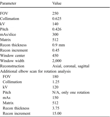
Table 2 Suggested CT parameters in arthroplasty Parameter Value FOV 250 Collimation 0.625 kV 140 Pitch 0.426 mAs/slice 300 Matrix 512 Recon thickness 0.9 mm Recon increment 0.45 Window center 450 Window width 2,000
Reconstruction Axial, coronal, sagittal Additional elbow scan for rotation analysis
FOV 180
Collimation 1.25
kV 120
Pitch N/A, only one rotation
mAs 150
Matrix 512
Recon thickness 3.75 Recon increment 15.00
Parameters are proposed for a 40-row CT scanner adapted to postoperative shoulders with metallic components
FOV field of view, N/A not applicable
Fig. 6 Coronal proton density (PD)-weighted turbo spin echo image after total shoulder arthroplasty with craniocaudal frequency-encoding direction shows an intact supraspinatus tendon inferior to the acromion (white arrowheads). (TR 2,960 ms, TE 8 ms, ET 9, FOV 18×18 cm, 256×212 matrix, 4-mm slice thickness)
Table 3 Checklist for improving CT of shoulder prostheses Increase tube voltage
Increase tube current
Use soft tissue reconstruction algorithm
Suggested MR protocols and a checklist are presented in Tables4and5.
Complications of shoulder arthroplasty
Although shoulder arthroplasty is generally successful [26], complications may occur. A revision rate of 7% after 13.4 years has been reported [27]. Complication rates, patient readmission rates, and duration of hospitalization vary between institutions [28,29].
A meta-analysis by Bohsali has demonstrated a compli-cation rate of 14.7% in a series of 2,810 total shoulder replacements [27]. The most common complications were (in order of decreasing frequency): prosthetic loosening, glenohumeral instability, periprosthetic fracture, rotator cuff tears, neural injury, infection, and deltoid muscle dysfunction. Less frequent complications include fracture
of the acromion, the scapular spine or the coracoid process, glenoid notching, hematoma, and periarticular soft tissue calcification. Sometimes postoperative persistent shoulder pain or shoulder pain after a longer asymptomatic interval is the leading problem without the presence of the previously mentioned complications. If so, possible causes to be considered are cartilage wear over the glenoid in hemiarthroplasty and synovitis.
Prosthetic loosening
Prosthetic loosening is the most common complication of shoulder arthroplasty accounting for approximately 40% of all complications [27, 30]. Loosening typically is diag-nosed several years after surgery and predominantly involves the glenoid component. There is an association between glenohumeral instability and glenoid component loosening [31, 32]. Radiographic signs of loosening are migration (translation and tilting) and the appearance of a radiolucent line at the bone–implant or bone–cement interface [33] (Figs. 8 and 9). Presence of radiolucent lines around the humeral stem can be assessed with a scoring system adapted from the hip [34].
In this system the outline of the humeral prosthesis component is divided into zones numbered from 1 to 7 in anteroposterior and cross-table radiographs. On anteropos-terior films zone 1 is located at the proximal third of the prosthesis, laterally, zone 4 at the tip of the prosthesis and zone 7 at the proximal humeral shaft, medially. The same system is applied on cross-table views from anterior to posterior [35]. Sperling et al. defined an additional zone 8 at the base of the head of the humeral component [34].
Glenoid radiolucent lines can be assessed similarly by defining four zones: 1, around superior baseplate; 2, around inferior baseplate (without notch if present); 3, around central pillar; 4, around the screws [36].
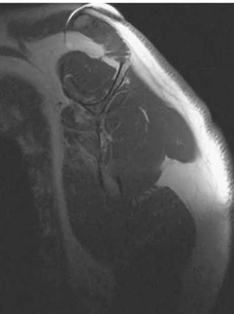
Lucent lines may be present without clinically signifi-cant loosening and do not necessarily correlate with pain [37]. Sanchez-Sotelo and coworkers [38] found that prevalence and extent of humeral radiolucent lines were significantly higher in total shoulder arthroplasty than in hemiarthroplasty. The most reliable radiological evidence Fig. 7 Sagittal T1-weighted spin echo image after total shoulder
arthroplasty with craniocaudal frequency-encoding direction de-monstrates rotator cuff musculature and fatty atrophy of subscapu-laris muscle (Goutallier grade 3) and normal muscle quality of supraspinatus and infraspinatus muscle. (TR 311 ms, TE 5.1 ms, ET 1, FOV 18×18 cm, 256×256 matrix, 5-mm slice thickness)
Table 4 Suggested MR parameters in shoulder arthroplasty (1.5 T)
Sequence TR/TE FOV (mm) Matrix Time (min:s) Paracoronal TSE PD-weighted fat sat 2,640/15 160 256×512 2:29 Paracoronal STIR 5,590/35/160 160 256×512 3:34 Parasagittal STIR 5,590/35/160 160 256×512 3:34 Parasagittal SE T1-weighted 539/15 160 256×512 4:05 Axial TSE PD-weighted fat sat 2,640/15 160 256×512 3:20 Section thickness = 4 mm for all sequences
Abbreviations: FOV field of view, PD proton density, SE spin echo, STIR short tau inversion recovery, Time acquisition time, TR/TE repetition time/echo time, TSE turbo spin echo
of loosening include radiolucent lines measuring 2 mm or more around the whole implant as well as subsidence and tilt of the component [34].
Bone resorption at the glenoid can be reduced by preservation of the subchondral bone plate, concentric glenoid reaming [39], and avoiding a mismatch between the radius of the glenoid and the humeral head [40, 41] which increases polyethylene wear.
Notching
Notching is found in semiconstrained inversed total shoulder arthroplasty. It relates to bone resorption at the
inferior scapular neck caused by impingement of the humeral cup during adduction (Fig.9). Notching has been classified as grade 0 (no notch), grade 1 (small notch stopping short of inferior screw), grade 2 (medium notch reaching inferior screw), and grade 3 (large notch extending beyond inferior screw) [36]. Mild notching is not typically symptomatic. However, advanced notching is associated with glenoid component loosening.
Glenohumeral instability
Glenohumeral instability is the second leading cause of dysfunction after arthroplasty. The prevalence is 4% which corresponds to 30% of all complications [27,30,31].
Instability after shoulder arthroplasty is typically ante-rior (80% of unstable shoulders). Anteante-rior subluxation of the humeral head by more than 5 mm as seen on cross-table radiographs or CT suggests anterior instability. Anterior instability may have multiple causes including subscapu-laris and anterior capsular abnormalities, anteversion of the glenoid component, oversized humeral head component, anterior placement of the humeral component [42], and decreased humeral retroversion (<20° retroversion). Ante-rior dislocation may occur when the subscapularis tendon is torn.
The diagnosis of superior instability is made when the acromiohumeral distance is less than 5 mm on an anteroposterior view. Superior instability is associated with anterior instability, rotator cuff tears [43], cranial
Table 5 Checklist for reduction of implant-related artifacts in MR imaging
Position long axis of prosthesis parallel to the direction of the main magnetic field (B0)
Avoid gradient echo sequences, use spin echo sequences Use fast/turbo spin echo instead of conventional spin echo
sequences
Starting from the prosthesis, the region of interest should not be in the phase-encoding direction
Replace frequency-selective fat-saturated T2-weighted spin echo images by STIR sequence
Reduce echo spacing in fast/turbo spin echo sequences Increase echo train length
Reduce slice thickness Increase sampling bandwidth Increase matrix size (e.g., 512×512)
Fig. 8 Loosening of the humeral component: A radiolucent line is visible around the entire humeral implant. Varus tilting of the prosthesis and lateral cortical bulging with periosteal reaction (white arrowheads)
Fig. 9 Fracture and mild inferior dislocation of the acromion (white arrows) after semiconstrained inversed total prosthesis. Notching grade II is noted (black arrowheads) with a radiolucent line (black arrow) at the central pillar of the glenoid component
placement of the humeral component with relative lengthening of the humerus, and superior tilting of the glenoid component.
In posterior instability, a wide dorsal capsule, anterior soft tissue contracture, infraspinatus tendon deficiency, malrotation of humeral component (retroversion >45°), dorsal glenoid defect, and retroversion of glenoid compo-nent (>20°) may be responsible.
Inferior instability is typically found when the humerus is shortened which may occur after proximal humeral fractures or humeral defects in tumor surgery. Deltoid muscle weakness also leads to inferior instability. This may be found in axillary nerve palsy or deltoid muscle detachment. Elevation of the arm above the horizontal plane is practically impossible when the humerus is shortened.
Preoperative imaging of the contralateral side for comparison is used for planning of surgery.
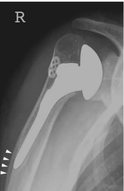
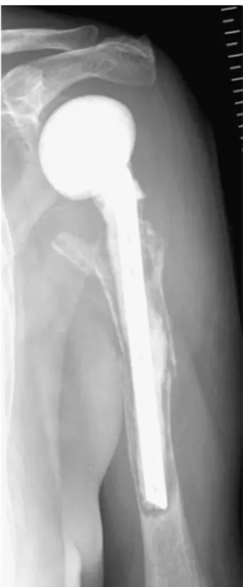
Periprosthetic fracture
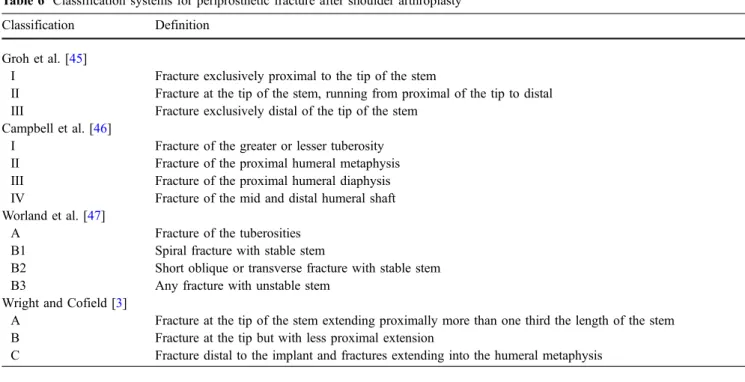
Periprosthetic fractures have a prevalence of about 1.5–3% (11% of all complications associated with shoulder arthro-plasty) [27]. Fractures of the humerus may occur intraoperatively and later due to acute trauma or as a fatigue fracture (Fig.10). Risk factors include osteoporosis, female sex, increased age, and a tendency to fall. Trans-verse and short oblique fractures may demonstrate delayed healing or even result in non-union. Different classification systems for periprosthetic fractures are in use (Table 6)
[44]. The most commonly used classification system by Wright and Cofield [3] divides fractures into three types according the position of the fracture in relation to the tip of the humeral component. Groh et al. [45] use similar criteria. Campbell et al. [46] divide fractures according to the position with the humerus. Worland et al. [47] include implant stability.
Treatment of periprosthetic humeral fractures depends on the exact location and course of the fracture. Conser-vative treatment is preferred in elderly patients with comorbidities. In most cases, surgical treatment with or without replacement of the humeral shaft is necessary [3].
Fractures that are not directly related to the prosthesis
Fractures of the acromion may be found in inversed prosthesis. The mechanism of fracture is unknown. Fatigue fracture due to increased deltoid tension or acute trauma has been discussed. Because surgical fixation of acromial fractures may not be successful and because the fracture is rather well tolerated by the patients conservative treatment of acromion fractures not extending to the scapular spine is preferred at the authors’ institution. Acromial fractures can be differentiated from Os acromiale based on their commonly more medial position and the irregular borders, often associated with reactive sclerosis.
Fractures of the coracoid process are even less common than acromial fracture. Repetitive pulling by the conjoined tendon (coracobrachialis, short head of biceps) and impingement of the humeral head may be responsible for acute or chronic coracoid fractures. Treatment is usually conservative.
Rotator cuff tears
Rotator cuff tears are the fourth most common complica-tion after shoulder arthroplasty [27]. They are relevant for the survival of the implant because asymmetric load of prosthetic components leads to increasing wear and loosening. Postoperative rotator cuff tears are associated with insufficient fixation of the tendon after arthroplasty, oversized prosthesis, malrotation of the humeral compo-nent, multiple surgery, aggressive physiotherapy involving external rotation during the early postoperative period, and tendon compromise in humeral lengthening [48–50].
The subscapularis tendon is the most commonly torn tendon after shoulder arthroplasty. Tuberculum minus osteotomy instead of tenotomy can reduce postoperative subscapularis insufficiency considerably [51].
Supraspinatus tears are suspected when the distance between the top of the humeral prosthesis and the acromion is less than 5 mm on standard radiographs. However, this distance may also appear to be reduced in posterior subluxation of the humeral head after shoulder arthroplasty. Fig. 10 Example of an
antibi-otic-impregnated cement spacer after explantation of total shoulder arthroplasty due to in-fection. Fracture around the spacer is noted
Ultrasonography may be used for the diagnosis of rotator cuff tears. MR imaging may not be able to demonstrate the entire rotator cuff due to the susceptibility artifacts associated with implants. CT may be useful for the demonstration of fatty degeneration of the rotator cuff as a reliable indirect sign of a rotator cuff tear.
Infection
Postoperative infection is a rare but severe complication of shoulder arthroplasty. Bohsali et al. reported a prevalence of 0.7% [27]. Periprosthetic infections can occur directly after implantation or with a delayed onset (3 months or later) [52, 53]. Infection with Staphylococcus aureus is most commonly encountered, followed by coagulase-negative staphylococcus and Propionibacterium acnis. Immunosuppression, diabetes, rheumatoid arthritis and other systemic inflammatory disease, remote infections, previously performed surgery, chemotherapy, corticoste-roid medication, and repeated intra-articular stecorticoste-roid injec-tions represent risk factors [31, 54,55]. Clinical findings, laboratory test, radiography, scintigraphy, and joint aspi-ration are either insensitive or non-specific, or both [5]. Pain and reduced function are the most common symp-toms. Skin reddening and swelling may be present but are frequently not. Laboratory parameters include increased C-reactive protein, erythrocyte sedimentation rate, and white blood cell count [31,52–54].
On standard radiographs, infection can only be diag-nosed late in the course of disease. Progressive loosening is the most conspicuous sign. Skeletal scintigraphy
demon-strates massively increased tracer uptake in infection. However, there is an overlap with normal postoperative uptake which is seen during the first postoperative year. During this same period of time, about two-thirds of prosthetic infections occur. MR imaging and sonography may be useful for the demonstration of soft tissue abnormality associated with implant infection. Immediate revision, aggressive debridement, saving or exchange of the prosthesis depending on the onset of infection, and appropriate intravenous antibiotic therapy is generally rated to be the best treatment, but not feasible in all patients. Two-stage replacement with antibiotic-impreg-nated cement spacer (Fig.10) has been employed. Sperling et al. concluded that two-stage reimplantation offers the best outcome [56]. Coste et al. [57] stated that antibiotics or debridement alone are ineffective. Rarely, arthrodesis or even amputation have been used [31,52–54,56–62]. Nerve injury/deltoid muscle dysfunction
Intraoperative injury to the brachial plexus and axillary nerve is rare. It leads to deltoid muscle dysfunction with reduced abduction and commonly with inferior instability. Detachment is another reason for deltoid muscle dysfunc-tion. The risk for deltoid muscle detachment depends on the surgical approach. For revision arthroplasty with massive rotator cuff tears deltoid muscle function is crucial. If deltoid muscle function is insufficient not even inversed shoulder prosthesis can be used. Ultrasound and MR imaging may be used for the diagnosis of deltoid muscle abnormalities.
Table 6 Classification systems for periprosthetic fracture after shoulder arthroplasty Classification Definition
Groh et al. [45]
I Fracture exclusively proximal to the tip of the stem
II Fracture at the tip of the stem, running from proximal of the tip to distal III Fracture exclusively distal of the tip of the stem
Campbell et al. [46]
I Fracture of the greater or lesser tuberosity II Fracture of the proximal humeral metaphysis III Fracture of the proximal humeral diaphysis IV Fracture of the mid and distal humeral shaft Worland et al. [47]
A Fracture of the tuberosities B1 Spiral fracture with stable stem
B2 Short oblique or transverse fracture with stable stem B3 Any fracture with unstable stem
Wright and Cofield [3]
A Fracture at the tip of the stem extending proximally more than one third the length of the stem B Fracture at the tip but with less proximal extension
Heterotopic ossification
Heterotopic ossification develops early postoperatively [63]. It can be graded according to Kjaersgaard et al. [64], i.e.,by evaluating the space between the medial humeral shaft and the lateral glenoid. Grade 0 means no ossification, grade 1 ossification occupying <50% of the space, grade 2 ossification occupying >50% of the space, and grade 3 bridging of the space. Ossifications are typically low grade and are often not clinically important [63]. In higher grades joint mobility is reduced. Standard radiographs are typically used for this diagnosis. CT may be used to demonstrate additional details.
Implant failure
Implant failure is rare. Subluxation or dislocation of polyethylene inlays [65,66], broken fixations screws [67, 68], fracture of the keel or metal glenoid backing [68,69], and dissociation of the polyethylene glenoid insert from its metal tray [69, 70] have been described. Standard radio-graphs demonstrate part of these complications. CT may provide additional information.
Progressive wear of the glenoid after hemiarthroplasty
After hemiarthroplasty degeneration on the glenoid side of the joint progresses. This is a problem in young, active
patients. Surgical revision with total arthroplasty may be necessary. Standard radiographs are typically used for this diagnosis.
Conclusion
Shoulder prostheses are now widely used. Clinical results and patient satisfaction are good. The most commonly used types are humeral hemiarthroplasty or resurfacing, un-constrained total shoulder arthroplasty, and semicon-strained inversed shoulder prosthesis.
Complications of shoulder arthroplasty depend on the prosthesis type used. The most common complications are prosthetic loosening, glenohumeral instability, peripros-thetic fracture, rotator cuff tears, nerve injury, infection, and deltoid muscle dysfunction.
Standard radiographs are the basis of both pre- and postoperative imaging. Skeletal scintigraphy has a rather limited role because there is overlap between postoperative changes which may persist for up to 1 year and early loosening and infection. Sonography is most commonly used postoperatively in order to demonstrate complications (hematoma and abscess formation) but may also be useful for the demonstration of rotator cuff tears occurring during follow-up. CT is useful for the demonstration of bone details both pre- and postoperatively. MR imaging is mainly used preoperatively, for instance for demonstration of rotator cuff tears.
References
1. Lugli T (1978) Artificial shoulder joint by Pean (1893): the facts of an excep-tional intervention and the prosthetic method. Clin Orthop Relat Res 133:215–218
2. Péan JE (1894) Des moyens prosthe-tiques destinés à obtenir la reparation des parties osseuses. Gaz des Hôp 67:291 Reprinted in English (1973) Clin Orthop 94:4
3. Wright TW, Cofield RH (1995) Hu-meral fractures after shoulder arthro-plasty. J Bone Jt Surg Am
77:1340–1346
4. Palestro CJ, Love C, Miller TT (2006) Infection and musculoskeletal condi-tions: imaging of musculoskeletal in-fections. Best Pract Res Clin Rheumatol 20:1197–1218 5. Love C, Tomas MB, Marwin SE,
Pugliese PV, Palestro CJ (2001) Role of nuclear medicine in diagnosis of the infected joint replacement. Radio-graphics 21:1229–1238
6. Pakos EE, Koumoulis HD, Fotopoulos AD, Ioannidis JP (2007) Osteomyelitis: antigranulocyte scintigraphy with 99mTC radiolabeled monoclonal anti-bodies for diagnosis - meta-analysis. Radiology 245(3):732–741
7. Friedman RJ, Hawthorne KB, Genez BM (1992) The use of computerized tomography in the measurement of glenoid version. J Bone Jt Surg Am 74:1032–1037
8. Nyffeler RW, Jost B, Pfirrmann CW, Gerber C (2003) Measurement of gle-noid version: conventional radiographs versus computed tomography scans. J Shoulder Elb Surg 12:493–496 9. Hernigou P, Duparc F, Hernigou A
(2002) Determining humeral retrover-sion with computed tomography. J Bone Jt Surg Am 84-A:1753–1762 10. Goutallier D, Postel JM, Bernageau J,
Lavau L, Voisin MC (1994) Fatty muscle degeneration in cuff ruptures. Pre- and postoperative evaluation by CT scan. Clin Orthop Relat Res 304:78–83
11. Ohashi K, El-Khoury GY, Bennett DL, Restrepo JM, Berbaum KS (2005) Orthopedic hardware complications di-agnosed with multi-detector row CT. Radiology 237:570–577
12. Haramati N, Staron RB, Mazel-Sperling K et al (1994) CT scans through metal scanning technique ver-sus hardware composition. Comput Med Imaging Graph 18:429–434 13. White LM, Buckwalter KA (2002)
Technical considerations: CT and MR Imaging in the postoperative orthopedic patient. Sem Musculoskelet Radiol 6:5–17
14. Vande Berg B, Malghem J, Maldague B, Lecouvet F (2006) Multi-detector CT imaging in the postoperative or-thopedic patient with metal hardware. Eur J Radiol 60:470–479
15. Barrett JF, Keat N (2004) Artifacts in CT: recognition and avoidance. Radio-graphics 24:1679–1691
16. Wilting JE, Timmer J (1999) Artefacts in spiral-CT images and their relation to pitch and subject morphology. Eur Radiol 9:316–322
17. Zanetti M, Hodler J (2004) MR imag-ing of the shoulder after surgery. Magn Reson Imaging Clin N Am 12:169–183 18. Owen RS, Iannotti JP, Kneeland JB,
Dalinka MK, Deren JA, Oleaga L (1993) Shoulder after surgery: MR imaging with surgical validation. Ra-diology 186:443–447
19. Gaenslen ES, Satterlee CC, Hinson GW (1996) Magnetic resonance imag-ing for evaluation of failed repairs of the rotator cuff. J Bone Jt Surg Am 78:1391–1396
20. Suh JS, Jeong EK, Shin KH et al (1998) Minimizing artifacts caused by metallic implants at MR imaging: experimental and clinical studies. AJR Am J Roentgenol 171:1207–1213 21. Port JD, Pomper MG (2000)
Quantifi-cation and minimization of magnetic susceptibility artifacts on GRE images. J Comput Assist Tomogr 24:958–964 22. Hilfiker P, Zanetti M, Debatin JF,
McKinnon G, Hodler J (1995) Fast spin-echo inversion-recovery imaging versus fast T2-weighted spin-echo im-aging in bone marrow abnormalities. Invest Radiol 30:110–114
23. Hauger O, Dumont E, Chateil JF, Moinard M, Diard F (2002) Water excitation as an alternative to fat satu-ration in MR imaging: preliminary results in musculoskeletal imaging. Radiology 224:657–663
24. Merkle EM, Dale BM, Thomas J, Paulson EK (2006) MR liver imaging and cholangiography in the presence of surgical metallic clips at 1.5 and 3 Tesla. Eur Radiol 16:2309–2316 25. Pauchard Y, Smith MR, Mintchev MP
(2005) Improving geometric accuracy in the presence of susceptibility differ-ence artifacts produced by metallic implants in magnetic resonance imag-ing. IEEE Trans Med Imaging 24:1387–1399
26. White CB, Sperling JW, Cofield RH, Rowland CM (2003) Ninety-day mor-tality after shoulder arthroplasty. J Ar-throplasty 18:886–888
27. Bohsali KI, Wirth MA, Rockwood CA Jr (2006) Complications of total shoul-der arthroplasty. J Bone Jt Surg Am 88:2279–2292
28. Hasan SS, Leith JM, Smith KL, Matsen FA (2003) The distribution of shoulder replacement among surgeons and hos-pitals is significantly different than that of hip or knee replacement. J Shoulder Elb Surg 12:164–169
29. Hammond JW, Queale WS, Kim TK, McFarland EG (2003) Surgeon experi-ence and clinical and economic out-comes for shoulder arthroplasty. J Bone Jt Surg Am 85-A:2318–2324
30. Deshmukh AV, Koris M, Zurakowski D, Thornhill TS (2005) Total shoulder arthroplasty: long-term survivorship, functional outcome, and quality of life. J Shoulder Elb Surg 14:471–479 31. Wirth MA, Rockwood CA Jr (1996)
Complications of total shoulder-repla-cement arthroplasty. J Bone Joint Surg Am 78:603–616
32. Gruen TA, McNeice GM, Amstutz HC (1979)“Modes of failure” of cemented stem-type femoral components: a ra-diographic analysis of loosening. Clin Orthop Relat Res 141:17–27
33. Martin SD, Zurakowski D, Thornhill TS (2005) Uncemented glenoid com-ponent in total shoulder arthroplasty. Survivorship and outcomes. J Bone Jt Surg Am 87:1284–1292
34. Sperling JW, Cofield RH, O’Driscoll SW, Torchia ME, Rowland CM (2000) Radiographic assessment of ingrowth total shoulder arthroplasty. J Shoulder Elb Surg 9:507–513
35. Torrens C, Martinez-Diaz S, Ruiz A, Gines A, Caceres E (2007) Assessment of radiolucent lines in cemented shoul-der hemi-arthroplasties: study of con-cordance and reproducibility. Int Orthop doi:10.1007/s00264-007-0452-4 36. Boileau P, Watkinson D, Hatzidakis
AM, Hovorka I (2006) Neer Award 2005: The Grammont reverse shoulder prosthesis: results in cuff tear arthritis, fracture sequelae, and revision arthro-plasty. J Shoulder Elb Surg 15(5):527– 540
37. Farron A (2006) Die Schultergelenk-prothese. Schweiz Med Forum 6:53–58 38. Sanchez-Sotelo J, O’Discroll SW,
Torchia ME, Cofield RH, Rowland CM (2001) Radiographic assessment of cemented humeral components in shoulder arthroplasty. J Shoulder Elb Surg 10:526–531
39. Collins D, Tencer A, Sidles J, Matsen F 3rd (1992) Edge displacement and deformation of glenoid components in response to eccentric loading. The effect of preparation of the glenoid bone. J Bone Jt Surg Am 74:501–507
40. Walch G, Edwards TB, Boulahia A, Boileau P, Mole D, Adeleine P (2002) The influence of glenohumeral pros-thetic mismatch on glenoid radiolucent lines: results of a multicenter study. J Bone Jt Surg Am 84-A:2186–2191 41. Iannotti JP, Gabriel JP, Schneck SL,
Evans BG, Misra S (1992) The normal glenohumeral relationships. An ana-tomical study of one hundred and forty shoulders. J Bone Jt Surg Am 74:491– 500
42. Warren RF, Coleman SH, Dines JS (2002) Instability after arthroplasty: the shoulder. J Arthroplasty 17:28–31 43. Boyd AD Jr, Aliabadi P, Thornhill TS
(1991) Postoperative proximal migra-tion in total shoulder arthroplasty. In-cidence and significance. J Arthroplasty 6:31–37
44. McDonough EB, Crosby LA (2005) Periprosthetic fractures of the humerus. Am J orthop 34(12):586–591
45. Groh G, Heckman M, Curtis R et al (1994) Treatment of fractures adjacent to humeral prosthesis. In: Annual meeting of the American Academy of Orthopaedic Surgeons. New Orleans, LA
46. Campbell JT, Moore RS, Iannotti JP, Norris TR, Williams GR (1998) Peri-prosthetic humeral fractures: mechan-isms of fracture and treatment options. J Shoulder Elb Surg 7:406–413 47. Worland RL, Kim DY, Arredondo J
(1999) Periprosthetic humeral fractures: management and classification. J Shoulder Elb Surg 8:590–594 48. Brems JJ (2002) Complications of
shoulder arthroplasty: infections, in-stability, and loosening. Instr Course Lect 51:29–30
49. Bonutti PM, Hawkins RJ (1992) Frac-ture of the humeral shaft associated with total replacement arthroplasty of the shoulder. A case report. J Bone Jt Surg Am 74:617–618
50. Miller BS, Joseph TA, Noonan TJ, Horan MP, Hawkins RJ (2005) Rupture of the subscapularis tendon after shoulder arthroplasty: diagnosis, treat-ment, and outcome. J Shoulder Elb Surg 14:492–496
51. Gerber C, Pennington SD, Yian EH, Pfirrmann CA, Werner CM, Zumstein MA (2006) Lesser tuberosity osteoto-my for total shoulder arthroplasty. Surgical technique. J Bone Jt Surg Am 88(Suppl 1 Pt 2):170–177
52. Wolfe SW, Figgie MP, Inglis AE, Bohn WW, Ranawat CS (1990) Management of infection about total elbow prosthe-ses. J Bone Jt Surg Am 72:198–212 53. Yamaguchi K, Adams RA, Morrey RF
(1998) Infection after total elbow ar-throplasty. J Bone Jt Surg Am 80:481– 491
54. Matsen FA 3rd, Rockwood CAJ, Wirth MA, Lippitt SB, Parsons M (2004) Glenohumeral arthritis and its manage-ment. In: Rockwood CAJ (ed) The shoulder, 3rd edn. Saunders, Philadel-phia, pp 879–1008
55. Rosenberg N, Neuann L, Modi A, Mersich IJ, Wallace AW (2007) Im-provements in survival of the unce-mented Nottingham total shoulder prosthesis: a prospective comparative study. BMC Musculoskelet Disord 8:76 56. Sperling JW, Kozak TK, Hanssen AD,
Cofield RH (2001) Infection after shoulder arthroplasty. Clin Orthop Relat Res 382:206–216
57. Coste JS, Reig S, Trojani C, Berg M, Walch G, Boileau P (2004) The man-agement of infection in arthroplasty of the shoulder. J Bone Jt Surg Br 86:65– 69
58. Proubasta IR, Itarte JP, Lamas CG, Escriba IU (2005) Permanent articulat-ed antibiotic-impregnatarticulat-ed cement spacer in septic shoulder arthroplasty: a case report. J Orthop Trauma 19:666– 668
59. Ince A, Seemann K, Frommelt L, Katzer A, Loehr JF (2005) One-stage exchange shoulder arthroplasty for periprosthetic infection. J Bone Jt Surg Br 87:814–818
60. Seitz WH Jr, Damacen H (2002) Staged exchange arthroplasty for shoulder sepsis. J Arthroplasty 17:36–40 61. Ramsey ML, Fenlin JM Jr (1996) Use
of an antibiotic-impregnated bone ce-ment block in the revision of an infected shoulder arthroplasty. J Shoulder Elb Surg 5:479–482 62. Loebenberg MI, Zuckerman JD (2004)
An articulating interval spacer in the treatment of an infected total shoulder arthroplasty. J Shoulder Elb Surg 13:476–478
63. Sperling JW, Cofield RH, Rowland CM (2000) Heterotopic ossification after total shoulder arthroplasty. J Arthro-plasty 15:179–182
64. Kjaersgaard-Andersen P, Frich LH, Sojbjerg JO, Sneppen O (1989) Het-erotopic bone formation following total shoulder arthroplasty. J Arthroplasty 4:99–104
65. Brenner BC, Ferlic DC, Clayton ML, Dennis DA (1989) Survivorship of unconstrained total shoulder arthro-plasty. J Bone Jt Surg Am 71:1289– 1296
66. Clayton ML, Ferlic DC, Jeffers PD (1982) Prosthetic arthroplasties of the shoulder. Clin Orthop Relat Res 164:184–191
67. McElwain JP, English E (1987) The early results of porous-coated total shoulder arthroplasty. Clin Orthop Relat Res 218:217–224
68. Martin SDSC, Thomas WH, Thornhill TS (1995) Total shoulder arthroplasty with an uncemented glenoid compo-nent. Paper presented at the annual meeting of the American Shoulder and Elbow Surgeons, Orlando
69. Cofield RHDP (1992) Total shoulder arthroplasty with a tissue-ingrowth glenoid component. J Shoulder Elb Surg 1:77–85
70. Driessnack RP, Ferlic DC, Wiedel JD (1990) Dissociation of the glenoid component in the Macnab/English total shoulder arthroplasty. J Arthroplasty 5:15–18