HAL Id: hal-03004564
https://hal.archives-ouvertes.fr/hal-03004564
Submitted on 13 Nov 2020
HAL is a multi-disciplinary open access archive for the deposit and dissemination of sci-entific research documents, whether they are pub-lished or not. The documents may come from teaching and research institutions in France or abroad, or from public or private research centers.
L’archive ouverte pluridisciplinaire HAL, est destinée au dépôt et à la diffusion de documents scientifiques de niveau recherche, publiés ou non, émanant des établissements d’enseignement et de recherche français ou étrangers, des laboratoires publics ou privés.
Digitization of fossils from the Fezouata Biota (Lower
Ordovician, Morocco): Evaluating computed
tomography and photogrammetry in collections
enhancement
Khaoula Kouraïss, Khadija El Hariri, Abderrazak El Albani, Abdelfattah
Azizi, Arnaud Mazurier, Bertrand Lefebvre
To cite this version:
Khaoula Kouraïss, Khadija El Hariri, Abderrazak El Albani, Abdelfattah Azizi, Arnaud Mazurier, et al.. Digitization of fossils from the Fezouata Biota (Lower Ordovician, Morocco): Evaluating computed tomography and photogrammetry in collections enhancement. Geoheritage, Springer, 2019. �hal-03004564�
Digitization of fossils from the Fezouata Biota (Lower
Ordovician, Morocco): Evaluating computed tomography
and photogrammetry in collections enhancement.
Khaoula Kouraissa, Khadija El Hariria, Abderrazak El Albanib, Abdelfattah Azizia, Arnaud Mazurierb, Bertrand Lefebvrec
.
a Département des sciences de la terre, Faculté des Sciences et Techniques, Université Cadi-Ayyad, BP 549, 40000 Marrakesh, Morocco
b Institut IC2MP UMR CNRS 7285 (HydrASA), Université de Poitiers, 5 rue Albert Turpin, Bâtiment B8, TSA 51106, 86073 Poitiers Cedex, France
c UMR CNRS 5276, Université Lyon 1, 2, rue Raphaël Dubois, 69622 Villeurbanne Cedex, France. E-mail addresses: [email protected](K. Kouraiss), [email protected] (K. El Hariri),
[email protected](A. El Albani), [email protected] (A. Azizi), [email protected](A. Mazurier), [email protected] (B. Lefebvre).
Abstract
Palaeontological collections housing material from the Fezouata Shale Lagerstätte (Lower Ordovician, Morocco) are of a high scientific interest as they testify to the existence of Burgess Shale-type taxa in one of the most critical Palaeozoic period: the Cambrian–Ordovician transition. The preservation of this unique patrimony can benefit from the emergence of imaging methodologies that have provided innovative ways in three-dimensional (3D) digitization. Computed tomography and photogrammetry were applied in order to create 3D models of fossils from the Fezouata Biota. Tomographic results show the exciting potential of these techniques in internal investigation of fossils, while photogrammetric method enables surface reconstructions with great accuracy in terms of texture, color and morphology, and can be convenient when internal exploration is not required. Three-dimensional digitization techniques thus seem to be reliable methods suited to highlight the potential of palaeontological data housed in museums and make easier the scientific dissemination of information.
1. Introduction
Morocco has a vastly varied landscape, as well as numerous fossiliferous sites constituting a very rich and diversified palaeontological heritage worldwide. In the central Anti-Atlas, the Fezouata Shale (Zagora region, Fig. 1) is one of the most fascinating illustrations of this heritage; it encloses an extraordinary record of marine life in the Early Ordovician yielding thousands of spectacular, exceptionally well-preserved specimens (e.g. Van Roy et al., 2010, 2015; Lefebvre et al., 2013, 2014, 2019; Gutiérrez-Marco et al., 2017; Vinther et al., 2017) . This material is housed in natural history collections and holds a great scientific and authentic heritage value.
Palaeontological collections are often deemed to be a dry and dustycultural heritage,with limited access to fossil specimens (Cunningham et al., 2014). However, in recent years, the discipline benefits from the emergence of powerful 3D digitization methods, making easier the communication of findings (Milory et al., 2015) as well as enabling to build flexible databases to
enrich heritage inventories (Goswami, 2015; De Paor, 2016; Boyer et al., 2017; Lebrun et Orliac, 2017). Indeed, 3D acquisition techniques such as photogrammetry and computed tomography (CT) are increasingly used in palaeontology. X-ray CT is a non-invasive tool based on the differential attenuation recording of an X-ray beam through a sample from different angles allowing the qualitative and quantitative analyses of the internal and external structures of an object in two-and three-dimensions without any risks for the original sample, which could be too rare or fragile for destructive investigation (Sutton, 2008; Racicot, 2016; Sutton et al., 2016). As for photogrammetry, which is one of the easiest and less expensive among all 3D surface digitization techniques (Cunningham et al., 2014), it requires only a conventional camera and specific software to reconstruct the three-dimensional geometric properties of an object of interest (Falkingham, 2012; Mallison et Wings, 2014; Evin et al., 2016; Wilson et Varricchio, 2019). The present contribution illustrates the results of 3D digital modelling of Fezouata fossils offered by both photogrammetry and CT, and evaluates their performances in the valorization of Fezouata Biota collections as well as in the exploration of its fossils.
Fig. 1. A, Location of studied area in Africa. B, Map of the Anti-Atlas showing the location of Zagora and the distribution of Ordovician outcrops. C, General view of a part of the excavation site of Bou Izargane, lower part of the Fezouata Shale (late Tremadocian), about 20 km N. of Zagora.
2. The Fezouata Biota Database in Marrakesh
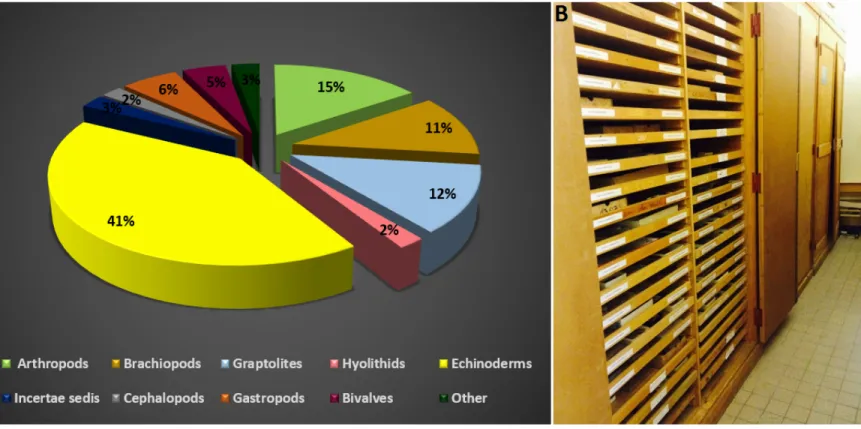
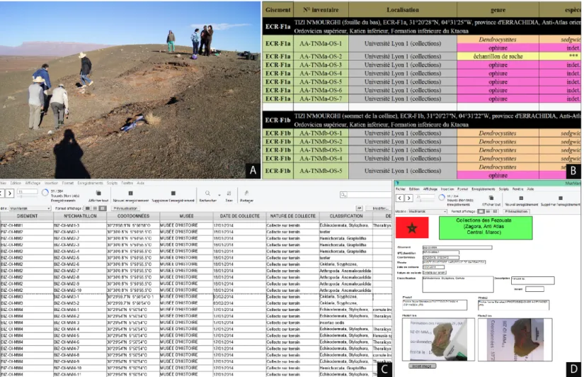
Since the original discovery of the Fezouata Biota by Mohamed ‘Ou Saïd’ Ben Moula in the early 2000s(Lefebvre et al., 2016a), to date, thousands of spectacular specimens were collected in situ (Figs. 2A-F and 4A), allowing the constitution of rich reference collections in institutions around the world, based mainly in France, Morocco, Switzerland, and the United States. The Cadi Ayyad University in Marrakesh, Morocco, owns one of the largest collections of fossils from the Fezouata Shale. Part of these collections is already in Marrakech, part is temporarily housed in the palaeontological collections of Lyon 1 University, France, pending its repatriation to Morocco (Fig. 3B), and finally, another part is on loan for scientific purposes in more than 15 institutions worldwide. The establishment of Fezouata collections at Marrakesh University was one of the primary aims of the collaboration initiated in 2002 between the Universities of Burgundy (Dijon) and Cadi-Ayyad (Marrakesh) by Bertrand Lefebvre and Khadija El Hariri (Lefebvre et al., 2016a, b). In this context, digital acquisition of Fezouata fossils was performed to better manage the collection and make it available for research. At first time, the digitization was designed using Microsoft Excel (Fig. 4B) and then this file was imported under the FileMaker Pro software (FileMaker Inc.), a cross-platform application, which allows the creation of customizable databases to suit the user requirements. Specimens are stored in the program as text records containing all the corresponding informations: inventory number, inventory status, systematics, stratigraphy, locality information, images, collection information (period, collector), loans, figurations, coordinates of the host organizations and any additional details. Data into FileMaker Pro can be visualized in three different ways as a form, as a list, and as a table (Figs. 4C-D). According to the last update in late October 2018, the full Marrakesh collection encompasses an estimated 4241 inventoried specimens, with 300 are figured and 12 are type specimens. This total includes annelids, arthropods, brachiopods, bryozoans, cnidarians, echinoderms, eldoniids, graptolites, hyolithids, lobopodians, molluscs, palaeoscolecidan worms, sponges, and other unidentified specimens (Fig. 3A). Any record that had no taxonomic classification, was assigned to the category ‘incertae sedis’. The Fezouata Biota database in Marrakesh is regularly updated and improved to meet the demands of a constantly evolving research. The value of a collection depends on the contribution of its specimens to the scientific accumulation of knowledge (Balzeau et al., 2010; Bradley et al., 2014). This is substantially illustrated in the specific case of the Fezouata Biota, by the involvement of over 50 scientists of 13 different nationalities in its study (Lefebvre et al., 2016a, b). Accordingly, Fezouata fossils remain in increase demand for use in research projects on a national and international scale. This means that institutions that house this heritage regularly loan material to researchers, and the specimens are transported all over the world, exposing them to damage or even loss. Hence, curators are confronting a double challenging mission which consists in preserving this unique heritage as well as paving the way for its scientific prosperity. In this sense, it is necessary to discuss the possibility of increasing the utility and the sustainability of Fezouata Biota collections by combining recent advances in 3D imaging methods, illustrated herein by computed microtomography and photogrammetry.
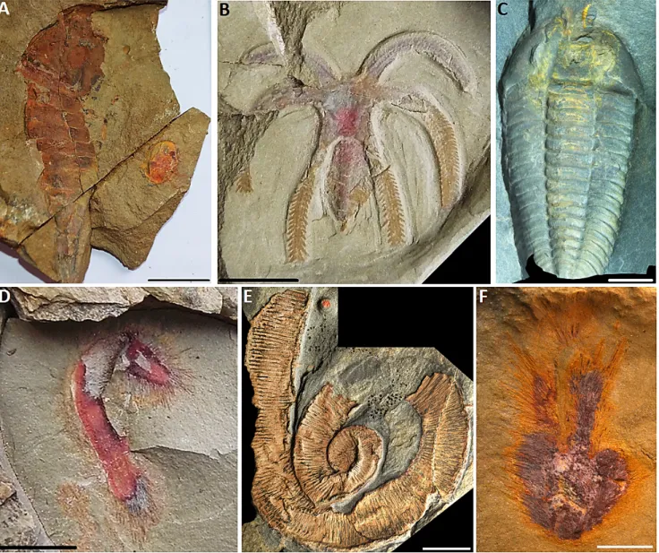
Fig. 2. Examples of exceptionally preserved fossils from the Fezouata Biota. A, Tremaglaspis sp. aglaspidid and a xiphosurid (AA-TER-OI-16). B, Furca sp. marrellomorph (AA-TER-OI-14). C, Bavarilla sp. trilobite (AA-BIZ15-OI-16). D, Pirania auraeum demosponge (AA-TER-OI-15). E, undetermined palaeoscolecid worm (AA-BGF2-OI-1). F, Pirania auraeum demosponge (AA-JBZ-OI-115). C, E, F, from Martin et al., 2016. Scale bars: A, 20 mm; B, D, 10 mm; C, E, F, 5 mm.
Fig 3. A, Distribution of fossiliferous groups from the Fezouata Shale belonging to the Marrakesh collections. B, Marrakesh collections temporarily deposited at Lyon 1 University, France.
Fig. 4. A, Excavation site of Bou Izargane, in the Fezouata Shale. B-D, Screenshots of the Fezouata Biota Database in Excel format (B) and FileMaker Pro layouts (C, D).
3. Material and Methods:
Material
Studied material consists of five specimens from the Fezouata Shale: AA.OBZ2.OI.2, AA.OBZ2.OI.3 and AA.OBZ2.OI.4 were collected from a site located in the North West of Tinzouline, Zagora (Central Anti-Atlas, Morocco), section [Z-F5(2)] wich is a lateral equivalent of [Z-F5] Oued Beni Zoli, within the A. murrayi Zone, whereas AA-TER-OI-5, AA-TER-OI-11 are from an unknown locality.
All specimens used in this study are housed in the collections of Cadi Ayyad University (Faculty of Sciences and Techniques), Marrakesh, Morocco.
Microtomography
Two specimens have been imaged at the University of Poitiers, France, with a laboratory device, the EasyTom XL Duo (RX-Solutions), set at the PLATINA platform of the IC2MP. According to the size of specimens, a microfocus x-ray source (Hamamatsu L8121-03) has been used, coupled with a flat panel imager (Varian PaxScan 2520 DX). The acquisition parameters are as follows: AA.OBZ2.OI.3 (8.5cm x 4.5cm): 75kV voltage, 333µA current, 1mm Al filter, stack acquisition with 2880 projections in 2 turns to acquire the whole specimen, 10 frames per second, average of 18 frames per projection, acquisition time of 1h45, anti-ring shift procedure and a spatial resolution of 30.5µm; AA.OBZ2.OI.4 (16cm x 10.5cm): 90kV, 460µA, 1mm Al filter, 2976 proj. in 3 turns, 12.5 fr. p. s., average of 20 fr. p. proj., acquisition time of 1h20, anti-ring shift procedure, and spatial resolution of 41.8µm. The weight of the raw data are 16 Go for both specimens. The data reconstructions (i.e. serial slices in 16bits uncompressed tif format) were computed using the software XAct (RX Solutions) with a filtered back projection algorithm (based on the Feldkamp method for cone beam geometry with Tukey filter) and attenuation of the beam hardening artefacts through linearization with a polynomial function. The weight of the reconstructed data are 5 Go and 8 Go for AA.OBZ2.OI.3 and AA.OBZ2.OI.4, respectively. Segmentation of the data, two and three dimension elaborations (virtual sections, 3D rendering and movies, wavefront objects) were performed using the software AVIZO v.9.7 (Thermo FisherTM-FEI).
Photogrammetry:
The software program used in this study for the 3D reconstructions is the commercial Agisoft PhotoScan Professional 1.4.4. Agisoft PhotoScan is a stand-alone 3D scanning software package, and is among the most reliable digitization programs (Kwan & Kwan, 2017),offering an easy-to-use interface that can be followed by non-experts. Photographs were taken using a Canon EOS 1300Dfitted with an EF-S 18–55 mm. The number of photographs required varies with the size and the detail level of the specimen; 36 to 47 photos were taken from different angles with only the natural light of the room, keeping a constant distance between the focus point and the specimen. A significant overlap between images was obtained to ensure the best coverage of the fossil.
The process of 3D model building from a physical specimen consists firstly of loading photographs into the program after checking their accuracy and then running the alignment. At this step, the software finds common points on photographs, matching them and determines the camera location of each photograph (Fig. 5A). Based on the estimated camera positions, the program reconstructs the depth information of each photo to create a dense point cloud that is very close to the appearance of the real object (Fig. 5B). This cloud is subsequently used to build a polygonal mesh, which consists of triangles that connect the 3 coordinates X, Y, Z (Fig. 5C). The program calculates then the texture of the mesh and constructs a 3D model of the fossil surface (Fig. 5D). The models were made with the default parameters of the program, except for the quality setting of “build dense cloud” that was changed to medium.
Fig.5. Main steps of photogrammetric reconstruction process (AA-TER-OI-5). A, Aligned images. B, Dense cloud generation, C, Polygon mesh construction. D, Textured model.
4. Results
Microtomography
For each analyzed sample, the reconstructed dataset can be visualized and operated through various ways: series of parallel slices along the orthogonal planes, oblique slices, surface rendering, volume rendering with or without false color, with or without transparency (see attached movies created with Avizo in supplementary data). CT provides reconstructed images whose contrasts are related to the differential attenuation of x-rays by materials. Indeed, the gray levels intensity of a given voxel in CT data is linked to the mean absorption of the X-ray beam by the corresponding volume of matter (i.e. linear attenuation coefficient, µ) which is a function of material density, atomic number Z, and energy of the incident beam (Kak and Slaney, 2001; Stock, 2009). Thereby, the gray scale allows relative comparison of densities: the denser the material is, the more strongly it attenuates the beam, and the higher the pixels intensity is in the reconstructed data. Conversely, the low density materials are the darkest in the reconstructions. For the sample AA.OBZ2.OI.3, microtomographic images revealed a clear clustering of the rhombiferan echinoderm Macrocystella bohemica and trilobites embodied in the matrix (Figs. 6B-G). Specimens are dense and exhibit good contrast with the hosting rock, bioturbations are also present (Fig. 7A, E). Some brighter elements can correspond to the preservation of an undissolved mineralized organism (7E). A colour code has been used in the segmentation according to the taxonomic attribution (Fig. 7). In AA.OBZ2.OI.4, the orthoconic cephalopod presents less, almost no contrast with the surrounding matrix (Fig. 8E) and is difficult, even sometimes impossible, to be successfully extracted. Unfortunately, the color structures visible to the naked eye on the external surface do not show any contrast in cross sections. However, transverse sections revealed a pronounced feature in the body chamber that is not visible on the surface (Figs.8C-D). Furthermore, CT have revealed the occurrence of dense ovoid structures with regular shape, varying in size but no granular texture is visible in their interiors. These structures may correspond to a secondary filling of voids (i.e. dense minerals).
In the two cases presented, the hidden structures/specimens have been segmented by the combination of digital image processing tools available within the software package Avizo 9.7 (e.g., Non-local mean filtering, Top-Hat filter, and manual corrections with the Segmentation Editor).
Fig. 6. Macrocystella bohemica sample (AA.OBZ2.OI.3). A, General view of the studied material. B, 3D rendering of the external morphology. C-E, Virtual transverse sections showing “hidden” specimens. F-G, 3D modelling using transparency. B–F, Microtomographic images. Scale bars: A, F, G, 10 mm; B, 5 mm; C-E, 2.5 mm.
Fig. 7. Macrocystella bohemica sample in transparency (AA.OBZ2.OI.3). A colour code has been used according the taxonomic attribution (A). Macrocystella bohemica fragments in green (B); trilobites in blue (C) and (D); Bioturbations in pink and other elements in beige (E); (F) profile view. Scale bars: A-E, 10 mm; F, 15 mm.
Fig.8. Orthoconic cephalopod (AA.OBZ2.OI.4). A, General view of the studied material. B, 3D surface rendering. C, D, Transverse sections revealing a feature in the living chamber (boxed area). E, 3D modelling using transparency. B–F, Microtomographic images. Scale bars: A, 2 cm; B-E, 10 mm.
Photogrammetry
Photogrammetric process has produced manipulable 3D digital models of the aglaspidid Tremaglaspis sp, a new xiphosurid, and the trilobite Asaphellus jujuanus (Figs. 9A-C).
Reconstructed models display satisfying informations about morphological details in terms of texture, color and shape. The resolution and accuracy of these models are not the best that can be obtained and may be further improved by adapting the camera and reconstruction parameters. The models were saved and exported in Wavefront (*.obj) and 3D pdf formats. (3D Pdfs are
Fig. 9. Final models of three specimens from Fezouata Biota, obtained with photogrammetry. A, Tremaglaspis sp., aglaspidid (AA-TER-OI-5). B, xiphosurid indet. (AA-TER-OI-11). C, Asaphellus jujuanus trilobite (AA.OBZ.OI.2).
5. Discussion
Computed tomography has been increasingly employed by palaeontologists to investigate internal structures and obtain the maximum knowledge about the fossils without destroying them (Tate & Cann, 1982; Ketcham & Carlson, 2001; Carlson et al., 2003; El Albani et al., 2010, 2014, 2019; Meyer et al., 2014). For example, Gaspard (2013) demonstrated the performance of this technique in the investigation of brachiopod shell, leading to the improvement of taxonomic precision in brachiopods. Likewise, a number of authors illustrated the great possibilities offered by microtomography in the study of fossils, revealing wealth of taxonomical detail, e.g. in tetrapods, (Anderson et al., 2003) bryozoans (Koromyslova & Pakhnevich, 2016; Koromyslova et al., 2018), or palaeoscolecids, (Kouraiss et al., 2018), allowing in some cases a detailed description and formal assignment to a species level (e.g., fossils in amber, Dierick et al., 2007). Likewise, 3D models of Fezouata fossils generated herein by microtomography (micro-CT) illustrate how informations that are optically blocked from view can be revealed, which added considerable scientific value to these samples. Micro-CT have gleaned the clustering within the rock of columnals, stem portions and isolated thecal plates of M. bohemica, as well as several trilobites fragments including 2 trilobites mostly complete (Figs. 6C-G; 7B-D, F). The only specimen of M. bohemica mostly articulated, with complete stem still in connection with the theca, is the one visible on the surface (Figs. 6A-B). Tinzouline section [Z-F5 (2)], is the lateral equivalent of Oued Beni Zoli section [Z-F5], which has yielded extremely abundant, exquisitely preserved specimens of the rhombiferan M. bohemica. Although they have been crushed nearly flat, most individuals are fully articulated, with complete stems and brachioles still in connection with the theca (Lefebvre et al., 2016c). This remarkable preservation requires the rapid, in situ entombment of alive individuals (Brett et al., 1997), probably by distal storm events (Ebbestad et Lefebvre, 2015). In contrast, some thin levels are crowded with skeletal elements (e.g., thecal plates of Macrocystella, trilobite fragments), and contain few complete specimens. Such levels can be interpreted as resulting from the downslope transport and accumulation of skeletal elements by stronger storm events (Ebbestad et Lefebvre, 2015; Lefebvre et al., 2016c). Similar preservation is illustrated herein in tomographic results which have gleaned the clustering of disarticulated specimens enduring likely, after death, some possible reworking before entombment (Ebbestad and Lefebvre, 2015). In the orthoconic cephalopod, micro-CT results in transparency mode, did not produce satisfying results (Fig. 8E), which would have permitted a meaningful comparison with the taxonomic characters used for species described previously from the Fezouata Biota (Kröger & Lefebvre, 2012). In a broader context, the results illustrate the potential of micro-CT in the investigation of hidden specimens and inner structures, as well as in the creation of virtual specimens, that can be manipulated digitally and easily shared without any risk for the original, often unique, specimens. Indeed, we predict a more widespread and systematic use of the method during the coming years in the exploration of new, but also ancient, palaeontological discoveries.
Despite its many advantages, the method has some limitations in palaeontological research. Some fossils cannot be investigated by (micro-) CT scanner due to a high degree of mineralization or
low absorption contrast (but synchrotron radiation and phase contrast tomography could be the solution; Taforeau et al., 2006; Bidola et al., 2015). The size and morphology of specimens may also be an obstacle. If the specimen is too thick and/or dense, the transmission rate of the x-ray beam will be too low and no contrast will be seen (Stock, 2009). If the specimen (or the rock hosting it) is too large, the spatial resolution will be too coarse to detect the presence of thin features or small fossils due to the partial volume effect (Mazurier et al., 2016; but local tomography can partially resolve this point). For instance, in the present study, the magnification used for scanning the entire AA.OBZ2.OI.3 sample is too weak to study the hidden trilobites in detail (Fig. 6, 7). Thereby, even it is a non-invasive technique, it is sometime necessary to cut samples to reach a higher magnification and a better transmission rate (Reid et al., 2019). Moreover, most of the fossil samples have a non-cylindrical morphology and a heterogeneous mineralogical composition. They are then far from the optimum conditions for computed tomography, especially with laboratory devices using conical and polychromatic x-ray beams, and so subject to many artefacts (e.g. beam hardening, cone beam, metal and streak artefacts), which can hide or deform structures of interest (Ketcham and Carlson, 2001; Cnudde and Boone, 2013; but computed laminography, for instance, can be a satisfactory solution for flat sample; Hann et al., 2017). Furthermore, the cost of this technique is reasonably high, which restricts its application and requires a fine selection of fossils to be scanned.
Regarding photogrammetry, it is a three-dimensional (3D) surface digitization method. Agisoft photoscan constructs 3D models of studied fossil from a series of overlapping photographs taken at different orientations. Generated 3D models provide information about the shape, texture and color, representing original specimens with great objectivity (Figs. 9A-C) even in limited light conditions. Yet, this method is scale-less, which requires to include a scale bar on the images, or object with known dimensions that can serve as a scale (Falkingham, 2012). Photogrammetric reconstruction is relatively easy as tutorials explaining the whole process are available online, along with many early studies, which have applied and discussed widely the method (Falkingham, 2012; Kwan & Kwan, 2017; Mallison & Wings, 2014; Milroy et al., 2015). Nonetheless, the photography phase is in many cases challenging. For instance, photographing large specimens without sacrificing accuracy can be complicated, because it is difficult to capture the whole sample and bringing out at the same time good details, if a high resolution of the model is required. Similarly, the small size of specimens makes them quite difficult to photograph adequately, as the closer an object is to the camera, the more it blurs, and both resolution and contrast suffer. The photography phase has an enormous impact on the final model, hence it was necessary to reshoot studied samples multiple times, until obtaining images with the best possible quality. Besides, the accuracy of the final model relies also on parameters fixed at each stage of the process (lowest, low, medium, high, and ultra-high). The construction time increases when the detail level and the image resolution are high (Falkingham, 2012; Fau et al., 2016). Accordingly, the specific requirement of a digital specimen determines ultimately the appropriate parameters to use. In this respect, it is often helpful to avoid high options when a ‘nice-looking’’ 3D model for visualization purposes is required, while for documentation, high accuracy is necessary (Remondino, 2006).
Photogrammetry is an inexpensive, portable, flexible and easy to use approach to obtain 3D models (Evin et al., 2016; Fau et al., 2016). For these reasons, the method has seen a growing application among palaeontologists and archaeologists over the past decade (Falkingham, 2012; Kersten & Lindstaedt, 2012). According to Petti et al. (2008),the photogrammetric 3D models of dinosaur tracks have provided accurate morphometric details, as well as several interesting information not recognizable in the field, leading to new interpretations about dinosaur walking dynamics. In the same way, photogrammetry performance was evaluated by many other authors as an accurate tool to produce reliable 3D models with satisfying levels of morphological detail in terms of texture, coloration and geometry (e.g. palaeobotanic specimens, Milroy et al., 2015;
extinct giant ratite bird, Fau et al., 2016; wolf crania, Evin et al., 2016). Getting the high
performance computer required by Agisoft Photoscan (Li et al., 2016), the main limitations met in this study are recognized in the large file sizes corresponding to the 3D models and the relatively low size of RAM, which has limited the general performance of the system and increased significantly the processing time.
In summary, digital models of specimens of the Fezouata Biota either produced through micro-CT or photogrammetry,underline the great capability of these 3D imaging techniques in research field and collection enhancement. On the one hand, generated 3D models could be stored in the Fezouata Biota database, providing a permanent digital record of palaeontological findings, as well as helping to reduce damages resulting from excessive handling of original specimens and therefore contributing to long-term conservation and preservation of more delicate specimens including type and figured ones. On the other hand, these methods facilitate the scientific communication, as they enable different researchers to exchange the virtual models of the studied specimens, and solve in some cases the problem of loans, especially if targeted specimens are required and cannot be transported because of their weight, administrative obstacles, or as they are of great value, it can be necessary to avoid any risk – even minimal- of loss or destruction. Further, such digital models make easier the dissemination of technical information to non-specialists audience (e.g. Bates et al., 2009, 2010), allowing them to interact with manipulable virtual specimens as well as giving the chance to those who consider fossils as very old and dusty objects, to see them in an artistic way and discover the ‘beauty’ of the iconic specimens.
Finally, it should be emphasized that the direct interaction with the physical specimen cannot be entirely replaced by the virtual model, notably in the case of palaeontological researches, which depend strongly on physical samples and require in some cases many analyses of the rock. Interestingly, these digital models can constitute an alternative of the physical specimens in some studies, if only they are accurate enough and display the level of details sought by the researcher.
6. Conclusion
Specimens of the Fezouata Biota are the core of a wide collaborative research program in continuous evolution and we should make our best to preserve this unique heritage. The examples presented here demonstrate that the Fezouata fossils could be more exciting and may yield additional informations, if they are digitally reconstructed in 3D. However, the adoption of 3D imaging methods, such as microtomography and photogrammetry, in palaeontology is still far from ubiquitous and the full potential of these techniques remains to be further explored. We
believe that the use of these digital techniques would certainly be beneficial, if it is extended to wider practices, stressing that the virtual 3D models could give an additional value to the fossil record in terms of internal investigation, scientific exchange, virtual documentation, conservation and valorization of collections.
Supplementary data can be found in the online version at http://dx. doi.org Acknowledgments
We are grateful to the Académie Hassan II des Sciences et Techniques (Morocco) that largely funded this work. The authors acknowledge Dr. Fabrice Monna (University of Burgundy, Dijon) for the valuable discussion about the photogrammetric software. Dr. Abel Prieur is deeply thanked for the database software handling. Special thanks go also to Youssef Taib, Najib Akka and Fatim-zahra Ihbach for the kind help with specimens photography. Agisoft Photoscan team is also thanked to have provided us the License Key. We thank the PLATeforme INstrumentale d’Analyse of the IC2MP for the access to the microCT device of the University of Poitiers. This paper is an outcome of the cooperation program VALORIZ (2012-2015) funded by both the CNRST and the CNRS. Finally, the French region La Nouvelle Aquitaine and the FEDER are also thank here for their support.
References
Anderson, J. S., Carroll, R. L., & Rowe, T. B. (2003). New information on Lethiscus stocki (Tetrapoda: Lepospondyli: Aistopoda) from high-resolution computed tomography and a phylogenetic analysis of Aistopoda. Canadian Journal of Earth Sciences, 40(8), 1071–1083. Balzeau, A., Grimaud-Hervé, D., Crevecoeur, I., Rougier, H., Froment, A., Gilissen, E., Mennecier, P., & Semal, P. (2010). Applications des
méthodes d’imagerie en paléoanthropologie: apports en termes de préservation, gestion et développement des collections. Comptes Rendus - Palevol, 9(6–7), 265–275.
Bates, K., Falkingham, P., & Rarity, F., Hodgetts, D., Purslow, A., & Manning, P. L. (2010). Application of high-resolution laser scanning and photogrammetric techniques to data acquisition, analysis and interpretation in palaeontology. International Archives of Photogrammetry, Remote Sensing and Spatial Information Sciences, XXXVIII(5), 68–73.
Bates, K. T., Falkingham, P. L., Hodgetts, D., Farlow, J. O., Breithaupt, B. H., O’Brien, M., Matthews, N., Sellers, W. I., & Manning, P. L. (2009). Digital imaging and public engagement in palaeontology. Geology Today, 25(4), 134–139.
Bidola, P., Stockmar, M., Achterhold, K., Pfeiffer, F., Pacheco, M.L.A.F, Soriano, C., Beckmann, F., & Herzen, J. (2015). Absorption and phase contrast X-ray imaging in paleontology using laboratory and synchrotron sources. Microscopy and Microanalysis, 21, 1288-1295. Boyer, D.M., Gunnell, G.F., Kaufman, S., & McGeary T.M. (2017). Morphosource: archiving and sharing 3-D digital specimen data. The
Paleontological Society Papers, 22, 157-181.
Bradley, R., D., Bradley, L.C., Garner, H.J., & Baker, R.J. (2014). Assessing the value of natural history collections and addressing issues regarding long-term growth and care. BioSciences, 64(13), 1150-1158.
Brett, C.E., Moffat, H.A., Taylor, W.L., 1997. Echinoderm taphonomy, taphofacies, and Lagersta¨tten. Paleontological Society Papers 3, 147–190. Carlson, W. D., Rowe, T., Ketcham, R. a, & Colbert, M. W. (2003). Applications of high-resolution X-ray computed tomography in petrology,
meteoritics and palaeontology. Geological Society Special Publication, 215, 7–22.
Cnudde, V., & Boone, M.N. (2013). High-resolution X-ray computed tomography in geosciences: A review of the current technology and applications. Earth-Science Reviews, 123, 1-17.
Cunningham, J. A., Rahman, I. A., Lautenschlager, S., Rayfield, E. J., & Donoghue, P. C. J. (2014). A virtual world of paleontology. Trends in Ecology and Evolution, 29(6), 347–357.
De Paor, D. G. (2016). Virtual Rocks. GSA Today, 26(8), 4–11.
Dierick, M., Cnudde, V., Masschaele, B., Vlassenbroeck, J., Van Hoorebeke, L., & Jacobs, P. (2007). Micro-CT of fossils preserved in amber. Nuclear Instruments and Methods in Physics Research, Section A: Accelerators, Spectrometers, Detectors and Associated Equipment, 580(1 SPEC. ISS.), 641–643.
Ebbestad, J. O. R., & Lefebvre, B. (2015). An unusual onychochilid mollusc from the Ordovician (Tremadocian) Fezouata Formation, Morocco. Geobios, 48(6), 427-438.
El Albani, A., Mangano, M. G., Buatois, L. A., Bengtson, S., Riboulleau, A., Bekker, A., et al. (2019). Organism motility in an oxygenated shallow-marine environment 2.1 billion years ago. Proceedings of the National Academy of Sciences, 116(9), 3431-3436.
El Albani, A., Bengtson, S., Canfield, D.E., Bekker, A., Macchiarelli, R., Mazurier, A., Hammarlund, E.U., Boulvais, P., Dupuy, J.-J., Fontaine, Claude, Fürsich, F.T., Gauthier-Lafaye, F., Janvier, P., Javaux, E., Ossa, F.O., Pierson-Wickmann, A.-C., Riboulleau, A., Sardini, P., Vachard, D., Whitehouse, M., Meunier, A. (2010). Large colonial organisms with coordinated growth in oxygenated environments 2.1 Gyr ago. Nature 466, 100–104.
El Albani, A., Bengtson, S., Canfield, D.E., Riboulleau, A., Rollion-Bard, C., Macchiarelli, R., Ngombi Pemba, L., Hammarlund, E., Meunier, Alain, Moubiya Mouele, I., Benzerara, K., Bernard, S., Boulvais, P., Chaussidon, M., Cesari, C., Fontaine, C., ChiFru, E., Garcia Ruiz, J.M., Gauthier-Lafaye, F., Mazurier, A., Pierson-Wickmann, A.C., Rouxel, O., Trentesaux, A., Vecoli, M., Versteegh, G.J.M., White, L., Whitehouse, M., Bekker, A. (2014). The 2.1 Ga old Francevillian biota: biogenicity, taphonomy and biodiversity. PLoS One e99438, 9. Evin, A., Souter, T., Hulme-Beaman, A., Ameen, C., Allen, R., Viacava, P., Larson, G., Cucchi, T., & Dobney, K. (2016). The use of close-range
photogrammetry in zooarchaeology: Creating accurate 3D models of wolf crania to study dog domestication. Journal of Archaeological Science: Reports, 9, 87–93.
Falkingham, P. L. (2012). Acquisition of high resolution three-dimensional models using free, open-source, photogrammetric software. Palaeontologia Electronica, 15(1), 1–15.
Fau, M., Cornette, R., & Houssaye, A. (2016). Apport de la photogrammétrie à la numérisation 3D d’os de spécimens montés : potentiel et limites. Comptes Rendus - Palevol, 15(8), 968–977.
Gaspard, D. (2013). X-ray computed tomography: A promising tool to investigate the brachiopod shell interior. Effects on 3D modelling and taxonomy. Comptes Rendus - Palevol, 12(3), 149–158.
Gutiérrez-Marco, J.C., García-Bellido, D.C., Rábano, I., & Sá, A.A. (2017). Digestive and appendicular soft-parts, with behavioural implications, in a large Ordovician trilobite from the Fezouata Lagerstätte, Morocco. Scientific Reports, 7, 39728.
Hann, G., Mao, F., Bi, S., Wang, Y., & Meng, J. (2017). A Jurassic gliding euharamiyidan mammal with an ear of five auditory bones. Nature, 551, 451-456.
Kak, A.C., & Slaney, M. (2001). Principles of Computed Tomography Imaging: Philadelphia, Pennsylvania, SIAM (Society for Industrial and Applied Mathematics), 327 p.
Kersten, T. P., & Lindstaedt, M. (2012). Image-Based Low-Cost Systems for Automatic 3D Recording and Modelling of Archaeological Finds and Objects. In : Proceedings of Euromediterranean conference, 1–10.
Ketcham, R. A., & Carlson, W. D. (2001). Acquisition, optimization and interpretation of x-ray computed tomographic imagery: Applications to the geosciences. Computers and Geosciences, 27(4), 381–400.
Koromyslova, A. V., & Pakhnevich, A. V. (2016). New species of Pachydermopora Gordon, 2002 and Beisselina Canu, 1913 (Bryozoa: Cheilostomida) from a Campanian erratic block (Belarus) and their micro-CT investigation. Paleontological Journal, 50(1), 41–53. Koromyslova, A. V, Pakhnevich, A. V, & Martha, S. O. (2018). Summary of micro-CT studies on Late Cretaceous bryozoans. In: Abstr. Bruker
Micro-CT Users Meeting, Ghent, 160–164.
Kouraiss, K., El Hariri, K., El Albani, A., Azizi, A., Mazurier, A., & Vannier, J. (2018). X-ray microtomography applied to fossils preserved in compression: Palaeoscolescid worms from the Lower Ordovician Fezouata Shale. Palaeogeography, Palaeoclimatology, Palaeoecology, 508, 48–58.
Kröger, B., & Lefebvre, B. (2012). Palaeogeography and palaeoecology of early Floian(Early Ordovician) cephalopods from the Upper Fezouata Formation, Anti-Atlas, Morocco. Fossil Record, 15(2), 61–75.
Kwan, D. H., & Kwan, J. M. (2017). Empowering cultural preservation in China through participatory digitization. Journal of Archaeological Science: Reports, 12, 161–164.
Lebrun, R., & Orliac, M.J. (2017). Morphomuseum: an online platform for publication and storage of virtual specimens. The Paleontological Society Papers, 22, 183-195.
Lefebvre, B., Guensburg, T.E., Martin, E., Milne, C.H., Mooi, R., Noailles, F., Vannier, J. (2013). Soft-part preservation in a solutan echinoderm from the Fezouata Biota (Lower Ordovician, Morocco). Abstr. 57th Ann. Meeting Palaeont. Ass., Zurich, pp. 44–45.
Lefebvre, B., El Hariri, K., Kouraïss, K., Martin, E., Noailles, F. (2014). Préservation exceptionnelle de parties molles chez des échinodermes stylophores de l'Ordovicien inférieur de la région de Zagora (Anti-Atlas central, Maroc). J. Assoc. Paléontol. Fr. 66, 42.
Lefebvre, B., El Hariri, K., Lerosey-Aubril, R., Servais, T. & Van Roy, P. (2016a). The Fezouata Shale (Lower Ordovician, Anti-Atlas, Morocco): A historical review. Palaeogeography, Palaeoclimatology, Palaeoecology, 460, 7-23.
Lefebvre, B., Lerosey-Aubril, R., Servais, T., & Van Roy, P. (2016b). The Fezouata Biota: An exceptional window on the Cambro-Ordovician faunal transition. Palaeogeography, Palaeoclimatology, Palaeoecology, 460, 1–6.
Lefebvre, B., Allaire, N., Guensburg, T.E., Hunter, A.W., Kouraïss, K., Martin, E.L.O., Nardin, E., Noailles, F., Pittet, B., Sumrall, C.D., Zamora, S. (2016c). Palaeoecological aspects of the diversification of echinoderms in the Lower Ordovician of central Anti-Atlas, Morocco. Palaeogeogr. Palaeoclimatol. Palaeoecol. 460, 97–121.
Lefebvre, B., Guensburg, T.E., Martin, E.L.O., Mooi, R., Nardin, E., Nohejlová, M., Saleh., F., Kouraïss, K., El Hariri, K., & David, B. (2019). Exceptionally preserved soft pars in fossils from the Lower Ordovician of Morocco clarify stylophoran affinities within basal deuterostomes. Geobios, 52, 27-36.
Li, X. quan, Chen, Z. an, Zhang, L. ting, & Jia, D. (2016). Construction and Accuracy Test of a 3D Model of Non-Metric Camera Images Using Agisoft PhotoScan. Procedia Environmental Sciences, 36, 184–190.
Mallison, H., & Wings, O. (2014). Photogrammetry in paleontology - A practical guide. Journal of Paleontological Techniques, 12(12), 1–31. Martin, E., Pittet, B., Gutiérrez-Marco, J.C., Lefebvre, B., Vannier, J., El Hariri, K., LeroseyAubril, R., Masrour, M., Nowak, H., Servais, T.,
Vandenbroucke, T., Van Roy, P., Vaucher, R., 2016a. The Lower Ordovician Fezouata Konservat-Lagerstätte from Morocco: age, environment and evolutionary perspectives. Gondwana Resarch, 34, 274–283.
Mazurier, A., Sardini, P., Rossi, A.M., Graham, R.C., Hellmuth, K.-H., Parneix, J.-C., Siitari-Kauppi, M., Voutilainen, M., & Caner, L. (2016). Development of a fractcture network in crystalline rocks during weathering: Study of Bischop Creek chronosequence using X-ray computed tomography and 14C-PMMA impregnation method. Geological Society of America Bulletin, 128(9-10), 1423-1438.
McKenzie-Clark, J., & Magnussen, J. (2016). Real and virtual: the role of computed tomography and 3D imaging in museum practice. In: Simpson A. and Hammond G. (eds) : A Cultural Cacophony: Museum Perspectives and Projects., Museums Galleries Australia, Sydney, pp. 208-221.
S. (2014). Three-dimensional microCT analysis of the Ediacara fossil Pteridinium simplex sheds new light on its ecology and phylogenetic affinity. Precambrian Research, 249, 79–87.
Milroy, A., Rozefelds, A. C., Coghlan, S., Holmes, A., & Hocknull, S. (2015). Digitising the collection evaluating photogrammetry as a means of producing a digital, three-dimensional model. Jouranl of Natural Science Illustration, 47, 3-10.
Petti, F., Avanzini, M., Belvedere, M., De Gasperi, M., Ferretti, P., Girardi, S., Remondino, F., & Tomasoni, R. (2008). Digital 3D modelling of dinosaur footprints by photogrammetry and laser scanning techniques: integrated approach at the Coste dell’Anglone tracksite (Lower Jurassic, Southern Alps, Northern Italy). Studi Trentini Di Scienze Naturali, Acta Geologica, 83, 303–315.
Racicot, R. (2016). Fossils secret revealed: x-ray scanning and applications in paleontology. The Paleontological Society Papers, 22, 21-38. Reid, M., Bordy, E.M., Taylor, W.L., le Roux, S.G., & du Plessis A. (2018). A micro X-ray computed tomography dataset of fossil echinoderms
in an ancient obrution bed: a robust method for taphonomic and palaeoecologic analyses. GigaScience, giy156. Remondino, F., & El Hakim, S. (2006). Image-based 3D modelling: a review. The Photogrammetric Record, 21(115), 269–291. Stock, S.R. (2009). MicroComputed Tomography: Methodology and Applications. Boca Raton, Florida, CRC Press, 331 p.
Sutton, M. D. (2008). Tomographic techniques for the study of exceptionally preserved fossils. Proceedings of R. Soc. B, 275, 1587-1593. Sutton, M., Rahman, I., & Garwood, R. (2016). Virtual Paleontology—an Overview. The Paleontological Society Papers, 22, 1–20.
Taforeau, P., Boistel, R., Boller, E., Bravin, A., Brunet, M., Chaimanee, Y., Cloetens, P., Feist, J., Hoszowska, J., Jaeger, J.-J., Kay, R.F., Lazzari, L., Marivaux, L., Nel A., Nemoz, C., Thibault, X., Vignaud, P., & Zabler S. (2006). Applications of X-ray synchrotron microtomography for non-destructive 3D studies of paleontological specimens. Applied Physics A., Materials Science and Processing, 83(2), 195-202. Tate, J. R., & Cann, C. E. (1982). High-resolution computed tomography for the comparative study of fossil and extant bone. American Journal of
Physical Anthropology, 58(1), 67–73.
Van Roy, P., Briggs, D. E. G., & Gaines, R. R. (2015). The Fezouata fossils of Morocco; an extraordinary record of marine life in the Early Ordovician. Journal of the Geological Society, 172(5), 541–549.
Van Roy, P., Orr, P. J., Botting, J. P., Muir, L. A., Vinther, J., Lefebvre, B., El Hariri, K., Briggs, D. E. G. (2010). Ordovician faunas of Burgess Shale type. Nature, 465(7295), 215–218.
Vinther, J., Parry, L., Briggs, D. E., & Van Roy, P. (2017). Ancestral morphology of crown-group molluscs revealed by a new Ordovician stem aculiferan. Nature, 542(7642), 471.
Wilson, J. P., & Varricchio, D. J. (2019). Photogrammetry of the Oryctodromeus cubicularis type locality burrow and the utility of preexisting, standard field photographs for three dimensional digital reconstruction. Historical Biology, 1-8.