HAL Id: inserm-02467823
https://www.hal.inserm.fr/inserm-02467823
Submitted on 5 Feb 2020
HAL is a multi-disciplinary open access
archive for the deposit and dissemination of
sci-entific research documents, whether they are
pub-lished or not. The documents may come from
teaching and research institutions in France or
abroad, or from public or private research centers.
L’archive ouverte pluridisciplinaire HAL, est
destinée au dépôt et à la diffusion de documents
scientifiques de niveau recherche, publiés ou non,
émanant des établissements d’enseignement et de
recherche français ou étrangers, des laboratoires
publics ou privés.
expansion
Matthias Jacquet-Lagrèze, Nourredine Bouhamri, Philippe Portran, Rémi
Schweizer, Florent Baudin, Marc Lilot, William Fornier, Jean-Luc Fellahi
To cite this version:
Matthias Jacquet-Lagrèze, Nourredine Bouhamri, Philippe Portran, Rémi Schweizer, Florent Baudin,
et al.. Capillary refill time variation induced by passive leg raising predicts capillary refill time response
to volume expansion. Critical Care, BioMed Central, 2019, 23 (1), pp.281.
�10.1186/s13054-019-2560-0�. �inserm-02467823�
R E S E A R C H
Open Access
Capillary refill time variation induced by
passive leg raising predicts capillary refill
time response to volume expansion
Matthias Jacquet-Lagrèze
1,2*, Nourredine Bouhamri
1, Philippe Portran
1,2, Rémi Schweizer
1,2, Florent Baudin
3,2,
Marc Lilot
4,5,6,7, William Fornier
1,2and Jean-Luc Fellahi
1,2Abstract
Background: A peripheral perfusion-targeted resuscitation during early septic shock has shown encouraging results. Capillary refill time, which has a prognostic value, was used. Adding accuracy and predictability on capillary refill time (CRT) measurement, if feasible, would benefit to peripheral perfusion-targeted resuscitation. We assessed whether a reduction of capillary refill time during passive leg raising (ΔCRT-PLR) predicted volume-induced peripheral perfusion improvement defined as a significant decrease of capillary refill time following volume expansion.
Methods: Thirty-four patients with acute circulatory failure were selected. Haemodynamic variables, metabolic variables (PCO2gap), and four capillary refill time measurements were recorded before and during a passive leg raising test and after a 500-mL volume expansion over 20 min. Receiver operating characteristic curves were built, and areas under the curves were calculated (ROCAUC). Confidence intervals (CI) were performed using a bootstrap analysis. We recorded mortality at day 90.
Results: The least significant change in the capillary refill time was 25% [95% CI, 18–30]. We defined CRT responders as patients showing a reduction of at least 25% of capillary refill time after volume expansion. A decrease of 27% inΔCRT-PLR predicted peripheral perfusion improvement with a sensitivity of 87% [95% CI, 73– 100] and a specificity of 100% [95% CI, 74–100]. The ROCAUCofΔCRT-PLR was 0.94 [95% CI, 0.87–1.0]. The ROCAUC of baseline capillary refill time was 0.73 [95% CI, 0.54–0.90] and of baseline PCO2gap was 0.79 [0.61–0.93]. Capillary refill time was significantly longer in non-survivors than in survivors at day 90.
Conclusion:ΔCRT-PLR predicted peripheral perfusion response following volume expansion. This simple low-cost and non-invasive diagnostic method could be used in peripheral perfusion-targeted resuscitation protocols. Trial registration: CPP Lyon Sud-Est II ANSM: 2014-A01034-43
Clinicaltrial.gov,NCT02248025, registered 13th of September 2014
Keywords: Capillary refill time, Fluid responsiveness, Passive leg raising, Peripheral perfusion, Microcirculation, Circulatory shock, PCO2gap
© The Author(s). 2019 Open Access This article is distributed under the terms of the Creative Commons Attribution 4.0 International License (http://creativecommons.org/licenses/by/4.0/), which permits unrestricted use, distribution, and reproduction in any medium, provided you give appropriate credit to the original author(s) and the source, provide a link to the Creative Commons license, and indicate if changes were made. The Creative Commons Public Domain Dedication waiver (http://creativecommons.org/publicdomain/zero/1.0/) applies to the data made available in this article, unless otherwise stated.
* Correspondence:matthias.jl@gmail.com
1
Département d’Anesthésie Réanimation, Centre Hospitalier Louis Pradel, Hospices Civils de Lyon, 59 Boulevard Pinel, 69500 Bron, France
2Université Claude-Bernard, Lyon 1, Campus Lyon Santé Est, 8 avenue
Rockefeller, 69008 Lyon, France
Introduction
Shock is one of the most common life-threatening con-ditions in critical care and a frequent cause of admission to intensive care units [1,2]. Variables related to macro-circulation, such as mean arterial pressure and central venous pressure, are used in the haemodynamic assess-ment of critically ill patients [1, 2]. These variables are considered as good surrogates to guide haemodynamic resuscitation [1]. However, macrohaemodynamic vari-ables may poorly reflect tissue perfusion and microcircu-lation [3]. Lactate reflects tissue perfusion and lactate-targeted resuscitation is the gold standard under current guidelines [1, 2], but lactate increase can have various explanation, and its decrease can be prolonged com-pared to peripheral perfusion [4]. Peripheral perfusion evaluation reflects intra-abdominal visceral organ perfu-sion [5]. A mottled skin and an increased capillary refill time (CRT) attest peripheral perfusion. CRT is defined as the time taken for a distal capillary bed to regain its colour after pressure has been applied to cause blanch-ing [6]. CRT has an acceptable prognostic value [7, 8]. Abnormal peripheral perfusion after initial resuscitation is associated with increased morbidity and mortality [9– 11]. CRT is widely used in critically ill paediatric and adult patients [14, 15]. Some authors praise to use per-ipheral perfusion-targeted therapy [12, 13]. Peripheral perfusion-targeted resuscitation is enticing as it might provide a real-time response to increases in flow. This could accelerate the decision to stop resuscitation and avoid the risks of fluid overload [14]. Recent studies have tested this hypothesis but have failed to show superiority against lactate-targeted resuscitation [15]. Many studies have focused on the prediction of cardiac index (CI) changes of a volume expansion while few have investi-gated the effects of volume expansion on tissue perfu-sion [16]. Passive leg raising (PLR) predicts fluid responsiveness based on cardiac output changes [17]. PLR has also been reported as a surrogate of volume ex-pansion to assess the effect of volume exex-pansion on the microcirculation [18]. As peripheral perfusion-targeted therapy is gaining importance, and other studies are ex-pected in this scope [19], a direct prediction of the effect of fluid on peripheral perfusion improvement could be helpful to tailor further studies.
Therefore, we hypothesised that a rigorous protocol to measure CRT variation in association with standardised PLR would be discriminating to predict peripheral perfu-sion response to fluid in adult patients with circulatory shock.
Materials and methods
Ethics
The study protocol was approved by our Institutional Review Board (IRB) for human projects (CPP Lyon
Sud-Est, ANSM: 2014-A01034-43), and the protocol was published a priori (Clinicaltrial.gov: NCT02248025). Oral and written information was given to all patients or rela-tives. Signed consent was waived by the ethics commit-tee. To allow our readers to assess the risk of bias, we followed the Standards for Reporting Diagnostic Accur-acy (STARD) statement to design and report the study [19].
Patients
This prospective observational study was conducted in a 20-bed adult cardiothoracic intensive care unit in a ter-tiary teaching hospital (Louis Pradel Hospital) in Lyon between September 2014 and December 2016. All pa-tients diagnosed with acute circulatory failure to whom the attending anaesthesiologist decided to administer a volume expansion could be included. Eligibility criteria were as follows: the patient required an arterial and a central venous catheter, a CRT had to be measurable, a cardiac output monitoring by transpulmonary thermodi-lution (PiCCO™ PULSION Medical Systems, Munich, Germany), and a 500-mL volume expansion needed to be prescribed by the attending physician. We defined acute circulatory failure according to the ESICM guide-lines [1]. We excluded patients with the following char-acteristics: pregnancy, cardiogenic pulmonary oedema with acute respiratory failure, mechanical circulatory support, a moribund state, intra-abdominal hyperten-sion, and lower limb amputation or compression stockings.
Study protocol and measurements
The study protocol encompassed four steps: baseline (T1), during PLR (T2), at return to baseline (T3), and after volume expansion (T4). At each of these steps, the following macrohaemodynamic variables were collected: systolic, diastolic, mean, arterial, and central venous pressure; heart rate; and cardiac output (CO), and we calculated the cardiac index (CI) as CO divided by the body surface area. Four consecutive CRT measurements were assessed at each time T1, T2, and T4. Mottling score [11] and metabolic variables (arterial and venous blood gases including arterial lactate) were also collected at T1 and T4, enabling us to calculate oxygen delivery, oxygen uptake, venous-to-arterial difference in carbon dioxide partial pressure (PCO2gap), modified respiratory
quotient, and oxygen extraction ratio (formulae of calcu-lation detailed in Additional file 1: Annex 3). We col-lected respiratory rate and pulse oximetry, and in cases of mechanical ventilation, we also assessed end-expira-tory pressure, plateau pressure, tidal volume, and
pres-sure support variables (Sequential Organ Failure
Assessment score [20] at inclusion and new Simplified Acute Physiology Score (SAPS2) [21]). Patients were a
posteriori sorted into two groups: capillary refill time re-sponders (CRT-R) and non-rere-sponders (CRT-NR), ac-cording to the reduction of at least 25% of CRT following volume expansion or not. Patients were also a posteriori sorted into two other groups: cardiac index re-sponders (CI-R) and non-rere-sponders (CI-NR), according to the increase of at least 15% of CI following volume expansion or not.
We recorded CRT with a smartphone’s video camera iPhone 6™ (Apple Inc., Cupertino, CA) characteristics: 8-megapixel iSight™ camera with 1.5 micropixels, auto-focus with auto-focus pixels,ƒ/2.2 aperture with video record-ing (1080p HD video recordrecord-ing, time-lapse video with stabilisation, cinematic video stabilisation, 30 images/s, with locked continuous autofocus). We controlled light-ing conditions uslight-ing the flashlight system. We made a calibrated compression of the skin using a piston for seven seconds (Additional file 1: Annex 2). The piston characteristics were as follows: a 10-ml syringe (BD Plas-tipak™, Plymouth, MI) filled in with 10 ml of air and closed with a plug (Vygon™, Ecouen, France). We had chosen the duration of compression according to a pre-vious publication [6], and the pressure was chosen to de-crease intra-observer variability (personal data). We applied the piston on the skin; the 10 ml of air was com-pressed to fit a 7-ml volume, generating a pressure at the surface of the skin of 176 mmHg on a 2.5-cm2 sur-face (personal data). Four CRT acquisitions were made on the thorax at each haemodynamic condition in less than 3 min by a single investigator (MJL) and subse-quently averaged and analysed a posteriori by 2 readers (MJL and NB) using the freeware Kinovea™ ( www.kino-vea.org). The video was seen several times to determine the end of the CRT, and the chronometer of the soft-ware was used to assess CRT. The readers were blinded to the clinical condition of the patients, to the evaluation of the index test (ΔCRT-PLR), and to the reference standard (ΔCRT-VE). In 20 patients, 4 CRT were ana-lysed by 2 observers (MJL and NB) to evaluate both intra- and inter-observer reproducibility. As recom-mended, we performed PLR from a semi-recumbent position at 45° [22]. Volume expansion consisted of a 500-mL lactate Ringer administration over 20 min. No modifications to the administration rate or new drug ad-ministration have occurred in the study period.
Endpoints
The primary endpoint of the study was to determine the diagnostic ability of ΔCRT-PLR to predict peripheral perfusion response, defined as a CRT decrease of at least 25% following volume expansion (VE). Secondary
end-points were to compare ΔCRT-VE, ΔCRT-PLR,
meta-bolic and macrocirculatory variables, and prognostic
markers and to measure the inter- and intra-observer variabilities.
Statistical analysis
Free Software Foundation’s R packages were used to compute descriptive and analytical statistical analyses. Sample size calculation was based on our primary end-point using Obuchowsky’s method [24], and 34 patients were needed to detect an area under the curve of the re-ceiver operating characteristic (ROC) curve of 0.8 with a power of 0.9 and an alpha risk of 0.05. The ratio be-tween CRT-R and CRT-NR in our population was hypothesised to be 0.5. A Shapiro-Wilk test was used to test the normal distribution of the data. Data were expressed as mean ± standard deviation or median [25th–75th interquartile range (IQR)] according to their distribution. The inter- and intra-observer reproducibil-ity for CRT measurements were evaluated by the coeffi-cient of variation. The definition of CRT-R was a CRT reduction of at least 25%. This was based on the least significant change (LSC) of the CRT of previous non-published personal data and then challenged by the LSC of the CRT in this cohort. As the LSC arbitrarily defines this threshold, we also displayed the results for different thresholds to define CRT-R and CRT-NR (Add-itional file 1: Annex 1). Pairwise comparisons of data were done with the paired Student’s t test or Wilcoxon test. The two-tailed Student t test or Mann-Whitney U test compared CRT-R and CRT-NR. Fisher test and χ2 were used appropriately to compare categorical data. To compare the effect of the group (CRT-R/CRT-NR) and time (T1, T2, T3, T4) on haemodynamic variables, we used a linear mixed-effect model using time as a variable with a fixed effect, and patient and group as variables with a random effect for intercept and slopes, respect-ively. Visual inspection of residual plots assessed the ab-sence of deviations from homoscedasticity or normality. Dunnett’s test enabled multiple comparisons to the
base-line for each haemodynamic variable. CRT was
expressed as a variation from baseline, computed as the difference between final and baseline value divided by the baseline value. Pearson’s correlation coefficient tested the linear correlations. ROC curves were built, and AUC was expressed with 95% confidence interval (CI) calculated with a bootstrap method using 2000 rep-etitions. Best thresholds were determined by the“closest top-left” method, and sensitivity, specificity, and positive and negative predictive values were expressed with 95% CI. The grey zone was determined with a two-step method: First, a bootstrap resampling method was ap-plied on ΔCRT-PLR and basal CRT and PCO2gap data.
The best threshold and its 95% CI were calculated for each variable using a bootstrap technique with 2000 rep-etitions to define a first inconclusive zone. Secondly, we
determined the cut-off values with a sensitivity less than 90% or specificity less than 90% defining a second incon-clusive zone. The larger of the two zones were retained as the grey zone. All the tests were two-sided, and a p value < 0.05 was considered statistically significant.
Results
Patient characteristics
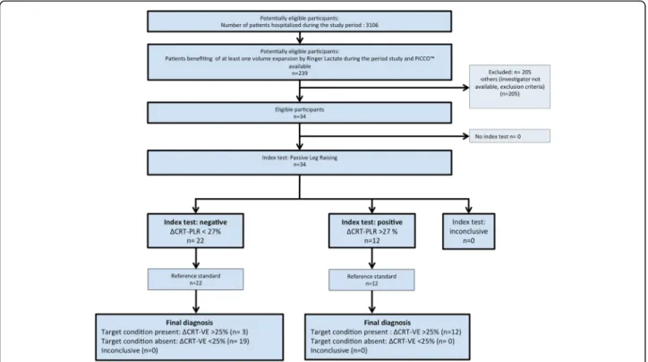
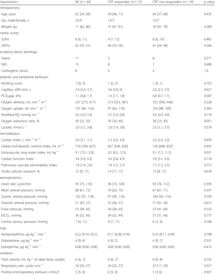
We included 34 non-consecutive patients in the study period (Fig. 1). Fifteen (44%) patients were CRT-R, and 19 (56%) were CRT-NR. The main characteristics of the patients’ population are shown in Table 1. Ten patients died before day 90 (29.4%). We assessed CRT with 4 vid-eos in each of the 3 steps of the study, except for 1 pa-tient who had only 9 over 12 video acquisitions due to a technical issue; their data were included in the final ana-lysis. Blood gases were missing in 6 patients due to transport and analytical issues. CRT and PCO2gap only
were significantly different between R and CRT-NR (Table 1). Using a response based on the cardiac index (CI), and defined as an increase of at least 15% fol-lowing VE, 13 (38%) patients were cardiac index re-sponders (CI-R) while 21 (62%) were not (CI-NR). Comparing CI-R and CI-NR for the same characteristics and haemodynamic variables, we did not find any signifi-cant differences, except for CI, oxygen delivery, and CRT at baseline: 2.7 s [IQR, 2.3–2.9] in CI-NR vs. 3.7 s
[IQR, 3.1–4.7] in CI-R (p = 0.018) (Additional file 1: Annex 5). We did not observe any adverse event from performing both CRT and PLR. Macrocirculatory, per-ipheral perfusion, and metabolic variables in CRT-R and CRT-NR at the different steps of the experimental protocol are shown in Table2. Only CRT changed sig-nificantly during PLR and volume expansion in the CRT-R compared to the CRT-NR.
The relationship between ΔCRT-PLR and ΔCRT after
volume expansion is depicted in Fig. 2 (r2= 0.62; p < 0.001). Individual CRT in CRT-R and CRT-NR, at base-line, during PLR, and after volume expansion are depicted in Additional file 1: Annex 4. We did not find any significant correlation between the changes in CRT and changes in macrocirculatory and metabolic variables induced by VE. However, the PCO2gap was higher in
CRT-R and the oxygen extraction ratio was almost nificantly higher in CRT-R at baseline and decreased sig-nificantly only in CRT-R following volume expansion. Median CRT at baseline was 2.7 s [IQR, 2.4–3.5] in sur-vivors and 4.4 s [IQR, 3.1–6.3] in non-sursur-vivors (p = 0.021).
Relationship between CRT responders and CI responders to volume expansion
Twenty-one patients were CI-NR, and 13 were CI-R. Seven patients (54%) were CRT-R in the 13 CI-R.
Fig. 1 Flow chart of the study. CRT, capillary refill time;ΔCRT-PLR, capillary refill time variation induced by passive leg raising; ΔCRT-PLR > 27%, positive index test defined as a decrease of capillary refill time induced by passive leg raising of at least 27%;ΔCRT-VE > 25%, CRT response defined as a decrease of capillary refill time induced by volume expansion of at least 25%; PLR, passive leg raising; VE, volume expansion
Table 1 Patients’ demographic and clinical characteristics in capillary refill time responders and non-responders to volume expansion
Characteristics All (n = 34) CRT responders (n = 15) CRT non-responders (n = 19) p value Anthropometry Age, years 62 [54, 69] 59 [46, 71] 64 [57, 68] 0.435 Sex, male/female, n 25/9 13/2 12/7 Weight, kg 71 [62, 80] 75 [67, 81] 70 [61, 78] 0.289 Severity scores SOFA 8 [6, 11] 9 [7, 12] 8 [6, 10] 0.485 SAPS2 42 [33, 51] 44 [33, 56] 41 [34, 48] 0.266 Circulatory failure aetiology
Sepsis 11 3 8 0.271
SIRS 15 8 7 0.489
Cardiogenic shock 8 4 4 1.0
Metabolic and peripheral perfusion
Mottling score 1 [0, 2] 1 [0, 2] 1 [0, 1] 0.702 Capillary refill time, s 2.9 [2.4, 3.7] 3.6 [2.8, 6] 2.6 [2.3, 3.3] 0.021 PCO2gap, kPa 1.1 [0.8, 1.7] 1.4 [1.1, 1.8] 0.8 [0.7, 1.1] 0.007
Oxygen delivery, mL min−1m−2 337 [272, 417] 313 [253, 361] 355 [309, 440] 0.228 Oxygen uptake, mL min−1m−2 101 [84, 116] 97 [64, 119] 103 [88, 109] 0.383 Modified RQ, mmHg mL−1 2.6 [2.0, 5.4] 3.5 [2.4, 6.8] 2.6 [2.0, 4.0] 0.118 Oxygen extraction ratio, % 30 [25, 35] 35 [26, 40] 28 [23, 30] 0.051 Lactate, mmol L−1 2.0 [1.2, 3.8] 2.0 [1.4, 3.8] 2.0 [1.1, 3.5] 0.578 Thermodilution
Cardiac index, L min−1m−2 2.6 [2.1, 3.1] 2.3 [2.0, 3.0] 2.6 [2.4, 3.2] 0.499 Global end-diastolic volume index, mL m−2 710 [549, 827] 667 [506, 826] 726 [606, 823] 0.492 Extravascular lung water index, mL kg−1 9.1 [7.0, 12.0] 9.2 [6.5, 12.5] 9.1 [7.2, 11.5] 0.931 Cardiac function index 3.6 [2.9, 4.3] 3.6 [2.8, 4.3] 3.6 [3.1, 4.3] 0.718 Pulmonary vascular permeability index 1.8 [1.4, 2.6] 1.8 [1.5, 2.7] 1.7 [1.3, 2.5] 0.772 Stroke volume variation, % 13 [9, 17] 14 [11, 17] 13 [8, 17] 0.676 Haemodynamics
Heart rate, cycle/min 95 [75, 110] 96 [73, 104] 93 [78, 112] 0.395 Mean arterial pressure, mmHg 68 [61, 72] 70 [62, 75] 67 [61, 71] 0.357 Systolic arterial pressure, mmHg 102 [91, 118] 97 [90, 116] 104 [94, 116] 0.795 Diastolic arterial pressure, mmHg 51 [43, 57] 52 [46, 57] 51 [41, 56] 0.615 Pulse pressure, mmHg 53 [40, 65] 49 [36, 65] 53 [41, 64] 0.532 EtCO2, mmHg 36 [32, 45] 34 [32, 44] 37 [31, 46] 0.737
Central venous pressure, mmHg 7 [4, 11] 8 [7, 11] 6 [3, 9] 0.186 Drugs
Norepinephrine,μg kg−1min−1 0.22 [0.10, 0.51] 0.17 [0.08, 0.76] 0.23 [0.11, 0.49] 0.798 Dobutamine,μg kg−1min−1 4 [0, 6] 4 [0, 5] 4 [0, 7] 0.531 Epinephrine,μg kg−1min−1 0.00 [0.00, 0.04] 0.00 [0.00, 0.00] 0.00 [0.00, 0.05] 0.472 Ventilation
Tidal volume, mL kg−1of ideal body weight 6 [6, 7] 6 [6, 7] 6 [6, 8] 0.702 Respiratory rate, cycles min−1 24 [20, 27] 24 [23, 27] 22 [17, 29] 0.357 Positive end-expiratory pressure, cmH2O 5 [5, 6] 6 [5, 6] 5 [5 6] 0.937
Thirteen patients (61%) were CRT-NR in the 21 CI-NR patients, and the odds ratio was 1.86 [95% CI, 0.38; 9.63]. Among the 19 patients with a CRT of less than 3 s, only 3 were CI-R. Among the 15 patients with a CRT of more than 3 s, 10 were CI-R and 9 were CRT-R. Reproducibility measurements
In 11 patients, 10 consecutive CRT were collected to as-sess the intra-observer variability. The coefficient of vari-ation for intra-observer variability was 17.6% [95% CI, 14.4–20.9] for a single CRT measurement and 8.8% [95% CI, 6.7–11.0] for 4 CRT measurements. The LSC for 4 measurements was 25.0% [95% CI, 17.7–29.6]. In 20 patients, CRT was analysed 4 times by 2 observers (MJL and NB). The coefficient of variation for inter-ob-server variability was 7.3% [95% CI, 3.9–10.2].
Prediction of CRT responsiveness
A 27% decrease in CRT-PLR predicted CRT responsive-ness with a sensitivity of 87% [95% CI, 73–100] and a specificity of 100% [95% CI, 74–100]. The ROCAUC of
ΔCRT-PLR was 0.94 [95% CI, 0.87–1.0]. Using the grey zone approach, inconclusive values ranged from − 30 to − 18% for ΔCRT-PLR to predict CRT responsiveness, in-cluding 24% of the patients. Using different thresholds to define CRT responsiveness, namely 15%, 20%, 30%, and 35%, ROCAUC were 0.97 [95% CI, 0.93–1.00], 0.97
[95% CI, 0.92–1.00], 0.96 [95% CI, 0.88–1.00], and 0.94 [95% CI, 0.84–1.00], respectively (Additional file 1: Annex 1).
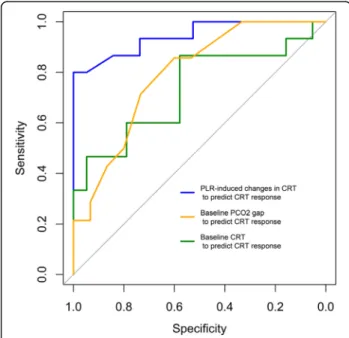
Two other variables predicted CRT responsiveness:
baseline CRT with a ROCAUC of 0.73 [95% CI, 0.54–
0.90] and PCO2gap with a ROCAUC of 0.79 [95% CI,
0.61–0.93]. Using the grey zone approach, inconclusive values ranged from 2.6 to 4.1 s for baseline CRT (includ-ing 44% of the patients) and from 0.9 to 1.7 kPa for PCO2gap (including 32% of the patients). Comparative
abilities of ΔCRT-PLR, baseline CRT, and PCO2gap to
predict CRT responsiveness are shown in Fig. 3 and
Table3.
Discussion
The main results are as follows: (1) changes in CRT dur-ing PLR predicts CRT responsiveness with a good
accuracy in acute circulatory failure, and the best thresh-old to assess CRT responsiveness is a CRT decrease by 27% during PLR; (2) baseline CRT and PCO2gap are also
able to predict CRT responsiveness; (3) baseline CRT is longer in non-survivors than in survivors; and (4) per-ipheral perfusion, macrocirculatory, and metabolic vari-ables are poorly correlated. The originality of the present study was to investigate a method predicting the effect of volume expansion on peripheral perfusion by using a simple, non-invasive, costless, static, and dynamic clin-ical sign, namely the CRT.
To carry out this work, we used a rigorous approach. First, to increase both precision and reproducibility of the CRT, we used a piston to calibrate the compression and we recorded the CRT on a video with controlled lu-minosity [23], enabling a blind lecture. Second, we
aver-aged four measurements for each haemodynamic
condition. Thus, we minimised the inter- and intra-ob-server variabilities compared with previous reports [24]. As we studied the variation in the same patients, we controlled other variables which might have influenced the CRT values, such as ambient temperature [25] and inter-patient variability linked to age or sex [24]. Third, to assess the precision of our measurement, we calcu-lated the LSC of CRT. It helped us to define a threshold to differentiate random variation due to fluctuation in the measurement and real change of the CRT that de-fined CRT responsiveness. The same method was used to define a fluid responder regarding cardiac output [26]. The assessment of cardiac index by transpulmonary thermodilution was performed as recommended. This technique provides a LSC of 12% [26], which is below our definition of cardiac index response. CRT values were significantly longer in patients dying in an intensive care unit than in survivors, as previously described [12]. Finally, diagnostic accuracy studies are at high risk of biases [27], but we minimised them by applying STARD guidelines [19] and reporting our adhesion to nearly all items (Additional file1: Annex 6).
Improvement of CRT by using volume expansion was reported [28], and a dissociation between macrocircula-tion and microcirculamacrocircula-tion or peripheral perfusion is well known [4, 29]. The dissociation may also be due to the lack of precision of both techniques, as the LSC of
Table 1 Patients’ demographic and clinical characteristics in capillary refill time responders and non-responders to volume expansion (Continued)
Characteristics All (n = 34) CRT responders (n = 15) CRT non-responders (n = 19) p value FiO2, % 40 [30, 60] 40 [36, 51] 40 [30, 60] 0.607
Driving pressure, cmH2O 12 [9, 15] 12 [10, 14] 12 [9, 15] 0.806
Values are median [percentile 25–75] or number. Wilcoxon test and Fisher’s exact test were used to calculate p value. CRT responders are defined as patients showing a decrease of at least 25% of the capillary refill time after volume expansion
CRT capillary refill time, EtCO2end-tidal carbon dioxide, FiO2fraction of inspired dioxygen, Modified RQ modified respiratory quotient defined as PCO2gap/
difference in arterio-venous content in oxygen, PCO2gap veno-arterial difference in carbon dioxide partial pressure, SAPS2 Simplified Acute Physiology Score, SIRS
Table 2 Changes in haemodynamic (A) and metabolic (B) parameters over the study period A Baseline (T1) During PLR (T2) Baseline (T3) After VE (T4) Group R/ NR Study period R/NRstudy period CRT, s CRT-R (n = 15) 3.6 [2.8, 6.0] 2.3 [1.6, 3.6] NA 2.1 [1.8, 2.9]* < 0.001 0.972 < 0.001 CRT-NR (n = 19) 2.6 [2.3, 3.3] 2.6 [2.0, 3.1] NA 2.6 [2.0, 3.1] CI, L min−1m−2 CRT-R (n = 15) 2.5 [2.0, 3.1] 2.8 [2.5, 3.3] 2.5 [2.1, 3.2] 3.0 [2.7, 3.2] 0.987 < 0.001 0.829 CRT-NR (n = 19) 2.5 [2.2, 3.1] 2.8 [2.5, 3.5] 2.4 [2.1, 3.3] 3.1 [2.6, 3.4] SVi, mL m−2 CRT-R (n = 15) 31 [24, 35] 34 [27, 37] 30 [24, 36] 33 [30, 40] 0.787 0.001 0.997 CRT-NR (n = 19) 27 [22, 35] 31 [30, 37] 27 [21, 36] 32 [27, 36] HR, min−1 CRT-R (n = 15) 96 [73.0, 104] 97 [72, 105] 96 [73, 106] 93 [70, 105]* 0.700 0.674 0.537 CRT-NR (n = 19) 93 [78, 112] 94 [78, 110] 95 [79, 110] 91 [80, 108] SAP, mmHg CRT-R (n = 15) 97 [90, 116] 116 [102, 136] 107 [94, 132.] 118 [105, 136] 0.624 0.477 0.554 CRT-NR (n = 19) 104 [94, 116] 112 [99, 116] 98 [94, 110] 112 [104, 119] DAP, mmHg CRT-R (n = 15) 52 [46, 57] 53 [46, 64] 49 [43, 54] 57 [45, 66] 0.958 0.524 0.249 CRT-NR (n = 19) 51 [41, 56] 51 [47, 56] 51 [46, 54] 51 [46, 57] MAP, mmHg CRT-R (n = 15) 70 [62, 75.] 71 [65, 82] 68 [60, 73] 76 [69, 81] 0.714 0.101 0.932 CRT-NR (n = 19) 67 [61, 70] 67 [63, 76.] 64 [58, 70] 68. [63, 75] PP, mmHg CRT-R (n = 15) 49 [36, 65.] 55 [46, 83] 58 [44, 83] 60 [46, 75] 0.321 0.051 0.548 CRT-NR (n = 19) 53 [41, 64] 56 [49, 63] 51 [44, 58] 58 [50, 72] SpO2, % CRT-R (n = 15) 98 [97, 100] 99 [97, 100] 99 [97, 100] 100 [97, 100] 0.353 0.143 0.575 CRT-NR (n = 19) 99 [95,100] 98 [95, 100] 97 [93, 100] 99 [96, 100] EtCO2, mmHg CRT-R (n = 15) 34 [32, 44] 36 [32, 45] 34 [30, 43] 34 [29.7, 45.5] 0.708 0.175 0.548 CRT-NR (n = 19) 37 [31, 46] 36 [32, 44] 35 [30, 43] 36 [31, 43] CVP, mmHg CRT-R (n = 15) 8 [7, 11] 11 [9, 14] 8 [6, 11] 10 [7, 13] 0.678 0.906 0.543 CRT-NR (n = 19) 6 [3, 9] 7 [5, 12] 5 [2, 8] 6 [4, 9] B
Baseline (T1) After VE (T4) p value Oxygen delivery, mL min−1m−2 CRT-R (n = 14) 313 [254–361] 370 [317–399] 0.002 CRT-NR (n = 14) 355 [309–440] 397 [327–458] 0.003 p value 0.24 0.697 Oxygen uptake, mL min−1m−2 CRT-R (n = 14) 103 [88–109] 100 [90–109] 0.357 CRT-NR (n = 14) 97 [64–119] 104 [80–133] 0.375 p value 0.401 0.697
Oxygen extraction ratio CRT-R (n = 14) 0.34 [0.26–0.40] 0.28 [0.22–0.33] 0.024 CRT-NR (n = 0.28 [0.23–0.30] 0.24 [0.19–0.33] 0.583
transpulmonary thermodilution cardiac index was up to 17% [26] (Table3). Preload modifications, as volume ex-pansion and PLR, can lead to changes in cardiac output [16], but also in arterial pressure according to patients’
dynamic elastance [30], and in central venous pressure and blood viscosity [31]. A reduction of sympathetic tone can increase microvascular flow [18]. The signifi-cant decrease in heart rate in CRT-R in the current study supports that idea. Though preload modification may change arterial pressure, venous pressure, sympa-thetic tone, and fluid administration can change blood viscosity. All those changes may have an opposing effect on microcirculation and peripheral perfusion. An ap-proach based directly on peripheral perfusion such as the CRT could be interesting in that context. Initial CRT predict the reduction of CRT after volume expansion, and this finding is consistent with the microvascular flow index findings, analysed with videomicroscopy, where an initial low microvascular flow index predicts an increase of microvascular flow index after volume ex-pansion [15]. The absence of correlation with metabolic variables could be explained by the lack of precision of
the biological tests [32]. The inherent natural variability of PaO2is important [17]. This could explain the
inabil-ity of oxygen uptake, oxygen delivery, and the modified respiratory quotient [33] to be linked with CRT respon-siveness in our study. Contrariwise, PCO2gap has less
variability.
We did not expect to have that much CRT-R patients among CI-NR patients. In peripheral perfusion-targeted therapy, only the patient showing fluid responsiveness on CI had a fluid load, and this strategy was stopped when peripheral perfusion targets were obtained [15]. We are not sure that patients who are CRT-R and CI-NR would benefit from a volume expansion, and those results should be interpreted cautiously. Noteworthy, the median value of CRT before volume expansion in CI-NR is normal (less than 3 s) [15] and significantly lower than in CI-R. A restrictive approach on volume expansion that select patient with CRT of more than 3 s and that
Table 2 Changes in haemodynamic (A) and metabolic (B) parameters over the study period (Continued)
14)
p value 0.051 0.608
Values are median [IQR]. A mixed-effect linear model was used to compute p value. Dunnett’s test was performed for multiple comparisons to the baseline *p < 0.05
CRT capillary refill time, CI cardiac index, CVP central venous pressure, DAP diastolic arterial pressure, EtCO2end-tidal carbon dioxide, HR heart rate, IQR 25th–75th
interquartile range, MAP mean arterial pressure, CRT-R responders to volume expansion defined as patients showing a decrease in CRT of at least 25% after volume expansion, CRT-NR non-responders to volume expansion define as patients showing a decrease in CRT after VE of less than 25%, PLR passive leg raising, PP pulse pressure, SAP systolic arterial pressure, DAP diastolic arterial pressure, SpO2dioxygen pulse saturation, SVi stroke volume index, VE volume expansion
Fig. 2 Scatter plot of capillary refill time variation induced by passive leg raising vs. by volume expansion. CRT, capillary refill time; PLR, passive leg raising; VE, volume expansion
Fig. 3 ROC curves of CRT andΔCRT-PLR to predict CRT response to volume expansion. CRT, capillary refill time; CRT responders, response to volume expansion defined as patients showing a decrease in CRT after VE of at least 25%; PCO2gap, central venous-to-arterial carbon dioxide difference; PLR, passive leg raising; VE, volume expansion
decrease significantly CRT during PLR could reasonably be tested and may reduce further fluid overload [14].
We acknowledge that our method to assess CRT is time-consuming and may be unrealistic in routine clin-ical practice. Future developments to digitalise CRT, providing real-time measurements, could facilitate its routine use [37].
Our study contains limitations. First, we did not have a gold standard to define microcirculation improvement. Such a standard does not presently exist, and each method explores a single window of the microvascular bed [34]. A way to validate the relevance of a microcir-culation assessment technique is to check the link with outcomes and mortality [35]. This has been done with CRT [12]. Second, the studied population is heteroge-neous with a majority of surgical patients experiencing a systemic inflammatory response syndrome due to car-diopulmonary bypass. This model of acute circulatory failure is not so far from the sepsis model, including vasoplegia, capillary leak, and contractility. Third, we are not sure that improvement of peripheral perfusion leads to less organ dysfunction and improved survival, as dif-ferent microcirculatory beds may behave difdif-ferently [36]. Goal-directed therapy protocols based on capillary refill time assessment have been tested and tend to be super-ior to protocols based on lactate assessment [15]. In this context, studies confirming our diagnostic method or other approaches predicting the effect of volume expan-sion on capillary refill time will be required. Fourth, the effect of PLR and volume expansion may have a different effect on tissue perfusion as blood viscosity evolution during PLR and VE was not the same and may alter the prediction accuracy of our method. Fifth, the sample size is quite small, and even if the study is positive for the primary endpoints, it impedes the valid interpretation of the association with other haemodynamic variables.
Conclusion
We report above an original method to predict the effect of volume expansion on peripheral tissue perfusion, based on CRT measurement coupled to a passive leg
raising manoeuvre. The method was accurate to predict the improvement in peripheral tissue perfusion of vol-ume expansion. This method could be implemented in peripheral perfusion-targeted therapy leading to a re-strictive fluid therapy approach. To be clinically feasible, this strategy needs to be confirmed using devices to as-sess an accurate, real-time digitalised CRT.
Additional file
Additional file 1:Annex 1. AUC ROC according to the different thresholds to define CRT responders. Annex 2. Presentation of the piston to perform a calibrated compression of the skin before analysing the capillary refill time. Annex 3. Formulae used. Annex 4. Capillary refill time at the different time courses of the study in CRT responders and non-responders. Annex 5. Patients’ demographic and clinical characteristics in cardiac index responders and non-responders to volume expansion. Annex 6. Adhesion of our study to the STARD statement. (DOCX 923 kb)
Abbreviations
ANSM:Agence National de Sécurité du Médicament et des Produits de Santé; CI: Cardiac index; CI: Confidence interval; CI-NR: Cardiac index non-responders; CI-R: Cardiac index non-responders; CPP: Comité de Protection des Personnes; CRT: Capillary refill time; CRT-NR: Capillary refill time non-responders; CRT-R: Capillary refill time non-responders; ESICM: European Society of Intensive Care Medicine; IQR: Interquartile range; LSC: Least significant change; PCO2gap: Venous-to-arterial carbon dioxide tension difference; PLR: Passive leg raising; ROCAUC: Receiver operating characteristic curves were built and areas under the curves; SAPS2: Simplified acute physiology score; STARD: Standards for Reporting Diagnostic Accuracy; ΔCRT-PLR: Variation of CRT following passive leg raising;ΔCRT-VE: Variation of CRT following volume expansion
Acknowledgements Not applicable
Authors’ contributions
MJL was responsible for the study concept and design, acquisition and interpretation of the data, drafting of the manuscript, statistical analysis, and study supervision. NB, PP, RS, FB, ML, and WF were responsible for the acquisition and interpretation of the data and critical revision of the manuscript. JLF was responsible for the study concept and design, interpretation of the data, critical revision of the manuscript, and study supervision. All authors read and approved the final manuscript.
Funding
There was no funding source. The Department of Anaesthesiology and Intensive Care of Louis Pradel Hospital took care of the extra cost. Table 3 Diagnostic performances to predict CRT and cardiac index responsiveness
Index test AUC [95% CI] Best threshold
Specificity Sensitivity PPV NPV Youden index Peripheral perfusion response (decrease of
25% of CRT after VE) ΔCRT-PLR 0.94 [0.87– 1.0] − 27% [− 30, − 18] 1.00 [0.741.00] – 0.87 [0.73– 1.0] 1.0 [0.72– 1.0] 0.91 [0.79– 1.0] 0.87 PCO2gap 0.79 [0.61– 0.93] 1.2 kPa [0.9– 1.6] 0.73 [0.53– 0.93] 0.79 [0.57– 1.00] 0.72 [0.58– 0.92] 0.79 [0.62– 1.00] 0.52 Baseline CRT 0.73 [0.54– 0.90] 2.7 s [2.6–4.1] 0.74 [0.47– 1.0] 0.80 [0.47– 1.00] 0.69 [0.54– 1.00] 0.67 [0.8– 1.00] 0.49 Cardiac index response (increase of 15% of CI) ΔCI-PLR 0.95 [0.86–
1.00] 9.0% [8.7– 13.7] 0.90 [0.71– 1.00] 1.00 [0.77– 1.00] 0.85 [0.68– 1.00] 1.00 [0.86– 1] 0.90
ROCAUCarea under the receiving operating characteristic curves, CRT capillary refill time, VE volume expansion,ΔCRT-PLR variation of CRT induced by passive leg
Availability of data and materials
The datasets used and analysed during the current study are available from the corresponding author on reasonable request.
Ethics approval and consent to participate
Comité de Protection des Personnes Lyon Sud-Est II ANSM: 2014-A01034-43.
Clinicaltrial.gov: NCT02248025. Consent for publication Not applicable Competing interests
The corresponding author has created a medtech company called DICARTECH which is developing a device to assess capillary refill time. All other authors declare that they have no competing interests.
Author details
1Département d’Anesthésie Réanimation, Centre Hospitalier Louis Pradel,
Hospices Civils de Lyon, 59 Boulevard Pinel, 69500 Bron, France.2Université
Claude-Bernard, Lyon 1, Campus Lyon Santé Est, 8 avenue Rockefeller, 69008 Lyon, France.3Département de Réanimation Pédiatrique, Centre Hospitalier
Femme mère enfant, Hospices Civils de Lyon, 59 Boulevard Pinel, 69500 Bron, France.4Département d’Anesthésie Pédiatrique, Centre Hospitalier
Femme Mère Enfant, Hospices Civils de Lyon, 59 Boulevard Pinel, 69500 Bron, France.5Centre Lyonnais d’Enseignement par Simulation en Santé,
SAMSEI, Université Claude Bernard Lyon 1, Lyon, France.6Health Services and
Performance Research Lab (EA 7425 HESPER), Université Claude Bernard Lyon 1, Lyon, France.7EPICIME-CIC 1407 de Lyon, Inserm, Hospices Civils de Lyon,
F-69677 Bron, France.
Received: 1 April 2019 Accepted: 31 July 2019
References
1. Cecconi M, De Backer D, Antonelli M, Beale R, Bakker J, Hofer C, et al. Consensus on circulatory shock and hemodynamic monitoring. Task Force of the European Society of Intensive Care Medicine. Intensive Care Med. 2014;40:1795–815.
2. Rhodes A, Evans LE, Alhazzani W, Levy MM, Antonelli M, Ferrer R, et al. Surviving Sepsis Campaign: international guidelines for management of sepsis and septic shock. Crit Care Med. 2017;1.
3. De Backer D, Creteur J, Preiser J, Dubois M, Vincent J. Microvascular blood flow is altered in patients with sepsis. Am J Respir Crit Care Med. 2002;166:98–104. 4. Garcia-Alvarez M, Marik P, Bellomo R. Sepsis-associated hyperlactatemia. Crit
Care. 2014;18:503.
5. Brunauer A, Koköfer A, Bataar O, Gradwohl-Matis I, Dankl D, Bakker J, et al. Changes in peripheral perfusion relate to visceral organ perfusion in early septic shock: a pilot study. J Crit Care. 2016;35:105–9.
6. Pickard A, Karlen W, Ansermino JM. Capillary refill time: is it still a useful clinical sign? Anesth Analg. 2011;113:120–3.
7. Ait-Oufella H, Bige N, Boelle PY, Pichereau C, Alves M, Bertinchamp R, et al. Capillary refill time exploration during septic shock. Intensive Care Med. 2014;40:958–64.
8. Fleming S, Gill P, Jones C, Taylor JA, Van den Bruel A, Heneghan C, et al. The diagnostic value of capillary refill time for detecting serious illness in children: a systematic review and meta-analysis. PLoS One. 2015;10. 9. Hernandez G, Pedreros C, Veas E, Bruhn A, Romero C, Rovegno M, et al.
Evolution of peripheral vs metabolic perfusion parameters during septic shock resuscitation. A clinical-physiologic study. J Crit Care. 2012;27:283–8. 10. de Moura EB, Amorim FF, da Cruz Santana AN, Kanhouche G, de Souza
Godoy LG, de Jesus Almeida L, et al. Skin mottling score as a predictor of 28-day mortality in patients with septic shock. Intensive Care Med. 2016;42: 479–80.
11. Ait-Oufella H, Lemoinne S, Boelle PY, Galbois A, Baudel JL, Lemant J, et al. Mottling score predicts survival in septic shock. Intensive Care Med. 2011;37: 801–7.
12. Hernandez G, Bruhn A, Castro R, Regueira T. The holistic view on perfusion monitoring in septic shock. Curr Opin Crit Care. 2012.
13. Dünser MW, Takala J, Brunauer A, Bakker J. Re-thinking resuscitation: leaving blood pressure cosmetics behind and moving forward to
permissive hypotension and a tissue perfusion-based approach. Crit Care. 2013;17:326.
14. van Genderen ME, Engels N, van der Valk RJP, Lima A, Klijn E, Bakker J, et al. Early peripheral perfusion-guided fluid therapy in patients with septic shock. Am J Respir Crit Care Med. 2015;191:477–80.
15. Hernández G, Ospina-Tascón GA, Damiani LP, Estenssoro E, Dubin A, Hurtado J, et al. Effect of a resuscitation strategy targeting peripheral perfusion status vs serum lactate levels on 28-day mortality among patients with septic shock: the ANDROMEDA-SHOCK randomized clinical trial. JAMA. 2019;321:654–64.
16. Pranskunas A, Koopmans M, Koetsier PM, Pilvinis V, Boerma EC.
Microcirculatory blood flow as a tool to select ICU patients eligible for fluid therapy. Intensive Care Med. 2013;39(4):612-9.
17. Monnet X, Marik P, Teboul J-L. Passive leg raising for predicting fluid responsiveness: a systematic review and meta-analysis. Intensive Care Med. 2016.
18. Pottecher J, Deruddre S, Teboul J-L, Georger J-F, Laplace C, Benhamou D, et al. Both passive leg raising and intravascular volume expansion improve sublingual microcirculatory perfusion in severe sepsis and septic shock patients. Intensive Care Med. 2010;36:1867–74.
19. Pettilä V, Merz T, Wilkman E, Perner A, Karlsson S, Lange T, et al. Targeted tissue perfusion versus macrocirculation-guided standard care in patients with septic shock (TARTARE-2S): study protocol and statistical analysis plan for a randomized controlled trial. Trials. 2016;17:384.
20. Bossuyt PM, Reitsma JB, Bruns DE, Gatsonis CA, Glasziou PP, Irwig L, et al. STARD 2015: an updated list of essential items for reporting diagnostic accuracy studies. BMJ. 2015:h5527.
21. Vincent JL, Moreno R, Takala J, Willatts S, De Mendonça A, Bruining H, et al. The SOFA (Sepsis-related Organ Failure Assessment) score to describe organ dysfunction/failure. On behalf of the Working Group on Sepsis-Related Problems of the European Society of Intensive Care Medicine. Intensive Care Med. 1996;22:707–10.
22. Le Gall JR, Lemeshow S, Saulnier F. A new Simplified Acute Physiology Score (SAPS II) based on a European/North American multicenter study. JAMA. 1993;270:2957–63.
23. Monnet X, Teboul J-L. Passive leg raising: five rules, not a drop of fluid! Crit Care Lond Engl. 2015;19:18.
24. Obuchowski NA. Sample size calculations in studies of test accuracy. Stat Methods Med Res. 1998;7:371–92.
25. Brown LH, Prasad NH, Whitley TW. Adverse lighting condition effects on the assessment of capillary refill. Am J Emerg Med. 1994;12:46–7.
26. Anderson B, Kelly A-M, Kerr D, Clooney M, Jolley D. Impact of patient and environmental factors on capillary refill time in adults. Am J Emerg Med. 2008;26:62–5.
27. Schriger DL, Baraff L. Defining normal capillary refill: variation with age, sex, and temperature. Ann Emerg Med. 1988;17:932–5.
28. Monnet X, Persichini R, Ktari M, Jozwiak M, Richard C, Teboul J-L. Precision of the transpulmonary thermodilution measurements. Crit Care. 2011;15:R204.
29. Jacquet-Lagrèze M, Izaute G, Fellahi J-L. Diagnostic accuracy studies: the methodologic approach matters! Anesthesiology. 2017;127:728–9. 30. Maitland K, Pamba A, Newton CRJC, Levin M. Response to volume resuscitation
in children with severe malaria. Pediatr Crit Care Med. 2003;4:426–31. 31. De Backer D, Ortiz JA, Salgado D. Coupling microcirculation to systemic
hemodynamics. Curr Opin Crit Care. 2010;16:250–4.
32. De Backer D, Donadello K, Sakr Y, Ospina-Tascon G, Salgado D, Scolletta S, et al. Microcirculatory alterations in patients with severe sepsis: impact of time of assessment and relationship with outcome. Crit Care Med. 2013;41:791–9.
33. García MIM, Romero MG, Cano AG, Aya HD, Rhodes A, Grounds RM, et al. Dynamic arterial elastance as a predictor of arterial pressure response to fluid administration: a validation study. Crit Care. 2014;18:626. 34. Guérin L, Teboul J-L, Persichini R, Dres M, Richard C, Monnet X. Effects of
passive leg raising and volume expansion on mean systemic pressure and venous return in shock in humans. Crit Care. 2015;19.
35. Klijn E, Niehof S, Johan Groeneveld AB, Lima AP, Bakker J, van Bommel J. Postural change in volunteers: sympathetic tone determines microvascular response to cardiac preload and output increases. Clin Auton Res. 2015;25: 347–54.
36. Mallat J, Lazkani A, Lemyze M, Pepy F, Meddour M, Gasan G, et al. Repeatability of blood gas parameters, PCO2 gap, and PCO2 gap to
arterial-to-venous oxygen content difference in critically ill adult patients. Medicine (Baltimore). 2015;94.
37. Blaxter LL, Morris DE, Crowe JA, Henry C, Hill S, Sharkey D, et al. An automated quasi-continuous capillary refill timing device. Physiol Meas. 2016;37:83–99.
Publisher’s Note
Springer Nature remains neutral with regard to jurisdictional claims in published maps and institutional affiliations.


![Table 2 Changes in haemodynamic (A) and metabolic (B) parameters over the study period A Baseline (T1) During PLR(T2) Baseline(T3) After VE (T4) Group R/NR Study period R/NRstudyperiod CRT, s CRT-R ( n = 15) 3.6 [2.8, 6.0] 2.3 [1.6, 3.6] NA 2.1 [1.8, 2.9]*](https://thumb-eu.123doks.com/thumbv2/123doknet/14654432.552439/8.892.86.790.169.1095/table-changes-haemodynamic-metabolic-parameters-baseline-baseline-nrstudyperiod.webp)

![Table 3 Diagnostic performances to predict CRT and cardiac index responsiveness Index test AUC [95%CI] Best threshold](https://thumb-eu.123doks.com/thumbv2/123doknet/14654432.552439/10.892.84.817.148.317/table-diagnostic-performances-predict-cardiac-responsiveness-index-threshold.webp)