HAL Id: hal-00528347
https://hal.archives-ouvertes.fr/hal-00528347
Submitted on 21 Oct 2010HAL is a multi-disciplinary open access archive for the deposit and dissemination of sci-entific research documents, whether they are pub-lished or not. The documents may come from teaching and research institutions in France or
L’archive ouverte pluridisciplinaire HAL, est destinée au dépôt et à la diffusion de documents scientifiques de niveau recherche, publiés ou non, émanant des établissements d’enseignement et de recherche français ou étrangers, des laboratoires
Quantum efficiency of energy transfer in noncovalent
carbon nanotube/porphyrin compounds
Cyrielle Roquelet, Damien Garrot, Jean-Sébastien Lauret, C. Voisin, Valérie
Alain-Rizzo, Philippe Roussignol, Jacques Delaire, Emmanuelle Deleporte
To cite this version:
Cyrielle Roquelet, Damien Garrot, Jean-Sébastien Lauret, C. Voisin, Valérie Alain-Rizzo, et al.. Quan-tum efficiency of energy transfer in noncovalent carbon nanotube/porphyrin compounds. Applied Physics Letters, American Institute of Physics, 2010, 97 (14), pp.141918. �10.1063/1.3496470�. �hal-00528347�
Quantum efficiency of energy transfer in non-covalent carbon
nanotube/porphyrin compounds
C. Roquelet,1 D. Garrot,1 J.S. Lauret,1, ∗ C. Voisin,2 V.
Alain-Rizzo,3 Ph. Roussignol,2 J. A. Delaire,3 and E. Deleporte1
1Laboratoire de Photonique Quantique et Mol´eculaire,
Institut d’Alembert, CNRS, ENS Cachan, 94235 Cachan, France
2Laboratoire Pierre Aigrain, Ecole Normale Sup´erieure,
UPMC, Universit´e Denis Diderot, CNRS, 75005 Paris, France
3Laboratoire de Photophysique et Photochimie Supramol´eculaires et Macromol´eculaires,
Institut d’Alembert, CNRS, ENS Cachan, 94235 Cachan, France
(Dated: August 11, 2010)
Abstract
We report on the quantum yield of excitation energy transfer in non-covalently bound nan-otube/porphyrin compounds. Evidence for energy transfer is gained from photoluminescence exci-tation experiments. We perform a quantitative evaluation of the transfer quantum yield in the case of (6,5) nanotubes through three independent methods : quantitative PLE measurements, evalu-ation of the luminescence quenching of the donor (porphyrin) and ultrafast transient absorption measurements. The latter shows a tremendous increase of the porphyrin recovery rate upon incor-poration in the compound. All these measurements consistently lead to an exceptional quantum yield efficiency: 1-10−3 ≤ η ≤ 1-10−5.
Functionalization of single-wall carbon nanotubes (SWNTs) is a rapidly developing field in view of applications such as biology imaging [1] or optoelectronics [2–4]. In particular, the coupling of dye molecules with nanotubes is of great interest regarding possible photovolta¨ıc and other optoelectronic applications [2, 3, 5]. The general motivation is to combine the exceptional transport properties of carbon nanotubes with the versatility of the optical properties of the chromophores. In this context, it is of particular importance to preserve the intrinsic properties of carbon nanotubes such as their high mobility. Therefore usual covalent chemistry, which leads to deep defects formation due to local destruction of the
π-electronic system, should be avoided. In contrast, the use of non-covalent chemistry (such
as π-stacking interaction) has proven to be compatible with high quality transport and optical properties of the nanotubes [6–8]. On the other hand it usually leads to poorly stable compounds [6–8]. Recently, an original method based on micelle encapsulation of the nanotube/molecule compound has been demonstrated that allows both stability and minimal perturbation to the nanotube properties [8]. Depending on the band alignment, this coupling can lead to either charge transfer (CT) or excitation energy transfer (EET). In view of applications, it is important to show that although the compound stability relies on weak interactions, it is possible to have an efficient coupling between the nanotube and the organic molecule.
In this Letter, we study SWNT/porphyrin compounds which are known for showing EET [6–8] and we perform a quantitative investigation of the strength of this coupling in the case of (6,5) nanotubes by measuring the quantum yield and the dynamics of the energy transfer. Photoluminescence (PL) experiments conducted on carbon nanotube/porphyrin compounds show unambiguous evidence for EET : the emission of light on the near infra-red (NIR) transitions of the nanotube is strongly enhanced when the excitation is in resonance with absorption lines of the porphyrin molecule. Concomitantly, the fluorescence of the porphyrin is strongly suppressed. In the time domain, the complex shows a reduction by several orders of magnitude of the porphyrin ground state recovery time. Those three independent measurements consistently lead to an exceptional EET quantum yield comprised between 1-10−3 and 1-10−5.
Purified CoMoCAT nanotubes (SouthWest Nanotechnologies, SG65) containing mainly the (6,5) chirality were functionalized with base-free tetraphenyl porphyrin (TPP) by means of the micelle swelling method (see [8] for details). The output of a monochromator
illumi-nated by a UV-VIS Xenon lamp served as a tunable light source for the photoluminescence excitation experiments (PLE). A step of 10 nm is used for the excitation in the PLE map. The ultrafast pump/probe experiments were conducted in a degenerate configuration at 2.952 eV and 2.831 eV with a frequency doubled mode-locked Ti:Saphire laser which deliv-ers ∼80 fs pulses at a 86 MHz repetition rate.
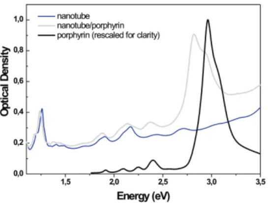
FIG. 1: Optical absorption spectra of a water suspension of nanotube in micelles (blue) and of a water suspension of nanotube/porphyrin compounds (grey). Optical absorption spectrum of a micellar suspension of porphyrins rescaled for clarity (black).
Carbon nanotubes display NIR absorption from S11 excitons (around 1.2 eV) and visible
absorption from S22 excitons near 2 eV (Figure 1, blue line). For the SWNT/TPP
com-pounds (Figure 1, grey line), these lines are red-shifted of about 20 meV as discussed in [6–8]. The main absorption line of the compound (the so-called Soret absorption band of the porphyrin) is observed at 2.831 eV. It corresponds to ground state absorption to the second electronic excited state of the porphyrin in the compound. The shoulder at 2.952 eV is a signature of residual free porphyrins embedded in micelles (to be compared to free por-phyrin absorption spectrum, black line in Fig. 1) [8]. The transitions to the first electronic excited states (Q bands, around 2 eV) are barely visible in the compound since they are much weaker and further overlap with the S22 excitonic absorption of the nanotubes.
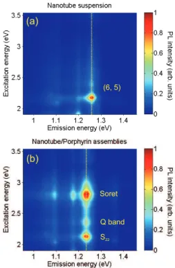
Figure 2a) displays the PLE map of the nanotube suspension. One spot predominates in this map. The semi-empirical formula of Bachilo et al. allows to assign this spot to
the (6,5) family [9]. Additional faint spots are present that correspond to weakly abundant chiral families. The PLE map of the nanotube/porphyrin compound suspension is shown in Figure 2b). The spot corresponding to (6,5) nanotubes is red-shifted both in excitation and emission energies. This shift results from a dielectric screening of the excitonic transition by the porphyrin molecules [10]. Compared to the PLE map of nanotubes, additional spots are visible along the vertical dashed line for excitation energies of 2.805 and 2.357 eV. The spot at (x=1.240 eV, y=2.805 eV) corresponds to absorption on the Soret-band and emission by nanotubes. This brings evidence for excitation energy transfer from the porphyrin to the nanotube: when photons are absorbed by the porphyrin molecules, the luminescence of the nanotube is enhanced [6–8]. We assign the PLE feature at an excitation energy of 2.357 eV to EET upon absorption on the porphyrin higher Q band, red shifted of about 50 meV in comparison with free porphyrins. The other Q bands could not be evidenced in PLE measurements, most probably because of spectral overlap with the nanotubes S22
transitions. Nevertheless, the observation of the 2.357 eV PLE feature means that the EET occurs at least partially through the Q bands of the porphyrin molecules.
(a) (b) Soret Q band S22 (6, 5)
FIG. 2: PLE maps of a) the nanotube suspension; b) the nanotube/porphyrin compound suspension. The yellow dashed line corresponds to a PLE line-cut.
Importantly, the intensity of the intrinsic PLE peak on the S22 transition of (6,5)
nan-otubes is on the same order than the PLE peak through EET from the Soret band (upper and lower peaks along the PLE line-cut in Fig. 2b). This allows quantitative investigation of the transfer yield. The PLE peak amplitude of nanotubes is related to their absorption cross-section σS22 (1.1 ± 0.1×10⁻¹² cm2.µm−1) for (6,5) nanotubes [11], since it is well
es-tablished that the internal conversion from S22 to S11 is ultrafast and efficient [12, 13]. On
the other hand, the PLE peak at 2.805 eV (EET) is related to the absorption cross-section of the porphyrin (σT P P = 7 × 10⁻¹⁶ cm2) [14], the surface coverage of the nanotube by TPP
molecules (α) and the transfer yield (η). The equal amplitude of the two PLE peaks leads to the following relationship:
σT P P × N × η = σS22 with N = α
SN T ST P P
where ST P P ' 1.22 nm2 is the surface of one TPP molecule and SN T is the wall surface per
micron length of a (6,5) nanotube. We find that ηα = 0.95 ± 0.10. The synthesis process ensuring that the coverage is close to one [8], we deduce that the transfer yield must also be on the order of one.
Another independent manner for evaluating this quantum efficiency is given by the quenching of the luminescence of the donor. Figure 3a) shows the visible luminescence of free porphyrins (black curve) and of the complex (grey curve) with the same porphyrin concentration and incident power. Both spectra display identical bands at 1.719 eV and 1.900 eV but the photoluminescence of the complex is reduced by a factor 750 for the complex. However, the position of the lines in the compound shows that the residual lu-minescence actually arises from left after porphyrin rather than from the compound itself since the bands of the latter should be red shifted by ∼ 50 meV. This assignment is fur-ther confirmed by PLE measurements: again the excitation spectrum of this residual PL (Figure 3b)) is identical to the one of free porphyrin whereas the redshift of the Soret band is of 120 meV for the compounds (See Figure 1). When looking specifically for PL of the compound by tuning the excitation at 2,818 eV, we cannot detect any signal within our detectivity which is limited by the tail emission of free porphyrin. Assuming that any signal from the compound would be detected if of intensity comparable to this tail emission, we deduce Id−a
Id ≤ 9.10
−4 where I
d−a (resp. Id) is the integrated PL intensity of the donor in
2,0 2,5 3,0 3,5 0,0 0,5 1,0 1,7 1,8 1,9 2,0 104 105 106 107 108 P L E In te n s it y (n o rm .) Energy (eV) (a) (b) P L In te n s it y (a rb . u n it s ) Energy (eV) 0 200 400 600 800 1000 0,0 0,5 1,0 porphyrinnanotube/porphyrin (c) D T /T (n o rm a li ze d ) Delay (fs)
FIG. 3: a) Photoluminescence spectra of free porphyrins (black curve) and of the nan-otube/porphyrin compounds (grey curve) excited on their respective Soret band. b) Normalized PLE detected 1.722 eV in free porphyrins (black curve) and in the nanotube/porphyrin compound (grey curve). c) Relative change of transmission as a function of the time delay for pump and probe in resonance with the Soret band of the free porphyrins (black curve) or in resonance with the Soret band of the compounds (grey curve). Green and blue dashed lines are bi- and mono-exponential fits respectively.
can be written:
η = 1 −Id−a
Id
we can give a lower bound of η : η ≥ 1 − 10−3.
Finally, ultrafast pump/probe experiments allowed us to gain further insight into the dynamics and therefore into the transfer yield of this EET. In these experiments, both pump and probe were tuned in resonance with the Soret band of the porphyrin (at 2.952 eV for the free porphyrin or at 2.831 eV for the compound). Free porphyrin shows a bi-exponential recovery dynamics (Figure 3c) with a main very slow component (unresolved here) that was reported to be on the order of 12 ns in previous studies [15]. This component corresponds
to the ground state recovery. In nanotube/porphyrin compounds, this recovery time is dramatically reduced down to ∼120 fs. This decrease of the donor life-time is typical of an efficient donor-acceptor coupling. The ratio τd−a
τd , where τd (resp. τd−a) is the recovery
time of the free (resp. in compound) porphyrin is on the order of 10−5 and is related to the
efficiency η by:
η = 1 − τd−a
τd = 1 − 10 −5
In summary, we have reported on efficient and ultrafast excitation energy transfer in nanotube/porphyrin assemblies. The quenching of the porphyrin luminescence and the tremendous increase of its relaxation rate show that the efficiency of the EET process is very high. Cross-estimates of this quantum yield lead to 1 − 10−3 ≤ η ≤ 1 − 10−5. This result
is especially strong for non-covalently bound compounds. This opens avenues for creating molecular assemblies based on nanotubes that preserve the nanotube intrinsic properties and nevertheless show efficient coupling to the chromophore for light-harvesting or photovolta¨ıc applications.
This work was supported by the GDR-E ”nanotube” (GDRE2756), grant ”C’Nano IdF EPONAD” from ”R´egion Ile de France” and ANR grant ”CEDONA”.
[1] Z. Liu, S. Tabakman, S. Sherlock, X. Li, Z. Chen, K. Jiang, S. Fan, and H. Dai, Nano Research 3, 222 (2010).
[2] C. Ehli, C. Oelsner, D.M. Guldi, A. Mateo-Alonso, M. Prato, C. Schmidt, C. Backes, F. Hauke, A. Hirsch., Nature Chemistry 1, 243 (2009).
[3] S. Campidelli, B. Ballesteros, A. Filoramo, D. Daz Daz, G. de la Torre, T. Torres, G. M. Aminur Rahman, C. Ehli, D. Kiessling, F. Werner, V. Sgobba, D.M. Guldi, C. Cioffi, M. Prato, and J.P. Bourgoin, J. Am. Chem. Soc. 130, 11503 (2008).
[4] M. Alvaro, P. Atienzar, P. de la Cruz, J. L. Delgado, V. Troiani, H. Garcia, F. Langa, A. Palkar, and L. Echegoyen., J. Am. Chem. Soc. 128, 6626 (2006).
[5] C. Ehli, G. M. Aminur Rahman, N. Jux, D. Balbinot, D. M. Guldi, F. Paolucci, M. Marcaccio, D. Paolucci, M. Melle-Franco, F. Zerbetto, S. Campidelli, and M. Prato, J. Am. Chem. Soc. 128, 11222 (2006).
[6] G. Magadur, J.S. Lauret, V. Alain-Rizzo, C. Voisin, Ph. Roussignol, E. Deleporte, and J.A. Delaire., ChemPhysChem 9, 1250 (2008).
[7] J.P. Casey, S.M. Bachilo, and R.B. Weisman, Journal of Material Chemistry 18, 1510 (2008). [8] C. Roquelet, J.S. Lauret, V. Alain-Rizzo, C. Voisin, R. Fleurier, M. Delarue, D. Garrot, A. Loiseau, Ph. Roussignol, J. A. Delaire, and E. Deleporte, ChemPhysChem 11, 1667 (2010). [9] S.M. Bachilo, M.S. Strano, C. Kittrell, R.H. Hauge, R.E. Smalley, and R.B. Weisman., Science
298, 2361 (2002).
[10] S. Berger, F. Iglesias, P. Bonnet, C. Voisin, G. Cassabois, J.S. Lauret, C. Delalande, and P. Roussignol, J. App. Phys. 105, 094323 (2009).
[11] S. Berciaud, L. Cognet and B. Lounis, Phys. Rev. Lett. 101, 077402 (2008).
[12] J.S. Lauret, C. Voisin, G. Cassabois, C. Delalande, Ph. Roussignol, L. Capes, O. Jost, Phys. Rev. Lett. 90, 057404 (2003).
[13] J. S. Lauret, C. Voisin, S. Berger, G. Cassabois, C. Delalande, Ph. Roussignol, L. Goux-Capes, A. Filoramo, Phys. Rev. B 72, 113413 (2005).
[14] J. B. Kim, J. J. Leonard and F. R. Longo, J. Am. Chem. Soc. 94, 3986 (1972). [15] J. S. Baskin, H.Z. Yu and A. H. Zewail, J. Phys. Chem. A 106, 9837 (2002).