doi:10.1093/intimm/dxt026
TGF-β type II receptor expression in thymic
epithelial cells inhibits the development of Hassall’s
corpuscles in mice
Chikako Odaka
1, Mathias Hauri-Hohl
2,6, Kazuya Takizawa
1, Yomiko Nishikawa
4, Masashi Yano
4,
Mitsuru Matsumoto
4, Richard Boyd
5and Georg A. Holländer
2,31Department of Safety Research on Blood and Biological Products, National Institute of Infectious Diseases, Tokyo 208-0011, Japan 2Department of Biomedicine, Laboratory of Pediatric Immunology, University of Basel and the University Children’s Hospital,
4058 Basel, Switzerland
3Developmental Immunology, Department of Paediatrics, University of Oxford, Oxford, UK
4Division of Molecular Immunology, Institute for Enzyme Research, University of Tokushima, Tokushima 770-8503, Japan 5Monash Immunology and Stem Cell Laboratories, Monash University, Clayton, Victoria 3800, Australia
6Present address: Benaroya Research Institute, Virginia Mason Hospital, Seattle, WA 98101, USA
Correspondence to: C. Odaka, Department of Safety Research on Blood and Biological Products, National Institute of Infectious Diseases, Tokyo 208-0011, Japan. E-mail: [email protected]
Received 23 January 2013, accepted 15 May 2013 Abstract
Hassall’s corpuscles are concentric clusters of keratinized epithelial cells located within the thymic medulla of humans and guinea pigs but are scant in mouse and rat. They are considered to be the terminally differentiated stages of medullary thymic epithelial cells (mTECs) but the mechanisms of their origin are unclear. We have previously deleted the TGF-β type II receptor (TGFβRII) specifically in mouse TECs and reported that these mice have mitigated thymic involution and exhibit earlier reconstitution post-irradiation. In this study, we analyzed the differentiation of mTECs in the TGFβRII-knockout mice. Interestingly, the TGFβRII-TGFβRII-knockout mice display enhanced development of Hassall’s corpuscles. The expression of Aire, stromal-cell-derived factor 1 and thymic stromal lymphopoietin in the thymi of the TGFβRII-knockout mice was similar to that previously reported for the human thymus. In addition, the putative epithelial progenitor markers MTS20 and MTS24 labeled Hassall’s corpuscles in normal mice, but the extent and intensity of this staining were greatly enhanced in Hassall’s corpuscles of the TGFβRII-knockout mice. The phosphorylated forms of ERK and JNK were also found in Hassall’s corpuscles of the TGFβRII-knockout mice. Taken together, we suggest that TGFβRII-mediated signaling in TECs inhibits their development into Hassall’s corpuscles in mice. Keywords: epithelial cells, Hassall’s corpuscle, MTS24, TGF-β type II receptor, thymus
Introduction
The thymus provides a specialized environment uniquely adept in attracting lymphoid precursor cells and inducing their pro-liferation, differentiation and selection into functionally mature T cells, which are ultimately exported to peripheral lymphoid tissues. Within this microenvironment, thymic epithelial cells (TECs) constitute the most abundant stromal component and are arranged both in the cortex and in the medulla as a three-dimensional scaffold (1–3). Recent reports have indicated that both different types of TECs, cortical TECs and medullary TECs (mTECs), may be derived from common progenitors in ontogeny (4–7). The MTS20 and MTS24 monoclonal antibod-ies recognize an antigen expressed on a population contain-ing high-efficiency TEC progenitor cells (4, 5). Accumulating
evidence indicates that mTECs comprise heterogeneous populations.
Hassall’s corpuscles, also known as Hassall’s bod-ies, are found in thymic medulla and form characteristic swirled epithelial structures. In addition to their distinctive histologic appearance, Hassall’s corpuscles express the antigens that are detectable in the terminally differentiated upper layers of the epidermis and are therefore thought to be composed of terminally differentiated mTECs (8–10). Hassall’s corpuscles are well developed in humans and guinea pigs, but, interestingly, they are not typically seen in mice or rats (2). Hassall’s corpuscles have been proposed to act in both the removal of dead thymocytes and the
For permissions, please e-mail: [email protected] doi:10.1093/intimm/dxt026
maturation of medullary thymocytes (11–13). Other studies have provided evidence that Hassall’s corpuscles express cytokines, such as IL-7, CD30 ligand, stromal-cell-derived factor 1 (SDF-1), macrophage-derived chemokine and thymic stromal lymphopoietin (TSLP) (14–18). Recently, TSLP produced by Hassall’s corpuscles was shown to educate dendritic cells to induce the development of CD4+CD25+ regulatory T cells (18). These data suggest
that Hassall’s corpuscles actively communicate with devel-oping T cells within the thymus.
TGF-β superfamily members exert their effects primarily via a receptor complex comprising type I and type II receptors (TGFβRI and TGFβRII) (19, 20). To date, TGFβRII has been identified to be essential for TGF-β binding and for complex assembly with TGFβRI. When TGF-β superfamily members bind to the TGFβRI and TGFβRII complex, activated TGFβRI propagates the signals downstream via phosphorylation of specific receptor-regulated Smad proteins. Phosphorylated Smad2 (p-Smad2) and p-Smad3 form heteromeric complexes with Smad4 and translocate into the nucleus to regulate β-responsive genes. Several Smad-independent TGF-β signaling pathways have also been identified, including MAPK pathways (19, 20). How they regulate thymopoiesis and the constitution of the TEC compartment is largely unknown. Mice deficient for the expression of TGFβRII die around embryonic day 11.5. Therefore, we have conditionally inactivated TGFβRII using Cre/Lox technology on TECs to identify whether the TGF-β signaling pathway plays a role in TEC development and function (21). We previously demonstrated that the disruption of TGFβRII expression on TECs results in a mitigated thymic involution and an early post-irradiation reconstitution (21).
Here, we performed a detailed analysis of thymic epi-thelium in the mice deficient for TGFβRII. We found the enhanced development of Hassall’s corpuscles in the thymic medulla, suggesting that TGFβRII expression on TECs restricts their cell progression into Hassall’s corpus-cles in mice. Although previous studies have demonstrated that MTS20 or MTS24 immunostaining is found on a rare subset of mTECs in adult mouse thymus (4, 5), we show that most of the MTS20 or MTS24 staining in the thymus of postnatal mice is found in Hassall’s corpuscles. On the basis of our results, we discuss the role of Hassall’s corpus-cles in thymogenesis.
Methods Mice
The generation of TGFβRIIlox/lox and TGFβRIIlox/lox::Foxn1-Cre
mice has been previously reported (21). Mice were housed at the center’s animal facility in accordance with Institutional and Cantonal review boards and were used at the age of 8 weeks.
Antibodies and reagents
The following antibodies and reagents were used: rabbit anti-keratin 5 (Covance, Berkeley, CA, USA), rabbit anti- involucrin (Covance), rat anti-E-cadherin (clone ECCD2) (a gift
from Dr M. Takeichi at the Riken Center for Developmental Biology or a product of Takara Bio Inc., Shiga, Japan), rabbit anti-CXCL12α subunit (SDF-1α) (eBioscience), rat monoclo-nal anti-Aire (22), rabbit anti-TSLP (Sigma–Aldrich, St Louis, MO, USA), rat monoclonal antibody MTS20 (4, 5), rat mono-clonal antibody MTS24 (4, 5), rabbit anti-phospho-p44/42 MAPK (ERK1/2) (Thr202/Tyr204) (Cell Signaling Technology, Inc., Danvers, MA, USA), rabbit anti-phospho-SAPK/JNK (Thr183/Tyr185) (Cell Signaling Technology, Inc.), rabbit anti-phospho-p-38 MAPK (Thr180/Tyr182) (Cell Signaling Technology, Inc.) and Alexa Fluor-labeled donkey second-ary antibodies (Molecular Probes, Eugene, OR, USA). The binding to biotinylated Ulex europaeus agglutinin-1 (UEA-1) (Vector Laboratories, Burlingame, CA, USA) or biotinylated
Tetragonolobus purpureas agglutinin (TPA) (Sigma–Aldrich)
was followed by FITC- or PE-conjugated streptavidin (eBioscience).
Immunofluorescence staining and confocal microscopy
Frozen thymic sections were prepared and immunofluo-rescence staining was performed on thymic sections as previously described (21, 23, 24). Confocal laser-scanning microscopy analysis was performed on a Zeiss LSM 510 (Carl Zeiss, Oberkochen, Germany). Negative controls were performed by replacement of first-step antibodies with iso-type-matched monoclonal antibodies or species-matched antibodies. Representative images were chosen from each experiment (n = 6 for TGFβRIIlox/lox; n = 6 for TGFβRIIlox/lox::
Foxn1-Cre mice) for figure preparation.
Results
Hassall’s corpuscles are developed in mice deficient for TGFβRII on TECs
To clarify whether the disruption of TGFβRII expression on TECs affects the differentiation of mTECs, we stained thy-mus tissues from 8-week-old TGFβRIIlox/lox and TGFβRIIlox/lox::
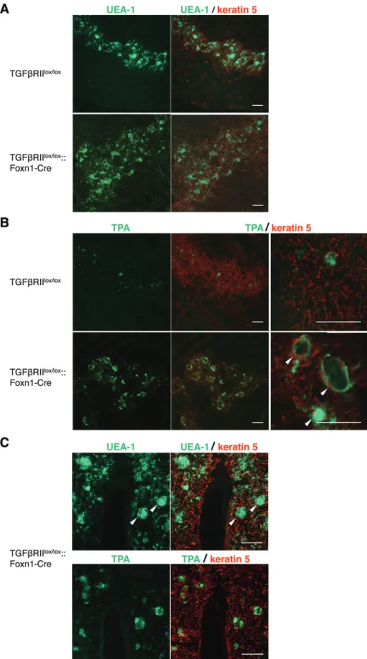
Foxn1-Cre mice with the reagents recognizing mTECs, and we analyzed them by confocal microscopy. The distribu-tion of keratin 5 allows distincdistribu-tion of mTECs, and the mature populations of mTECs bind the lectin UEA-1 (23, 24). It has been demonstrated that the lectin TPA particularly binds to Hassall’s corpuscles (25). In TGFβRIIlox/lox mice, UEA-1+
epithelial cells formed a network of stellate cells that builds the thymic medulla (Fig. 1A). As expected, the binding of TPA was also restricted to mTECs, and the fractions of TPA-binding TECs were occasionally detected in small globular cell bodies (Fig. 1B). Surprisingly, many TPA-binding spheres were detectable in the thymic medulla of TGFβRIIlox/lox
::Foxn1-Cre mice (Fig. 1B). These Hassall’s corpuscles in TGFβRIIlox/ lox::Foxn1-Cre mice varied in size and expressed the ligands
for both UEA-1 and TPA (Fig. 1C). Immunohistochemistry of human thymus using anti-involucrin antibody is known to stain Hassall’s corpuscles (26). When thymus sections from TGFβRIIlox/lox::Foxn1-Cre mice were stained with
anti-involucrin antibody, we found larger anti-involucrin-expressing structures with a hyalinized degenerated core in the thymic medulla (Fig. 2). Furthermore, immunoglobulins are present in Hassall’s corpuscles of the human thymus (27), and we
Fig. 1. Hassall’s corpuscles are developed in mice deficient for TGFβRII on TECs. Immunofluorescence staining in thymic sections of TGFβRIIlox/lox
also detected IgG in Hassall’s corpuscles of TGFβRIIlox/lox::
Foxn1-Cre mice (Fig. 2).
Phenotypic changes of mTEC subsets in mice deficient for TGFβRII on TECs
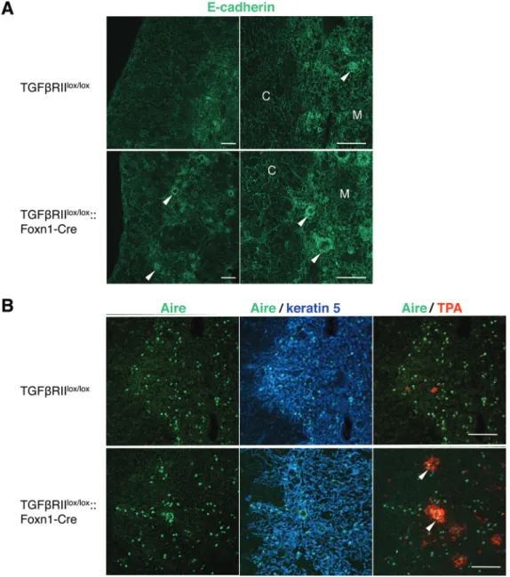
We further examined the expression of E-cadherin, Aire, SDF-1 and TSLP in the thymi of TGFβRIIlox/lox and TGFβRIIlox/ lox::Foxn1-Cre mice. As reported previously (28), E-cadherin
was expressed in all TECs of both cortex and medulla in the
thymi of TGFβRIIlox/lox mice, and Hassall’s corpuscles with
globular cell bodies in the medulla were strongly reactive with antibodies against E-cadherin (Fig. 3A). In TGFβRIIlox/ lox::Foxn1-Cre mice, the epithelial cells composing the outer
layers of Hassall’s corpuscles showed high expression of E-cadherin. A small population of mTECs was shown to express the autoimmune regulator Aire, which is crucial in the induction of T-cell tolerance toward tissue-restricted anti-gens (29). Previous observations reported that Aire+ mTECs
Fig. 2. Hassall’s corpuscles in mice deficient for TGFβRII on TECs. Immunofluorescence staining in thymic sections of TGFβRIIlox/lox and
TGFβRIIlox/lox::Foxn1-Cre mice was performed to detect involucrin (red) and the binding to IgG (green). Negative controls were performed by
replacement of first-step antibodies with isotype-matched antibodies. IgG binds to involucrin-expressing Hassall’s corpuscles (arrowheads). Data are representative of three independent experiments with six mice per group. Bars = 100 μm.
(C) Immunofluorescence staining in serial thymic sections of TGFβRIIlox/lox::Foxn1-Cre mice was performed to detect keratin 5 (red) and the
binding to UEA-1 or TPA (green). TPA binds to Hassall’s corpuscles (arrowheads) and TPA-binding TECs are concentrically arranged around degenerating cells. Data are representative of three independent experiments with six mice per group. Bars = 100 μm.
are accumulated around Hassall’s corpuscles of normal human thymus (30). In wild-type mice, Aire+ cells dispersed
within the thymic medulla, whereas a concentration of Aire+
cells was frequently detectable at the margins in the periph-ery of Hassall’s corpuscles in TGFβRIIlox/lox::Foxn1-Cre mice
(Fig. 3B).
SDF-1 is expressed in the thymus and has been reported as a chemoattractant for immature T-cell progenitors in mouse thymus (31). The distribution of SDF-1 was detected in mTEC of TGFβRIIlox/lox mice (Fig. 4A). On the other hand, in mutant
littermates, high levels of SDF-1 expression were detected in the outer walls of Hassall’s corpuscles and the adjacent mTECs as previously reported in human thymus (17). TSLP is
a cytokine that was originally identified as a growth factor from mTECs (32). TSLP is produced by stromal cells, epithelial cells and mast cells, but not other hematopoietic cell types or endothelial cells (33). In the mutant mice, TSLP was detectable in the outer layer of Hassall’s corpuscles (Fig. 4B).
The putative epithelial progenitor markers MTS20 and MTS24 are expressed in Hassall’s corpuscles
Furthermore, the reactivity of MTS20 or MTS24 in the thymi of TGFβRIIlox/lox and TGFβRIIlox/lox::Foxn1-Cre mice was
inves-tigated by immunofluorescence staining. As shown in Fig. 5, MTS20 or MTS24 staining was detected in a rare population of
Fig. 3. Localization of E-cadherin and Aire in the thymi of mice deficient for TGFβRII on TECs. (A) Immunofluorescence staining in thymic sec-tions of TGFβRIIlox/lox and TGFβRIIlox/lox::Foxn1-Cre mice was performed to detect E-cadherin (green). E-cadherin is highly expressed in cells
forming the outer walls of Hassall’s corpuscles (arrowheads) in TGFβRIIlox/lox::Foxn1-Cre mice. (B) Immunofluorescence staining in thymic
sec-tions of TGFβRIIlox/lox and TGFβRIIlox/lox::Foxn1-Cre mice was performed to detect Aire (green), keratin 5 (blue) and the binding to TPA (red). Note
the concentration of Aire+ cells in the periphery of Hassall’s corpuscles in TGFβRIIlox/lox::Foxn1-Cre mice (arrowheads). Data are representative
mTECs and restricted in Hassall’s corpuscles of TGFβRIIlox/lox
mice. Surprisingly, TGFβRIIlox/lox::Foxn1-Cre mice showed the
pres-ence of a high number of MTS20+ or MTS24+ cells in Hassall’s
corpuscles. The epithelial cells composing the outer layers of invo-lucrin-expressing Hassall’s corpuscles were labeled with MTS24 (Fig. 5B).
Activation of MAPK signaling in the thymus deficient for TGFβRII on TECs
Previous studies in human thymus have reported that Hassall’s corpuscles are active in ERK and p-38 kinase signaling (34). Activation of MAPK in the thymi of TGFβRIIlox/lox and TGFβRIIlox/lox::
Foxn1-Cre mice was investigated by immunofluorescence
staining using antibodies that specifically recognize the phosphorylated forms of ERK, JNK or p-38. As shown in
Fig. 6, Hassall’s corpuscles in the mutant mice frequently displayed positive staining for p-ERK and p-JNK, whereas p-38 kinase was hardly detected in the thymi of TGFβRIIlox/lox::
Foxn1-Cre mice (data not shown). The phosphorylated forms of these three MAPKs were hardly detectable in the thymi of wild-type mice (Fig. 6 and data not shown).
Discussion
In the present study, we found that thymi deficient for the expression of TGFβRII on TECs exhibit markedly enhanced development of Hassall’s corpuscles, these structures being Fig. 4. Localization of SDF-1 and TSLP in the thymi of mice deficient for TGFβRII on TECs. (A) Immunofluorescence staining in thymic sections of TGFβRIIlox/lox and TGFβRIIlox/lox::Foxn1-Cre mice was performed to detect SDF-1 (green). While SDF-1 is expressed in mTECs of TGFβRIIlox/lox
mice, it is seen in Hassall’s corpuscles (arrowheads) and the adjacent mTECs of TGFβRIIlox/lox::Foxn1-Cre mice. (B) Immunofluorescence
stain-ing in thymic sections of TGFβRIIlox/lox and TGFβRIIlox/lox::Foxn1-Cre mice was performed to detect the binding to TPA (green) and TSLP (red).
Note the expression of TSLP in the outer layer of Hassall’s corpuscles (arrowheads). Data are representative of three independent experiments with six mice per group. Bars = 100 μm.
rare or often non-existent in normal mice. The corollary is that TGFβRII-mediated signaling on TECs inhibits the devel-opment of Hassall’s corpuscles in mice. The distributions of Aire, SDF-1 and TSLP in the thymus of TGFβRIIlox/lox
::Foxn1-Cre mice were similar to those described previously in normal human thymus (17, 18, 30).
The putative epithelial progenitor markers MTS20 and MTS24 were also found to be expressed in Hassall’s corpus-cles. A high number of MTS20+ or MTS24+ cells was observed
in Hassall’s corpuscles of TGFβRIIlox/lox::Foxn1-Cre mice.
Indeed, this may be linked to the finding that TGFβRIIlox/lox::
Foxn1-Cre mice display a decelerated age-related thymic involution (21). In addition, the mutant mice exhibit accelerated
kinetics in thymopoiesis after irradiation (21). Thymus tissues from immunodeficient patients with a reduced thymopoiesis totally lack Hassall’s corpuscles (35–37). Collectively, this implies a role for Hassall’s corpuscles in thymic maintenance in the postnatal thymus; these mice will be a vehicle for explor-ing this possibility. TSLP is also reported to play an important role in expansion of thymocyte progenitors (38, 39), and TSLP was clearly evident in Hassall’s corpuscles of TGFβRIIlox/ lox::Foxn1-Cre, potentially contributing to the enhanced
thy-mopoiesis. Nevertheless, we cannot exclude the possibility that Hassall’s corpuscles may produce other factors induc-ing thymopoiesis. In human thymus, mTECs of Hassall’s cor-puscles produce TSLP, which acts on thymic dendritic cells, Fig. 5. Immunoreactivity of MTS20 and MTS24 in Hassall’s corpuscles. (A) Immunofluorescence staining in thymic sections of TGFβRIIlox/lox and
TGFβRIIlox/lox::Foxn1-Cre mice was performed with MTS20 or MTS24 (green). (B) Immunofluorescence staining in thymic sections of TGFβRIIlox/lox and
TGFβRIIlox/lox::Foxn1-Cre mice was performed to detect the binding to MTS24 (red) and TPA (green). Negative controls were conducted by
replace-ment of first-step antibodies with isotype-matched antibodies and FITC-conjugated streptavidin alone. Note the positive staining of MTS20 and MTS24 in Hassall’s corpuscles (arrowheads). Data are representative of three independent experiments with six mice per group. Bars = 100 μm.
and these activated dendritic cells subsequently prime dif-ferentiation of CD4+CD8−CD25− thymocytes into regulatory T
cells (18). We examined the localization of regulatory T cells and dendritic cells within the thymi of TGFβRIIlox/lox::Foxn1-Cre
mice, but we could not observe the accumulation of Foxp3+
thymocytes and CD11c+ dendritic cells in Hassall’s
corpus-cles (data not shown).
Interestingly, the phosphorylated forms of ERK and JNK were frequently detectable in Hassall’s corpuscles of TGFβRIIlox/lox::Foxn1-Cre mice. It has been unknown why
Hassall’s corpuscles are poorly developed in mice and rats. Our findings imply that the absence of the activation
of these MAPKs may be contributory. Additional studies will be required to determine whether TGF-β signaling could negatively regulate other signaling-induced MAPK activation in mouse TECs. Since the lack of TGFβRII expression on TECs resulted in the enhanced develop-ment of Hassall’s corpuscle in mice, it will be interesting to clarify the relationship between TGFβRII expression on TECs and the formation of Hassall’s corpuscles in the thymi of patients with primary immunodeficiencies who lack thymopoiesis.
In summary, a striking finding in the loss of TGFβRII expres-sion on TECs was the progressive change in the differentiation Fig. 6. Phosphorylation of MAPK in Hassall’s corpuscles. Immunofluorescence staining in thymic sections of TGFβRIIlox/lox and TGFβRIIlox/lox::
Foxn1-Cre mice was performed to detect the binding to TPA (green) and phosphorylated forms of ERK (A, red) and JNK (B, red). TGFβRIIlox/lox::
Foxn1-Cre mice display an increased proportion of cells staining positive for p-ERK and p-JNK in Hassall’s corpuscles. Data are representative of three independent experiments with six mice per group. Bars = 100 μm.
of mTECs leading to the formation of Hassall’s corpuscles. To our knowledge, this is the first observation of significant development of Hassall’s corpuscles in mice. Further studies will be required to elucidate the mechanism by which TGF-β signaling regulates TEC differentiation, via engagement of other signaling pathways.
Funding
Swiss National Science Foundation (Berne, Switzerland; 3100-68310.02 to G.A.H.); European Community 6th Framework Programs Euro-Thymaide Integrated Project (Liege, Belgium; G.A.H.); National Institutes of Health (Bethesda, USA; ROI-A1057477-01 to G.A.H); Roche Research Foundation (Basel, Switzerland; M.H.-H.).
Acknowledgement
We are grateful to Dr M. Takeichi for providing anti-E-cadherin antibody.
Conflict of interest: The authors declare no financial or commercial conflict of interest.
References
1 van Ewijk, W., Wang, B., Hollander, G. et al. 1999. Thymic micro-environments, 3-D versus 2-D? Semin. Immunol. 11:57.
2 Farr, A. G., Dooley, U. L. and Erickson, M. 2002. Organization of thymic medullary epithelial heterogeneity: implications for mecha-nisms of epithelial differentiation. Immunol. Rev. 189:20. 3 Blackburn, C. C. and Manley, N. R. 2004. Developing a new
para-digm for thymus organogenesis. Nat. Rev. Immunol. 4:278. 4 Bennett, A. R, Farley, A., Blairm, N. F., Gordon, J., Sharp, L. and
Blackburn, C. C. 2002. Identification and characterization of thymic epithelial progenitor cells. Immunity 16:803.
5 Gill, J., Malin, M., Holländer, G. A. and Boyd, R. 2002. Generation of a complete thymic microenvironment by MTS24+ thymic epi-thelial cells. Nat. Immunol. 3:635.
6 Rossi, S. W., Jenkinson, W. E., Anderson, G. and Jenkinson, E. J. 2006. Clonal analysis reveals a common progenitor for thymic cortical and medullary epithelium. Nature 441:988.
7 Bleul, C. C., Corbeaux, T., Reuter, A., Fisch, P., Mönting, J. S. and Boehm, T. 2006. Formation of a functional thymus initiated by a postnatal epithelial progenitor cell. Nature 441:992.
8 Lobach, D. F., Scearce, R. M. and Haynes, B. F. 1985. The human thymic microenvironment: phenotypic characterization of Hassall’s bodies with the use of monoclonal antibodies. J. Immunol. 134:250.
9 Lobach, D. F., Itoh, T., Singer, K. H. and Haynes, B. F. 1987. The thymic microenvironment: characterization of in vitro differentia-tion of the IT26R21 rat thymic epithelial cell line. Differentiadifferentia-tion 34:50.
10 Patel, D. D., Whichard, L. P., Radcliff, G., Denning, S. M. and Haynes, B. F. 1995. Characterization of human thymic epithelial cell surface antigens: phenotypic similarity of thymic epithelial cells to epidermal keratinocytes. J. Clin. Immunol. 15:80.
11 Blau, J. N. 1967. Antigen and antibody localization in Hassall’s corpuscles. Nature 215:1073.
12 Blau, J. N. and Veall, N. 1967. The uptake and localization of pro-teins, Evans Blue and carbon black in the normal and pathologi-cal thymus of the guinea-pig. Immunology 12:363.
13 Senelar, R., Escola, M. J., Escola, R., Serrou, B. and Serre. A. 1976. Relationship between Hassall’s corpuscles and thymocytes fate in guinea-pig fetus. Biomedicine 24:112.
14 He, W., Zhang, Y., Deng, Y. and Kabelitz, D. 1995. Induction of TCR-γδ expression on triple-negative (CD3− 4− 8−) human
thy-mocytes. Comparative analysis of the effects of IL-4 and IL-7. J. Immunol. 154:3726.
15 Romagnani, P., Annunziato, F., Manetti, R. et al. 1998. High CD30 ligand expression by epithelial cells and Hassall’s corpuscles in the medulla of human thymus. Blood 91:3323.
16 Annunziato, F., Romagnani, P., Cosmi, L. et al. 2000. Macrophage-derived chemokine and EBI1-ligand chemokine attract human thymocytes in different stage of development and are produced by distinct subsets of medullary epithelial cells: possible implications for negative selection. J. Immunol. 165:238.
17 Zaitseva, M., Kawamura, T., Loomis, R., Goldstein, H., Blauvelt, A. and Golding, H. 2002. Stromal-derived factor 1 expression in the human thymus. J. Immunol. 168:2609.
18 Watanabe, N, Wang, Y. H., Lee, H. K. et al. 2005. Hassall’s cor-puscles instruct dendritic cells to induce CD4+CD25+ regulatory T
cells in human thymus. Nature 436:1181.
19 Massagué, J. and Chen, Y. G. 2000. Controlling TGF-beta signal-ing. Genes Dev. 14:627.
20 Derynck, R. and Zhang, Y. E. 2003. dependent and Smad-independent pathways in TGF-beta family signalling. Nature 425:577.
21 Hauri-Hohl, M. M., Zuklys, S., Keller M. P. et al. 2008. TGF-beta signaling in thymic epithelial cells regulates thymic involution and postirradiation reconstitution. Blood 112:626.
22 Nishikawa, Y., Hirota, F., Yano, M. et al. 2010. Biphasic Aire expression in early embryos and in medullary thymic epithe-lial cells before end-stage terminal differentiation. J. Exp. Med. 207:963.
23 Odaka, C. 2009. Localization of mesenchymal cells in adult mouse thymus: their abnormal distribution in mice with disorgani-zation of thymic medullary epithelium. J. Histochem. Cytochem. 57:373.
24 Bravo-Nuevo, A., O’Donnell, R., Rosendahl, A., Chung, J. H., Benjamin, L. E. and Odaka, C. 2011. RhoB deficiency in thymic medullary epithelium leads to early thymic atrophy. Int. Immunol. 23:593.
25 Farr, A. G. and Anderson, S. K. 1985. Epithelial heterogeneity in the murine thymus: fucose-specific lectins bind medullary epithe-lial cells. J. Immunol. 134:2971.
26 Hale, L. P. and Markert, M. L. 2004. Corticosteroids regulate epi-thelial cell differentiation and Hassall body formation in the human thymus. J. Immunol. 172:617.
27 Henry, L. and Anderson, G. 1990. Immunoglobulins in Hassall’s corpuscles of the human thymus. J. Anat. 168:185.
28 Lee, M. G., Sharrow, S. O., Farr, A. G., Singer, A. and Udey, M. C. 1994. Expression of the homotypic adhesion molecule E-cadherin by immature murine thymocytes and thymic epithelial cells. J. Immunol. 152:5653.
29 Anderson, M. S., Venanzi, E. S., Klein, L. et al. 2002. Projection of an immunological self shadow within the thymus by the aire protein. Science 298:1395.
30 Kekäläinen, E., Tuovinen, H., Joensuu, J. et al. 2007. A defect of regulatory T cells in patients with autoimmune polyendo-crinopathy-candidiasis-ectodermal dystrophy. J. Immunol. 178:1208.
31 Kim, C. H., Pelus, L. M., White, J. R. and Broxmeyer, H. E. 1998. Differential chemotactic behavior of developing T cells in response to thymic chemokines. Blood 91:4434.
32 Friend, S. L., Hosier, S., Nelson, A., Foxworthe, D., Williams, D. E. and Farr, A. 1994. A thymic stromal cell line supports in vitro development of surface IgM+ B cells and produces a novel
growth factor affecting B and T lineage cells. Exp. Hematol. 22:321.
33 Soumelis, V., Reche, P. A., Kanzler, H. et al. 2002. Human epithe-lial cells trigger dendritic cell mediated allergic inflammation by producing TSLP. Nat. Immunol. 3:673.
34 Nishio, H, Matsui, K., Tsuji, H., Tamura, A. and Suzuki, K. 2001. Immunolocalization of the mitogen-activated protein kinase sig-nalling pathway in Hassall’s corpuscles of the human thymus. Acta Histochem. 103:89.
35 Hale, L. P., Buckley, R. H., Puck, J. M. and Patel, D. D. 2004. Abnormal development of thymic dendritic and epithelial cells in human X-linked severe combined immunodeficiency syndrome. Clin. Immunol. 110:63.
36 Gosseye, S., Diebold, N., Griscelli, C. and Nezelof, C. 1983. Severe combined immunodeficiency disease: a pathological analysis of 26 cases. Clin. Immunol. Immunopathol. 29:58. 37 Neuhaus, T. J. and Briner, J. 1986. Morphology of original and
transplanted thymuses in severe combined immunodeficiency. Pediatr. Pathol. 5:251.
38 Jiang, Q., Coffield, V. M., Kondo, M. and Su, L. 2007. TSLP is involved in expansion of early thymocyte progenitors. BMC Immunol. 8:11.
39 Jensen, C. T., Böiers, C., Kharazi, S. et al. 2008. Permissive roles of hematopoietin and cytokine tyrosine kinase receptors in early T-cell development. Blood 111:2083.