European Journal of Orthodontics 29 (2007) 426–429 © The Author 2007. Published by Oxford University Press on behalf of the European Orthodontic Society. All rights reserved. For permissions, please email: [email protected].
doi:10.1093/ejo/cjm055
Advance Access publication 4 September 2007
Adenoid face
The effect of mode of breathing on craniofacial growth has
been a widely debated and controversial issue within
orthodontics for decades. It has classically been maintained
that because of large adenoids, nasal breathing is (partially)
obstructed leading to mouth breathing and the stereotype of
the adenoid face ( Subtelny, 1954 ; Linder-Aronson, 1970 ),
however, the complexity of this association has also been
discussed ( McNamara, 1981 ; Warren and Spalding, 1991 ;
Trotman et al. , 1997 ; Vig, 1998 ). The adenoid face is
characterized by an incompetent lip seal, a narrow upper
dental arch, retroclined mandibular incisors, increased
anterior face height, a steep mandibular plane angle, and
retrognathic mandible compared with faces of healthy
controls ( Linder-Aronson, 1970 ). Comparable changes in
the craniofacial structure have been described in a group of
subjects with large tonsils ( Behlfelt et al. , 1990 ). This
development has been explained in a ‘ mechanistic ’ way as
occurring by changes in the muscular balance. Because of
mouth breathing, the tongue position in the oral cavity is
low and the balance between forces from the cheeks and
tongue is different compared with healthy children. This
leads to a lower mandibular position and extended head
posture with all the above-mentioned dental and skeletal
consequences ( Solow and Kreiborg, 1977 ; Linder-Aronson,
1979 ; Solow et al. , 1984 ; Figure 1 ).
Consequences after adenoidectomy
After adenoidectomy and facilitation of nasal breathing,
accelerated mandibular growth and closure of the mandibular
plane angle, but not the maxillary plane angle, have been
reported, however, with a large variation in response
( Linder-Aronson et al. , 1986 ; Woodside et al. , 1991 ). In a
more detailed analysis, anterior face height was found to be
unaffected and remained longer in the initially large adenoid
subjects than in healthy controls 5 years after adenoidectomy.
In the same study, growth of the mandibular ramus and
condylar process of adenoidectomy patients was found to
be greater than that in the control subjects ( Kerr et al. ,
1989 ). The changes have, as a rule, been explained by
alteration in tongue position and autorotation of the mandible
( Linder-Aronson, 1979 ; Figure 2 ). However, a decrease in
the mandibular plane angle necessitates more growth in
posterior face height/ramus height than anteriorly, since
intrusion of maxillary teeth may only be possible with the
use of intrusive devices or maxillary impaction with surgery
( Woodside et al. , 1991 ).
Obstructive sleep apnoea
In all individuals, muscular activity is reduced and upper
airway resistance increased during sleep compared with
wakefulness ( Worsnop et al. , 2000 ). This does not have a
The effect of mode of breathing on craniofacial growth —
revisited
Timo Peltomäki
Clinic for Orthodontics and Pediatric Dentistry, Center for Dental and Oral Medicine, University of Zurich, Switzerland
SUMMARY
It has been maintained that because of large adenoids, nasal breathing is obstructed leading to
mouth breathing and an ‘adenoid face’, characterized by an incompetent lip seal, a narrow upper dental
arch, increased anterior face height, a steep mandibular plane angle, and a retrognathic mandible. This
development has been explained as occurring by changes in head and tongue position and muscular
balance. After adenoidectomy and change in head and tongue position, accelerated mandibular growth
and closure of the mandibular plane angle have been reported. Children with obstructive sleep apnoea
(OSA) have similar craniofacial characteristics as those with large adenoids and tonsils, and the fi rst
treatment of choice of OSA children is removal of adenoids and tonsils. It is probable that some children
with an adenoid face would nowadays be diagnosed as having OSA. These children also have abnormal
nocturnal growth hormone (GH) secretion and somatic growth impairment, which is normalized following
adenotonsillectomy.
It is hypothesized that decreased mandibular growth in adenoid face children is due to abnormal
secretion of GH and its mediators. After normalization of hormonal status, ramus growth is enhanced by
more intensive endochondral bone formation in the condylar cartilage and/or by appositional bone growth
in the lower border of the mandible. This would, in part, explain the noted acceleration in the growth of
the mandible and alteration in its growth direction, following the change in the mode of breathing after
adenotonsillectomy.
427
MODE OF BREATHING AND CRANIOFACIAL GROWTH
notable effect on breathing in anatomically and functionally
‘
healthy
’
individuals. On the other hand, reduction of
muscular tonus in children with large adenoids and tonsils,
or with other underlying abnormal upper airway anatomy,
may lead to airway obstruction and eventually to obstructive
sleep apnoea (OSA). Interestingly, these children have been
found to have similar craniofacial characteristics as adenoid
face children ( Guilleminault et al. , 1996 ; Shintani et al. ,
1997 ; Agren et al. , 1998 ; Zucconi et al. , 1999 ; Kawashima
et al. , 2000 , 2002 ; Zettergren-Wijk et al. , 2006 ). The fi rst
treatment of choice of OSA children is removal of adenoids
and tonsils ( Nieminen, 2002 ; Guilleminault et al. , 2004 ). It
can thus be postulated that some children with a clinical
diagnosis of an adenoid face could nowadays be diagnosed
as having OSA. Of particular interest is the recent
cephalometric study on 5-year-old children with
polysomnographically verifi
ed OSA (
Zettergren-Wijk
et al. , 2006 ). This study showed that OSA children have a
different facial morphology compared with age-matched
controls. The mandibular plane angle was found to be
posteriorly inclined, anterior face height to be greater, and
posterior face height smaller, in the OSA than in the control
children. At the 5-year follow-up after adenotonsillectomy,
no major craniofacial differences were noted. In a closer
look at the growth changes it becomes evident that anterior
face height remained greater in the OSA children than in
the control children (difference on average 2.5 mm), but it
increased on average by a comparable amount in both
groups of children. Yet, the mandibular plane angle was
decreased in the OSA children. This may be explained by
the described greater posterior face height growth (ramus
growth) in the OSA than in the control children (OSA
children 5 mm, control children 3 mm).
OSA children with large adenoids and tonsils have also
been found to have somatic growth impairment due to
abnormal nocturnal growth hormone (GH) secretion
( Goldstein et al. , 1987 ; Bar et al. , 1999 ; Nieminen et al. ,
2002 ). Following adenotonsillectomy, a signifi cant increase
in the serum levels of GH mediators, i.e. insulin-like growth
factor I (IGF I) and its binding protein, has been reported.
Consequently, normalization and even catch-up of somatic
growth have been observed ( Bar et al. , 1999 ; Nieminen
et al. , 2002 ). Could the craniofacial characteristics, particularly
the height of the mandibular ramus, in adenoid face children
and changes after removal of adenoids and tonsils, be partly
explained by changes found in the hormonal status?
Growth of the mandibular ramus
Endochondral bone formation in the condylar cartilage and
bone apposition in the lower border of the mandible (gonial
region) contribute to the growth in height of the mandibular
ramus. Studies on mandibular condylar cartilage have
shown that the cartilage not only is a passive growth site,
but also is endowed with some tissue-separating potential
( Copray et al. , 1986 ; Rönning and Peltomäki, 1991 ). It has
also been maintained to be active in displacing the condylar
process downwards ( Kantomaa, 1984 ). In addition, the
mandibular condylar cartilage seems to be a target and
Figure 2 Tracing of a child after adenotonsillectomy. Because of normalization of breathing and tongue position, the mandibular plane angle has been found to decrease and mandibular growth accelerated with no changes in anterior face height (adapted from Linder-Aronson et al. , 1986 ; Woodside et al. , 1991 ). In addition, according to the present hypothesis, because of normalization of secretion of growth hormone and its mediators, accelerated mandibular growth and change in its growth direction can be explained particularly by increased ramus growth. In other words, more intensive growth in the condylar cartilage and/or in the lower border of the mandible at the muscular attachment area.
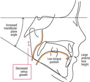
Figure 1 Tracing of a child with large adenoids. Because of mouth
breathing, tongue position in the oral cavity is low and the balance between forces from the cheeks and tongue is different compared with healthy children. This leads to a lower mandibular position and extended head posture. Cephalometrically a large anterior face height and increased mandibular plane angle can be noted (adapted from Linder-Aronson, 1970 , 1979 ). In addition, according to the present hypothesis, because of abnormal nocturnal growth hormone secretion, ramus growth is less than that in healthy subjects.
T. PELTOMÄKI
428
production site of hormonal agents as evidenced by IGF I
receptor and IGF I messenger RNA expression in the
cartilage ( Visnapuu et al. , 2001 , 2002 ). Patients with GH
defi ciency have been shown to have a small posterior face
height compared with age and gender-matched healthy
controls ( Pirinen et al. , 1994 ; Karsila-Tenovuo et al. , 2001 ).
Furthermore, administration of GH to patients with GH
defi ciency, such as those with Turner syndrome or in bone
marrow transplant patients, has clearly shown that
mandibular growth, and particularly mandibular ramus
growth, is accelerated compared with control children
( Dahllöf et al. , 1991 ; Rongen-Westerlaken et al. , 1993 ;
Simmons, 1999 ; Forsberg et al. , 2002 ). The increase in
mandibular ramus height by GH can be explained by two,
possibilities. Firstly, increased endochondral bone formation
in the condylar cartilage and secondly, increased bone
apposition in the lower border of the mandible through the
anabolic effects of GH on the masseter and medial pterygoid
muscles ( Vogl et al. , 1993 ).
Conclusion
Taking into account the recent evidence from children with
OSA, it can be postulated that the craniofacial structure before,
and its change after adenotonsillectomy, in patients with large
adenoids and tonsils (classically, regarded as mouth breathing
patients) are not only caused by a mechanistic alteration in the
muscular balance and head and tongue position due to the
change in the mode of breathing but also caused by a more
complex sequence of epigenetic events. Because of abnormal
nocturnal secretion of GH and its mediators in children with
obstructed breathing, mandibular ramus growth is less than
that in healthy subjects ( Figure 1 ). After normalization of
hormonal status, ramus growth is enhanced by more intensive
endochondral bone formation in the condylar cartilage and/or
by appositional bone growth in the lower border of the
mandible at the muscular attachment area ( Figure 2 ). This
growth enhancement would, in part, explain the noted
acceleration in the growth of the mandible and change in its
growth direction after alteration in the mode of breathing
following adenotonsillectomy. Finally, it is noteworthy that in
many cases the growth acceleration is not suffi cient to solve
the already formed malocclusion and skeletal discrepancy,
and therefore, orthodontic treatment is also indicated.
Address for correspondence
Timo Peltomäki
Clinic for Orthodontics and Pediatric Dentistry
Center for Dental and Oral Medicine
University of Zurich
Plattenstrasse 11
CH-8032 Zurich
Switzerland
E-mail: [email protected]
References
Agren K , Norlander B , Linder-Aronson S , Zettergren-Wijk L , Svanborg E 1998 Children with nocturnal upper airway obstruction: postoperative orthodontic and respiratory improvement . Acta Otolaryngologica (Stockholm) 118 : 581 – 587
Bar A , Tarasiuk A , Segev Y , Phillip M , Tal A 1999 The effect of adenotonsillectomy on serum insulin-like growth factor-I and growth in children with obstructive sleep apnea syndrome . Journal of Pediatrics 135 : 76 – 80
Behlfelt K , Linder-Aronson S , McWilliam J , Neander P , Laage-Hellman J 1990 Cranio-facial morphology in children with and without enlarged tonsils . European Journal of Orthodontics 12 : 233 – 243
Copray J C , Jansen H W , Duterloo H S 1986 Growth and growth pressure of mandibular condylar and some primary cartilages of the rat in vitro . American Journal of Orthodontics and Dentofacial Orthopedics 90 : 19 – 28
Dahllöf G et al. 1991 Craniofacial growth in bone marrow transplant recipients treated with growth hormone after total body irradiation . Scandinavian Journal of Dental Research 99 : 44 – 47
Forsberg C M , Krekmanova L , Dahllöf G 2002 The effect of growth hormone therapy on mandibular and cranial base development in children treated with total body irradiation . European Journal of Orthodontics 24 : 285 – 292
Goldstein S J , Wu R H , Thorpy M J , Shprintzen R J , Marion R E , Saenger P 1987 Reversibility of defi cient sleep entrained growth hormone secretion in a boy with achondroplasia and obstructive sleep apnea . Acta Endocrinologica (Copenhagen) 116 : 95 – 101
Guilleminault C , Li K K , Khramtsov A , Pelayo R , Martinez S 2004 Sleep disordered breathing: surgical outcomes in prepubertal children . Laryngoscope 114 : 132 – 137
Guilleminault C , Pelayo R , Leger D , Clerk A , Bocian R C 1996 Recognition of sleep-disordered breathing in children . Pediatrics 98 : 871 – 882 Kantomaa T 1984 The role of the mandibular condyle in the facial growth .
Proceedings of the Finnish Dental Society 80 : 1 – 57
Karsila-Tenovuo S et al. 2001 Disturbances in craniofacial morphology in children treated for solid tumors . Oral Oncology 37 : 586 – 592 Kawashima S et al. 2000 Cephalometric comparisons of craniofacial and
upper airway structures in young children with obstructive sleep apnea syndrome . Ear, Nose, and Throat Journal 79 : 499 – 506
Kawashima S , Peltomäki T , Sakata H , Mori K , Happonen R P , Rönning O 2002 Craniofacial morphology in preschool children with sleep-related breathing disorder and hypertrophy of tonsils . Acta Paediatrica 91 : 71 – 77
Kerr W J S , McWilliam J S , Linder-Aronson S 1989 Mandibular form and position related to changed mode of breathing — a fi ve-year longitudinal study . Angle Orthodontist 59 : 91 – 96
Linder-Aronson S 1970 Adenoids: their effect on mode of breathing and nasal airfl ow and their relationship to characteristics of the facial skeleton and the dentition . Acta Oto-Laryngologica. Supplementum 265:pp.1-132
Linder-Aronson S 1979 Respiratory function in relation to facial morphology and the dentition . British Journal of Orthodontics 6 : 59 – 71 Linder-Aronson S , Woodside D G , Lundström A 1986 Mandibular growth direction following adenoidectomy . American Journal of Orthodontics 89 : 273 – 284
McNamara Jr J A 1981 Infl uence of respiratory pattern on craniofacial growth . Angle Orthodontist 51 : 269 – 300
Nieminen P 2002 Snoring and obstructive sleep apnea in young children . Thesis, University of Oulu , Oulu, Finland
Nieminen P , Löppönen T , Tolonen U , Lanning P , Knip M , Löppönen H 2002 Growth and biochemical markers of growth in children with snoring and obstructive sleep apnea . Pediatrics 109 : e55
Pirinen S , Majurin A , Lenko H L , Koski K 1994 Craniofacial features in patients with defi cient and excessive growth hormone . Journal of Craniofacial Genetics and Developmental Biology 14 : 144 – 152
429
MODE OF BREATHING AND CRANIOFACIAL GROWTH
Rongen-Westerlaken C et al. 1993 Effect of growth hormone treatment on craniofacial growth in Turner’s syndrome . Acta Paediatrica 82 : 364 – 368
Rönning O , Peltomäki T 1991 Growth potential of the rat mandibular condyle as an isogeneic transplant traversing the interparietal suture . Archives of Oral Biology 36 : 203 – 210
Shintani T , Asakura K , Kataura A 1997 Evaluation of the role of adenotonsillar hypertrophy and facial morphology in children with obstructive sleep apnea . Journal for Oto-Rhino-Laryngology and its Related Specialties 59 : 286 – 291
Simmons K E 1999 Growth hormone and craniofacial changes: preliminary data from studies in Turner’s syndrome . Pediatrics 104 : 1021 – 1024 Solow B , Kreiborg S 1977 Soft-tissue stretching: a possible control factor
in craniofacial morphogenesis . Scandinavian Journal of Dental Research 85 : 505 – 507
Solow B , Siersbæk-Nielsen S , Greve E 1984 Airway adequacy, head posture, and craniofacial morphology . American Journal of Orthodontics 86 : 214 – 223
Subtelny J D 1954 The signifi cance of adenoid tissue in orthodontia . Angle Orthodontist 24 : 59 – 69
Trotman C A , McNamara Jr J A , Dibbets J M , van der Weele L T 1997 Association of lip posture and the dimensions of the tonsils and sagittal airway with facial morphology . Angle Orthodontist 67 : 425 – 432
Vig K W L 1998 Nasal obstruction and facial growth: the strength of evidence for clinical assumptions . American Journal of Orthodontics and Dentofacial Orthopedics 113 : 603 – 611
Visnapuu V , Peltomäki T , Rönning O , Vahlberg T , Helenius H 2001 Growth hormone and insulin-like growth factor I receptors in the temporo-mandibular joint of the rat . Journal of Dental Research 80 : 1903 – 1907 Visnapuu V , Peltomäki T , Rönning O , Syrjänen S 2002 Distribution of
insulin-like growth factor-I mRNA in the mandibular condyle and rib cartilage of the rat during growth . Archives of Oral Biology 47 : 791 – 798
Vogl C , Atchley W R , Cowley D E , Crenshaw P , Murray J D , Pomp D 1993 The epigenetic infl uence of growth hormone on skeletal development . Growth, Development, and Aging 57 : 163 – 182
Warren D W , Spalding P M 1991 Dentofacial morphology and breathing: a century of controversy . In: Melsen B (ed.). Current controversies in orthodontics. Quintessence Publishing , Chicago , pp. 45 – 76
Woodside D G , Linder-Aronson S , Lundström A , McWilliam J 1991 Mandibular and maxillary growth after changed mode of breathing . American Journal of Orthodontics and Dentofacial Orthopedics 100 : 1 – 18
Worsnop C , Kay A , Kim Y , Trinder J , Pierce R 2000 Effect of age on sleep onset-related changes in respiratory pump and upper airway muscle function . Journal of Applied Physiology 88 : 1831 – 1839
Zettergren-Wijk L , Forsberg C M , Linder-Aronson S 2006 Changes in dentofacial morphology after adeno-/tonsillectomy in young children with obstructive sleep apnoea — a 5-year follow-up study . European Journal of Orthodontics 28 : 319 – 326
Zucconi M 1999 Craniofacial modifi cations in children with habitual snoring and obstructive sleep apnoea: a case-control study . European Respiratory Journal 13 : 411 – 417