International Immunology, Vol. 6, No. 9, pp. 1333-1343 © 1994 Oxford University Press
cAMP up-regulates IL-4 and IL-5 production
from activated CD4
+
T cells while
decreasing IL-2 release and NF-AT
induction
Marc Lacour, Jean-Francois Arrlghl, Kai M. MQIIer, Carsten Carlberg,
Jean-Hilaire Saurat and Conrad Hauser
Department of Dermatology, Hflpital Cantonal Universitaire de Geneve, 24, rue Micheli-du-Crest, 1211 Geneva 14, Switzerland
Key words: cAMP, CD4+, cholera toxin, IL-2, IL-4, NF-AT, regulation, T helper cell subsets
Abstract
Seven days after activation with concanavalln A and irradiated spleen cells, murine CD4+ T cells were re-stlmulated with lonomycln and phorbol 12-myristate 13-acetate (PMA). IL-2 and IL-4 were determined In the supernatant. When cholera toxin, forskolin together with phosphodlesterase Inhibitors or dlbutyryl-cAMP were added at the time of re-stimulation, a dose-dependent increase of IL-4 and IL-5 release was noted. IL-2 was down-regulated as reported before. The
up-regulatlon of IL-4 and the down-regulation of IL-2 correlated with an increase of IL-4 mRNA and a decrease of IL-2 mRNA as determined by seml-quantltative reverse trarwcrlptase polymerase chain reaction. Similar results were found with prostaglandin E, using PMA and tonomycln or plate-bound antl-CD3 antibody as re-stimulants. These results suggest that, In activated CO4+ T cells, cAMP-elevating agents induce a switch of lymphoklne production towards a T^-JIke phenotype through regulation at the transcrlptlonal level. This Is supported by the fact that complex formation between a synthetic nuclear factor of activated T cells (NF-AT) binding site from the IL-2 promoter and nuclear extracts was decreased when cholera toxin was added to re-actlvated CD4* T cells, suggesting that cholera toxin and cAMP down-regulate IL-2 expression via decreased NF-AT binding. Finally, since IL-4 has been reported to amplify IL-4 release from activated CD4+ T cells, the autolnductlon of IL-4 may very well function via cAMP.
Introduction
It is well documented that the function of Th cells depends on their secreted rymphokines (1). On this basis, Th cells have been classified in two subsets, Th1 and T ^ , each of them playing a specific role in the generation of an immune response. Activated Th1 cells produce mainly IL-2 and IFN-7 and induce cell-mediated immune responses, such as delayed-type hyper-sensitivity reactions, whereas activated T£ cells, which produce IL-4 and IL-5, will rather promote a humoral-type response (2). Furthermore, this functional separation of Th cells has been shown to be of great biological relevance since Th1 and Th2 cells were demonstrated to be differentially involved in both murine and human immunopathologic processes (3,4). Several authors have therefore attempted to elucidate the factors that regulate the differentiation of primary Th cells into Th1 and 1^2
cells, and to analyze the regulation of their lymphokine production.
It was shown that, upon antigenic stimulation, Th cells first acquire an unrestricted pattern of lymphokine production (1L-2*. IFN-7+. IL-4* and IL-5+) (4,5). At the clonal level, T cells with an unrestricted lymphokine pattern are named Th0 cells. These ceils are able to differentiate into both Th1 and T ^ subsets (6,7). Factors which promote the induction of Th1 cells are transform-ing growth factor-0 and IL-12 (8,9). The presence of IL-4 (7,10,11) and TCR occupancy (12) are required for Th2 cell induction.
Several groups have studied the role of various intracellular signalling pathways for the expression of Th subset typical rymphokines. Gajewski ef a/, showed that lymphokine production by Th1 clones was more sensitive to inhibition by cholera toxin
Correspondence to: C. Hauser
(CT) and cAMP than was lymphokine production of T ^ clones (13). Moreover, it was shown that CT and forskolin (FSK) inhibited TCR-triggered IL-2 production and proliferation in Th1 cells, while the same reagents failed to block IL-4 production and proliferation in T ^ cells (14). Both IL-2 and IFN-7 production were reduced in the presence of the cAMP-elevating agents prostaglandin E2 (PGE2) when added to Th0 clones or short-term T cell lines. IL-4 levels were not affected by PGE2 (15). Similar results were obtained with the EL-4 cell line: cAMP diminished IL-2 mRNA but did not affect IL-4 mRNA levels (16). These results suggested that cAMP-dependent signaling events may selectively regulate Th1 lymphokines.
Because most previous work was done with cells that possess a fixed lymphokine pattern, we investigated the effect of cAMP and cAMP-elevating agents on IL-4 production by polyclonal CD4+ T cells expressing an unrestricted and non-fixed lymphokine pattern. Here we show that, when CD4+ T cells were re-activated with phorbo) 12-myristate 13-acetate (PMA) and ionomycin, both cAMP and cAMP-elevating agents could indeed affect Th2 lymphokines: these reagents increased IL-4 bioactivity released into the supernatant and IL-4 mRNA accumulation as determined by semi-quantitative reverse transcriptase polymerase chain reaction (RT-PCR). IL-5 production was similarly increased by cAMP-elevating agents. We also show that increased IL-4 production was induced by the naturally occurring cAMP-elevating agent PGE2 in conjunction with either PMA and ionomycin or anti-TCR stimulation. Finally a complex from nuclei of activated CD4+ T cells binding to an oligonucleotide containing the nuclear factor of activated T cells (NF-AT) binding sequence from the IL-2 promoter was decreased by cAMP, suggesting that cAMP may down-regulate IL-2 by decreasing nuclear factors binding to the NF-AT site.
Methods
Culture medium and reagents
Cells were cultured in Iscove's modified Dulbecco's medium with L-glutamine, supplemented with 1 % MEM non-essential amino acids, 100 U/ml penicillin, 100 /ig/ml streptomycin and 2 mM L-glutamine (all from Gibco, Grand Island, NY), 10% heat-inactivated FCS, (Gibco, Munich, Germany), 1 mM sodium pyruvate (Fluka, Buchs, Switzerland) and 2 x 1 0 ~5 M 2-mercaptoethanol (Sigma, St Louis, MO). PMA, CT, dibutyryl-cAMP (Bt2-cAMP), 3-isobutyM-methylxanthine (IBMX), PGE2 and FSK were purchased from Sigma; concanavalin A (Con A) was from P-L Biochemicals (Milwaukee, IL) and ionomycin from Calbiochem (La Jolla, CA). The phosphodiesterase (PDE) inhibitor RO 20-1724 (17) was kindly provided by Hoffmann-La Roche (Basel, Switzerland). The mAbs to MHC class II (M5/114.15.2), CD8 (5.3-6.72) and mouse anti-rat x chain (MAR 18.5) were obtained from the American Type Culture Collection (Rockville, MD). For phenotyping, cells were stained with mAb anti-CD4 - phycoerythrin conjugate and anti-CD8 - FITC (Becton-Dickinson, Mountain View, CA) and analysed on a FACScan (Becton-Dickinson).
Preparation of CD4+ activated T cells
Female BALB/c mice (IFFA-Credo, Arbresle, France) were used in all experiments. Activated CD4+ T cells were obtained in 7
days of culture as follows. CD4+ T cells were purified as described (18). Briefly, lymph node cells were purified by passage over a nylon wool column, and incubated on ice for 30 min with mAb to MHC class II and CD8, followed by MAR-18.5 and rabbit tow-tox M complement (1/10; Cedarlane, Hornby, Ontario, Canada). CD4+ T cells (5X10⁶) ( > 9 8 % CD4+, < 1 % CD8+, < 1 % MHC class II+ and non-responsive to Con A atone) were then stimulated in 10 ml of culture medium with Con A (2 /ig/ml), 50 U/ml human rhlL-2 (a generous gift from Dr C. R. Franks, Eurocetus, Amsterdam, Netherlands) and 107 3000 rad-irradiated splenocytes. Three days later, the culture was divided in six parts and supplemented with fresh medium containing rhlL-2. On day 6, cells were washed and cultured in rhll_2-con-taining medium for a further 24 h. On day 7, these activated CD4+ T cells were washed again and re-stimulated for lymphokine production.
T cell stimulation
Activated CD4+ T cells (lOS/well) were seeded in 96-well tissue culture plates (Costar, Cambridge, MA) filled with 200 /J culture medium ± stimulator/inhibitor at the indicated final concentra-tions. After 30 min incubation at 37°C, PMA (1 ng/ml) and ionomycin (1 /tg/ml) were added for stimulation. Supernatants were harvested after 24 h and stored at - 20°C until lymphokine analysis. For anti-CD3 stimulation, wells were coated with 50 /J of affinity purified mAb 145 2C11 (19) 3 /ig/ml in PBS for 2 h at 37°C. Wells were then washed three times with 150 /J of sterile PBS before addition of culture medium ± the indicated reagents and lastly cells. For RNA analysis, 4 - 1 0 x 106 activated T cells were first incubated for 30 min at 37°C at a density of 2 - 3 x i O6 cells/ml with the indicated reagents, and then stimulated for 6 h (unless stated otherwise) with PMA and ionomycin. The 6 and 24 h supernatants (obtained from parallel cultures) were kept for lymphokine analysis.
Bioassays
To measure the production of IL-2 and IL-4, a specific lymphokine assay was used (18). Briefly, 5 x 10³ CTLL cells responding to both IL-2 and IL-4 (a kind gift of Dr E. M. Shevach, LI, NIAID, NIH, Bethesda, MD) were incubated in flat-bottomed 96-well tissue culture plates (Costar) with appropriate serial dilutions of lymphokine-containing culture supernatants in the presence of either S4B6, a neutralizing mAb to mouse IL-2, kindly provided by Dr T. Mosmann (DNAX, Palo Alto, CA) (20), as a 1/1000 dilution of ascites or a 1/100 dilution of a Tecnomouse™ (Tecnomara, Switzerland) supernatant, 11B11, a neutralizing mAb to mouse IL-4, kindly provided by Dr W. E. Paul, LI, NIAID, NIH, Bethesda, MD (21), as a 1/1000 dilution of ascites, both mAb or no mAb. Internal standards for IL-2 and IL-4 calibrated with rhlL-2 or with murine rlL-4 (obtained from Dr W. E. Paul), respectively, were incubated in parallel. Proliferation was assessed by [3H]methyl-thymidine incorporation after pulsing the cultures (0.5 pCi/well) for the final 6 - 8 h of the 24 h culture period. Sample points lying on the linear part of the dose - response curve were used to calculate IL-2 and IL-4 activities. The detection of IL-2 and IL-4 by the CTLL cells was not affected by PMA and ionomycin, nor by the presence of CT, FSK, IBMX, RO-20-1724 or Bt2-cAMP at working concentra-tions. IL-5 in culture supernatants was determined by ELISA,
cAMP up-regulates IL-4 in activated T cells 1335
using the anrj-IL-5 rat mAb TRFK4 (5 pg/ml for coating) and TRFK5 (biotin-labeled, 1 pg/ml, kindly donated by Dr R. L. Coffman, DNAX, Palo Alto, CA) (22), as previously described (23). The Student's f-test was used for statistical analysis.
RNA analysis
At the indicated time points, CD4+ T cells were harvested, transferred to Eppendorf tubes and centrifuged. Total cellular RNA was recovered after cell lysis in 1 ml of guanidine thio-cyanate buffer by acid phenol extraction and isopropanol precipitation (24). The yield was 8 ± 2 pg of total RNA/106 CD4+ T cells. The integrity of the RNA was assessed by analysis of ribosomal RNA on 1 % agarose gel electrophoresis. The efficiency of the reverse transcription (RT) was verified by detection of the dihydrofolate reductase (DHFR) mRNA (25) with RT-PCR (26). In brief, RT-PCR was done as follows for all the mRNA tested: - 2 pg of total cellular RNA was retrotranscribed after heat denaturation and annealing with oligo(dT) primers (15-mer, Boehringer Mannheim, Rotkreuz, Switzerland) in the presence of 200 U Moloney murine leukemia virus reverse transcriptase (Gibco), 40 U RNAsin (Promega, Madison, W1), 5 mM DTT and 200 pM of each deoxynucleoside triphosphate in 50 pi for 1 h at 37°C. The reactions were stopped by heating inactivation for 5 min at 95°C, diluted 1:10 with water and stored at - 2 0 ° C until use. Either 5 pi (DHFR) or 1 pi (IL-2 and IL-4) of one-tenth diluted cDNA were then amplified in a DNA thermo-cycler (Perkin-Elmer, Norwalk, CT) with 200 pM of each deoxynucleoside triphosphate, 50 U/ml of Taq polymerase (Promega) diluted in the buffer supplied by the manufacturer, and 500 nM of both upstream and downstream primer, in a volume of 20 pi. Each PCR cycle consisted of a denaturation step (94°C, 30 s), an annealing step (60°C, 30 s) and an elongation step (72°C, 30 s). For the first cycle only, the duration of the denaturation step was 3 min and for the final step only, the duration of the processing step was 10 min. A total of 35 cycles for DHFR and 30 cycles for IL-2 and IL-4 detection, corres-ponding to the linear portion of the amplification curve, was used in all experiments.
Each RT-PCR included negative controls for (i) RNA extraction: lysis buffer alone treated as a normal sample; (ii) RT: RT reactants without RNA; (iii) PCR: PCR reactants without cDNA.
All the necessary precautions against contamination of PCRs were rigorously observed (27). The primers had the following sequences: DHFR upstream 5'-CTCAGGGCTGCGATTTCGC-GCCAAACT-3', downstream 5'-CTGGTAAACAGAACTGCC-TCCGACTATC-3', amplifying a 447 nucleotide sequence; IL-2 upstream 5'-GTCAACAGCGCACCCACTTCAAGC-3', down-stream 5'-GCTTGTTGAGATGATGCTTTGACA-3', amplifying a 450 nucleotide sequence; IL-4 upstream 5'-ACGGAGATGGA-TGTGCCAAACGTC-3', downstream 5'-CGAGTAATCCATTT-GCATGATGC-3', amplifying a 279 nucleotide sequence. Amplimer sequences were chosen from separate exons of the genes so that products from cDNA amplification could be distinguished from products derived from any contaminating genomic DNA. The specificity of the PCR was confirmed by restriction site analysis of the amplified cDNA, which generated restriction fragments of the expected size for all cytokines tested. RT-PCR amplified products of the DHFR house-keeping gene were first loaded on a gel to allow normalization using scanning
densitometry. Shown gels represent the reloaded normalized amounts of DHFR amplified products together with equal amounts of IL-2 and IL-4 amplifications for each culture condition.
DNA binding assays
Nuclear extracts from reactivated CD4+ T cells were prepared as described (28). The oligonucleotides comprising the murine IL-2 NF-AT and the NF-IL-2-A site (29) as well as an oligonucleotide containing an AP-1 consensus sequence, underlined (30): NFVkT (-285O -270) 5'caglCCCCAAAGAGGAMATTTGTTTCATACAGl 3-3- • GGGGTTTCTCCTTTTAAACAAAGTATGT(*gac5' 5'caglGAAAATATGTGTAATATGTAAAACATCGt 3-y aCTTTTATACACATTATACATTTTGTAGCqafcS' NR.-S-A (-W ID -66) yqaglGCTCGGGTAGGGGTGACTCACCGGQTGAACGGGGGCATCTCGACTCG t 3* 31 •CGAGCCCATCCCCACTGAGTGGCCCACTTGCCCCCGTAGAGCTGAGC *•£ y
The oligonucleotides were purified, annealed to yield a double-stranded DNA fragment and subsequently labeled by a fill-in reaction using [a-32P]dCTP and T7 DNA polymerase (Pharmacia).
Nuclear extracts (5 pi) from re-activated CD4+ T cells were incubated with ~ 1 ng of labeled probe (25,000 c.p.m.) for 30 min at room temperature in a total volume of 20 pi binding buffer [10 mM HEPES, pH 7.9, 80 mM KCI, 1 mM DTT, 0.2 pg/pl pory(dl-dC) and 5% glycerol]. The protein-DNA complexes were resolved on a 5% non-denaturing potyacrylamide (29:1) gel at room temperature in 0.5 x TBE (pre-run for 2 h), and the dried gel was exposed to a Kodak XAR film.
Results
Freshly isolated and highly purified resting CD4+ cells ( > 9 8 % by flow cytometry) were stimulated with Con A and irradiated syngeneic spleen cells in the presence of IL-2. On day 6, activated CD4+ cells were washed and incubated for a further 24 h in medium supplemented with rhlL-2. This procedure lead to an 8- to 12-fokJ increase of cells that were > 9 8 % CD4+. These cells are subsequently referred to as 'activated CD4+ T cells'. When stimulated with PMA and ionomycin, they produced an unrestricted rymphokine pattern with regard to IL-2, IFN-7, IL-4 and IL-5 (data not shown). PMA alone (1 ng/ml) was ineffective for the production of either cytokine and ionomycin (1 pg/ml) alone induced lower IL-4 production than in conjunction with PMA (data not shown). Together, these results indicated that in activated CD4+ T cells, high production of both IL-2 and IL-4 was achieved through the pharmacological triggering of both the protein kinase C (PKC) and calcium-dependent pathways, as described before (31).
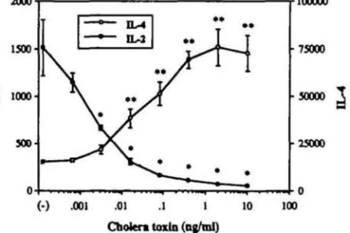
In PMA- and ionomycin-re-stimulated CD4+ T cells, CT dramatically decreased IL-2 production (Fig. 1). In contrast, PMA-and ionomycin-induced IL-4 production was enhanced. This increase of IL-4 production was dose-dependent and repro-ducible in more than five similar experiments, in which a maximal 3- to 8-fold increase of IL-4 was observed at 1 ng/ml of CT. The presence of CT, PMA or ionomycin in the supernatant did not interfere with the biological detection of IL-2 and IL-4, even at the highest tested concentration (supernatant dilution 1:20). This was also the case for all modulators subsequently used in this report.
Since lymphokine production in T cells was shown to be regulated at the transcriptional level, we verified whether CT affected IL-2 and IL-4 mRNA accumulation (Fig. 2). In parallel to cytokine production, IL-2 mRNA was clearly decreased in cells incubated in the presence of CT, whereas IL-4 mRNA was increased. Although the RT-PCR method used here is only
semi-2000
1500-2
10
°°-100000 75000 30000
3
25000 .001 .01 .1 1 Cholera toxin (ng/ml) 10 100Fig. 1. CT differentially regulates IL-2 and IL-4 production in activated CD4+ T cells. Activated CD4+ T cells (10s) were resfmulated with PMA
and ionomycin in 200 fii culture medium containing CT at the indicated final concentrations. Lymphokine concentrations represent the means ± SD of the 24 h supernatants of triplicate cultures and are expressed as units per 106 ceils. Presented data are representative of five similar
experiments. 'P < 0.05, "P < 0.01.
quantitative, the opposite effects of CT on IL-2 and IL-4 mRNA were clear, reproducible in all experiments, and correlated with IL-2 and IL-4 biological activities.
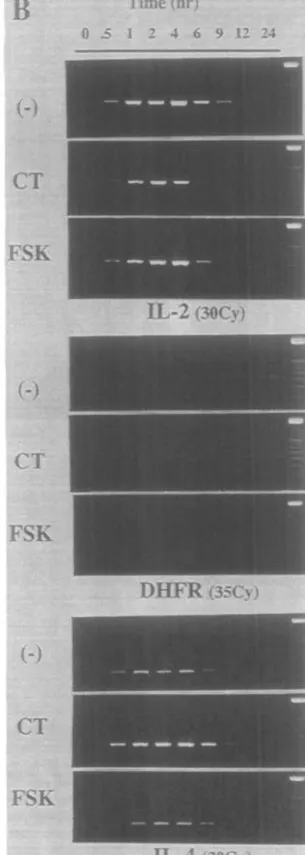
Most effects of CT have been shown to be mediated through an increase of cAMP. The ADP ribosylation by CT of the Gs, protein of the adenylate cyclase complex induces a prolonged activation of the adenylate cyclase and thus the hydrolysis of ATP leading to increased levels of intracellular cAMP (32). In order to confirm that our findings in activated CD4+ T cells stimulated in the presence of CT were also mediated by an increase of cAMP, we next used FSK, a direct activator of adenylate cyclase. In dose-response experiments, FSK decreased IL-2 in PMA-and ionomycin-reactivated CD4+ T cells with a 50% inhibition at - 3 - 1 0 fiM, whereas IL-4 production was slightly increased only at doses from 10 to 30 /iM (data not shown). Higher doses of FSK were toxic for the cells and resulted in complete inhibition of both cytokines. We further investigated whether CT and FSK altered the kinetics of IL-2 and IL-4 mRNA and protein production (Fig. 3). At indicated time points, cultured wells containing 2 x 1 0 * activated CD4+ T cells/ml were harvested, and assayed for lymphokine production and mRNA expression. CT and FSK, used here at concentrations (1 ng/ml and 30 /»M respectively) previously determined to maximally influence the lymphokine pattern of activated CD4+ T cells, similarly reduced IL-2 production. The increase of IL-4 by FSK was, however, far less than with CT (Fig. 3A). IL-2 mRNA transcripts were diminished in the presence of CT (Fig. 3B). Decreased IL-2 mRNA in the presence of FSK was clear only at 1 and 6 h. An increase of
Protein
mRNA
« »•• IL-2 (30cj) DHFR (JOtjr) IL-4 Ofejr) (-) P/I 10 1 0.1 CTFlfl. 2. CT increases IL-4 mRNA expression In activated CD4+ T cells. Cell (2x 10⁶) were stimulated with PMA and ionomycin in the absence
(P/I) or presence of 10, 1 or 0.1 ng/ml CT. Protein: lymphokine levels are from 24 h supernatants of parallel cultures and are expressed as units per 10* cells. Standard deviation of triplicate lymphokine determination was <9W) of the mean. Results are repesentative of three similar experiments. mRNA: after 6 h, cells were harvested for total RNA extraction and reverse transcription. cDNA corresponding to 20 ng of total RNA was then used tor PCR amplification with specific primers. After normalization with PCR amplification of the DHFR gene, as described in Methods, aliquots of the amplified cDNA were electrophoresed on 2% agarose gets in the presence of ethidium bromide.
B
cAMP up-regulates IL-4 in activated T cells 1337
" * Time (hr)
0 . 5
I 2 4 6 9 12 24A
300 2 0 0 1 0 0 -Ch. Toxin FonkofinCT
FSK
IL-2 (30Cy)
0 2 4 6 8 10 12 14 16 18 20 22 24 Tlme(hr)CT
FSK
10000 7SO05 0 0 0 2 5 0 0-D H F R (35Cy)
CT
FSK
0 2 4 6 8 10 12 14 16 18 20 22 24 Timed)!-)IL-4 (30Cv
Fig. 3. Effect of FSK and CTon the productionof IL-2 and IL-4 from actrvatedOD4+Tc»fls. Activated CD4+T cells (2x10^ cells/mO were stimulate
with PMA and ionomycin in the absence of in the presence of 30 pM FSK or 1 ng/ml CT. Results represent the lymphokine content of supematants (A) and RT-PCR amplifications of the corresponding mRNA (B), harvested at indicated times. RT-PCR amplification of the DHFR mRNA was used for normalization, as described in Methods. Differences in lymphokine content of CT- and FSK-treated versus untreated culture supernatants were statistically significant: P < 0.05 at 2 h and P < 0.01 at 4 h and after. Data are representative of three experiments for CT. For FSK, three experiments for lymphokine analysis and two for RNA yielded similar results.
20000-15000. 10000. 5000. I
.ll
p<0.01 p<aosmil inn
I
600. 450. 300. 150,lib
(-).03.3 3 10 (-) 1 3 1030 (-) 1 3 1030 (-) 1 3 1030 A B C DFig. 4. Synergistic influence of FSK and the POE inhibitor RO 20-1724
on IL-2 and IL-4 production. Activated CD4+ T cells (105) were re-stimulated with PMA and tonomydn in 200 /J culture medium containing (A) CT (ng/ml), (B) FSK fciM), (C) RO 20-1724 0»M) or (D) 10 pM FSK combined with RO 20-1724 at the indicated concentrations 0«M). Lymphokine concentrations represent the means ± SD of the 24 h supernatants of triplicate cultures and are expressed as units per 106 ceils. One of three similar experiments is shown. *P < 0.05, "P < 0.01, as compared with the negative control of each group.
IL-4 mRNA was seen with CT at all time points. However, the 2-fdd increase of IL-4 bioactivity in FSK-incubated wells was not detectable by semi-quantitative RT-PCR. To determine whether the discrepancy between the effects of CT and FSK on IL-4 modulation could be explained by a difference in cAMP accumulation or by other effects of CT such as its effect on the non-catalytic B subunit (33,34), activated CD4+ T cells were re-stimulated in the presence of the PDE inhibitor RO 20-1724, highly specific for cAMP PDE (17). As expected, 1-30 /iM RO 20-1724 decreased IL-2 in a dose-dependent manner. IL-4 production was, however, left unchanged (Fig. 4C). When the combination of both FSK and RO 20-1724 (Fig. 4D) was used, a synergistic effect was seen since the combination could enhance IL-4 to levels close to those observed in the presence of CT (Fig. 4A) and abrogated IL-2 release. A statistically signifi-cant synergy on IL-2 reduction and IL-4 increase was also observed with the combination of FSK and the less specific PDE inhibitor IBMX (data not shown). Since the combination of an activator of the adenylate cyclase combined with PDE inhibitors could reproduce the CT effect, it is likely that the modification of IL-2 and IL-4 production we observed with CT was mainly mediated through an increase of cAMP. These experiments also
indicated that IL-2 was more sensitive than IL-4 to cAMP-etevating agents in activated CD4+ T cells, thus explaining why CT, through its sustained binding on the G-stimulatory protein of the adenylate cyclase complex, was more effective than FSK alone at raising IL-4 production.
To confirm that cAMP was the main mediator of the opposite regulation of IL-2 and IL-4 in activated CD4+ T cells, we supplemented cultures with Bt2-cAMP, a cAMP analog know to rapidly diffuse into cells. In dose - response experiments, Bt2-cAMP increased IL-4 production and reduced IL-2 release from PMA- and ionomycin-re-stimulated CD4+ T cells to an extent comparable with levels obtained with CT (data not shown). A time-course experiment with Bt2-cAMP is shown in Fig. 5. It shows that IL-2 mRNA levels were lower and IL-4 mRNA higher in Bt2-cAMP treated cells than in cells without Bt2-cAMP. At all time points, the lymphokine levels in the supernatants correlated with IL-2 and IL-4 mRNA accumulation. The parallel increase of IL-4 and decrease of IL-2 lymphokine accumulation and mRNA production mediated by cAMP-elevating agents (Figs 2, 3 and 5) indicate that cAMP is likely to influence lymphokine production directly at the transcriptional level. It is however possible that the effect on steady state mRNA levels could also be explained by effects of cAMP on mRNA stability, as reported for IFN-y (35). IL-5, a mediator of eosinophilia in vivo (36), is another lymphokine typical for Th2 cells. Since a cAMP-dependent increase of IL-5 production was previously reported in short-term T cell lines (15), we examined the effect of cAMP elevation on IL-5 produced by activated CD4+ T cells re-activated by PMA and ionomycin. The presence of CT (Fig. 6A) or Bt2-cAMP (Fig. 6B) both up-regulated IL-5 production but suppressed IL-2 release in the experiments.
Having found that pharmacological cAMP-elevating agents differentially modulated IL-2 and IL-4 together with IL-5 in CD4+ T cells re-activated with PMA and ionomycin, we then further assessed whether the lymphokine production of re-activated CD4+ T cells was similarly regulated in a more physiologic situation, using PGE2 as a cAMP-elevating agent and re-activation through the TCR-CD3 complex. In PMA and ionomycin-re-activated CD4+ T cells (Fig. 7A), PGE2 repro-duced results obtained with CT and Bt2-cAMP: it inhibited IL-2 and increased IL-4 production in a dose-dependent manner. We next verified whether activated CD4+ T cells re-activated through the TCR - CD3 complex were also susceptible to PGE2. We therefore used plate-bound anti-CD3 to re-stimulate activated CD4+ T cells. Again, PGE2 up-regulated IL-4 release and suppressed IL-2 production (Fig. 7B). The effect on IL-4 was less pronounced than with PMA- and ionomycin-stimulated cells, but reproducible and significant. Similar results were obtained with CT and Bt2-cAMP when plate-bound anti-CD3 re-stimulation was used (data not shown)
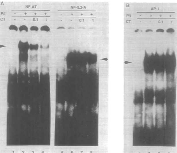
As the second messenger cAMP may directly influence IL-2 release in re-activated CD4+ T cells at the transcriptional level, we finally investigated whether cAMP-elevating agents could modify known nuclear factors initiating IL-2 transcription. We therefore prepared nuclear extracts from re-activated CD4+ T cells stimulated with PMA and ionomycin in the presence of CT, and performed gel retardation analysis using a-13P-labeled DNA fragments comprising binding sites for NF-AT and NF-IL-2-A from the murine IL-2 promoter. CT clearly inhibited the binding of nuclear factors to the NF-AT binding site, while no modification
400
300-cAMP up-regulates IL-4 in activated T cells . 1339
r40000 cAMP(lOOuM) cAMP(30uM) 30000 20000 10000 Time(hr) 0 2 4 6 8 10 12 14 16 18 20 22 24 0 2 4 6 8 10 12 14 16 18 20 22 24 Time (hr) 0 .5 1 2 4 6 9 12 24
cAMP
(KK^iM) 0 .5 1 2 4 6 9 12 24 0 .5 1 2 4 6 9 12 24IL-2
DHFR
IL-4
Fig. 5. The opposite effect of Btj-cAMP on the production of IL-2 and IL-4 is reflected at the mRNA level. Cells (2 x 10⁶) were stimulated with PMA
and ionomycin in the presence or absence of Bt2-cAMP. At indicated time points, cultures were harvested and immediately centrifuged. Supematants were assayed for lymphokine production and total mRNA was extracted from pelleted cells. Lymphokine levels are expressed as means =t SD of triplicate cultures (A). RT-PCR amplifications of specific mRNAs were electrophoresed on 2% agarose gels in the presence of ethidium bromide (B), after normalization with DHFR amplification products. Differences in lymphokine content of cAMP-treated versus untreated culture supematants were statistically significant after 4 h (P < 0.01). Lymphokine analysis of four similar experiments and RNA analysis of two similar experiments gave comparable results.
was seen with the protein complexes on the NF-IL-2-A site (Fig. 8A). Because the AT complex that is generated with the NF-AT site of the IL-2 promotor contains a pre-existing unit (NF-NF-ATp) in association with Jun and Fos family proteins, it is possible that decreased NF-AT binding reflects a decrease in the level of FosAJun proteins. To test this possibility, we performed a gel shift assay with nuclear extracts and an oligonucleotide containing an AP-1 consensus sequence (30), because AP-1 consensus sequences form complexes containing Jun and Fos family proteins in nuclear extracts of activated T cells (37). However, when complexes between an oligonucleotide containing an AP-1 consensus sequence and nuclear extracts from PMA- and tonomycin-stjmulated T cells were compared with complexes from PMA, ionomycin and CT incubated cells, no difference was observed (Fig. 8B). This renders unlikely the possibility that decreased binding to the NF-AT site induced by CT was due to diminished Jun/Fos family proteins. Specificity of the DNA probes was assessed in previous inhibition experiments and has been previously published (29,30).
Discussion
Here, we have clearly shown that cAMP and cAMP-increasing reagents differentially regulate IL-2 and the T ^ lymphokines IL-4 and IL-5. The fact that we found up-regulation of IL-4 by cAMP, in contrast to previous studies, may be due to the use of different cell sources. In earlier reports, mainly cell lines with a fixed lymphokine pattern were used (13-16). In these cells, activation in the absence of cAMP may already induce maximal IL-4 production and cAMP may not be able to further augment the release of IL-4. Alternative explanations may come from differences in working concentrations of cAMP-elevating agents (16) or from different kinds of stimulation, such as antigen plus antigen presenting cell on short-term lines (15). Indeed, since antigen presenting cell-derived co-stimulatory signals can selectively regulate IL-2 and IL-4 production (38), it is likely that cAMP-elevating agents differently affect lymphokine production in a system where antigen plus antigen presenting cell, rather than PMA and ionomycin or anti-TCR - CD3 stimulation is used.
PMA-induced IL-5 production has very recently been reported to be increased by cAMP in EL-4 cells (39). In contrast to most previous experiments, we used a polyclonal population of previously activated CD4+ T cells that may represent an intermediate stage in T ^ development. In our hands, these activated CD4+ T cells with an unrestricted lymphokine pattern could be induced to differentiate into 1^2 cells when Con A and IL-2 were used as previously described (18). Pharmacological activation or activation via the TCR - CD3 complex leads to IL-4 and IL-5 production that can be further stimulated by cAMP. Our hypothesis that in vitro activated CD4+ T cells represent a valid model for in vivo activated CD4+ T cells is supported by our observation that freshly isolated CD4+ T cell blasts show the same susceptibility to cAMP and cAMP-elevating reagents as in vitro activated CD4+ T cells (M. Lacour and C. Hauser, manuscript in preparation). cAMP seems also to promote the development of a T^-fike lymphokine pattern in vivo as recently
400 300.
A
—— IL-2 — ° — IL-5*
••
A;
*
•4000 •3000 2000 J 1000 100. .0000) (-) .001 .01 .1 1 10 Cholera toxin (ng/ml) 230 200. 150. 100. 5000 '4000 •3000 •2000 1000 .1 (-) 1 10 Dibutyryl-cAMP (|iM)Rfl. 6. IL-5 production from activated C D 4+T cells is up-regulated by
CT and Bt2-cAMP. Activated CD4+ T cells (10s) were re-stimulated with
PMA and ionomycin in 200 ?l of culture medium containing the indicated concentrations of (A) CT or (B) Btj-cAMP. Results represent the lymphokine content of 24 h supernatarrts and are expressed as means ± SD of triplicate cultures. As compared with controls, IL-4 production was increased by a ratio of 5.81 at 1 ng/ml CT (A) and a ratio of 6.72 at 50 pM Btj-cAMP (B). Data are representative of three similar experiments. *P < 0.05, "P < 0.01.
shown by Xu-Amano et al. (40) who used CT as an adjuvant in an oral immunization protocol.
PGE2 is an extracellular ligand that has been demonstrated to increase cAMP in CD4+ T cells, and to exert many immuno-suppressive functions by counteracting T cell activation and proliferation (41). Here, we show that PGE2 increases IL-4 release in re-activated CD4+ T cells. Most likely, this increase of IL-4 by PGE2 was due to an elevation of intracellular cAMP levels. It has recently been reported that some IL-4-induced effects on endothelial cells are dependent on cAMP (42), suggesting that cAMP may be a second messenger of the IL-4 receptor, as has been shown in human tonsillar B cells (43). We therefore added IL-4 during re-stimulation of activated CD4+ T cells by PMA and ionomycin, and found that IL-4 production was indeed up-regulated (data not shown). Because IL-4 has been well documented to autoinduce its production (10,11,44,45), it is thus possible that IL-4 may autoamplify its expression via a cAMP-dependent pathway.
Our findings that cAMP-elevating agents promote a Th2-like pattern of lymphokine production in activated CD4+ T cells also
60000 .001 (-) .01 .1 1 Prostaglandin E2 (uM) 60000 •45000 30000 -J 15000 .001 (-) .01 .1 1 Prostaglandin E2 (|iM)
Fig. 7. PGE2 up-regulates IL-4 and decreases IL-2 in
PMA/ionomycin-and anti-CD3-re-stimulated CD4+ T cells. Cells (10s) were placed in
200 /il of culture medium containing graded doses of PGE2, and
re-stimulated with either PMA and ionomycin (A) or coated anti-CD3 (B). Results represent the lymphokine content of 24 h supernatarrts and are expressed as mean ± SD of triplicate cultures. Data are representative of three (A) and two (B) experiments. 'P < 0.05, "P < 0.01.
cAMP up-regulates IL-4 in activated T cells . 1341
confirm that PGE2 may play a major role in the regulation of the immune response. Indeed, PGE2 induces a cascade of events that all lead to Th2 cell induction and high IgE production by B cells (reviewed in 46). It may therefore be significant that monocytes from patients with either hyper-lg E syndrome or atopic dermatitis secrete abnormally high levels of PGE2 (47,48). However, it remains unclear whether these findings reflect a primary or secondary event.
Finally, what are the intracellular cAMP targets relevant for IL-2 and IL-4 production? The decrease of IL-2 gene nuclear trans-cription induced by PGE2 has been reported to be reverted by H-8 (49), a competitor for. the ATP-binding site of the catalytic subunft of the cAMP-dependent protein kinase, PKA (50). In addition, the same group has recently given evidence that cAMP inhibits IL-2 promoter activity by interfering with cydosporin A-sensitive and calcineurin-dependent pathways (51). Another mechanism of mRNA regulation has been shown by Kaldy et al. They found that cAMP regulated lympnokine mRNA levels by modulating their stability (35).
Caldneurin has been demonstrated to be the target of the IL-2 suppressing property of the cyclosporin A - cyclophilin complex (52,53). It has previously been shown that NF-AT, composed of Jun and Fos as well as a recently cloned component (54,55), is also cyclosporin A sensitive (56). Calcineurin was then shown to be a substrate of NF-AT (57). Calcineurin probably regulates NF-AT by controlling its translocation from the cytoplasm to the nucleus. The NF-AT complex plays an important role for IL-2 gene expression by binding to a proximal promoter element 5' upstream from the transcription initiation site of IL-2. This NF-AT site, when occupied by NF-AT, confers increased promoter activity (58). We therefore tested the hypothesis whether cAMP suppresses IL-2 expression via a modification of NF-AT. Gel retardation assays performed with nuclear extracts of activated T cells and a synthetic NF-AT site demonstrated that CT, when added to stimulated CD4+ T cells, caused a decrease in NF-AT binding. Although no functional data are presented with the presently used system that consists of non-immortalized T cells, our results suggest that cAMP inhibits IL-2 gene expression via
NF-AT P/l CT 0.1 NF-IL2-A 0.1 AP-1 P/l CT 0.1 1 1
Fig. 8. Interaction of nuclear proteins present in nuclear extracts from activated CD4+ T cells with the NF-AT and NF-IL-2-A binding sites of the murine IL-2 promoter and an AP-1 consensus sequence. (A) Nuclear extracts from non-stimulated activated CD4+ T cells (lanes 1 and 5) or from activated CD4+ T cells stimulated with PMA and ionomycin (lanes 2 - 4 and 6-8) in the presence of 0.1 ng/ml CT (lanes 3 and 7) or 1 ng/ml CT (lanes 4 and 8) were incubated with ot-^P-labeted DNA fragments containing the binding sites for NF-AT (lanes 1 -4) or NF-IL-2-A (lanes 5-8) respectively. Arrows indicate the respective specific complexes. (B) Nuclear extracts from non-stimulated activated CD4+ T cells (lane 1) or from activated CD4+ T cells stimulated with PMA and ionomyctn (lanes 2-4) in the presence of 0.1 ng/ml CT (lane 3) or 1 ng/ml CT (lane 4) were Incubated with oe^P-labeled DNA fragments containing the binding site for AP-1 (lanes 1 - 4).
decreased NF-AT binding. To rule out the possibility that decreased binding to NF-AT was due to diminished Jun/Fos family proteins that bind to AP-1 and form a part of the complex that bind to NF-AT, we compared complexes binding to an AP-1 consensus sequence from CT-treated and control cells. As no difference was observed, it is unlikely that decreased NF-AT binding can be explained by decreased Jun/Fos family proteins. We are currently using immortalized cell lines in order to obtain functional evidence for this hypothesis.
At present, we have no experimental data to explain as to how cAMP up-regulates the expression of IL-4. However, it is interesting to note that several NF-AT-like sites in the IL-4 promoter have been described (59-61). NF-AT protein complexes bind to these sites and confer increased promoter activity. It is of particular interest that in activated Th1 and T ^ cell clones, an
NF-AT species without AP-1 has been identified that binds to an NF-AT site within the - 7 4 to - 6 7 region of the IL-4 promoter (62). This site can confer increased IL-4 promoter activity. It is therefore tempting to speculate that CT caused a shift from NF-AT containing AP-1 to NF-AT without AP-1, and thus explain the suppression of IL-2 production and the enhancement of IL-4 release. However, our data with the gel shift experiments using an AP-1 consensus site cannot support this hypothesis because Th 2 cells are reported to contain nuclear factors binding to AP-1
sites. To test the possibility that some factors binding to the mentioned NF-AT site within the IL-4 promoter changed with CT treatment, we compared complexes forming with an oligo-nucleotide containing the - 86 to - 60 site of the IL-4 promoter. However, CT treatment resulted in a decrease of both binding complexes (data not shown), again not supporting the hypothesis that CT may shift NF-AT protein from AP-1 rich to AP-1 poor.
Acknowledgements
We thank F. Jaunln and C. Tougne for expert technical assistance, and Drs U. Lang, G. del Giudice and D. Church for very helpful discussions. This work was supported in part by grants from the Swiss National Foun-dation for Scientific Research to C. H. (32-27/59.89 and 31-30930.91).
Abbreviations Bt2-cAMP Con A CT DHFR FSK IBMX NF-AT PDE PGE2 PMA RT-PCR References dibutyryl-cAMP concanavalin A cholera toxin dihydrofolate reductase forskolin 3-isobutyl-1 -methytxanthine nuclear factor of activated T cells phosphodiesterase
prostaglandin E2
phorbol 12-myrtetate 13-acetate reverse transcriptase - polymeras
1 Mosmann, T. R. and Coffmann, R. L. 1989. Th1 and Th2: different patterns of rymphokine secretion lead to different functional properties. Annu. Rev. Immunol. 7:145.
2 Powrie, F. and Coffman, R. L. 1993. Cytokine regulation of T-cell function: potential for therapeutic intervention. Immunol. Today 14:270. 3 Muller, K. M. and Hauser, C. 1993. The biological significance of
helper T cell subsets in the human. Exp. Dermatol. 1:161. 4 Mosmann, T. R., Schumacher, J. H., Street, N. F., Budd, R.,
O'Garra, A., Fong, T. A. T., Bond, M. W., Moore, K. W. M., Sher, A.
and Forentino, D. F. 1991. Diversity of cytokine synthesis and function of mouse CD4+ T cells. Immunol. Rev. 123:209.
5 Firestetn, G. S., Roeder, W. D., Laxer, J. A., Townsend, K. S., Weaver, C. T., Horn, J. T., Linton, J., Torbett, B. E. and Glasebrook, A. L. 1989. A new murine CD4+ T cell subset with an
unrestricted cytokine profile. J. Immunol. 143:518.
6 Rocken, M., Saurat, J.-H. and Hauser, C. 1992. Common precursor for CD4+ T cells producing IL-2 or IL-4. J. Immunol. 148:1031.
7 Abehsira-Amar, O., Gibert, M., JoSy, M., Theze, J. and Jankovic, D. L 1992. IL-4 plays a dominant role in the differential development of Th0 into Th1 and T ^ cells. J. Immunol. 148:3820.
8 Swain, S. L , Huston, G., Tonkonogy, S. and Weinberg, A. 1991. Transforming growth factor-/3 and IL-4 cause helper T cell precursors to develop into distinct effector helper cells that differ in rymphokine secretion pattern and cell surface phenotype. J. Immunol. 147:2991. 9 Trinchieri, G. 1993. lnterleukin-12 and its rote in the generation of Th1
cells. Immunol. Today 14:335.
10 Seder, R. A., Paul, W. E., Davis, M. M. and Fazekas de St. Groth, B. 1992. The presence of interleukin 4 during in vitro priming determines the lymphokine-producing potential of CD4+ T cells from T cell
receptor transgenic mice. J. Exp. Med. 176:1091.
11 Swain, S. L , Weinberg, A. D., English, M. and Huston, G. 1990. IL-4 directs the development of T^-like helper effectors. J. Immunol. 145:3796.
12 Rocken, M., Muller, K. M., Saurat, J.-H., MOIIer, I., Louis, J. A., Cerottini, J.-C. and Hauser, C. 1992. Central role for TCR/CD3 ligation in the differentiation of CD4+ T cells toward a Th1 or Th2 functional
phenotype. J. Immunol. 148:47.
13 Gajewski, T. F., Schell, S. R. and Fitch, F. W. 1990. Evidence implicating utilization of different T cell receptor-associated signaling pathways by Th1 and T ^ clones. J. Immunol. 144:4110.
14 Munoz, E., Zubtega, A. M., Merrow, M., Sauter, N. P. and Huber, B. T. 1990. Cholera toxin discriminates between T helper 1 and 2 cells in T cell receptor-mediated activation: rate of cAMP in T cell proliferation. J. Exp. Med. 172:95.
15 Betz, M. and Fox, B. 1991. Prostaglandin E2 inhibits production of
Th1 lymphokines but not of Th2 lymphokines. J. Immunol. 146:108.
16 Novak, T. J. and Rothenberg, E. V. 1990. cAMP inhibits induction of interleukin 2 but not of interteukin 4 in T cells. Proc. Natl Acad. Set. USA 87:9353.
17 Beavo, J. A. 1988. Multiple teozymes of cyclic nucleotlde phosphodiesterase. Adv. Sec. Mess. Phosphoprot. Res. 22:1. 18 Rocken, M., Muller, K. M., Saurat, J.-H. and Hauser, C.
1991.Lectin-medlated induction of IL-4-producing CD4+ T cells. J. Immunol.
146:577.
19 Leo, O., Foo, M., Sachs, D. H., Sametson, L E. and Bluestone, J. A. 1987. Identification of a monoclonal antibody specific for a murine T3 polypeptide. Proc. Natl Acad. Set. USA 84:1374.
20 Mosmann, T. R., Bond, M. W., Coffman, R. L., Ohara, J. and Paul, W. E. 1986. T-cell and mast cell lines respond to B-cell stimulatory factor 1. Proc. Natl Acad. Sd. USA 83:5654.
21 Ohara, J. and Paul, W. E. 1985. Production of a monoclonal antibody to and molecular characterization of B-cell stimulatory factor 1. Nature 315:333.
22 Schumacher, J. H., O'Garra, A., Shrader, B., van Kimmenade, A., Bond, M. W., Mosmann, T. R. and Coffman, R. L. 1988. The characterization of four monoclonal antibodies specific for mouse IL-5 and development of mouse and human IL-5 enzyme-linked rmmunosorbent. J. Immunol. 141:1576.
23 Grillot, D., Michel, M., Muller, I., Tougne, C , Renia, L , Mazier, D., Corradin, G., Lambert, P.-H., Louis, J. A. and De Giudice, G. 1990. Immune responses to defined epitopes of the drcumsporozoite protein of the murine malaria parasite. Plasmodium yoelii. Eur. J. Immunol. 20:1215.
24 Chomczynski, P. and Sacchi, N. 1987. Single-step method of RNA isolation by acid guanidium thkxyanate - chloroform extraction. Anal. Bkxhem. 162:156.
25 Crouse, G. F., Simonsen, C. C , McEwan, R. N. and Schimke, R. T. 1982. Structure of amplified normal and variant dihydrofolate reductase genes in mouse sarcoma S180 cells. J. Biol. Cnem. 257:7887.
26 Saiki, R. K., Gelfand, D. H., Stoffel, S., Scharf, S. J., Higuchi, R., Horn, G. T., Mulfis, K. B. and Erlich, H. A. 1988. Primer directed