M A J O R A R T I C L E
Infections With the Tick-Borne Bacterium
“Candidatus Neoehrlichia mikurensis” Mimic
Noninfectious Conditions in Patients With B Cell
Malignancies or Autoimmune Diseases
Anna Grankvist,1Per-Ola Andersson,2Mattias Mattsson,3Monica Sender,2Krista Vaht,2Linnea Höper,4Egidija Sakiniene,4 Estelle Trysberg,4Martin Stenson,5Jan Fehr,6Sona Pekova,7Christian Bogdan,8Guido Bloemberg,9and Christine Wennerås1,2
1Department of Clinical Microbiology/Infectious Diseases, Sahlgrenska Academy, Sahlgrenska University Hospital, Göteborg;2Department of Hematology
and Coagulation, Sahlgrenska University Hospital, Göteborg;3Department of Internal Medicine, Karlstad Hospital, Karlstad;4Department of
Rheumatology, Sahlgrenska University Hospital, Göteborg and5Department of Medicine, Kungälv Hospital, Kungälv, Sweden;6Division of Infectious
Diseases and Hospital Epidemiology, University Hospital Zurich, University of Zurich, Zurich, Switzerland;7Laboratory for Molecular Diagnostics,
CHAMBON Laboratories, Prague, Czech Republic;8Mikrobiologische Institut—Klinische Mikrobiologie, Immunologie und Hygiene, Friedrich Alexander
Universität Erlangen-Nürnberg and Universitätsklinikum Erlangen, Erlangen, Germany and9Institute of Medical Microbiology, University of Zurich, Zurich,
Switzerland
Background. Candidatus Neoehrlichia mikurensis is a newly discovered noncultivatable bacterium spread among ticks and rodents in Europe and Asia that can infect humans, particularly immunocompromised patients.
Methods. We compiled clinical and laboratory data from 11 patients with hematological malignances or auto-immune diseases who were diagnosed with Candidatus N. mikurensis infection in Europe 2010–2013. Both pub-lished (6) and unpubpub-lished cases (5) were included.
Results. The patients had a median age of 67, were mostly male (8/11), and resided in Sweden, Switzerland, Germany, and the Czech Republic. All but one had ongoing or recent immune suppressive treatment and a majority were splenectomized (8/11). Less than half of them recalled tick exposure. The most frequent symptoms were fever (11/11), localized pain afflicting muscles and/or joints (8/11), vascular and thromboembolic events (6/11), that is, deep vein thrombosis (4), transitory ischemic attacks (2), pulmonary embolism (1), and arterial aneurysm (1). Typ-ical laboratoryfindings were elevated C-reactive protein, leukocytosis with neutrophilia, and anemia. Median time from onset of symptoms to correct diagnosis was 2 months. In at least 4 cases, the condition was interpreted to be due to the underlying disease, and immunosuppressive therapy was scheduled. All patients recovered completely when doxycycline was administered.
Conclusions. Candidatus N. mikurensis is an emerging tick-borne pathogen that may give rise to a systemic in-flammatory syndrome in persons with hematologic or autoimmune diseases that could be mistaken for recurrence of the underlying disease and/or unrelated arteriosclerotic vascular events. Awareness of this new pathogen is warranted among rheumatologists, hematologists, oncologists, and infectious disease specialists.
Keywords. B-cell malignancies; human; infection, Neoehrlichia; tick-borne.
“Candidatus Neoehrlichia mikurensis” was first report-ed to be a human pathogen in 2010 [1–3]. Until now, a total of 15 cases have been reported from Sweden, [2] Germany, [3] Switzerland, [1,4] the Czech Republic, [5] and China [6]. A little more than half of the pub-lished cases concerned apparently healthy persons, [1,
3,6], whereas the remainder were immunocompro-mised patients [2–5].
Received 24 January 2014; accepted 12 March 2014; electronically published 18 March 2014.
Correspondence: Christine Wennerås, MD, PhD, Department of Clinical Microbi-ology, Guldhedsgatan 10, 413 46 Göteborg Sweden (christine.wenneras@microbio. gu.se)
Clinical Infectious Diseases 2014;58(12):1716–22
© The Author 2014. Published by Oxford University Press on behalf of the Infectious Diseases Society of America. All rights reserved. For Permissions, please e-mail: journals.permissions@oup.com.
Ca. N. mikurensis received its name in 2004, after it was dis-covered in ticks and rodents on the Japanese island of Mikura by means of polymerase chain reaction (PCR) directed against conserved genes of the bacterial genome, for example, 16SrRNA and groESL sequences [7]. Transmission electron microscopy of infected rats showed small cocci in the cytoplasm of endothelial cells [7]. Phylogenetic analyses reveal it to be a new species be-longing to the family Anaplasmataceae. Its closest relative is Candidatus Neoehrlichia lotoris, which primarily infects rac-coons [8]. Other related species are Ehrlichia ruminantium, Ehrlichia chaffeensis, and Anaplasma phagocytophilum [7,9]. All these bacterial species are strict intracellular pathogens that can only be cultivated in cell lines. N. mikurensis is denom-inated“Candidatus” because no one to our knowledge has yet reported its cultivation. The target cells of Neoehrlichia infec-tion in humans are yet to be defined although polymorphonu-clear granulocytes [5] and endothelial cells have been implicated [9]. At present, the only diagnostic option is either pan-bacterial PCR (targeting the 16S rRNA gene) followed by sequence anal-ysis, [2] or a specific real-time polymerase chain reaction (RT-PCR), [4] performed on whole blood, plasma, or bone marrow. No serological tests are available, and the lack of serological cross-reactivity with either Anaplasma phagocytophilum or Ehrlichia chaffeensis [3,7] precludes the use of Anaplasma- or Ehrlichia-based indirectfluorescence antibody tests.
In retrospect, it has become clear that others have reported on the same bacterial species prior to Kawahara’s original report from 2004 but under other names [10–15]. Ca. N. mikurensis is widely distributed among ticks (Ixodes ricinus, I. persulcatus, I. ovatus, I. frontalis), [8,16] rats,field mice, and voles in North-ern [12,17,18], Central [19–22], and Eastern Europe [16,23,24], Asia [7,25,26], and Africa [27]. No reports exist from the Amer-icas or Australia. Rodents appear to be healthy carriers of Ca. N. mikurensis and may be viewed as the zoonotic reservoir [17,28]. The only other animal species besides humans that has been shown to become sick due to Ca. N. mikurensis infection are dogs [29].
One peculiarity of Neoehrlichia infection in humans is the ac-cumulation of cases among patients with B-cell malignancies or rheumatological diseases, many of whom have been splenecto-mized [2,4,5]. Another distinguishing feature is the high prev-alence of thromboembolic complications among these patients [2–4]. Importantly, diagnosis of infection could be missed or se-verely delayed as the clinical picture of Neoehrlichia infection may be misinterpreted as noninfectious conditions, for exam-ple, arteriosclerotic thromboembolism with secondary fever, or systemic inflammation due to a new bout or recurrence of the underlying rheumatologic or hematologic disease.
The objective of this study was to provide a synopsis of new, unpublished cases of Neoehrlichia infection in patients with rheumatic/autoimmune diseases or hematologic malignancies
(n = 5) along with already published cases (n = 6) regarding host factors, clinical picture, and laboratoryfindings. The goal is to make rheumatologists, hematolo/oncologists, and infec-tious disease specialists aware of this new infecinfec-tious disease as patients may remain untreated, or even worse, be given chemo-therapy and/or immune suppressive chemo-therapy against the under-lying disease.
METHODS
Data Collection
All but 1 of the 6 Swedish patients reside on the west coast of Sweden, within a 100 km-radius from the city of Göteborg. They attend the rheumatology and/or hematology clinics at the Sahlgrenska University Hospital in Göteborg and Kungälv’s Hospital. One patient was treated at Karlstad’s Hospital. The Swedish patients have given oral or written consent to publish nonidentifiable data. Some of the patients participate in the “Neo-VÄST” study, which was approved by the Local Ethics Committee in Göteborg, Sweden.
The details on the origin, clinical recruitment, and follow-up of all other patients summarized here have been reported else-where [3–5].
Procedures
Clinical data have mainly been derived from the patients’ jour-nals and attending physicians in charge of the respective pa-tients; anamnestic data were derived from the patients themselves. Some data were obtained from published patient cases [2–5]. Laboratory data were also obtained from patient charts and published case reports. GraphPad prism 5.0 software was used to calculate medians and 25/75 percentiles.
The diagnosis of Ca. N. mikurensis infection was in all cases based on PCR analysis of peripheral blood samples followed by sequence analysis. Specimens of bone marrow, cerebrospinal fluid, and blood culture flask contents were also tested for the presence of Neoehrlichia DNA. Pan-bacterial PCR directed against either the V1–V4 region (Sweden, Germany, Switzer-land) [2–4] or V4–V8 region (Czech Republic) [5] of the 16S rRNA-gene was used to amplify bacterial DNA. Taqman-based real-time PCR assays targeting either parts of the groEL gene or the 16 SrRNA gene [2–4] of Ca. N. mikurensis, incor-porating internal control plasmids containing the same respec-tive gene sequences, were used to estimate the concentrations of bacterial gene copies in patients’ samples.
Statistical Analyses
Statistical analyses were not performed because there was no control group for comparative analyses.
Role of Funding Source
The funding sources (Västra Götaland Regional Research and Development Fund, ALF Research Fund, and Cancer and Aller-gy Foundation) are noncommercial organizations that have had no impact on the design of the present study.
RESULTS
Host Factors
The 11 patients were all middle-aged or elderly (median age 67 years, range 54–77) and had an underlying disease involving the adaptive immune system (Table1). More specifically, the pa-tients had either malignant clonal expansion of lymphocytes, in most cases malignant lymphoma or chronic lymphocytic leu-kemia engaging B cells, or clonal expansion of autoreactive lymphocytes, giving rise to autoimmune diseases such as rheu-matoid arthritis, systemic lupus erythematosus, or psoriasis (Table 1). Three patients first had an autoimmune disease and subsequently also developed a malignant hematological disease ( patients 3, 4, and 6). A surprisingly high fraction of the patients (73%) had no spleen; in most instances it had been removed to diagnose the suspected hematologic disease (Table1). Also, a majority of the patients had ongoing or recent ( preceding 3 months) chemotherapy or immunosuppressive
treatment with systemic corticosteroids and/or rituximab (anti-CD20 monoclonal antibody).
Symptoms and Clinical Signs
All patients had systemic inflammation, manifested as high fever, often spiking up to 40°C, with chills and nightly sweats (clinical signs are shown in Table2). Another prominentfinding was different types of localized pain, such as migrating muscular pain, stiff neck, tender subcutaneous veins, and joint pain engag-ing knees, mandibular/temporal joints, and elbows—2 of the pa-tients with myalgia and arthralgia required opiates because the pain was not relieved by corticosteroid treatment. All but one of the patients were hospitalized, and 2 were admitted more than once until the correct diagnosis was established.
The most strikingfinding was the high rate of vascular and thromboembolic events associated with this infectious disease. More than half of the patients were afflicted (6/11), and some of them severely: 2 patients had deep vein thrombosis above the knee, a third patient developed deep vein thrombosis twice (upper arm and leg), and another patient developed deep vein thrombosis, pulmonary embolism, and transitory ischemic at-tacks. The 2 patients with transitory ischemic attacks had re-peated episodes that engaged both sides of the body. One patient had severe arterial inflammation with aneurysms.
Table 1. Host Factors of 11 Patients Diagnosed With Candidatus Neoehrlichia mikurensis Infection
Patient Age Sex
Hematological
Malignancy Autoimmune disease Immune Suppression Asplenic
Date of
Diagnosis Country Reference
1 77 M B-CLL Corticosteroids Yes July 2009 Sweden [2]
2 75 M B-CLL Azathioprine
Corticosteroids
Yes July 2011 Sweden This study 3 67 F Later developed
Follicular lymphoma
SLE Corticosteroids Yes (inborn) July 2011 Sweden This study
4 67 F T-LGL Psoriasis artropathy Cyclophosphamide Corticosteroids
Yes January 2013 Sweden This study
5 54 M None Psoriasis Cyclophosphamide
Corticosteroids
No January 2013 Sweden This study 6 59 M DLCBL Rheumatoid arthritis Cyclophosphamide Yes June 2013 Sweden This study
7 68 M CLL Rituximab
Corticosteroids
Yes October 2011 Switzerland [4] 8 58 M Follicular
lymphoma
Rituximab No October 2011 Switzerland [4] 9 55 F Mantle cell lymphoma Rituximab Cytosinarabinoside Mitoxantrone Methothrexate
Yes March 2008 Czech Republic
[5]
10 58 M PTLD Sclerosing cholangitis Rituximab Tacrolimus Yes July 2009 Czech Republic
[5] 11 69 M None Chronic inflammatory
demyelinating polyneuropathy Rituximab Cyclophosphamide Corticosteroids No June 2007 Germany [3]
Abbreviations: B-CLL, B-cell chronic lymphocytic leukemia; CLL, Chronic Lymphocytic Leukemia; DLCBL, Diffuse large cell B-cell lymphoma; F, female; M, male; PTLD, post transplant lymfoproliferative disorder; SLE, Systemic lupus erythematosus; T-LGL, T-cell large granular lymphocyte lymphoma/leukemia.
Possibly, the ankle swelling observed among several patients re-flected local blood vessel inflammation. Two patients who did not develop any vascular or thromboembolic events were on prophylactic anticoagulation treatment at the time of diagnosis ( patient 4, low-dose aspirin because of atrialfibrillation; pa-tient 6, chronic low-molecular heparin because of repeated DVT and pulmonary embolism). Hence, multiple thromboem-bolic events seem to characterize Ca. N. mikurensis infection.
Less specific findings displayed by some patients were skin rashes of the lower extremities, that is, erythema nodosum and erysipelas, and diarrhea. Significant weight loss developed in a few patients, particularly those with systemic inflammation of long duration. Pulmonary symptoms such as cough were dis-crete and infrequent, and the majority of patients lacked pulmo-nary infiltrates on chest radiograms.
Laboratory Findings
The most abnormal laboratory findings among the routine parameters that are usually analyzed in patients with systemic inflammation are listed in Table3. The only consistentfinding among all patients was elevation of the acute phase reactant C-reactive protein in serum, with levels ranging from 4- to 74-fold higher than the cutoff level. Leukocytosis with neutrophilia and anemia were also typicalfindings. About half of the patients had at some point raised levels of lactate dehydrogenase in serum and hyponatremia, respectively. Lowered platelet counts and elevated transaminase concentrations were rarefindings.
Diagnostics and Bacterial Load
The diagnosis of Ca. Neoehrlichia infection was in all cases based on pan-bacterial PCR analysis of segments of the 16S rRNA gene followed by sequence and homology analyses. EDTA- or citrate-anticoagulated peripheral blood samples were uniformly used for diagnosis, but Neoehrlichia DNA was also detected in plasma specimens (most patients), blood cul-tureflask contents (patient 1) and in bone marrow samples ( patients 7, 8, and 11). However, Neoehrlichia was not detected in cerebrospinalfluid of patient 3 despite neurologic symptoms; these symptoms were interpreted to result from infection-related transitory ischemic attacks.
Indirect estimates of the concentrations of bacteria in patient samples were performed using real-time PCR and internal con-trol plasmids. If one assumes that Neoehrlichia contains only 1 copy of the 16S rRNA gene or heat shock protein gene groEL, bacterial concentrations were on the order of 106/mL blood among these patients (median value of 2 × 10⁶bacterial gene copies/mL in 5 assessed patients, 25/75 percentile = 0.05– 7.8 × 10⁶/mL). The levels were even higher in bone marrow: up to 35 × 107/mL ( patient 7).
Diagnostic Delay
Diagnostic delay was considerable, the median number of days from the onset of symptoms to diagnosis was 60 (25/75 percen-tile = 26–135). All patients were subjected to extensive microbi-ological investigations and given empiric antibiotics. Some patients received corticosteroids because of suspected“disease fever”, for example, “tumor fever” or “autoimmune fever.” At least 4 of the patients had been scheduled to receive chemotherapy but narrowly escaped such treatment once Ca. N. mikurensis in-fection was diagnosed. Five of the 11 patients recalled tick bites. Three patients fell ill during winter/early spring (January– March in Europe), one of whom (patient 4) received blood trans-fusions prior to development of infectious symptoms (Table1).
Response to Antibiotics
All patients had been given empiric antibiotics treatment other than doxycycline before the diagnosis of Ca. N. mikurensis in-fection was set. No response was seen to benzyl-penicillin or pi-peracillin/tazobactam, amoxicillin ± clavulanate, clindamycin, third-generation cephalosporines (cefotaxime, ceftazidime), aminoglycosides (gentamicin), or quinolones (ciprofloxacin and levofloxacin). A transient response to meropenem was seen in a few cases. A complete response was achieved in all pa-tients once they were treated with doxycycline (100 mg PO BID) for 3–6 weeks. Many patients experienced dramatic and rapid improvement within 1 day of initiation of doxycycline therapy. The median time to resolution of symptoms was 5 days (25/75 percentile = 2–8).
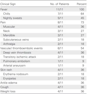
Table 2. Clinical Signs of 11 Patients Diagnosed With
Neoehrlichiosis
Clinical Sign No. of Patients Percent
Fever 11/11 100 Chills 7/11 64 Nightly sweats 5/11 45 Pain 8/11 73 Muscular 4/11 36 Neck 3/11 27 Mandible 3/11 27 Subcutaneous veins 2/11 18 Arthralgia 2/11 18
Vascular/ thromboembolic events 6/11 54
Deep vein thrombosis 4/11 36
Transitory ischemic attack 2/11 18
Pulmonary embolism 1/11 9 Arterial aneurysm 1/11 9 Skin rash 4/11 36 Erythema nodosum 2/11 18 Erysipelas 2/11 18 Ankle edema 4/11 36 Cough 4/11 36 Weight loss 4/11 36
DISCUSSION
The purpose of this case review study was to highlight one rea-son for why human cases of Neoehrlichia infection may go unrecognized: the aberrant clinical picture seen among 2 partic-ular groups of patients–middle-aged persons with autoimmune diseases or hematologic malignancies. First, myalgia, arthralgia, and/or fever are typicalfindings among patients with some of the autoimmune conditions described in this study, for exam-ple, psoriasis arthropathy, rheumatoid arthritis, or systemic lupus erythematosus. Second, if no infectious agents are discov-ered despite extensive microbiological investigations in patients with fever and systemic inflammation with a serious underlying condition, such as malignant lymphoma, chronic lymphatic leukemia, or autoimmune systemic rheumatic disease, the most common interpretation is that the condition reflects recur-rence/deterioration of the underlying morbidity. This may ulti-mately result in erroneous, and potentially dangerous, administration of immune suppressive therapy such as chemo-therapy, rituximab, and corticosteroids, which almost occurred in at least 4 of the cases documented in this study.
Another commonfinding among the Neoehrlichia-infected patients was the high incidence of thromboembolic/vascular complications; these were not interpreted to be the consequence of an infection but coincidental to a systemic inflammatory con-dition of unknown cause. In one case, fever was suspected to be secondary to widespread venous embolism [2]. As transitory is-chemic attacks are quite common among older persons, it is un-derstandable that this complication was initially seen as a separate phenomenon, not connected to the febrile episode. Finally, we report a case of arterial aneurysm in a Neoehrli-chia-infected patient with underlying autoimmune disease. Al-though it is impossible to attribute causality between the development of the aneurysm and Neoehrlichia infection, there has been a previous report of cerebral arterial aneurysm in a purported previously healthy person with suspected
Neoehrlichia infection, who actually died from intracerebral hemorrhage [3].
The pathogenic mechanisms behind the thromboembolic complications associated with Neoehrlichia infection are un-known. Is it a direct effect of infection? Does Neoehrlichia infect endothelial cells or exert toxic effects on the endothelium? Or is it an indirect effect of long-standing systemic inflammation with concomitant activation of coagulation [30]? Blood clot formation is a primitive infectious defense mechanism used by horse shoe crabs and other invertebrates to limit infectious processes [30]; the high rate of thromboembolic complications in our patients may have been the next best alternative for those with depressed B-cell immune responses to curb the infectious process.
The importance of the spleen in the infectious defense against Neoehrlichia is reflected by the high prevalence of splenectomized patients in this study. The well-known function of the spleen in the defense against encapsulated bacteria is not likely to be of rel-evance for Ca. Neoehrlichia mikurensis with its frail cell wall [7]. Instead, the spleen’s importance for the generation of “natural antibodies” [31] and maintenance of immunoglobulin M (IgM) memory B cells [32] is probably more pertinent. The very high loads of bacteria in the blood of patients most likely reflect their impaired immune state. However, no patient died despite the fact that diagnostic delay was significant. Moreover, the patients recovered within a week after doxycycline therapy was initiated. It appears as if 100 mg doxycycline twice daily for a 3-week period is sufficient to clear the infection.
There are several more reasons for the paucity of reported human Neoehrlichia cases: (1) the bacterium does not grow in cell-free media. In fact, to date no one to our knowledge has yet succeeded in cultivating the bacterium at all, which requires the use of eukaryotic cell lines. Hence, the diagnosis relies on pan-bacterial or specific PCR, which are not used as first-line diag-nostic procedures. (2) Awareness of this new microbe is very low among medical practitioners. (3) Some of the symptoms arising from a Neoehrlichia infection may be attributed to other
Table 3. Laboratory Parameters of 11 Patients Diagnosed With Neoehrlichiosis
Laboratory Parameter Level Range of Altered Parameter Reference Level Unit No. of Patients Percent
S-C-reactive protein ↑ 19–370 <5 g/L 11/11 100
White blood cell count ↑ 11–26 3.5–8.8 ×10⁹/L 9/11 82
B-Neutrophils ↑ NR 1.8–7.5 ×10⁹/L 9/11 82
B-Hemoglobin ↓ 85–119 117–170 g/L 9/11 82
S-Sodium ↓ 127–136 137–145 mmoles/L 6/11 54
S-Lactate dehydrogenase ↑ 3.9–15 1.8–3.4 µcat/L 5/11 45
B-platelet counts ↓ Slight decrease 145–348 ×109/L 2/11 18
S-Aspartate aminotransferase ↑ Slight increase (<2-fold) (0.25–0.75) µcat/L 2/11 18 S-Alanine aminotransferase ↑ Slight increase (<2-fold) (0.15–1.1) µcat/L 2/11 18 Abbreviations: B, blood; NR, not reported; S, serum.
infectious agents such as Borrelia, Anaplasma or a random “summer virus”. Doxycycline, the treatment of choice for Neo-ehrlichia infection, is equally efficient to treat infections per-ceived to be caused by Borrelia, Anaplasma, and Ehrlichia species, respectively. We propose the use of the term “neoehrli-chiosis” as a complement to the longer “Candidatus Neoehrli-chia mikurensis” infection, both in the interest of brevity, but also to focus on this new clinical infectious disease. Moreover, this nomenclature has already been used elsewhere [4].
The most likely route of transmission of the infection to hu-mans is direct inoculation of the bacteria through the skin via the bite of infected ticks. The incubation period is unknown but has been estimated to be 5–21 days for the other human path-ogenic ehrlichiae [33]. The fact that more than half of the pa-tients were unaware of tick bite is in agreement with studies of Borrelia, where it is well-known that tick bites may pass un-noticed by patients [34]. Three patients apparently fell ill during winter/early spring, which was surprising as ticks are less active during this period of the year. Because one of the patients had received blood transfusions prior to the development of fever, we investigated if this might have been the route of transmis-sion, as has been shown for Anaplasma [35] and Ehrlichia [36]. Unfortunately, no aliquot from the erythrocyte concen-trate had been stored. One of the 2 blood donors claimed to have had a flulike episode prior to donation of blood; this donor tested negative for Neoehrlichia DNA but had recovered completely and was asymptomatic at the time of testing. Thus, we were unable to determine if blood transfusions are a possible route of Neoehrlichia infection.
To conclude, we hope to raise the awareness of this newly dis-covered microbe that may give rise to an infectious disease in elderly persons with autoimmune conditions and hematological malignancies. Physicians within thefields of rheumatology, he-matology, oncology, and infectious diseases should be aware of this new infectious agent and their attention drawn to patients with suspected recurrence of the underlying autoimmune or he-matologic disease where the clinical picture is atypical.
Notes
Financial support. This work was supported by Västra Götaland Re-gional Research and Development Fund, ALF Foundation, Strategic ALF Grant for Transplantation, the Bavarian Ministry for Environment, Health and Consumer Protection and the Swedish Cancer and Allergy Foundation.
Potential conflicts of interest. All authors: No reported conflicts. All authors have submitted the ICMJE Form for Disclosure of Potential Conflicts of Interest. Conflicts that the editors consider relevant to the con-tent of the manuscript have been disclosed.
References
1. Fehr JS, Bloemberg GV, Ritter C, et al. Septicemia caused by tick-borne bacterial pathogen Candidatus Neoehrlichia mikurensis. Emerg Infect Dis2010; 16:1127–9.
2. Welinder-Olsson C, Kjellin E, Vaht K, Jacobsson S, Wenneras C. First case of human Candidatus Neoehrlichia mikurensis infection in a febrile patient with chronic lymphocytic leukemia. J Clin Microbiol2010; 48:1956–9.
3. von Loewenich FD, Geissdorfer W, Disque C, et al. Detection of “Can-didatus Neoehrlichia mikurensis” in two patients with severe febrile ill-nesses: evidence for a European sequence variant. J Clin Microbiol 2010; 48:2630–5.
4. Maurer FP, Keller PM, Beuret C, et al. Close geographic association of human neoehrlichiosis and tick populations carrying“Candidatus Neo-ehrlichia mikurensis” in Eastern Switzerland. J Clin Microbiol 2013; 51:169–76.
5. Pekova S, Vydra J, Kabickova H, et al. Candidatus Neoehrlichia mikur-ensis infection identified in 2 hematooncologic patients: benefit of mo-lecular techniques for rare pathogen detection. Diagn Microbiol Infect Dis2011; 69:266–70.
6. Li H, Jiang JF, Liu W, et al. Human infection with Candidatus Neoehr-lichia mikurensis, China. Emerg Infect Dis2012; 18:1636–9. 7. Kawahara M, Rikihisa Y, Isogai E, et al. Ultrastructure and phylogenetic
analysis of‘Candidatus Neoehrlichia mikurensis’ in the family Anaplas-mataceae, isolated from wild rats and found in Ixodes ovatus ticks. Int J Syst Evol Microbiol2004; 54(Pt 5):1837–43.
8. Yabsley MJ, Murphy SM, Luttrell MP, Wilcox BR, Ruckdeschel C. Rac-coons (Procyon lotor), but not rodents, are natural and experimental hosts for an ehrlichial organism related to“Candidatus Neoehrlichia mi-kurensis”. Vet Microbiol 2008; 131:301–8.
9. Rar V, Golovljova I. Anaplasma, Ehrlichia, and“Candidatus Neoehrli-chia” bacteria: Pathogenicity, biodiversity, and molecular genetic char-acteristics, a review. Infect Genet Evol2011; 11:1842–61.
10. Alekseev AN, Dubinina HV, Van De Pol I, Schouls LM. Identification of Ehrlichia spp. and Borrelia burgdorferi in Ixodes ticks in the Baltic re-gions of Russia. J Clin Microbiol2001; 39:2237–42.
11. Brouqui P, Sanogo YO, Caruso G, Merola F, Raoult D. Candidatus Ehr-lichia walkerii: a new EhrEhr-lichia detected in Ixodes ricinus tick collected from asymptomatic humans in Northern Italy. Ann N Y Acad Sci2003; 990:134–40.
12. Jenkins A, Kristiansen BE, Allum AG, et al. Borrelia burgdorferi sensu lato and Ehrlichia spp. in Ixodes ticks from southern Norway. J Clin Mi-crobiol2001; 39:3666–71.
13. Pan H, Liu S, Ma Y, Tong S, Sun Y. Ehrlichia-like organism gene found in small mammals in the suburban district of Guangzhou of China. Ann N Y Acad Sci2003; 990:107–11.
14. Schouls LM, Van De Pol I, Rijpkema SG, Schot CS. Detection and identification of Ehrlichia, Borrelia burgdorferi sensu lato, and Bartonel-la species in Dutch Ixodes ricinus ticks. J Clin Microbiol1999; 37: 2215–22.
15. Shpynov S, Fournier PE, Rudakov N, Tarasevich I, Raoult D. Detection of members of the genera Rickettsia, Anaplasma, and Ehrlichia in ticks collected in the Asiatic part of Russia. Ann N Y Acad Sci 2006; 1078:378–83.
16. Movila A, Alekseev AN, Dubinina HV, Toderas I. Detection of tick-borne pathogens in ticks from migratory birds in the Baltic region of Russia. Med Vet Entomol2013; 27:113–7.
17. Andersson M, Raberg L. Wild rodents and novel human pathogen Can-didatus Neoehrlichia mikurensis, Southern Sweden. Emerg Infect Dis 2011; 17:1716–8.
18. Fertner ME, Molbak L, Boye Pihl TP, Fomsgaard A, Bodker R. First de-tection of tick-borne“Candidatus Neoehrlichia mikurensis” in Denmark 2011. Euro Surveill2012; 17:1–3.
19. Capelli G, Ravagnan S, Montarsi F, et al. Occurrence and identification of risk areas of Ixodes ricinus-borne pathogens: a cost-effectiveness analysis in north-eastern Italy. Parasit Vectors2012; 5:61–70. 20. van Overbeek L, Gassner F, van der Plas CL, Kastelein P, Nunes-da
Rocha U, Takken W. Diversity of Ixodes ricinus tick-associated bacterial communities from different forests. FEMS Microbiol Ecol 2008; 66:72–84.
21. Vayssier-Taussat M, Le Rhun D, Buffet JP, et al. Candidatus Neoehrlichia mikurensis in bank voles, France. Emerg Infect Dis2012; 18:2063–5. 22. Lommano E, Bertaiola L, Dupasquier C, Gern L. Infections and
coinfec-tions of questing Ixodes ricinus ticks by emerging zoonotic pathogens in Western Switzerland. Appl Environ Microbiol2012; 78:4606–12. 23. Movila A, Toderas I, Uspenskaia I, Conovalov J. Molecular detection of
tick-borne pathogens in Ixodes ricinus from Moldova collected in 1960. Ticks Tick Borne Dis2013; 4:359–61.
24. Spitalska E, Boldis V, Kostanova Z, Kocianova E, Stefanidesova K. Inci-dence of various tick-borne microorganisms in rodents and ticks of cen-tral Slovakia. Acta Virol2008; 52:175–9.
25. Li H, Jiang J, Tang F, et al. Wide distribution and genetic diversity of “Candidatus Neoehrlichia mikurensis” in rodents from China. Appl En-viron Microbiol2013; 79:1024–7.
26. Rar VA, Epikhina TI, Livanova NN, et al. Study of the heterogeneity of 16s rRNA gene and groESL operone in the dna samples of Anaplasma phagocytophilum, Ehrlichia muris, and“Candidatus Neoehrlichia mi-kurensis” determined in the Ixodes persulcatus ticks in the area of Urals, Siberia, and far east of Russia. Mol Gen Mikrobiol Virusol 2011; 26:17–23.
27. Kamani J, Baneth G, Mumcuoglu KY, et al. Molecular detection and characterization of tick-borne pathogens in dogs and ticks from Nigeria. PLoS Negl Trop Dis2013; 7:e2108.
28. Silaghi C, Woll D, Mahling M, Pfister K, Pfeffer M. Candidatus Neoehr-lichia mikurensis in rodents in an area with sympatric existence of the
hard ticks Ixodes ricinus and Dermacentor reticulatus, Germany. Parasit Vectors2012; 5:285–92.
29. Diniz PP, Schulz BS, Hartmann K, Breitschwerdt EB.“Candidatus Neo-ehrlichia mikurensis” infection in a dog from Germany. J Clin Microbiol 2011; 49:2059–62.
30. Delvaeye M, Conway EM. Coagulation and innate immune responses: can we view them separately? Blood2009; 114:2367–74.
31. Ochsenbein AF, Fehr T, Lutz C, et al. Control of early viral and bacterial distribution and disease by natural antibodies. Science 1999; 286:2156–9.
32. Kruetzmann S, Rosado MM, Weber H, et al. Human immunoglobulin M memory B cells controlling Streptococcus pneumoniae infections are generated in the spleen. J Exp Med2003; 197:939–45.
33. Thomas RJ, Dumler JS, Carlyon JA. Current management of human granulocytic anaplasmosis, human monocytic ehrlichiosis and Ehrlichia ewingii ehrlichiosis. Expert Rev Anti Infect Ther 2009; 7:709–22.
34. Hengge UR, Tannapfel A, Tyring SK, Erbel R, Arendt G, Ruzicka T. Lyme borreliosis. Lancet Infect Dis2003; 3:489–500.
35. Alhumaidan H, Westley B, Esteva C, Berardi V, Young C, Sweeney J. Transfusion-transmitted anaplasmosis from leukoreduced red blood cells. Transfusion2013; 53:181–6.
36. Regan J, Matthias J, Green-Murphy A, et al. A confirmed Ehrlichia ewingii infection likely acquired through platelet transfusion. Clin Infect Dis2013; 56:e105–7.