Nephrol Dial Transplant (2013) 28: 505–517 doi: 10.1093/ndt/gfs526
Advance Access publication 12 December 2012
NDT Perspectives
Fabry nephropathy: indications for screening and guidance for
diagnosis and treatment by the European Renal Best Practice
Wim Terryn
1,
Pierre Cochat
2,
Roseline Froissart
3,
Alberto Ortiz
4,
Yves Pirson
5,
Bruce Poppe
6,
Andreas Serra
7,
Wim Van Biesen
8,
Raymond Vanholder
8and Christoph Wanner
91Division of Nephrology, Department of Internal Medicine,
Regional Hospital Jan Yperman, Ypres, Belgium,
2Centre de Référence des Maladies Rénales Rares, Hôpital
Femme-Mère-Enfant, Lyon, France,
3Laboratoire des Maladies Héréditaires du Métabolisme et Dépistage
Néonatal, Lyon, France,
4IIS-Fundacion Jimenez Diaz, U Autonoma de Madrid, Redinren,
FRIAT, Madrid, Spain,
5Division of Nephrology, Cliniques Universitaires Saint-Luc,
Université Catholique de Louvain, Brussels, Belgium,
6Medical Genetics, Ghent University Hospital, Ghent, Belgium, 7Division of Nephrology, University Hospital, Zürich, Switzerland, 8Nephrology Section, Department of Internal Medicine, Ghent
University Hospital, Ghent, Belgium and
9Division of Nephrology, University Hospital, Würzburg, Germany
Correspondence and offprint requests to: Wim Terryn; E-mail: guidelines@era-edta.org
Keywords: Fabry disease, Fabry nephropathy, screening
A B S T R AC T
Fabry disease (FD) is an X-linked disorder of glycosphingo-lipid catabolism resulting in the accumulation of glycoglycosphingo-lipids including globotriaosylceramide in cells of various tissues resulting in end-organ manifestations. Initially, FD is typi-cally characterized by angiokeratoma and recurrent episodes of neuropathic pain in the extremities occurring during childhood or adolescence. Most affected patients also exhibit a decreased ability to sweat. Later in life, FD results in left ventricular hypertrophy, proteinuria, renal failure and stroke. These later disease manifestations are non-specific and also common in diabetes, hypertension and atheroma-tosis and thus for most practitioners do not point into the direction of FD. As a consequence, FD is under-diagnosed and screening of high-risk groups is important for case finding, as is a thorough pedigree analysis of affected patients. In the nephrology clinic, we suggest to screen
patients for FD when there is unexplained chronic kidney disease in males younger than 50 years and females of any age. In men, this can be performed by measuring α-galacto-sidase A activity in plasma, white blood cells or dried blood spots. In women, mutation analysis is necessary, as enzyme measurement alone could miss over one-third of female Fabry patients. A multidisciplinary team should closely monitor all known Fabry patients, with the nephrologist screening kidney impairment (glomerularfiltration rate and proteinuria) on a regular basis. Transplanted Fabry patients have a higher mortality than the regular transplant popu-lation, but have acceptable outcomes, compared with Fabry patients remaining on dialysis. It is unclear whether enzyme replacement therapy (ERT) prevents deterioration of kidney function. In view of the lack of compelling evidence for ERT, and the low likelihood that a sufficiently powered ran-domized controlled trial on this topic will be performed, data of all patients with FD should be collected in a central registry.
INTRODUCTIO N
European Renal Best Practice (ERBP) is the official guideline body of the European Renal Association/European Dialysis and Transplant Association (ERA/EDTA). The mission of ERBP is to improve the outcome of patients with kidney disease in a sustainable way, through enhancing the accessi-bility of knowledge on patient care, in a format that stimu-lates its use in clinical practice. In line with this mission, and in view of its philosophy [1], the ERBP advisory board con-sidered it useful to develop guidance in the field of orphan diseases with nephrological relevance. Typical for these dis-eases are the rather low patient number, and consequently, the lack of large trials. As a consequence, formal evidence-based medicine is nearly impossible in this field. Neverthe-less, nephrologists need guidance on how to approach patients with these diseases. Therefore, ERBP decided to use the combination of formal systematic literature reviews, a consensus meeting with an international panel of experts and peer review as a suitable model to develop guidance in the field of orphan diseases. A first paper on oxalosis has already been published in this series [2]. This paper presents the results of a guidance process on the topic of Fabry disease (FD).
FD (OMIM ID #301500) is an X-linked inborn error of glycosphingolipid catabolism caused by quantitative or quali-tative defects in the lysosomal enzymeα-galactosidase A (α-Gal A). As a result, glycosphingolipids, mainly globotriaosyl-ceramide (Gb-3), accumulate in the lysosomes of different cells throughout the body, ultimately resulting in organ failure [3, 4]. Patients with FD have a markedly limited life expectancy due to cardiovascular, neurological and renal in-volvement. Enzyme replacement therapy (ERT) has been made available since 2001. Intravenous infusion every other week results in the removal of a part of the Gb-3 deposits, diminishes Fabry-related symptoms and possibly protects organs to a certain extent [5,6]. The effects of ERT on pro-gression of renal disease ( proteinuria and renal function) are unclear.
A I M S O F T H I S PU B LI CATIO N
The first aim of this paper is to review the current literature on renal disease in Fabry patients, in order to provide gui-dance to the nephrologist on when to screen for this disease and why, and to understand the preferred methods that should be used for screening.
The second aim is to provide guidance on the follow-up, prevention and treatment of renal disease, and its compli-cations ( proteinuria, renal failure). The role of ERT, angio-tensin-converting enzyme inhibitors (ACEi), angiotensin receptor blockers (ARB) and renal replacement therapy (RRT) is reviewed.
M E TH OD S
A literature search was conducted using the PubMed database (most recent search July 2012). The search term used was ‘Fabry Disease’ with limits: ‘Humans’, ‘Clinical Trial’, ‘Meta-Analysis’, ‘Practice Guideline’, ‘Randomized Controlled Trial’, ‘Review’, ‘English’, ‘French’. A total of 357 articles were re-trieved; the articles were classified to the following topics (one paper can be attributed to more than one classification):
(i) epidemiology, screening studies; (ii) diagnostic methods;
(iii) Fabry nephropathy: natural history, complications (hy-pertension), mechanisms, renal pathology;
(iv) treatment of Fabry nephropathy; with ERT, ACEi and ARB, RRT; efficacy and safety issues.
Articles out of scope and review articles that presented no new data were excluded. Articles on experimental, non-regis-tered treatments were also excluded.
The reference lists of the identified relevant studies were manually searched for additional citations.
After all relevant publications were retrieved, a consensus meeting was held with all co-authors. The resulting paper was sent for internal review before submission, as explained in the‘instructions to authors’ section of the ERBP website [7].
E P I D E M I O LOGY A N D TH E N E E D FO R S CR E E NI NG
1.1 We do not recommend screening in the general popu-lation. (Ungraded statement)
1.2 We recommend obtaining informed consent from the patient before screening, using an information form drafted in collaboration with a clinical geneticist. (Un-graded statement)
1.3 We recommend screening for FD in male chronic kidney disease (CKD) patients below 50 years of age in whom a reliable renal diagnosis is absent. (Ungraded statement) 1.4 We suggest screening for FD in females with unexplained
CKD, irrespective of age, with other unexplained symp-toms potentially associated with FD. (Ungraded statement) 1.5 We recommend discussing with the patient the impli-cations of diagnosing a genetic disease and the possible implications for the at-risk relatives. (Level 1C)
Rationale
Classical FD is a progressive multisystem disease predomi-nantly presenting in males, characterized by angiokeratoma, hypohidrosis and acroparesthesia (neuropathic pain) in child-hood, followed by renal failure, left ventricular hypertrophy (LVH), stroke and premature death in the fourth or fifth decade of life [8]. In male patients, levels ofα-Gal A activity
ND
T
P
ERSPECTIVES
are classically very low or undetectable. However, as a result of screening studies during the past decade, clinical variants of FD in male patients with varying degrees of residual activity of α-Gal A have been described. The first described was the ‘cardiac variant’ with isolated LVH and/or cardio-myopathy presenting in the sixth or seventh decade, lacking the classical disease symptoms and time course [9, 10]. Patients suffering from this variant may have proteinuria, but their renal function is typically normal for their age. Later a ‘renal variant’ phenotype was described in a screening study in a dialysis population, where patients again were lacking the classical manifestations. This phenotype was described as ‘intermediate’ between the cardiac variant and the classic phenotype [11]. These patients with cardiac and renal var-iants are called ‘atypical’ or ‘attenuated’ FD patients. The genetic basis of this variable penetrance and expression is unclear. It is believed that the atypical cases are the result of missense mutations that encode mutant enzyme protein or intronic lesions that reduce transcript levels, both resulting in a reduced but significant residual enzyme function (1–12% of normal) [12], although this has been debated, and others found no genotype–phenotype correlation [13]. Heterozygous women, in spite of having a mutation compatible with typical disease, can also present this attenuated phenotype as it was hypothesized that skewed X-inactivation can result in signi fi-cant residual enzyme function. However, it must be stressed that most females have the classical phenotype, but with a delayed and/or milder presentation of symptoms [14].
As a consequence, reported prevalence varies with the popu-lation studied and the test used for screening, and genetic screen-ing mightfind female index cases that are not found by enzyme-based methods [15]. The prevalence of classical FD has been es-timated at 1 in 117 000 births [14] and 1 in 40 000 males [8]. In several screening studies in high-risk populations, the frequency was up to 1% or even higher, especially in populations with un-explained LVH [16]. In newborns [17–19], the incidence of α-Gal A deficiency was 1 in 3100 with an 11 to 1 ratio of patients with the later-onset versus the classic phenotype. In the haemo-dialysis population, a prevalence of 0.33% in male and 0.10% in female patients has been found in a cross-sectional screening study [16]. Only two studies screened kidney transplant patients. In cryptogenic stroke, a prevalence of 0.8% [20] up to 2.4% [21] and 3.9% [22] was found; however, in the second study [21], half of the patients had the p.D313Y mutation, which is now generally regarded as a pseudo-deficiency, and in the last study [22], the specific mutations were not mentioned and could also have been polymorphisms. Many screening studies are not con-clusive for the female population, as they most frequently used α-Gal A activity screening, which is in women, as described above, not a sensitive screening tool.
Although there are no studies in the CKD population not on dialysis, we recommend screening for FD in patients with CKD without a clear diagnosis. In classical FD, most males reach CKD Stage 5 or die before the age of 50 [12,23]. As a consequence, we recommend screening in males only below the age of 50 years. We recommend screening even in the case of a negative family history as de novo mutations can occur, and the family history is not always suggestive for FD,
given the broad phenotypic spectrum of the disease. Arterial hypertension should not be an exclusion criterion as more than 50% of FD patients have mild to moderate hypertension, especially when estimated glomerularfiltration rate (eGFR) is <60 mL/min/1.73 m2 [23–25]. In women, disease onset can be later, so when there is unexplained kidney disease associ-ated with manifestations suggestive of FD, we suggest screen-ing for FD regardless of age.
The real prevalence should be derived from screening in the healthy population at a young age; this has been done in four studies in newborns [17–19,26]. However, this approach remains problematic for several reasons. The American College of Medical Genetics (ACMG) has proposed newborn screening for 29 disorders, but screening for FD was not in-cluded in this list (available online at:http://mchb.hrsa.gov/
screening/). Although measurement of α-Gal A has a good
sensitivity and specificity in males, it has a low positive pre-dictive value in the healthy population. This will result in unnecessary expensive tests. In addition, the majority of the detected cases in the newborn studies are‘atypical’ mutations, giving an attenuated phenotype or a cardiac variant. The finding of a genetic predisposition for a possible late-onset disease where the treatment effectiveness is unclear has ethical and legal implications that constrain a systematic screening of newborns. In these cases, it would be difficult to decide on ERT, as the natural history of patients carrying aty-pical mutations is poorly characterized, effects of ERT in mild cases have not been studied, and a lifelong treatment is a psychological burden for the patient and afinancial one for both the individual and society with, on top of that, uncertain results. As a consequence, we do not recommend screening for FD in the general population.
As FD is an X-linked disease with variable but significant morbidity both in males and females, its diagnosis might have profound consequences for the proband and his rela-tives. As a consequence, we recommend obtaining informed consent from the proband before screening, when possible in cooperation with an expert in genetic counselling. (Example in Supplementary appendix.)
Once the diagnosis is made, it is important to make up a pedigree in order to identify all relatives at risk. FD is an X-linked disease where all carriers can be symptomatic. It should be kept in mind that ‘skipping’ of a generation is possible because of variable expression.
The patient should receive further guidance in communi-cation with his family. He must be able to provide sufficient information (e.g. by using flyers written by the treating team), and one must anticipate a number of possible pro-blems in the communication with his family. Some people do not want a work-up to the diagnosis of FD, and it should be explained to the patient that they do have the right not to know their genetic status.
S C R E E N I N G ME T H O D S
2.1 We recommend using enzyme activity measurement for α-Gal A as a primary tool in males, followed by
ND
T
P
confirmation with mutation analysis when positive. (Un-graded statement)
2.2 We suggest using mutation analysis as a primary tool for screening in females. (Ungraded statement)
Measurement ofα-Gal A activity in leucocytes using the fluoro-genic substrate 4-methylumbelliferyl-α-D-galactopyranoside is
the gold standard for FD in men, with a sensitivity and speci-ficity of nearly 100%. Recently, a dried blood spot test (DBS) usingfilter paper has been proposed as an alternative to the leu-cocyte tests [27]. These samples are easy to transport and are stable at room temperature for many days, making it a most convenient screening tool in men, as it is a very sensitive tool with a negative predictive value reaching 100%.
In women, due to skewed X inactivation, enzyme activity measurement has a low sensitivity, as one in three women with FD have normal or nearly normalα-Gal A activity [15]. For this reason, enzymatic tests are less suitable and systema-tic genesystema-tic testing should be encouraged in females with un-explained CKD and manifestations suggestive for FD. As genetic testing is expensive (150–1000 Euro and more per test), a thorough anamnesis, family history and clinical inves-tigation could help to select female CKD patients in whom testing is cost-effective (Figure1).
In FD, gene mutation analysis is a way of confirming diag-nosis in male patients, subsequent to enzyme activity measurement. A fresh blood sample can be collected for this purpose, or polymerase chain reaction amplification can be performed on DNA eluted directly from thefilter paper used for the DBSα-Gal A measurement [28].
GLA gene mutations causing FD include single base changes leading to missense or nonsense mutations, or affect-ing consensus splice sites, small deletions or insertions, but also large gene rearrangements in <5% of the patients. Corre-lations between a specific mutation, i.e. the genotype, and the severity of the disease, the phenotype, are poor in FD. In a few cases, however, knowledge on the underlying mutation can provide information concerning prognosis and therapy and help the clinician in counselling. Some mutations are fre-quently associated with an attenuated phenotype, such as the mutation p.N215S, which gives a cardiac phenotype with only LVH [29]. These mutations are associated with a residual enzyme function [30]. A significant proportion of the mutations in men are, however, associated with a very low or absent enzyme function and the classic phenotype.
The GLA gene should be sequenced. As most of the mutations are ‘private’, i.e. unique to a family, it is always possible to completely identify a previously undetected mutation, and regular updates of such new mutations are available (http://www.hgmd.cf.ac.uk/ac/index.php). The pathogenicity of novel gene alterations such as missense or intronic mutations must always be evaluated. However, in females with normal biochemical tests, it may be difficult to confirm or exclude the diagnosis of FD when a variant of unknown significance is present.
In a suggestive clinical situation, most sequence alterations in exonic regions are pathogenic with very few exceptions.
One example of such inert exonic polymorphism is the p. ‘D313Y’ substitution (G to T at cDNA nucleotide 937); while the plasma enzyme activity towards the artificial sub-strate is significantly reduced, additional studies demonstrated high residual lysosomal enzyme activity and no pathologic excretion of urinary Gb-3. As a result, the p.D313Y substi-tution is now generally considered to be a so-called pseudo-deficiency.
If one finds a novel sequence variation in an intronic region or a novel missense mutation that is not known to be a polymorphism present in the general population, several methods allow non-invasive diagnostic analysis to establish whether it is disease causing. First, it should be checked whether these sequence variations exist in the normal lation (using electronic databases or an own control popu-lation). The second step is to check male relatives of the index case who are carriers of the sequence variation forα- A activity. If the sequence variation is present in some of them, despite a normal α-Gal A activity and absence of clinical manifestations of FD, the sequence variation can be con-sidered to be a polymorphism. If it coincides with a deficient α-Gal A in one or more of the male relatives, the possibility of a disease causing mutation is realistic, and in this case, a work-up of all carriers for the presence of (subclinical) FD disease manifestations should be considered.
Besides enzyme activity measurement and mutation analy-sis, detection of the accumulating substances (glycosphingoli-pids) has been studied as a tool for diagnosis. Globotriaosylceramide (Gb-3) is the most important glyco-sphingolipid, and it should be measured in urine rather than in plasma. Urinary Gb-3 can be a useful diagnostic tool in female heterozygotes with classical FD as it is increased in 95% of them. However, the proportion is much lower in het-erozygotes with variant forms. It can also be used in males as a surrogate marker to evaluate the response to ERT [31]. Mass spectrometric profiling of Gb-3 isoforms may also help to identify heterozygotes [32].
In plasma, deacylated Gb-3 (globotriaosylsphingosine, ‘lysoGb-3’) has been shown to have a better correlation with FD. It is elevated 200–400 times in males with classical disease, from an early phase in the disease, but its levels can remain low in asymptomatic females or in the ‘cardiac variant’ p.N215S in males [33–37]. The examination of the urinary sediment with phase-contrast microscopy under po-larized light shows tubular cells containing particles with bi-refringent Maltese Crosses, having a lamellated appearance with protrusions, and consisting of accumulated Gb-3. In the hands of Selvarajah et al. [38], this was a highly sensitive and specific tool for screening of FD, but its accuracy is strongly operator-dependent and therefore, it is probably an unrealis-tic option for large-scale screening studies.
WOR K- UP OF A PAT IE N T W I TH FD
3.1 We recommend that the detailed baseline and follow-up data of all patients with established FD should be trans-ferred to a central registry. (Ungraded statement)
ND
T
P
ERSPECTIVES
3.2 We recommend baseline and subsequent yearly evalu-ation by a multidisciplinary team, including kidney func-tion and albuminuria, in all patients with established FD (cardiology, neurology and nephrology). (Ungraded statement)
3.3 We recommend not considering female carriers for living donation, unless in exceptional cases. In these cases, we recommend a kidney biopsy to evaluate the risk for the donor and acceptor. (Ungraded statement)
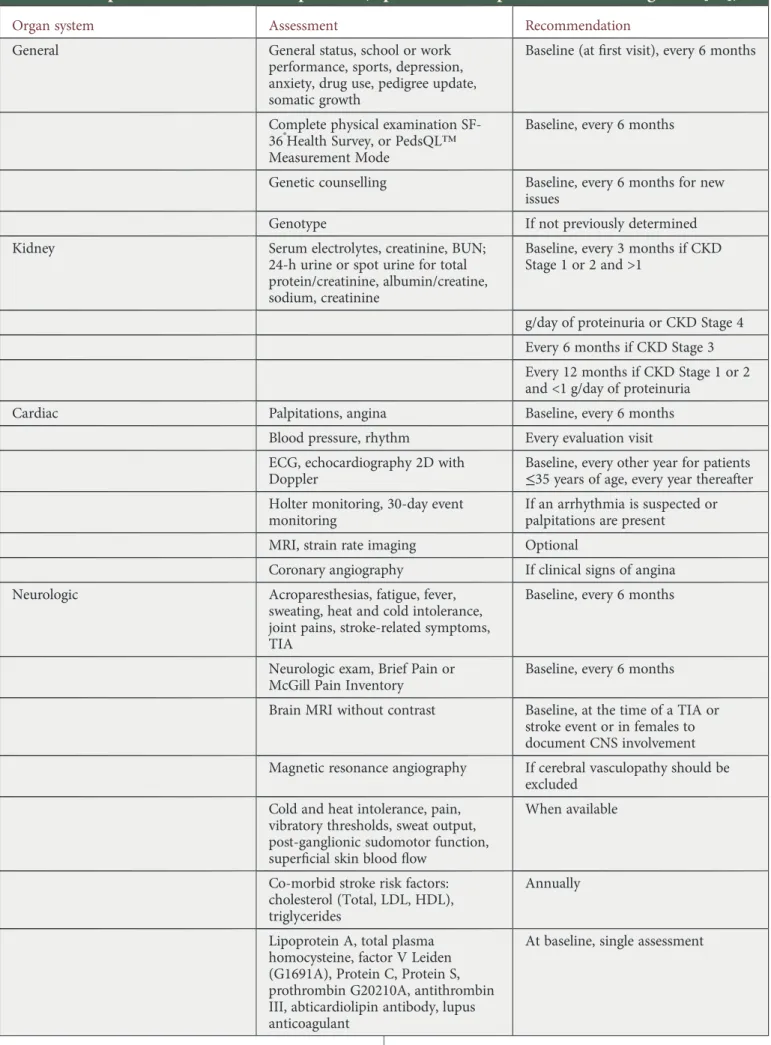
Once an index patient is diagnosed, a baseline evaluation is indicated. As FD is a progressive multisystem disease, base-line evaluation is optimally performed by a multidisciplinary team (Table 1, adapted from Eng et al. [39]). The baseline evaluation should be performed in male and all female car-riers, as the phenotype can be equally severe.
As this document is written from the nephrology perspective, we will focus on renal involvement in what follows. For evalu-ation and pathophysiology of other organs, we refer to the guidelines of the respective subspecialities.
Renal involvement is a cardinal feature of FD. Gb-3 depo-sition in renal cells is progressive and begins early in life. Besides these deposits, pathogenic mechanisms result in
glomerular ischaemia with subsequent glomerulosclerosis and tubular atrophy, even very early in the disease course. Vacuo-lization of podocytes and epithelial cells is a characteristic optical microscopy histological finding. These vacuoles are filled with deposits on electron microscopy, or following to-luidine blue staining of samples prepared for electron microscopy. At an early stage, hyperfiltration may, as in dia-betes, be thefirst sign of kidney damage.
As FD can progress subclinically, adolescent and adult patients should have urinary albumin measurement, as this is one of thefirst signs of Fabry nephropathy. We suggest asses-sing the amount of albumin normalized for creatinine on a fresh morning sample as diagnostic test. We suggest measur-ing urinary albumin rather than total protein, as it is more sensitive. Renal function can be assessed using serum creati-nine and eventually formulas to translate serum creaticreati-nine to estimated clearances. Even in the absence of albuminuria or renal failure, all these parameters should be re-evaluated at least yearly in order to detect progressive disease.
Renal intracellular Gb-3 deposits may be present even in young children with normal GFR and minimal or absent micro-albuminuria. In a recent study of 14 young Fabry patients aged 4–19 years with normal GFR, there was an
F I G U R E 1 : Flowchart for screening for FD in CKD patients.
ND
T
P
Table 1. Proposed assessments in FD patients (reproduced with permission from Eng
et al. [
39
])
Organ system Assessment Recommendation
General General status, school or work performance, sports, depression, anxiety, drug use, pedigree update, somatic growth
Baseline (atfirst visit), every 6 months
Complete physical examination SF-36®Health Survey, or PedsQL™ Measurement Mode
Baseline, every 6 months
Genetic counselling Baseline, every 6 months for new issues
Genotype If not previously determined Kidney Serum electrolytes, creatinine, BUN;
24-h urine or spot urine for total protein/creatinine, albumin/creatine, sodium, creatinine
Baseline, every 3 months if CKD Stage 1 or 2 and >1
g/day of proteinuria or CKD Stage 4 Every 6 months if CKD Stage 3 Every 12 months if CKD Stage 1 or 2 and <1 g/day of proteinuria
Cardiac Palpitations, angina Baseline, every 6 months
Blood pressure, rhythm Every evaluation visit ECG, echocardiography 2D with
Doppler
Baseline, every other year for patients ≤35 years of age, every year thereafter Holter monitoring, 30-day event
monitoring
If an arrhythmia is suspected or palpitations are present
MRI, strain rate imaging Optional
Coronary angiography If clinical signs of angina Neurologic Acroparesthesias, fatigue, fever,
sweating, heat and cold intolerance, joint pains, stroke-related symptoms, TIA
Baseline, every 6 months
Neurologic exam, Brief Pain or McGill Pain Inventory
Baseline, every 6 months
Brain MRI without contrast Baseline, at the time of a TIA or stroke event or in females to document CNS involvement Magnetic resonance angiography If cerebral vasculopathy should be
excluded Cold and heat intolerance, pain,
vibratory thresholds, sweat output, post-ganglionic sudomotor function, superficial skin blood flow
When available
Co-morbid stroke risk factors: cholesterol (Total, LDL, HDL), triglycerides
Annually
Lipoprotein A, total plasma homocysteine, factor V Leiden (G1691A), Protein C, Protein S, prothrombin G20210A, antithrombin III, abticardiolipin antibody, lupus anticoagulant
At baseline, single assessment
Continued ND T P ERSPECTIVES 510
association between the volume of Gb-3 deposition in the po-docytes, and age. The volume of Gb-3 deposition was also correlated with urinary protein excretion rates [40]. Tøndel et al. [41] found segmental foot process effacement in all young Fabry patients, despite the fact they were normo-albu-minuric (below 30 mg/day). Thus, in the case of patients at risk of FD, any albuminuria, even if in the ‘normal’ range, should be considered as suspect.
Proteinuria progresses and correlates with and probably also contributes to the decline in renal function, e.g. male Fabry patients with a proteinuria >1 g/24 h had a greater yearly decline in renal function (−6.9 mL/min/1.73 m2) than patients
with proteinuria between 0.1 and 1 g/24 h (−2.2 mL/min/1.73 m2) and patients with proteinuria <0.1 mg/24 h (−0.6 mL/min/ 1.73 m2) [42]. Other studies confirm that the urinary protein to urinary creatinine ratio (UP/Cr) is the most important indicator of renal disease progression [43]. The yearly decline in renal function also correlates with GFR at presentation (in males,−3 mL/min/1.73 m2 with GFR >60 mL/min/1.73 m2 versus –6.8 mL/min/1.73 m2 with GFR≤ 60 mL/min/1.73 m2; in females −0.9 mL/min/1.73 m2versus− 2.1 mL/min/1.73 m2) [42].
Most patients with CKD Stages 3–5 have some degree of proteinuria [23]. Proteinuria in the nephrotic range (>3.5 g/ 24 h) is, however, rarely seen (maximal 18% in [12]).
CKD Stage 5 usually develops between the third and the fifth decade, with a mean age at diagnosis of 38, but can appear as early as at the age of 16 [44,45]. Interestingly, the
mean age at initiation of RRT is similar for males and females, although the proportion of male versus female FD patients on RRT was 9 to 1 [42].
Living related donation in FD can pose a problem if ap-parently asymptomatic female carriers consider donating a kidney. Even in the case of a normal renal function and in the absence of albuminuria, significant Gb-3 deposits can be abundant in a renal biopsy [46] and thus female carriers are, in our opinion, not eligible for living kidney donation.
The Fabry population is small and heterogeneous which makes it difficult to study its natural course and to conduct larger-scale, placebo-controlled or open-label clinical trials. For these reasons, a high quality registry with all treated and untreated patients on a European scale, developed indepen-dently of industry, is highly desirable.
T R E AT M E N T O F FA B RY N E P H ROPAT H Y
4.1 We do not recommend starting ERT in patients with pro-teinuria [ protein-to-creatinine ratio >1 g/g (>0.1 = gram/ mol) creatinine] or eGFR <60 mL/min/1.73 m2, except for non-renal indications. (1D)
4.2 We recommend that when ERT is deemed indicated, it should be started as part of a well-designed clinical trial, either observational or interventional. (Ungraded statement)
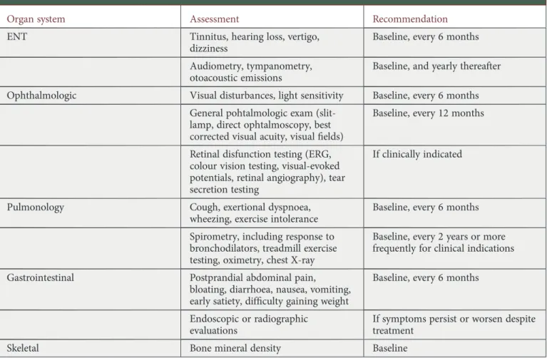
Table 1. Continued
Organ system Assessment Recommendation
ENT Tinnitus, hearing loss, vertigo, dizziness
Baseline, every 6 months
Audiometry, tympanometry, otoacoustic emissions
Baseline, and yearly thereafter
Ophthalmologic Visual disturbances, light sensitivity Baseline, every 6 months General pohtalmologic exam
(slit-lamp, direct ophtalmoscopy, best corrected visual acuity, visualfields)
Baseline, every 12 months
Retinal disfunction testing (ERG, colour vision testing, visual-evoked potentials, retinal angiography), tear secretion testing
If clinically indicated
Pulmonology Cough, exertional dyspnoea, wheezing, exercise intolerance
Baseline, every 6 months
Spirometry, including response to bronchodilators, treadmill exercise testing, oximetry, chest X-ray
Baseline, every 2 years or more frequently for clinical indications
Gastrointestinal Postprandial abdominal pain, bloating, diarrhoea, nausea, vomiting, early satiety, difficulty gaining weight
Baseline, every 6 months
Endoscopic or radiographic evaluations
If symptoms persist or worsen despite treatment
Skeletal Bone mineral density Baseline
ND
T
P
4.3 In a patient on haemodialysis, and when ERT is deemed indicated, we recommend administering the ERT during a haemodialysis session. (1A)
4.4 We recommend kidney transplantation as a valuable option in patients who are eligible for this intervention. (Ungraded statement)
4.5 After renal transplantation, we do not suggest ERT for renal indications, but it can be continued for non-renal indications. (Ungraded statement)
As discussed above, proteinuria is an important risk factor for the progression of renal FD. The use of ACE-i and ARB has been shown to be nephro-protective in other proteinuric renal diseases, and could thus be important in FD as well. As such, the use of ACE-i or sartane would be acceptable in FD. In a recent paper [47], it has been demonstrated that ERT interacts with ACE and inhibits its activity, possibly by re-moving the galactose residues from the enzyme. The clinical relevance of this observation is unclear, and should not be seen as a reason to prohibit the use of ACE-i.
Kidney Disease Improving Global Outcomes (KDIGO) guidelines suggest that in patients with CKD Stages 3–5, vitamin D deficiency be corrected [48]. Emerging evidence in patients with CKD show that vitamin D can reduce protei-nuria or albumiprotei-nuria even in the presence of angiotensin-converting enzyme inhibition [49]. Selective activation of the vitamin D receptor with paricalcitol lowered urinary albumin excretion, as was demonstrated in patients with Type 2 dia-betes in a recent randomized controlled trial [50]. In cultured human podocytes, vitamin D receptor activation prevented lyso-Gb-3-induced, TGFβ1-mediated, up-regulation of extra-cellular matrix proteins [51]. Even lacking more definitive evidence of a beneficial effect of vitamin D on Fabry nephro-pathy, it seems advisable to place particular emphasis in fol-lowing guidelines on vitamin D management in CKD patients in patients with FD.
Two forms of recombinant α-Gal A have been approved in Europe: agalsidase alpha (Replagal®; Shire Human Genetic Therapies, Boston, MA) and agalsidase beta (Fabrazyme®; Genzyme, Cambridge, MA). Agalsidase alpha is produced in a continuous human cell line and is administered as an intra-venous infusion over 40 min at a dose of 0.2 mg/kg body weight every 2 weeks. Agalsidase beta is produced in Chinese hamster ovary (CHO) cells and is given as an intravenous in-fusion over a 4-h period at a dose of 1.0 mg/kg body weight every 2 weeks.
According to a recent Cochrane review, the evidence base in favour of ERT is weak. Only five (total n = 187) poor quality randomized controlled trials are available. They all concern surrogate end points, such as decrease in plasma Gb-3 levels in plasma and tissues and evolution of renal function. According to the Cochrane review, these studies show no evi-dence for a clinical benefit of the use of agalsidase alpha or beta to treat Fabry nephropathy [52]. As there are at present no hard data that ERT alters the natural course of Fabry ne-phropathy (Table2), we recommend starting ERT only in the context of a clinical trial, interventional or observational. All
data from observational trials should be entered in a central registry.
Besides randomized controlled trials open-label studies and retrospective analyses have been performed. It is of inter-est to compare the evolution of renal disease in the historical untreated and treated cohorts of an international industry sponsored registry on FD [43, 53]. It is difficult to compare the data presented in both publications, as the design of the analyses and the presentation of data were different, and there was a substantial risk for selection bias, as only a minor proportion of all those enrolled could be evaluated because of missing data. Nevertheless, in both studies, patients were stra-tified into quartiles according to severity indices of renal in-volvement. The slope of change in GFR was similar in comparable quartiles of the treated and untreated cohorts, especially in men. Hence, one cannot deny the reflection that ERT might have no marked impact on the decline of kidney function. From this comparison, it is also clear that, irrespec-tive of ERT, proteinuria was the strongest predictor of outcome. In patients without proteinuria, renal function re-mained stable, equally in males as in females. In those with proteinuria, the slope of deterioration of eGFR appeared to be similar with or without ERT. It is unclear what the impli-cations of these observations are with regard to ERT: either it implies that ERT should be given before proteinuria develops (but these subjects have no deterioration of kidney function anyway) or that it should not be given for renal protection in those with already existing heavy proteinuria. It would be in-teresting to include complete data sets in a registry of patients developing proteinuria at early stages to see how the evol-ution of renal function is in this cohort. Remarkably, in the Fabry Registry, data on proteinuria were available in only 462 of 2850 (historical cohort) and 213 of 2887 (ERT cohort) patients [43].
Other observational studies in male FD patients showed that renal function remained stable under ERT during a follow-up period up to 54 months in the case of normal or near normal baseline function (CKD 1–2) and low protei-nuria (<1 g/g creatinine) in the majority of patients [54]. However, as only treated patients were observed, it cannot be excluded that these patients would have had no pro-gression even without therapy, as it is clear from registry data that proteinuria <0.3 g/g creatinine is a favourable prognostic marker. Other publications demonstrate that in FD patients with CKD Stage 4, or with glomerulosclerosis >50% or proteinuria >1 g/g creatinine, renal function con-tinues to deteriorate despite ERT (decline in renal function varying from 6.4 to 8.9 mL/min/1.73 m2/year [54, 55]. In the case of CKD Stage 3, the decline in eGFR seems to be attenuated by ERT in comparison with historical data [−3.0 (male) and −1.0 versus −6.8 mL/min/1.73 m2/year) [56]. Again, these data are small-scaled and use historic data as controls.
Few studies report on the effect of ERT on renal function in females. In a recent retrospective study of the Fabry Outcome Survey (FOS), the rate of decline in eGFR in females under ERT was similar to the normal expected age-related rate over a 4-year follow-up period, whereas the rate
ND
T
P
ERSPECTIVES
in men was approximately double the expected age-related rate of decline [57]. Another study reported on a stable renal function in female patients treated with ERT [58].
In summary, these studies suggest that, for the renal aspect of FD, treatment is at best only effective in CKD Stage 1 or 2, before the deterioration of renal function or onset of overt proteinuria, as it does not reduce proteinuria per se. Once proteinuria (>1 g/day) or CKD Stage 3 (eGFR <60 mL/ min/1.73 m2) develops, there are no data supporting a poten-tial protective effect of ERT. Taking this and the very high
cost (>200 000 Euro/year) into account, we do not rec-ommend treatment in these cases.
ERT has few side effects, except for mild infusion-related reactions consisting primarily of chills that can be treated with paracetamol, antihistamines or steroids. It has been shown that the infusions can be safely performed in a home setting [59,60].
The administration of ERT leads to the formation of anti-bodies in the majority of patients, and this is for both brands. These antibodies, especially the IgG, have inhibitory effects
Table 2. Randomized controlled trials in ERT; data concerning the kidney, reproduced from Dib
et al. [
52
]
Comparison I: Agalsidase alpha versus placebo Agalsidase alpha (n)Mean (SD) Placebo (n) Mean (SD) Mean difference, 95% CI Urinary sediment Gb3 Schiffmann 2001 up to 6 months 14 1683 (1657) 11 2495 (1104) −812,00 (−1897.83, 273.83) Kidney Gb3 Schiffmann 2001 up to 6 months 14 15.6 (5.98) 11 18.1 (10.54) −2.5 (−9.47, 4.47) Creatinine clearance Schiffmann 2001 up to 6 months 13 94.8 (27.76) 11 84.5 (35.15) 10.30 (−15.37, 35.97) Insulin clearance Schiffmann 2001 up to 6 months 13 71 (16.11) 11 71.5 (32.03) −0.50 (−21.36, 20.36) Mesangial widening Schiffmann 2001 up to 6 months 12 25.7 (20.78) 9 40.4 (28.5) −14.70 (−36.72, 7.32)
Glomeruli with segmental sclerosis
Schiffmann 2001 up to 6 months 12 6.8 (8.66) 9 3 (5.7) 3.80 (−2.35, 9.95) Obsolescent glomeruli Schiffmann 2001 up to 6 months 12 19.5 (20.78) 9 13 (15.3) 6.50 (−8.93, 21.93) Comparison II: Agalsidase beta versus placebo Agalsidase beta (n)
Mean (SD) Placebo (n) Mean (SD) Mean difference, 95% CI Renal microvascular endothelial deposits
Eng 2001 up to 6 months 29 0.4 (0.7) 29 2.1 (0.8) −1.7 (2.09, −1.31) Renal events Banikazemi 2007 intention-to-treat 10/51 7/31 13 (15.3) 0.87 (0.37, 2.04) ND T P ERSPECTIVES
on the enzyme activity in vitro [5,6,61, 62]. Although both agalsidase alpha and agalsidase beta have been associated with IgG formation, the reported incidence of antibodies has generally been higher for agalsidase beta [62]. In a study in 134 males and females, there was no correlation between anti α-Gal A IgG titres and the onset of clinical events or the rate in change in estimated GFR during treatment. However, a statistically significant association was found between anti-α-Gal A IgG titers and Gb-3 deposition in the dermal capillary endothelial cells during treatment, suggesting that Gb-3 clear-ance could be impaired [63]. In another study, there was less normalization of urinary Gb-3 in the seropositive patients compared with the seronegative ones [64,65].
Analysing the consequences of antibodies is challenging because the assays are not uniform and there are no inter-national antibody standards. Currently, numerous labora-tories are performing α-Gal A-antibody testing. Potential differences between antibody assays and their respective sen-sitivities make comparison of titre values across the Fabry community difficult. The objective of the Fabry Antibody Standardization Initiative is to identify differences in analyti-cal methods and to standardize α-Gal A antibody assays across the industry to allow the medical community involved in treatment to interpret antibody data equally [66].
We have very few data on the efficiency of higher doses than the ones registered for agalsidase alpha (0.2 mg/kg EOW) and agalsidase beta (1 mg/kg EOW). One open-label trial studied 11 adult male patients with FD who demon-strated a continuing decline in renal function despite 2–4 years of conventionally dosed agalsidase alpha therapy (0.2 mg/kg EOW) [67]. After switching to weekly dosing, three patients demonstrated an improvement in eGFR and six patients demonstrated a slow down in the rate of eGFR decline. Two patients failed to improve their eGFR slope. A multiple regression model confirmed that the weekly infusion regimen was the strongest explanatory variable for the change in eGFR, with a weaker contribution from the concomitant use of angiotensin-converting enzyme inhibitors/ARB, but the patient number was too low to allow meaningful conclusions.
We also have very few data comparing the two formulas. In a study by Vedder et al. [65], the low number of patients and the dose of agalsidase beta that was used (0.2 mg/kg instead of the licensed 1.0 mg/kg) precluded firm con-clusions. In a larger group of patients (n = 146), there was no difference in a composite outcome of renal, cardiac and neurological events after 30 months of treatment (West, Mol-ecular Genetics and Metabolism, 2011, abstract).
Tahir et al. found stabilization of renal function in a small open-label observational study in patients with CKD Stage 1– 2 (n = 4) and CKD Stage 3–4 (n = 6) treated with a combi-nation of agalsidase beta 1 mg/kg EOW and ACEi or ARB. The surprisingly favourable response in patients with GFR <60 mL/1.73 m2/min and proteinuria >1 g/day was unex-pected and should be confirmed in a larger study [68]. It is unclear in how far the positive effect, when confirmed, should be attributed to the ACE-i or the ERT. There is an on-going open-label, prospective, multi-centre study [The
Fabrazyme® and ARB’s and ACE Inhibitor Treatment (FAACET) Study, registered at ClinicalTrials.gov NCT00446862], with as primary hypothesis that titration of ACEi and ARBs to reduce urine protein excretion to <500 mg/day in Fabry patients receiving agalsidase beta therapy at 1 mg/kg every 2 weeks will slow the progression rate of decline of GFR compared with case–controls drawn from a Genzyme-sponsored Phase III extension study (GFR 60–125 mL/min/1.73 m2, urine protein >1 g/day) or the Phase IV study (GFR 20 to 60 mL/min/1.73 m2, urine protein >0.5 g/ day).
Survival of Fabry patients on RRT is poor, with a reported 3-year survival of 60–63%, which is lower than that of non-diabetic-matched controls [69]. There is no proof of an im-proved survival in RRT patients on ERT.
In patients with CKD Stage 5, where ERT is deemed to be an appropriate option, ERT can be performed during the haemodialysis sessions, which do not alter pharmacokinetics [70].
ERT diminished extra renal symptoms, and improved quality of life and in CKD Stage 5 patients on dialysis in a small (n = 9), non-placebo controlled cross-sectional study [71]. In another observational cross-sectional study (n = 16) on dialysis patients, with a mean follow-up of 45 months of ERT, mortality was very high (7/11), when patients were not transplanted [72]. These limited data suggest that, although typical Fabry symptoms such as pain crises can be controlled with ERT, we have no proof of improvement of cerebrovascu-lar or cardiac morbidity or mortality in CKD Stage 5. Instead, mortality remains high if these patients are not transplanted. Transplantation without ERT has shown acceptable results. In a retrospective study, patient and graft survival was good for thefirst 10 years, although this study was probably under-taken in a selected patient group with little co-morbidity. After 10 years, mortality increases very quickly, probably due to progression of FD [73]. Data from the organ procurement Transplant Network/United Network for Organ Sharing (n = 197) were compared with a matched cohort of non-Fabry and non-diabetic CKD Stage 5 patients; although 5-year graft survival was similar, Fabry patients had a higher risk of death [RR 2.15 (1.52–3.02)] [74]. All these data seem to indicate that transplantation can be successful in patients with Fabry nephropathy, and that transplanted patients have a stable kidney function without ERT.
AC KN OW L E DG E ME NTS
C.W., R.V. and W.V.B are members of the ERBP Advisory Board. Other members of the ERBP Advisory Board are J. Cannata-Andia, P. Cochat, A. Covic, K.U. Eckardt, D. Fouque, O. Heimburger, K. Jager, S. Jenkins, E. Lindley, F. Locatelli, G. London, A. MacLeod, A. Marti-Monros, J. Tattersall and A. Wiecek. This document has been ap-proved by the Advisory Board of ERBP and by the ERA– EDTA Council. ND T P ERSPECTIVES 514
CON FL IC T O F I NT E R E S T S TATE M E NT
The transparency declaration of each individual member can be found at:http://www.european-renal-best-practice.org. The present text is based upon the information available to the work group at the moment of the preparation of this publi-cation. It has been designed to provide information and assist decision-making, but is not intended to define a standard of care or to improve an exclusive course of diagnosis, preven-tion or treatment. Variapreven-tions in practice are inevitable when physicians take into account individual patient needs, avail-able resources and limitations specific for a geographic area, country, institution or type of practice. In addition, evidence may change over time as new information becomes available, so that practice may be modified subsequently. Every prac-titioner using this text is responsible for its application to any particular clinical situation. The work group members in-volved in the development of the present text have disclosed all actual and potential conflicts of interest that may arise as a result of an outside relationship or a personal, professional or business interest. W.T. is the recipient of research grants from Genzyme and Shire HGT ( pharmaceutical and biotechnology companies engaged in drug development programs for lyso-somal storage disorders). The department of R.V. received re-search grants from Genzyme. A.S. reports receiving consulting fees and travel and grant support from Genzyme and grant support from Shire.
R E F E R E N C E S
1. Zoccali C, Abramowicz D, Cannata-Andia JB et al. European best practice quo vadis? From European Best Practice Guidelines (EBPG) to European Renal Best Practice (ERBP). Nephrol Dial Transplant 2008; 23: 2162–2166
2. Cochat P, Hulton S-A, Acquaviva C et al. Primary hyperoxaluria Type 1: indications for screening and guidance for diagnosis and treatment. Nephrol Dial Transplant 2012; 27: 1729–1736 3. Kint JA. Fabry’s disease: alpha-galactosidase deficiency. Science
1970; 167: 1268–1269
4. Brady R, Gal A, Bradley R et al. Enzymatic defect in Fabry’s disease—ceramidetrihexosidase deficiency. N Engl J Med 1967; 276: 1163–1167
5. Eng CM, Guffon N, Wilcox WR et al. Safety and efficacy of re-combinant human alpha-galactosidase A—replacement therapy in Fabry’s disease. N Engl J Med 2001; 345: 9–16
6. Schiffmann R, Kopp JB, Austin HA et al. Enzyme replacement therapy in Fabry disease: a randomized controlled trial. JAMA 2001; 285: 2743–2749
7. www.european-renal-best-practice.org
8. Scriver CR. The Metabolic & Molecular Bases of Inherited Disease. 8th edition. McGraw-Hill, New York, 2001.
9. Scheidt von W, Eng CM, Fitzmaurice TF et al. An atypical variant of Fabry’s disease with manifestations confined to the myocardium. N Engl J Med 1991; 324: 395–399
10. Nakao S, Takenaka T, Maeda M et al. An atypical variant of Fabry’s disease in men with left ventricular hypertrophy. N Engl J Med 1995; 333: 288–293
11. Nakao S, Kodama C, Takenaka T et al. Fabry disease: de-tection of undiagnosed hemodialysis patients and identi fi-cation of a ‘renal variant’ phenotype. Kidney Int 2003; 64: 801–807
12. Branton M, Schiffmann R, Kopp JB. Natural history and treat-ment of renal involvetreat-ment in Fabry disease. J Am Soc Nephrol 2002; 13(Suppl 2): S139–S143
13. Altarescu GM, Goldfarb LG, Park KY et al. Identification of fifteen novel mutations and genotype–phenotype relationship in Fabry disease. Clin Genet 2001; 60: 46–51
14. Germain DP. Fabry disease. Orphanet J Rare Dis 2010; 5: 30
15. Linthorst GE, Vedder AC, Aerts JMFG et al. Screening for Fabry disease using whole blood spots fails to identify one-third of female carriers. Clin Chim Acta 2005; 353: 201–203
16. Linthorst GE, Bouwman MG, Wijburg FA et al. Screening for Fabry disease in high-risk populations: a systematic review. J Med Genet 2010; 47: 217–222
17. Spada M, Pagliardini S, Yasuda M et al. High incidence of later-onset Fabry disease revealed by newborn screening. Am J Hum Genet 2006; 79: 31–40
18. Hwu W-L, Chien Y-H, Lee N-C et al. Newborn screening for Fabry disease in Taiwan reveals a high incidence of the later-onset GLA mutation c.936+919G>A (IVS4+919G>A). Hum Mutat 2009; 30: 1397–1405
19. Lin H-Y, Chong K-W, Hsu J-H et al. High incidence of the cardiac variant of Fabry disease revealed by newborn screening in the Taiwan Chinese population. Circ Cardiovasc Genet 2009; 2: 450–456
20. Brouns R, Thijs V, Eyskens F et al. Belgian Fabry study: prevalence of Fabry disease in a cohort of 1000 young patients with cerebrovascular disease. Stroke 2010; 41: 863–868
21. Baptista MV, Ferreira S, Pinho-e-Melo T et al. Mutations of the GLA gene in young patients with stroke: the PORTYSTROKE study—screening genetic conditions in PORTuguese young STROKE patients. Stroke 2010; 41: 431–436
22. Rolfs A, Böttcher T, Zschiesche M et al. Prevalence of Fabry disease in patients with cryptogenic stroke: a prospective study. Lancet 2005; 366: 1794–1796
23. Ortiz A, Oliveira JP, Waldek S et al. Nephropathy in males and females with Fabry disease: cross-sectional description of patients before treatment with enzyme replacement therapy. Nephrol Dial Transplant 2008; 23: 1600–1607
24. Kleinert J, Dehout F, Schwarting A et al. Prevalence of uncon-trolled hypertension in patients with Fabry disease. Am J Hyper-tens 2006; 19: 782–787
25. Terryn W, Deschoenmakere G, De Keyser J et al. Prevalence of Fabry disease in a predominantly hypertensive population with left ventricular hypertrophy. Int J Cardiol 2012 Jul 15. [Epub ahead of print]
26. Mechtler TP, Stary S, Metz TF et al. Neonatal screening for lyso-somal storage disorders: feasibility and incidence from a nation-wide study in Austria. Lancet 2012; 379: 335–341
ND
T
P
27. Olivova P, Veen der KV, Cullen E et al. Effect of sample collec-tion on α-galactosidase A enzyme activity measurements in dried blood spots on filter paper. Clin Chim Acta 2009; 403: 159–162
28. Hagege AA, Caudron E, Damy T et al. Screening patients with hypertrophic cardiomyopathy for Fabry disease using a filter-paper test: the FOCUS study. Heart 2010; 97: 131–136
29. Eng CM, Desnick RJ. Molecular basis of Fabry disease: mutations and polymorphisms in the human alpha-galactosi-dase A gene. Hum Mutat 1994; 3: 103–111
30. Elstein D, Altarescu G, Beck M. Fabry Disease. Springer Dor-drecht Heidelberg London New York: Springer, 2010
31. Gal A, Hughes DA, Winchester B. Toward a consensus in the laboratory diagnostics of Fabry disease—recommendations of a European expert group. J Inherit Metab Dis 2011; 34: 509–514
32. Paschke E, Fauler G, Winkler H et al. Urinary total globo-triaosylceramide and isoforms to identify women with Fabry disease: a diagnostic test study. Am J Kidney Dis 2011; 57: 673–681
33. Vedder AC, Linthorst GE, van Breemen MJ et al. The Dutch Fabry cohort: diversity of clinical manifestations and Gb3 levels. J Inherit Metab Dis 2007; 30: 68–78
34. van Breemen MJ, Rombach SM, Dekker N et al. Reduction of elevated plasma globotriaosylsphingosine in patients with classic Fabry disease following enzyme replacement therapy. Biochim Biophys Acta 2011; 1812: 70–76
35. Auray-Blais C, Ntwari A, Clarke JTR et al. How well does urinary lyso-Gb3 function as a biomarker in Fabry disease? Clin Chim Acta 2010; 411: 1906–1914
36. Togawa T, Kawashima I, Kodama T et al. Tissue and plasma globotriaosylsphingosine could be a biomarker for assessing enzyme replacement therapy for Fabry disease. Biochem Biophys Res Commun 2010; 399: 716–720
37. Rombach SM, Dekker N, Bouwman MG et al. Plasma globo-triaosylsphingosine: diagnostic value and relation to clinical manifestations of Fabry disease. Biochim Biophys Acta 2010; 1802: 741–748
38. Selvarajah M, Nicholls K, Hewitson TD et al. Targeted urine microscopy in Anderson–Fabry disease: a cheap, sensitive and specific diagnostic technique. Nephrol Dial Transplant 2011; 26: 3195–3202
39. Eng CM, Germain DP, Banikazemi M et al. Fabry disease: guidelines for the evaluation and management of multi-organ system involvement. Genet Med 2006; 8: 539–548
40. Najafian B, Svarstad E, Bostad L et al. Progressive podocyte injury and globotriaosylceramide (GL-3) accumulation in young patients with Fabry disease. Kidney Int 2010; 79: 663–670
41. Tøndel C, Bostad L, Hirth A et al. Renal biopsy findings in chil-dren and adolescents with Fabry disease and minimal albumi-nuria. Am J Kidney Dis 2008; 51: 767–776
42. Schiffmann R, Warnock DG, Banikazemi M et al. Fabry disease: progression of nephropathy, and prevalence of cardiac and cer-ebrovascular events before enzyme replacement therapy. Nephrol Dial Transplant 2009; 24: 2102–2111
43. Wanner C, Oliveira JP, Ortiz A et al. Prognostic indicators of renal disease progression in adults with Fabry disease: natural history data from the Fabry registry. Clin J Am Soc Nephrol 2010; 5: 2220–2228
44. Meehan SM, Junsanto T, Rydel JJ et al. Fabry disease: renal in-volvement limited to podocyte pathology and proteinuria in a septuagenarian cardiac variant. Pathologic and therapeutic implications. Am J Kidney Dis 2004; 43: 164–171
45. Ortiz A, Cianciaruso B, Cizmarik M et al. End-stage renal disease in patients with Fabry disease: natural history data from the Fabry Registry. Nephrol Dial Transplant 2010; 25: 769–775
46. Kochar O, Wick MR, Kerr SE et al. Unexpected Fabry disease in a renal allograft kidney: an underrecognized cause of poor allo-graft function. Ultrastruct Pathol 2011; 35: 92–96
47. Batista EC, Carvalho LR, Casarini DE et al. ACE activity is modulated by the enzymeα-galactosidase A. J Mol Med 2010; 89: 65–74
48. Kidney Disease: Improving Global Outcomes (KDIGO) CKD-MBD Work Group. KDIGO clinical practice guideline for the diagnosis, evaluation, prevention, and treatment of Chronic Kidney Disease-Mineral and Bone Disorder (CKD-MBD). Kidney Int Suppl 2009; S1–S130
49. Agarwal R. Vitamin D, proteinuria, diabetic nephropathy, and progression of CKD. Clin J Am Soc Nephrol 2009; 4: 1523–1528
50. de Zeeuw D, Agarwal R, Amdahl M et al. Selective vitamin D receptor activation with paricalcitol for reduction of albuminuria in patients with type 2 diabetes (VITAL study): a randomised controlled trial. Lancet 2010; 376: 1543–1551
51. Sanchez-Niño MD, Sanz AB, Carrasco S et al. Globotriaosyl-sphingosine actions on human glomerular podocytes: impli-cations for Fabry nephropathy. Nephrol Dial Transplant 2011; 26: 1797–1802
52. Dib El RP, Pastores GM. Enzyme replacement therapy for An-derson–Fabry disease. Cochrane Database Syst Rev 2010; CD006663
53. Warnock DG, Ortiz A, Mauer M et al. Renal outcomes of agal-sidase beta treatment for Fabry disease: role of proteinuria and timing of treatment initiation. Nephrol Dial Transplant 2012; 27: 1042–1049
54. Germain DP, Waldek S, Banikazemi M et al. Sustained, long-term renal stabilization after 54 months of agalsidase beta therapy in patients with Fabry disease. J Am Soc Nephrol 2007; 18: 1547–1557
55. West M, Nicholls K, Mehta A et al. Agalsidase alfa and kidney dysfunction in Fabry disease. J Am Soc Nephrol 2009; 20: 1132–1139
56. Mehta A, Beck M, Elliott P et al. Enzyme replacement therapy with agalsidase alfa in patients with Fabry’s disease: an analysis of registry data. Lancet 2009; 374: 1986–1996
57. Hughes DA, Barba Romero M-Á, Hollak CEM et al. Response of women with Fabry disease to enzyme replacement therapy: comparison with men, using data from FOS—the Fabry Outcome Survey. Mol Genet Metab 2011; 103: 207–214 ND T P ERSPECTIVES 516
58. Baehner F, Kampmann C, Whybra C et al. Enzyme replacement therapy in heterozygous females with Fabry disease: results of a phase IIIB study. J Inherit Metab Dis 2003; 26: 617–627 59. Schiffmann R. Long-term therapy with agalsidase alfa for Fabry
disease: safety and effects on renal function in a home infusion setting. Nephrol Dial Transplant 2005; 21: 345–354
60. Linthorst GE, Vedder AC, Ormel EE et al. Home treatment for Fabry disease: practice guidelines based on 3 years experience in The Netherlands. Nephrol Dial Transplant 2006; 21: 355–360 61. Linthorst GE, Hollak CEM, Donker-Koopman WE et al. Enzyme
therapy for Fabry disease: neutralizing antibodies toward agalsi-dase alpha and beta. Kidney Int 2004; 66: 1589–1595
62. Deegan PB. Fabry disease, enzyme replacement therapy and the significance of antibody responses. J Inherit Metab Dis 2012; 35: 227–243
63. Bénichou B, Goyal S, Sung C et al. A retrospective analysis of the potential impact of IgG antibodies to agalsidaseβ on efficacy during enzyme replacement therapy for Fabry disease. Mol Genet Metab 2009; 96: 4–12
64. Ohashi T, Sakuma M, Kitagawa T et al. Influence of antibody formation on reduction of globotriaosylceramide (GL-3) in urine from Fabry patients during agalsidase beta therapy. Mol Genet Metab 2007; 92: 271–273
65. Vedder AC, Breunig F, Donker-Koopman WE et al. Treatment of Fabry disease with different dosing regimens of agalsidase: effects on antibody formation and GL-3. Mol Genet Metab 2008; 94: 319–325
66. Schellekens H. The immunogenicity of therapeutic proteins and the Fabry antibody standardization initiative. Clin Ther 2008; 30 (Suppl B): S50–S51
67. Schiffmann R, Askari H, Timmons M et al. Weekly enzyme re-placement therapy may slow decline of renal function in patients with Fabry disease who are on long-term biweekly dosing. J Am Soc Nephrol 2007; 18: 1576–1583
68. Tahir H, Jackson LL, Warnock DG. Antiproteinuric therapy and Fabry nephropathy: sustained reduction of proteinuria in patients receiving enzyme replacement therapy with agalsidase-beta. J Am Soc Nephrol 2007; 18: 2609–2617
69. Thadhani R, Wolf M, West ML et al. Patients with Fabry disease on dialysis in the United States. Kidney Int 2002; 61: 249–255
70. Pastores GM, Boyd E, Crandall K et al. Safety and pharmacoki-netics of agalsidase alfa in patients with Fabry disease and end-stage renal disease. Nephrol Dial Transplant 2007; 22: 1920–1925
71. Pisani A, Spinelli L, Sabbatini M et al. Enzyme replacement therapy in Fabry disease patients undergoing dialysis: effects on quality of life and organ involvement. Am J Kidney Dis 2005; 46: 120–127
72. Mignani R, Feriozzi S, Pisani A et al. Agalsidase therapy in patients with Fabry disease on renal replacement therapy: a na-tionwide study in Italy. Nephrol Dial Transplant 2008; 23: 1628–1635
73. Inderbitzin D, Avital I, Largiader F et al. Kidney transplantation improves survival and is indicated in Fabry’s disease. Transplant Proc 2005; 37: 4211–4214
74. Shah T, Gill J, Malhotra N et al. Kidney transplant outcomes in patients with Fabry disease. Transplantation 2009; 87: 280–285
Received for publication: 13.4.2012; Accepted in revised form: 26.9.2012
ND
T
P