The Acquisition of Large Datasets with Serial-Block Face Scanning Electron
Microscopy following Light Microscopy Image Acquisition: Workflows from
Sample Preparation to Image Acquisition
Christel Genoud, Adrian Wanner, Rainer Friedrich
Friedrich Miescher Institute for Biomedical Research, Basel, Switzerland
3D imaging of tissue ultrastructure by serial block face scanning electron microscopy (SBEM) is a promising approach to reconstruct large neuronal circuits in order to analyze their computational functions as well as to localize molecules in cells in culture. However, various steps in SBEM can still be improved to exploit the full potential of the method. In order to correlate light microscopy signals with ultrastructure to reconstruct for example synaptic connectivity among neurons in the zebrafish olfactory system or to identify the cell compartment in which a protein of interest is located, we have developed tools and refined various steps in the imaging pipeline for SBEM with an ultramicrotome in the vacuum chamber. These modifications are necessary to acquire a SBEM dataset corresponding to the field of view that has been imaged by fluorescence imaging in a reasonable time frame.
First, we optimized protocols to acquire stacks of EM images with high signal-to-noise ratio. The protocols include modifications of the protocol by Deerinck et al for SBEM [1] and the tiling of large fields of view.
Second, we developed software workflows for stitching of tiles, image registration, and other basic tasks based on TrakEM2 [2].
Third, software tools have been used to identify structures in stacks of EM images that were visualized by fluorescence microscopy in the same samples before fixation. Using Matlab and TrakEM2, a semiautomated approach has been used to strictly register in three dimensions 2 modalities of imaging, in our case fluorescent and SBEM stacks.
Forth, a company has been initiated that offers tracing and synapse annotation in stacks of EM images by highly trained tracers. Applications of these methods include the reconstruction of all neurons in the olfactory bulb of a zebrafish larva.
In summary, these methods have substantially increased the speed and quality of our SBEM approaches and are likely to be useful for a wide spectrum of applications in which acquisition of large datasets are necessary and the correlation with fluorescent data acquired in vivo are required.
Paper No. 0558 1117
doi:10.1017/S1431927615006376 © Microscopy Society of America 2015 Microsc. Microanal. 21 (Suppl 3), 2015
https://doi.org/10.1017/S1431927615006376
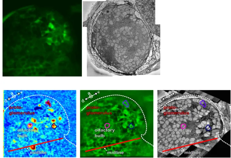
Figure 1. Once the fluorescent stack is acquired (upper left) and the SBEM stack is acquired and
stitched (upper right), the 2 modalities are registered (lower panel) in order to match the activity recorded in vivo and the ultrastructural morphology of the neurons
References:
[1] TJ Deerinck, E Bushong, A Thor, MH Ellisman, Microscopy (2010), p. 6-8
[2] A Cardona, S Saalfeld, J Schindelin, I Arganda-Carreras, S Preibisch, M Longair, P Tomancak, V Hartenstein, RJ Douglas, Plos ONE 7 (2012)
1118 Microsc. Microanal. 21 (Suppl 3), 2015
https://doi.org/10.1017/S1431927615006376