ﻲـﻣﻠﻌﻟا ثـﺣﺑﻟاو ﻲـﻟﺎﻌﻟا مــﯾﻠﻌﺗﻟا ةرازو
Ministère de l’Enseignement Supérieur et de la Recherche Scientifique
Centre Universitaire Abdelhafid BOUSSOUF de Mila
Institut des Sciences et de la Technologie Département des Sciences de la Nature et de la Vie
Mémoire préparé en vue de l’obtention du diplôme de
Master
Filière : Sciences Biologiques Spécialité : Biochimie Appliquée
Thème :
In silico characterization of Zika virus
envelope protein structure (2019)
Présentée par :-
BOULDJEDJE Sofia-
AMOURA Hamida-
LEBCIR RimaDevant le jury composé de :
Mr. BOUTALAA Saber Maitre de Conférences B Président Mr. BOUNAMOUS Azzedine Maitre de Conférences A Examinateur Mr. BOUBENDIR Abdelhafid Maitre de Conférences A Promoteur
Année Universitaire: 2019/2020
Acknowledgment
First and foremost, praises and thanks to Allah the almighty, for his
showers of blessings throughout our research work to complete the
research successfully And we do not forget the virtue, his mercy, his
Blessings are many on us, we do not count them and we do not count
praise on him and we cannot appreciate and he is glorified as he
praised himself and Glory be to him the worlds.
We would first like to thank my supervisor Mr.
BOUBENDIR
Abdelhafid for its teaching and its supervision Thanks to him, we
learned this new approach to working in silico and we started the path
of scientific research in Biology. Throughout this memory, his
relevant advice with listening, kindness and patience allowed our
work to succeed and see the light of day.
We offer our respects and thanks to the members of the jury who did
us the honor of evaluating this work Mr. BOUTALAA Saber
president of the jury and Mr. BOUNAMOUS Azzedine examiner,
who showed interest and availability for the examination of our
memory.
Many thanks to everyone who contributed to the success of this work
from far and near, whether material or moral.
Dedications
I dedicate this memory to the most loving mother in the world NADIA, little darling mom, every day you prove your love to me. My turn to give you the proof that I love you with all my heart. I promise to try to be more successful. I know that the greatest gift I can give you is
to make an effort and to love you very much, may Allah keep you for me. And to my darling father Mahfoud May God have mercy on you and make you dwell in Paradises. To my grandfather Ibrahim I may not have been born in the richest of families, but I grew up
with the sweetest of grandfathers. Thank you, grandfather, for this beautiful childhood that you gave me and for all this love that is priceless. Thank you for working hard with me for 21
years to study me and get me out of the pangs to the light. All I got today was thanks to you. And to my grandmother Yamina may god have mercy on you and make you dwell in
paradises.
I dedicate this work to my husband Bilal No words can express you my deep attachment and my gratitude for love, tenderness and kindness which you always surrounded me.Dear husband, I would like you to find in this work the expression of my feelings of gratitude the most sincere because thanks to your help and your patience with me that this work has seen
the light of day.
To all my brothers who have supported me throughout my life Djamal, Aymen, Fateh, Fares, Samir, and Mouloud, and to all my sisters Fatima, Rima,and Lamis. And to the wives of my
brothers Hayat, Yasmina, Fadila, and Tawas. To my loved cousins hassina, saida, adel, noureddine, ahmed, and bachir. To my all aunts and uncles, to my little chicks Adem, Tasnim,
and Iyad.
Special dedications for my dear sisters and friends at the same time nadia, nawal, and nourelhouda. To my girlfriends from primary to university Hadjer, Rima, Malika, Ismahane,
and Hadjer. And the great dedication with the great respect for my primary teachers Khalfa nawar and kouicem Abdelhak who taught me the real value of science and educated me on the
love of learning.
I dedicate this humble work....
✨ To my very dear mother Fatima zohra ✨
who worked for my success, with her love, her support, all the sacrifices made and her precious advice, for all her assistance and her presence in my life. May Almighty
God preserve you and grant you health, long life and happiness.
✨ To my very dear father Abdelkrim ✨
who can be proud and find here the result of long years of sacrifice and hardship to help me move forward in life. Thank you for the noble values, the education and the permanent support that came from you. Almighty God preserves you and gives you health, happiness
and long life.
✨ To my dear brother Aissa ✨
No dedication can express the love, esteem, dedication and respect that I have always had for you. I wish you a future full of success and serenity.
✨ To my sisters Aicha and Lamis ✨
Thank you, lovely sisters, for showing so much kindness and helpfulness towards me. May Allah, the Highest, grant you a happy life and a prosperous future.
✨ To all my family: uncles, aunts, cousins and cousins ✨
To all the members of my family, young and old Please find in this modest work the expression of my Affection.
✨ To my dear friends Houda and Dounia ✨
who have helped me throughout my student life.
✨ To my girlfriends at this work Rima and Sofia ✨
To those who have helped me from near or far.
To all the others that I have not mentioned but who I am also thinking of.
I dedicate this work to:
*God Almighty
my creator, my strong pillar for giving me this opportunity to do my Masters degree which
would not have been possible otherwise.
*To my source of inspiration and understanding; my loving parents whose affection, love,
encouragement ,prays and Douaa of day and night make me able to get such success and
honor.
My great beloved wonderful mother,who never stop giving of herself in countless ways, who
spread the happiness tenderness and warm. in my heart she is my teacher in life
My first and my forever hero, my precious father who leads me through the valley of
darkness with light of hope and support who stand by me when things look bleak.
He who always says : “I am here for you , I am living for you ”
*To my dearest sisters brothers Soumia Souria Bouchra Walid Badis also brother’s wife
Ilham and to your pure soul broth “OUSSAMA”
*To the sweet heart lovely princes” Nourssine” my niece.
*To the woman of Kindness and tenderness the iron woman my aunt Naima
*To the beautiful uant Noura.
And i would like to take this opportunity to say warm thanks to my sweet cousins Nassiha
Djenat Radia Ibtissam Ahlam Hayat Zahra Sana Mouna Nada Kamer Youssra Nedjela
Safa Maroua Abir Zainab and Meriem and special feeling of gratitude to Assaad a cousin
Profound gratitude to my teachers Mr.Zouaghi Mohammed ; Mrs Harrieche Ouahiba
and Mrs.Boucherit Hanane .They left a glowing imprint through my university carrer.
A deep feeling to all my friends far or near : Imane .Ikhlas never forget her help and
magnanimity.Maram.Micha Bouchra and her Her valuable advice. Aya wish to you all the
best Amel , Anfal and Khawla Saadi
*To thesis mate, Hamida and Sophia Thank you from the heart for the wonderful treatment
in the difficult circumstances I have been through.
*To the person who I can’t classify my relation with her: friend , dream mate or sister
Souad(Amina) Distance could never destroy the forever bond we have That you were never
not here, you were always here to talk to share to advice to hear me to understand and
believe in my dreams wish to you all the best God bless you.
*Thanks To the lost dream ,you opened new horizons for me.
Last but not least, deepest thanks go to all people who took part in making this
thesis real.
List of abbreviations ... I
List of figures ... III List of tables ... VI
Introduction ... 1
Part Ⅰ: Bibliographic search Chapter Ⅰ: Generalities ... 2
1. History of the Zika virus discovery... 2
2. Classification ... 3 2.1. Riboviria ...4 2.2. Flaviviridae ...4 2.3. Flavivirus ...4 3. Mechanism of infection ... 5 4. Target cells ... 5 4.1. Nerve cells ...5 4.2. Skin cells ...5 4.3. Placental cells ...6 4.4. Eye cells 7 4.5. Blood cells 7 5. Clinical presentation ... 7
5.1. Common signs and symptoms ...8
5.2. Guillain-Barré syndrome ...9
5.3. Microcephaly ...10
5.4. Other neurological complications ...11
6. Diagnosis... 11
7. Phylogeny ... 12
Chapter II: Molecular Biology ... 17
2. The viral genome ... 18
3. The viral proteins ... 20
3.1. Structural proteins ...20
3.1.1. Capsid (C) protein ... 20
3.1.2. Pre membrane (PrM) protein ... 21
3.1.3. Envelope (E) protein ... 22
3.2. Non-structural proteins ...23 3.2.1. NS1 ... 23 3.2.2. NS2A ... 25 3.2.3. NS2B ... 25 3.2.4. NS3 ... 25 3.2.5. NS4A ... 25 3.2.6. NS4B ... 26 3.2.7. NS5 ... 26 4. Replication cycle ... 27
5. Zika virus 3D protein envelope structure ... 28
5.1. 3D structure by microscopy...28
5.2. 3D structure by modeling ...30
Chapter Ⅲ: Epidemiology ... 34
1. Transmission of ZIKV ... 34
1.1. Vectoriel transmission ...34
1.2. Non vectoriel transmission ...35
1.2.1. Sexual transmission ... 35
1.2.2. Vertical transmission ... 36
1.2.3. Blood transfusion ... 36
1.2.4. Transmission by breast milk ... 36
2. Seroprevalence in Africa and Asia prior to the year 2000 ... 37
2.1. Senegal ...37
2.2. Sierra-Leon ...37
2.3. Gabon ...38
2.4. Central African Republic ...38
2.5. Egypt ...38
2.9. Malaysia ...39
3. The Yap outbreak and sporadic cases in Southeast Asia during the late 2000s to mid-2010s ... 40
3.1. Yap Island ...40
3.2. Cambodia ...40
4. The French Polynesia outbreak and spread in the Pacific Islands in the early 2010s ...40 4.1. French Polynesia...40 4.2. New Caledonia ...41 4.3. Easter Island ...41 4.4. Australia ...42 4.5. Italy ...42 4.6. Japan ...42
5. The Brazil outbreak and spread in the Americas in 2015–2016 ... 42
5.1. Brazil ...42
5.2. America ...42
Part Ⅱ: Experimental search 1. Material and Methods ... 45
1.1. Gathering of protein sequences from UniProt and alignment ...45
1.1.1. Presentation of UniProt ... 45
1.1.2. Steps of research on UniProt ... 46
1.1.3. Steps of alignment on UniProt ... 50
1.2. Phylogenetic analysis ...52
1.2.1. Presentation of MEGA 6 software ... 52
1.2.2. Aligning sequences by ClustalW ... 53
1.2.3. Estimating Evolutionary Distances Using Pairwise Distance ... 56
1.2.4. Constructing the phylogenetic tree ... 58
1.3. Protein structure homology-modelling ...59
1.3.1. Presentation of SWISS-MODEL ... 59
1.3.2. Start a new modelling project ... 60
1.3.3. Template search results and modelling ... 62
2.1. Gathering of data about ZIKV envelope protein ...65
2.2. Alignment of ZIKV envelope proteins...67
2.3. Phylogenetic analysis of ZIKV envelope proteins ...75
2.4. Three-dimensional structure of envelope protein of Senegalese strain A0A248T8F4 ...77
2.4.1. Alignment between the target Senegalese strain and the template. ... 78
2.4.2. The three-dimensional structure of envelope protein view ... 79
Conclusion ... 83 List of references
I A: Aedes
AXL: anexelekto
C: capsid
CF: complement fixation
Cryo-EM: cryo-electron microscopy
DB: dumbbell
DC-SIGN: Dendritic cell specific icam grabbing non-integrin
DENV: Dengue virus
E: envelope
ER: endoplasmic reticulum
GBS : Guillain Barré syndrom
GPI: glycosylphosphatidylinositol
HI: Hemagglutination inhibition
ICTV: International Committee on Taxonomy of Viruses
ID: identifiant
IFN: interferon
IgG: Immunoglobulin G
IgM: Immunoglobulin M
II NCR: non coding region
NS: non structural
NT: neutralization tests
ORF: open reading frame
prM: premembrane
RMSD: Root-Mean-Square Deviation
RT-PCR: Reverse transcription polymerase chain reaction
sHP-3′ SL: small hairpin 3′-stem-loop
SL: Stem loop
SPOV: spondweni virus
TAM: tyrosine 3 AXL and MER
TIM: T cell/transmembrane, immunoglobulin, and mucin
TLR3: Toll-like receptor 3
UTR: untraslated region
WNV: West Nile Virus
YF:yellow fever
YFV: yellow fever virus
III
Figure 1. A rooted phylogenetic tree based on the nucleotide sequence of complete or
near-complete genomes of all 46 available flaviviruses (Song et al., 2017). ... 3
Figure 2. Infected placental cells by ZIKV presented with red color (Tabata et al., 2016). ... 6 Figure 3. A- japan Rash case, B- Japan conjunctivitis case (Kutsuna et al., 2014). ... 9 Figure 4. Characteristic phenotype of fetal brain disruption sequence in infants with probable
congenital ZIKV syndrome: A) craniofacial disproportion and bipareital depression, B) prominent occiput (Moura da Saliva et al., 2016). ... 11
Figure 5. ZIKV nucleotide and amino acid alignments. Neighbor-joining phylogeny
generated from open reading frame nucleotide sequences of Zika virus strains. The tree was rooted with Spondweni virus (GenBank accession number DQ859064). The scale at the bottom of the tree represents genetic distance in nucleotide substitutions per site. Numbers at the nodes represent percent bootstrap support values based on 1,000 replicates. Isolates are represented according to strain name, country of origin, and year of isolation. The lineage of each virus is indicated to the right of the tree (Haddow et al., 2012)... 13
Figure 6. Maximum likelihood phylogenetic tree inferred for concatenated of sequences from
Envelope and NS5 genes of Zika virus Consensus tree summarized after 1000 non-parametric bootstrap replicates, with support values greater than 60% shown in the nodes. The cluster the Ugandan MR766 prototype strain was highlighted by the yellow sector and the Nigerian cluster was highlighted by the green sector. The strains from Senegal and Côte d'Ivoire are shown in green and orange, respectively. The tree has been rooted with the Spondweni lineage isolated in South Africa was used as outgroup to root the tree (Faye et al., 2014). .... 14
Figure 7. Phylogenetic relations between the envelope gene sequences of Suriname ZIKV
and other ZIKV (Enfissi et al., 2016). ... 16
Figure 8. Phylogenetic relationships among selected Zika virus strains belonging to the
African and Asian lineages based on complete genomic sequence (Maximum Likelihood analysis) (Charrel et al., 2016). ... 16
Figure 9. Structure of Zika virus (ZIKV) (Sirohi et Kuhn., 2017). ... 17 Figure 10. Genome structure, polyprotein processing of ZIKV (Yun et Lee., 2017). ... 18 Figure 11. Schematic representations of the Zika virus genome RNA secondary structures.
The short conserved 5′-ACAG-3′ sequences in the top loop of the sHP-3′ SL structure are indicated in red (Zhu et al., 2016). ... 19
IV
Figure 12. Zika genome. (a) A diagram of a flavivirus genomic RNA. (b) Processing strategy
and protein products. The polyprotein is processed at various sites by host (red arrow heads)
and viral (down black arrows) proteases. (Amorim L., 2018). ... 20
Figure 13. Structural proteins of Zika virus (Lin et al., 2018). ... 21
Figure 14. Overall Structure of the ZIKV-E Protein (Dai et al., 2016). ... 23
Figure 15. overall structure of protein NS1 (Song et al., 2016). ... 24
Figure 16. Replication cycle of ZIKV on the host cell (Gratton et al., 2019). ... 28
Figure 17. The structures of the ZIKV E and M proteins (Sirohi et al., 2016). ... 29
Figure 18. Three-dimensional (3D) dimer structure of ZIKV envelope protein. E protein sdimer is shown in ribbon form; E protein monomers are colored in light and dark green, and the transmembrane regions are colored in blue and purple. The N154 glycans on each monomer are labeled and shown projecting on the E protein surface (PDB: 5IRE) (Fontes Garfias et al., 2017). ... 30
Figure 19. Comparison of Zika and Dengue virion illustrations (Ekins et al., 2016). ... 31
Figure 20. Overlap of ZIKV homology models for glycoprotein E, Yellow = mature conformation (this study) compared with the immature conformation (red) (Ekins et al., 2016). ... 31
Figure 21. Represents the composition of secondary structure from amino acid residues of ZIKV envelope glycoprotein. Only 35,7% residues form sheet, 60,5% form helices and 3,8% residues form the turn region using an online server cfssp (Chou & Fasman Secondary Structure Prediction) (Alam et al., 2016). ... 33
Figure 22. (a) Modelled three‐dimenasional structure of ZIKV envelope glycoprotein; (b) Top three templates superimposed with our modelled protein with an average of RMSD = 0,481; (c) Ramachandran plot of our modelled protein showing the residues in allowed the region (Alam et al., 2016). ... 33
Figure 23. Vectoriel transmission (Petersen et al., 2016). ... 35
Figure 24. Geographic regions where ZIKV is caused epidemics (Gubler et al., 2017)... 37
Figure 25. Outbreak of ZIKV in Americas, brown color represents outbreak from January to October 2015 and dark red color represents outbreak from November 2015 to March 2016 (Petersen et al., 2016). ... 43
Figure 26. The Main MEGA Window. ... 53
Figure 27. Alignment by ClustalW. ... 55
Figure 28. The results of the alignment. ... 56
V
Figure 32. The access to SWISS-MODEL home page. ... 60
Figure 33. Start modelling. ... 60
Figure 34. Sequence(s) input. ... 61
Figure 35. Access code insertion. ... 61
Figure 36. Upload target sequence. ... 62
Figure 37. The search for template. ... 62
Figure 38. Template search running. ... 63
Figure 39. Template results. ... 63
Figure 40. Model results. ... 64
Figure 41. Amino-acids sequences alignment of 23 samples of ZIKV envelope glycoproteins from amino acid number 1 to 60.. ... 67
Figure 42. Amino-acids alignment from 61 to 120. ... 68
Figure 43. Amino-acids alignment from number 121 to 180. ... 69
Figure 44. Amino-acid alignment from 181 to 240. ... 70
Figure 45. Amino-acids alignment from number 241 to 300. ... 70
Figure 46. Amino-acids alignment from number 301 to 360. ... 71
Figure 47. Amino-acids alignment from number 361 to 420. ... 72
Figure 48. Amino-acids alignment from number 421 to 480. ... 73
Figure 49. Amino-acids alignment from number 481 to 504. ... 73
Figure 50. Phylogenetic tree of ZIKV Envelope glycoproteins from different geographical origins. Isolates are represented with access number, country of origin, and year of isolation, in which the red color indicates the African countries, blue color for Asian countries, American countries in green color, and black color for Oceanian countries. ... 75
Figure 51. Alignment result between the template 5iz7.2.1 and Senegalese strain A0A248T8F4. ... 78
Figure 52. Coverage between the template 5iz7.2.1 and Senegalese strain A0A248T8F4. ... 80
Figure 53. The three-dimensional structure of template 5iz7.2.1 E protein. ... 80
Figure 54. The predicted three-dimensional structure of E protein Senegalese strain (A0A248T8F4). ... 80
VI List of tables
Table 1. Taxonomy of Zika virus ((https://www.uniprot.org/taxonomy/64320), 2020). ... 4
Table 2. Comparison among dengue, chikungunya, and Zika symptoms (Amorim L., 2019). 8 Table 3. Countries and territories reporting Guillain-Barré syndrome (GBS) potentially associated with Zika virus infection (WHO, 2016). ... 10
Table 4. Oligonucleotide primers used for RT-PCR (Bhatnagar et al., 2017). ... 12
Table 5. Structural and non-structural proteins of ZIKA virus (Javed et al., 2017). ... 26
Table 6. Glycoprotein envelopes data gathered from UniProt. ... 65
Table 7. The length of Amino-acids sequences gathered and 3D structure. ... 66
Table 8. The whole of mutations in alignment. ... 74 Table 9. Alignment between the template (5iz7.2.1) and Senegalese strain (A0A248T8F4). 79
Introduction
1 Introduction
Zika virus (ZIKV) is an emerging virus that has been defined by the World Health Organization as a serious global biological-threat (Ramharack et al., 2016), it is a positive single-stranded RNA virus that is transmitted by mosquito bites (Ashfaq et al., 2016). A large number of serological studies in the half century since the discovery of ZIKV have revealed a broad but confined geographic distribution of human infection with the virus, across a relatively narrow equatorial belt running from Africa to Asia (Song et al., 2017).
The first isolation of zika virus was in Uganda in 1947(Faye et al., 2014) from Rhesus macaque and the first isolation from human was in Nigerian, Africa in 1954; it is originally transmitted in Africa through a sylvatic cycle involving mainly Aedes vectors and nonhuman primates, with humans being occasional hosts (Baronti et al., 2014).
This virus causes the neurodevelopmental congenital Zika syndrome and that has been linked to the neuroinflammatory Guillain–Barré syndrome. The absence of a vaccine or a clinically approved drug to treat the disease combined with the likelihood that another outbreak will occur in the future defines an unmet medical need (Bernatchez et al., 2019).
ZIKV Envelop protein is antigenic and is involved in fusion and entry of viral particles into the cell (Ashfaq et al., 2016). For this, the structural knowledge of the ZIKV proteins (exactly ZIKV Envelop protein) may allow us to understand exposed epitopes which will facilitate the development of specific diagnostic reagents that differentiate it from dengue and other flaviviruses. Furthermore, open sharing of the three-dimensional arrangement of viral surface proteins could allow the mapping of potential neutralizing epitopes, guiding efforts to rationally design effective vaccines (Ekins et al., 2016).
The objectives of the present study are:
• Gathering protein sequences in FASTA format from UniProt.
• Alignment of amino acids sequences using Clustal Omega on UniProt. • Phylogenetic analysis by MEGA6 software.
• Prediction of Zika virus 3D protein envelope structure using SWISS-MODEL online tool.
Part I
Chapter Ⅰ
Generalities
2 Chapter Ⅰ: Generalities
1. History of the Zika virus discovery
ZIKV was discovered in the Zika Forest of Uganda during research supported by the Rockefeller Foundation to study the enzootic or sylvatic cycle of yellow fever (YF) virus and to identify additional arboviruses (Weaver et al., 2016).
In brief, ZIKV was first isolated in 1947 from a febrile sentinel rhesus monkey (no. 766) held in a cage on a platform in the canopy of the Zika Forest in Uganda during studies to identify the vector of sylvatic YF. A blood sample from this monkey was collected on day 3 of fever and was inoculated intracerebrally into Swiss mice and into another rhesus monkey (no. 771). All mice showed signs of illness on day 10 post inoculation, and a filterable transmissible agent was isolated from the brains of these sick mice. Monkey 766 showed no other clinical signs or symptoms, and monkey 771 remained asymptomatic. The convalescent serum from both monkeys (766 and 771) neutralized the virus isolated from monkey 766 in mice, which was designated ZIKV 766. Preinfection sera from these monkeys did not neutralize ZIKV 766 (Gubler et al., 2017).
In January 1948, mosquitoes were collected in the Zika Forest in an attempt to isolate YFV. Eighty-six Aedes africanus mosquitoes were collected, and mice were inoculated with the Seitz filtrate of pools of these mosquitoes. One mouse died on day 6 after inoculation, and one appeared sick on day 14. The virus isolated from Ae. africanus was designated ZIKV(E/1strain). The remaining portion of the Seitz filtrate was inoculated subcutaneously into rhesus monkey no.758. This monkey remained asymptomatic, but two mice inoculated intracerebrally with blood taken from this monkey died and another became sick; ZIKV (758 strain) was isolated from its serum. Rhesus monkey no. 758 developed neutralizing antibodies to the agent isolated from its serum, to the strain of virus isolated from Ae. africanus (ZIKVE/1strain), and to the strain isolated from rhesus monkey no. 766 (the ZIKV 766 strain). Cross neutralization tests (NT) showed that ZIKV was different from YFV, DENV, and Theiler’s encephalomyelitis virus; NT with ZIKV and the antisera from other neurotropic viruses showed no relationship. Cross-reactions performed by complement fixation (CF) confirmed that ZIKV was a distinct virus (Musso et Gubler., 2016).
Part Ⅰ: bibliographic search
3
There is some controversy surrounding the first ZIKV isolate from humans. The first report was from serum of a 10 year old Nigerian female in 1954. The patient was clearly jaundiced, but interpretation of the clinical presentation was complicated by coinfection with malaria. In cross-neutralization tests with convalescent sera from monkeys infected with Bunyamwera, Bwamba, Mengo, Ntaya, Semliki Forest, Uganda S, West Nile, YF, and Zika viruses, only the serum from the monkey infected with ZIKV neutralized the virus isolated from the patient, strongly suggesting the girl was infected with ZIKV (Gubler et al., 2017).
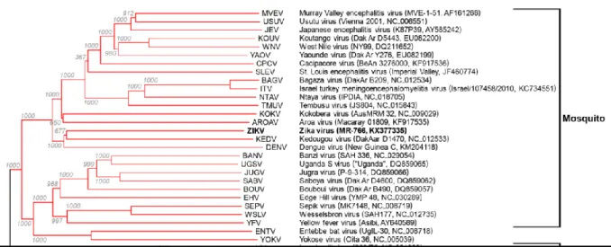
2. Classification
The virus is a member of the family Flaviviridae (Table 1), and is transmitted to humans by Aedes species mosquitoes (Wikan et Smith., 2016). It is a flavivirus and is related to other arboviruses such as YFV, Japanese Encephalitis virus, Dengue virus, and West Nile virus (Figure 1) (Shapshak et al., 2015).
Figure 1. A rooted phylogenetic tree based on the nucleotide sequence of complete or
4
Table 1. Taxonomy of Zika virus ((https://www.uniprot.org/taxonomy/64320), 2020).
Taxonomy Zika virus
Kingdom Virus Realm Riboviria
Family Flaviviridae
Genus Flavivirus
2.1. Riboviria
In February 2019 the International Committee on Taxonomy of Viruses (ICTV) has approved, by an absolute majority, the creation of the realm Riboviria, a likely monophyletic group encompassing all viruses with positive-strand, negative-strand and double-strand genomic RNA that use cognate RNA-directed RNA polymerases for replication (Walker et al., 2019).
2.2. Flaviviridae
The Flaviviridae is a family of small enveloped viruses with RNA genomes of 9000-13000 bases. Most infect mammals and birds. Many Flaviviruses are host-specific and pathogenic, such as hepatitis C virus in the genus Hepacivirus. The majority of known members in the genus Flavivirus are arthropod borne, and many are important human and veterinary pathogens (Simmonds et al., 2017).
2.3. Flavivirus
This genus consists primarily of more than 50 species of arthropod borne viruses, with distinct groups infecting mosquitoes or ticks. Mammals and birds are the usual primary hosts, in which infections range from asymptomatic to severe or fatal hemorrhagic fever or neurological disease. Important human pathogens include yellow fever virus, dengue virus, Japanese encephalitis virus, West Nile virus and tick-borne encephalitis virus. Other members cause economically important diseases in domestic or wild animals. Additional viruses
Part Ⅰ: bibliographic search
5
infecting only arthropods or only mammals have been described recently (Simmonds et al., 2017).
3. Mechanism of infection
Host-virus interactions that shape ZIKV infection remain poorly characterized. The human dermal fibroblasts, epidermal keratinocytes, and immature dendritic cells all were permissive to a ZIKV isolate from French Polynesia. TLR3 (Toll-like receptor 3) was identified as the initial immune receptor involved in the sensing of ZIKV infection in human fibroblasts leading to type I and type II interferon (IFN) responses. ZIKV also interacted with DC-SIGN (Dendritic cell specific icam grabbing non-integrin) to initiate infection of immature Mo-DCs (Monocyte and dendritic cells), whereas members of the TIM (T cell/transmembrane, immunoglobulin, and mucin) and TAM (tyrosine 3 AXL and MER) family of phosphatidylserine receptors possibly serve as receptors or attachment factors for other cells; in cutaneous fibroblasts and epidermal keratinocytes lacking expression of DC-SIGN, the TAM receptor AXL (anexelekto) facilitated ZIKV entry . The scientists examined the receptor repertoire of human radial glia cells in the fetal brain involved in ZIKV attachment and entry during neurogenesis. Distinct flavivirus entry receptor genes, including AXL receptor, were enriched in radial glia cells, astrocytes, endothelial cells, and microglia, suggesting that these cell populations may be particularly vulnerable to ZIKV infection in the developing brain (Olagnier et al., 2016).
4. Target cells
4.1. Nerve cells
Since the emergence of ZIKV virus, reports of microcephaly have increased considerably in the world. Studies of the effects of ZIKV infection in human neural stem cells growing as neurospheres and brain organoids, using immunocytochemistry and electron microscopy, showed that ZIKV targets human brain cells, reducing their viability, and neural stem cells can be infected and become apoptotic following infection. These results suggest that ZIKV abrogates neurogenesis during human brain development (Garcez et al., 2016).
4.2. Skin cells
Human skin cells are permissive for ZIKV infection and replication. Given the capacity of mosquitoes to inoculate ZIKV into the human skin during the blood-feeding process, the
6
potential target cells for infection with this virus are likely to be localized to the epidermis and dermis, which constitute the first line of defense. The skin fibroblasts, which have been recognized as a permissive target for various arboviruses, they were infected in vitro with ZIKV. The viral envelope protein was detected in several cells, 100% of the infected cells expressed ZIKV, the results show a gradual increase in the production of viral particles over time. Next, given the observation that the epidermal layer is comprised mainly of keratinocytes, these cells could also be a target for ZIKV. Dendritic cells have been reported to be permissive for ZIKV infection, and as such, they are recognized as an important target for propagation of this virus in the human skin, the capacity of ZIKV to replicate ex vivo in human skin cells was also studied (Hamel et al., 2015).
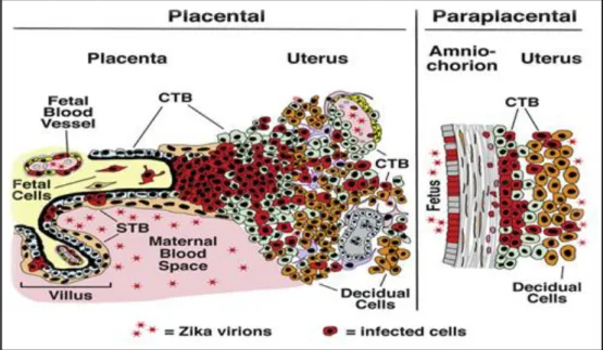
4.3. Placental cells
ZIKV infects primary human placental cells from mid and late gestation and chorionic villus explants from early gestation, these cells, along with a primary human umbilical vein endothelial cell line from umbilical cord, were infected with prototype ZIKV strain MR766 and were immunostained with monoclonal antibodies to E glycoprotein and nonstructural protein NS3, the results show that different types of primary cells from mid- and late-gestation placentas are permissive to infection with prototype and contemporary ZIKV strains. ZIKV virus infection during pregnancy is linked to severe birth defects, but mother to fetus transmission routes are unknown (Tabata et al., 2016).
Part Ⅰ: bibliographic search
7
4.4. Eye cells
Several clinical studies have observed eye malformations and pathology in neonates born to mothers infected with Zika during pregnancy. Manifestations of eye disease in newborns with zika include chorioretinal atrophy, intraretinal hemorrhages, and blindness. Viral infection in the eye can cause inflammation of uveal tissues (retina, choroid, iris, and ciliary body), also termed uveitis, which can lead to permenant vision loss if untreated. Zika causes conjunctivitis in most of infected adults. Fluid sampled from the anterior chamber of eye contained viral RNA, suggesting that Zika can replicate within the eye at some stage of clinical syndrome (Miner et al., 2016).
4.5. Blood cells
Some studies in macaques have allowed detected the presence of RNA ZIKV viral in whole blood and plasma, the animals experienced no clinical disease but developed short lived plasma viremias that cleared as neutralizing antibody developed. Despite no major histopathologic changes, many adults tissues contained RNA viral with highest levels in hemolymphatic tissues, these observations warrant further studies to investigate Zika persistence and its potential clinical implications for transmission via blood products or tissue and organ transplants (Coffey et al., 2017).
5. Clinical presentation
On 1 February 2016, the World Health Organization (WHO) declared that the recent cluster of microcephaly cases and other neurological disorders reported in the Americas, where an outbreak of Zika virus infection is ongoing, constitutes a public health emergency of international concern (Charrel et al., 2016).
ZIKV can produce a wide variety of clinical symptoms in humans. A growing body of evidence suggests that in some severe cases, ZIKV causes neurological diseases, such as Guillain-Barré syndrome in infected adults and microcephaly in infants born to ZIKV-infected women (Song et al., 2017).
ZIKV infection is not fatal. However, the first fatal case of ZIKV-associated encephalitis was reported in 2016 in a 47 year old non-pregnant woman, soon followed by the report of
8
three additional ZIKV-related fatalities with one of the patients being severely immunocompromised) (Gorshcov et al., 2019).
5.1. Common signs and symptoms
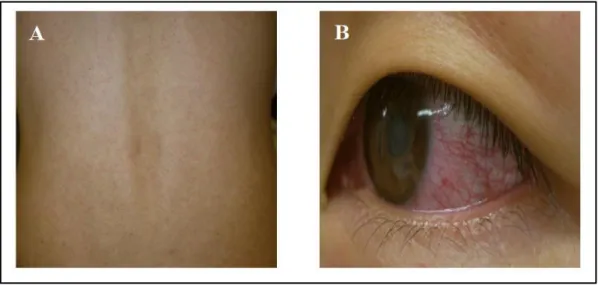
The clinical manifestations of Zika infection are very similar to those of other arboviruses such as dengue and chikungunya (Table 2) (Amorim L., 2019). ZIKV infections are symptomatic in only ~20-25% of the infected individuals who develop a mild and self-limited illness, with an incubation period of 4-10 days in symptomatic cases, the common symptoms are nonspecific and resemble those of a flu-like syndrome, including transient low-grade fever, itchy maculopapular rash (Figure 3 B), arthritis or arthralgia, and non-purulent conjunctivitis (Figure 3 A); at a lesser frequency, retro-orbital pain, headache, myalgia, edema, and vomiting are seen. Other clinical manifestations observed with acute ZIKV infection include hematospermia, hearing difficulties, thrombocytopenia, and subcutaneous bleeding. The symptoms generally appear along with the viremia and disappear spontaneously within a week, but arthralgia may persist for up to a month (Song et al., 2017).
Symptoms Dengue Chikungunya Zika
Fever ++++ +++ +++ Myalgia/arthralgia +++ ++++ ++ Edema of extremities 0 0 ++ Maculopapular rash ++ ++ +++ Retro-orbital pain ++ + ++ Conjunctivitis 0 + +++ Lymphadenopathies ++ ++ + Hepatomegaly 0 +++ 0 Leukopenia/thrombopenia +++ +++ 0 Hemorrhage + 0 0
0: absence of symptoms. (+) (++) (+++) (++++): density of symptoms
Part Ⅰ: bibliographic search
9
An additional risk of ZIKV in adults is damage to the testis. A study in mice reported the persistence of ZIKV in the testis and epididymis leading to extensive tissue damage. Male mice were reported to exhibit oligospermia, diminished testosterone. A more recent study reinforced these observations and revealed that peritubular spermatogonium cells are vulnerable to ZIKV infection. Furthermore, even an acute, uncomplicated, symptomatic ZIKV infection may result in microhematospermia in the absence of hematuria (Gorshcov et al., 2019).
Figure 3. A- japan Rash case, B- Japan conjunctivitis case (Kutsuna et al., 2014).
5.2. Guillain-Barré syndrome
Guillain-Barré syndrome is an autoimmune disease in which the immune system attacks part of the peripheral nervous system, causing tingling, muscle weakness, paralysis, and even death. Previously, this neuromuscular complication had been associated with infection by other arboviruses, such as DENV and Chikungunya virus. The temporal and geographic association of ZIKV with Guillain-Barré syndrome was initially observed during the 2013-2014 outbreak reported in French Polynesia, and subsequently during the 2015-2016 outbreak that is still ongoing in the Americas, during the previous French Polynesia outbreak, the incidence of Guillain Barré syndrome was estimated to be ~20-fold higher than its basal incidence of 1-2 cases per 100,000 population per year more definitively, a recent case-control study revealed that anti-ZIKV IgM or IgG was detected in 41 (98%) of 42 patients with Guillain-Barré syndrome, and all had neutralizing antibodies against ZIKV, as compared to 54 (55%) of 98 controls: age-, sex- and residence-matched patients with a non-febrile
10
illness. The same study also showed that patients with Guillain-Barré syndrome had electrophysiological characteristics consistent with the acute motor axonal neuropathy type of the disease. Thus far, ZIKV-induced Guillain Barré syndrome has been transient, and most patients have recovered fully. Currently, the mechanism by which ZIKV infection leads to Guillain-Barré syndrome is unknown but is under active investigation (Song et al., 2017). 18 countries and territories have reported an increased incidence of GBS and/or laboratory confirmation of a Zika virus infection among GBS cases (Table 3) (WHO, 2016).
Table 3. Countries and territories reporting Guillain-Barré syndrome (GBS) potentially
associated with Zika virus infection (WHO, 2016).
Classification Country/territory
Reported increase in incidence of GBS cases, with at least one GBS case with confirmed Zika virus infection
Brazil, Colombia, Dominican Republic, El Salvador, French Guiana, French Polynesia, Honduras, Jamaica, Martinique, Suriname, Venezuela.
No increase in GBS incidence reported, but at least one GBS case with confirmed Zika virus infection
Costa Rica, Grenada, Guadeloupe, Guatemala, Haiti, Panama, Puerto Rico.
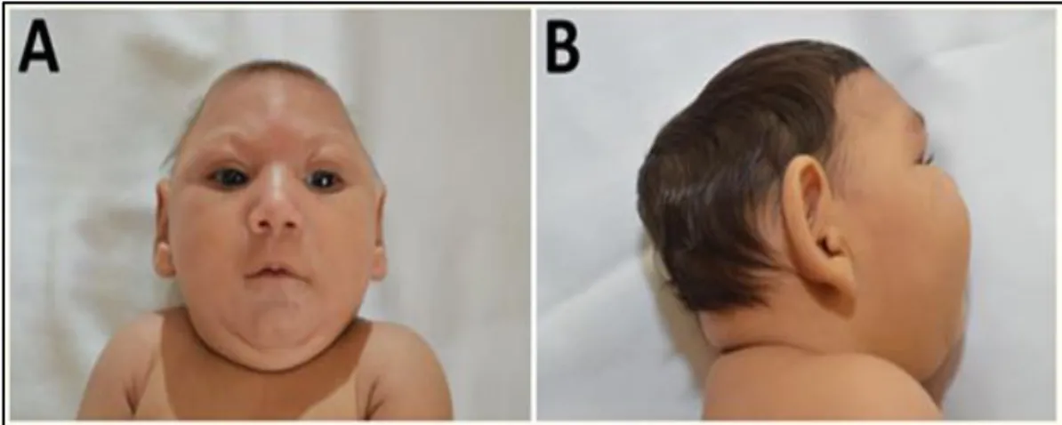
5.3. Microcephaly
Microcephaly is a neurological condition in which the brain of a baby does not develop properly, causing the head to be smaller than normal. It is divided into two types: primary or congenital microcephaly, which is present in utero or at birth; and secondary or postnatal microcephaly, which develops after birth While primary microcephaly is likely caused by a decrease in the number of neurons produced during neurogenesis, secondary microcephaly is presumably caused by a reduction in the number of dendritic processes and synaptic connections, microcephaly can be caused by a variety of genetic mutations, peri- and post-natal brain injuries, teratogenic agents, and congenital infections (Figure 4). For ZIKV, a causal relationship between prenatal infection and microcephaly emerged in Brazil, as the number of newborns with microcephaly began to rise in September 2015 (thereafter, 8,301 cases of microcephaly were recorded from November 2015 to July 2016 in that country Subsequently, the potential risk of microcephaly associated with ZIKV infection has been suggested by two retrospective studies from French Polynesia and one prospective study from
Part Ⅰ: bibliographic search
11
Brazil. In addition to this spatiotemporal association, ZIKV or its gene expression has been detected in the amniotic fluid and in various tissues of fetuses with microcephaly and those who died after birth or following abortion . Moreover, ZIKV-specific IgM has been identified in the cerebrospinal fluid and serum of neonates with microcephaly (Song et al., 2017).
Figure 4. Characteristic phenotype of fetal brain disruption sequence in infants with probable
congenital ZIKV syndrome: A) craniofacial disproportion and biparietal depression, B) prominent occiput (Moura da Saliva et al., 2016).
5.4. Other neurological complications
Case reports suggest that ZIKV infection may be associated with other neurological complications, including the case of an 81-year-old man who developed meningoencephalitis and the case of a 15-year-old girl who was diagnosed with acute myelitis research progresses, it is likely that other neurological and non-neurological complications caused by ZIKV infection will be identified in the future. Case control or cohort studies, together with well-characterized case reports, will be required in order for us to better understand the potential role of ZIKV infection in adults and newborns (Song et al., 2017).
6. Diagnosis
The diagnosis of infection by ZIKV is based on clinical, epidemiological and laboratorial criteria. Because the symptoms of ZIKV disease are nonspecific and can easily be confused with those of other arbovirus-induced diseases, such as Dengue and Chikungunya, in regions where those viruses co-circulate. Laboratorial diagnosis of ZIKV can be realized by the detection of virus, viral nucleic acid, viral antigen, or antibody or by a combination of these techniques. The choice of method depends on the purpose for which the test is performed
12
(clinical, epidemiological study, or vaccine development), the type of laboratory facilities and expertise available, and the sample collection time. When the sample is collected in the first few days after the onset of symptoms, a test detecting virus or viral nucleic acid may be performed. The virus detection is based on isolation from cell culture (using mosquito or mammalian cell lines), directly from mosquitoes, or intracerebrally from newborn mice. The low levels of viremia may explain this difficulty with the isolations. For the detection of viral RNA, molecular techniques, such as conventional or real-time RT-PCR, have been developed . These molecular techniques are the most widely used methods for ZIKV diagnosis, particularly because of the extensive antigenic cross-reactivity between flaviviruses (Zanulca et Santos., 2016). Two sets of primers that target the nonstructural 5 (NS5) and envelope (E) genes of Zika virus (Table 4) and developed RT-PCR assays for the detection of Zika virus RNA (Bhatnagar et al., 2017).
Table 4. Oligonucleotide primers used for RT-PCR (Bhatnagar et al., 2017).
Primers Sequence 5’3’ Gene
target Product size pb Annealing temperature Forward Reverse AAGTACACATACCAAAACAAAGTGGT TGTTAAGAGCGTAAGTGACAAC NS5 127 56 Forward Reverse TGCCCAACACAAGGTGAAGC ACTGACAGCATTATCCGGTACTC E 209 58 7. Phylogeny
ZIKV belongs to the Spondweni serocomplex, and phylogenetic analyses revealed the existence of two main virus lineages (African and Asian). The results suggest that a different ZIKV subtype of the West African circulated in the Aedes species in Central Africa. Molecular evolution studies indicated that ZIKV might have undergone several natural recombination events, which is an unusual feature among members of genus Flavivirus. A specific adaptive genetic change, the recurrent loss and gain of the N-linked glycosylation site in the E protein, was observed, and it has been suggested that this genetic alteration could be related to mosquito-cell infectivity. During the current epidemics in the Americas, a growing number of ZIKV genome sequences are being determined their phylogenetic relationship with other members of the Flavivirus genus revisited (Zanulca et Santos., 2016).
Part Ⅰ: bibliographic search
13
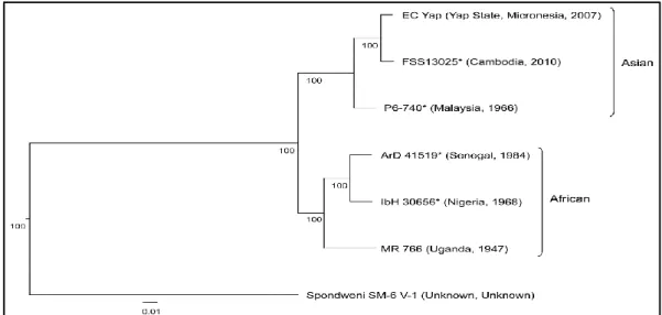
Several studies developed the phylogeny of ZIKV over the world, in this part examples of studies are provided with their corresponding phylogenetic trees. Haddow et al. (2012), reported that ZIKV is distributed throughout much of Africa and Asia. To elucidate the genetic relationships of geographically distinct ZIKV strains and the origin of the strains responsible for the 2007 outbreak on Yap Island and a 2010 Cambodian pediatric case of ZIKV infection, the nucleotide sequences of the open reading frame of five isolates from Cambodia, Malaysia, Nigeria, Uganda, and Senegal collected between 1947 and 2010 were determined. Phylogenetic analyses of these and previously published ZIKV sequences revealed the existence of two main virus lineages African and Asian (Figure 5) and that the strain responsible for the Yap epidemic and the Cambodian case most likely originated in Southeast Asia. Examination of the nucleotide and amino acid sequence alignments revealed the loss of a potential glycosylation site in some of the virus strains, which may correlate with the passage history of the virus.
Figure 5. ZIKV nucleotide and amino acid alignments. Neighbor-joining phylogeny
generated from open reading frame nucleotide sequences of Zika virus strains. The tree was rooted with Spondweni virus (GenBank accession number DQ859064). The scale at the bottom of the tree represents genetic distance in nucleotide substitutions per site. Numbers at the nodes represent percent bootstrap support values based on 1,000 replicates. Isolates are represented according to strain name, country of origin, and year of isolation. The lineage of each virus is indicated to the right of the tree (Haddow et al., 2012).
According to Faye et al. (2014), the trees for E, NS5 and the two concatenated genes (Figure 6) reinforced that ZIKV strains could be classified in three major clusters.
14
Accordingly, the African strains were arranged into two groups: the MR766 prototype strain cluster (yellow sector on Figure 6) and the Nigerian cluster (green sector on Figure 6); and the Micronesian and Malaysian strains clustered together forming the Asian clade (Figure 6), For West Africa, the strains from Ivory Coast and Senegal were found in both African clusters, suggesting that at least two distinct lineages of ZIKV circulated in these countries. Interestingly, we found that the position of the Senegalese cluster, comprising viruses isolated from 1998 to 2001 associated with Aedes dalzieli, branching as a sister group of HD78788 isolated in Senegal in 1991, was not simply explained by recombination or poor rooting of the tree, since it did not depend on the inclusion (Figure 6) or exclusion of the Spondweni, which is a bonafide outgroup.
Figure 6. Maximum likelihood phylogenetic tree inferred for concatenated of sequences from
Part Ⅰ: bibliographic search
15
bootstrap replicates, with support values greater than 60% shown in the nodes. The cluster the Ugandan MR766 prototype strain was highlighted by the yellow sector and the Nigerian cluster was highlighted by the green sector. The strains from Senegal and Côte d'Ivoire are shown in green and orange, respectively. The tree has been rooted with the Spondweni lineage isolated in South Africa was used as outgroup to root the tree (Faye et al., 2014).
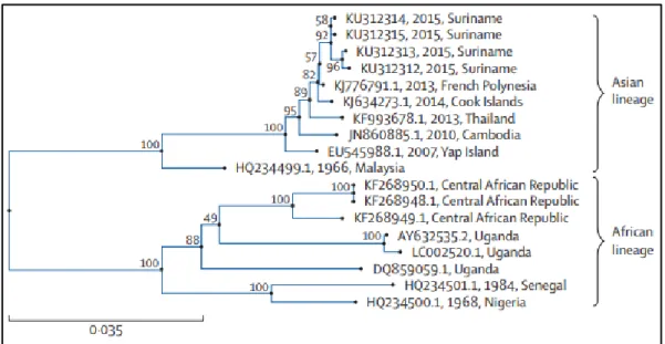
According to Enfissi et al. (2016), on October 1, 2015, a 52-year-old man was hospitalised with exanthema and conjunctivitis at the Academic Hospital in Paramaribo, Suriname. During the next few days, four patients were admitted with mild symptoms including exanthema. Sera from these patients were negative for dengue and chikungunya viruses but positive for ZIKV by specific real-time reverse transcription PCR. Soon after, the first evidence was found of the emergence of ZIKV in the Americas, in northeast Brazil in May, 2015. 4 autochthonous circulation of ZIKV in other countries started on Oct 16, 2015, in Colombia, followed by Suriname on Nov 12, 2015.The first five autochthonous cases detected in Suriname were confirmed by the French National Reference Centre for arboviruses, located at the Pasteur Institute in French Guiana. Viral sequencing was done directly from the sera of four of these viraemic patients. Complete coding of the ZIKV sequence was obtained for one patient and envelope protein coding sequences for the three others. Few complete genomes are available for ZIKV and, until this analysis, none for ZIKV circulating in the Americas. Phylogenetic analyses were conducted for the NS5 protein coding region, the envelope protein coding region, and the complete coding region, against the sequences available in databases: all the phylogenetic trees showed the same topology. The Suriname strains belong to the Asian genotype and seem to be most closely related to the strain that was circulating in French Polynesia in 2013, with which they share more than 99.7% and 99.9% of nucleotide and amino-acid identity, respectively (Figure 7).
16
Figure 7. Phylogenetic relations between the envelope gene sequences of Suriname ZIKV
and other ZIKV (Enfissi et al., 2016).
In another study conducted by Charrel et al. (2016), the phylogenetic analysis reveals the existence of two lineages the African lineage, which has shown no propensity to disseminate outside of Africa, and the Asian lineage, which continues to seed in previously unaffected regions of the world. All recently disseminated strains belong to the Asian lineage. ZIKV genomes from patients infected in Brazil and Suriname in 2015 are closely related to the strain that circulated in French Polynesia in 2013 (Figure 8), with more than 99.7% and 99.9% level of nucleotide and amino acid identities, respectively.
Figure 8. Phylogenetic relationships among selected Zika virus strains belonging to the
African and Asian lineages based on complete genomic sequence (Maximum Likelihood analysis) (Charrel et al., 2016).
Chapter II
17 Chapter II: Molecular Biology
1. The structure of the virion
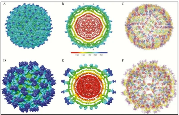
The Zika virions are 40–60 nm in diameter, spherical in shape and contain lipid envelope (Gurumayum et al., 2018). The structure of Zika virus is similar to other known flavivirus structures, except for the ~10 amino acids that surround the Asn154 glycosylation site (Sirohi et al., 2016).
Flaviviruses are enveloped viruses complexed with multiple copies of the capsid protein, surrounded by an icosahedral shell consisting of 180 copies each of the envelope (E) glycoprotein and the membrane (M) protein or precursor membrane(prM) protein, all anchored in a lipid membrane (Figure 9). During their life cycle, flavivirus virions exist in three major states— immature, mature, and fusogenic—which are non-infectious, infectious, and host membrane–binding states, respectively. The mature ZIKV structure is similar to mature DENV and WNV structures (Sirohi et al., 2016).
ZIKV particles may expand into smooth surfaced particles when incubated at higher temperatures, making the lipid envelope more fluid, and allowing the structure to revert to its normal state (Kostyuchenko et al., 2016).
Figure 9. Structure of Zika virus (ZIKV). A- C depict the mature form of ZIKV (3.8 Å
Part Ⅰ: bibliographic search
18
arrangement and Cα-backbone of the E and M proteins derived from the 3.8 Å density map of mature ZIKV (Protein Data Bank [PDB] 5IRE). Two asymmetric units related by 180° define the raft subunit of the virus consisting of 3 pairs of E and M homodimers. F shows the Cα-backbone of the DENV2 prM-E heterodimer (PDB 3C6E) and transmembrane domains of ZIKV E and M proteins (PDB 5IRE) fitted into the immature ZIKV map (PDB 5U4W). E protein is colored as follows: domain I (red), II (yellow) with fusion loop in green, III (blue) and stem-transmembrane helices (pink). pr peptide is shown in purple. The soluble region of M protein is displayed in magenta, and the stem-transmembrane helices are represented in cyan. The glycans projecting from the surface on prM and E proteins are highlighted (Sirohi et Kuhn., 2017).
2. The viral genome
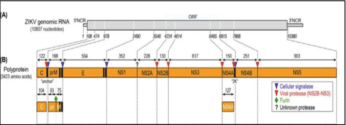
ZIKV contains a non-segmented, linear, single-stranded positive-sense ribonucleic acid (RNA) genome, typically 10,807 nucleotides in length (Figure 10.A), although some differences in length have been reported among different isolates and even among the prototype MR-766 strains with different passage histories. The genomic RNA has a type I cap structure (m7GpppAmG) at its 5’ end, followed by a 5’ non-coding region (NCR) of 106-107 nucleotides, a single open reading frame (ORF) of 10,272 nucleotides, and a 3’ NCR of 428-429 nucleotides, with no poly-A tail at the 3’ end (Figure 10.A) (Yun et Lee., 2017).
Figure 10. Genome structure, polyprotein processing of ZIKV. (A) Genome structure. The
positive-sense genomic RNA of ZIKV is composed of a 5’NCR, a single long ORF, and a 3’NCR. (B) Polyprotein processing. The viral ORF encodes a 3,423-amino-acid polyprotein, which is co- or post-translationally processed by host- and virus-encoded proteases (as indicated) into three structural (C, prM, and E) and at least seven nonstructural (NS1, NS2A, NS2B, NS3, NS4A, NS4B, and NS5) proteins. Vertical black bars represent one or two
19
transmembrane domains located at the junctions of C/prM (designated “anchor”), prM/E, E/NS1, and NS4A/NS4B (designated “2K”). Also indicated are the putative cleavage sites conserved among flaviviruses and the lengths of the cleavage products (yun et Lee., 2017).
The 3′ untranslated regions (UTR) is further divided into three domains, including the highly variable proximal domain 1 that directly follows the stop codon, the moderately conserved domain 2 that contains the stem-loop (SL) and dumbbell (DB) structures, and the highly conserved domain 3 that contains the complementary cyclization elements and the conserved sHP-3′ SL structure (Figure 11). Deletion of the SL sequences in the 5′- or 3′-UTR is lethal for flavivirus infectious clones.
A Y-shape stem-loop A (SLA) structure is found at the 5′-end of the ZIKV genome. At the 3′-end of the viral genome, a small hairpin 3′-stem-loop (sHP-3′ SL) structure, three additional SL structures, and a dumbbell (DB) structure are found (Figure 11). Notably, the external loop of the SLI in domain 1 of the 3′-UTR just distal to the stop codon of the NS5 in the 1947 prototype pre-epidemic strain is replaced by a large bulge of nine nucleotide bases (UAG UCA GCC) in the representative epidemic ZIKV strain. Short conserved sequences within the 3′ terminal SL structure include the terminal 5′-CU-3′ and a 5′-ACAG-3′ in the top loop of the sHP-3′ SL structure. There are three pairs of inverted complementary sequences (GAU CUG UG-CAC AGA UC, UGG AUU U-AAA UCC A and GAG UUU CUG GUC-GAC CAG AGA CUC and GAG UUU CUG GUC-GUC-GAC CAG AGA CUC that may mediate genome cyclization (Zhu et al., 2016).
Figure 11. Schematic representations of the Zika virus genome RNA secondary structures.
The short conserved 5′-ACAG-3′ sequences in the top loop of the sHP-3′ SL structure are indicated in red (Zhu et al., 2016).
Part Ⅰ: bibliographic search
20 3. The viral proteins
The RNA is translated into a single polyprotein (3423 amino acids in length) (Figure 10.B). This polyprotein is processed by host and viral proteases into three structural and seven non-structural proteins (Chambers et al., 2018). These structural proteins consist of capsid (C), pre-membrane (prM), and envelope (Env) proteins, which are predominantly involved in viral pathogenesis and virion structure. The seven non-structural proteins, NS1, NS2A, NS2B, NS3, NS4A, NS4B, and NS5 proteins, largely contribute towards the purposes of viral pathogenesis, replication, and immune evasion (Mohd Ropidi et al., 2020). The coding region orders and NS protein motifs of ZIKV are arranged in the order of 5′-Capsid (C)- pre-Membrane (prM)-Envelope (E)-NS1-NS2A-NS2B-NS3-NS4A-NS4B-NS5-3′ (Figure 12.b). The complete polyprotein sequences of ZIKV have low similarity with those of other human-pathogenic flaviviruses (DENV-2, 58.1% to 58.9%; SPOV, 68.3% to 69.0% nucleotide similarity) (Zhu et al., 2016).
Figure 12. Zika genome. (a) A diagram of ZIKV genomic RNA. (b) Processing strategy and
protein products. The polyprotein is processed at various sites by host (red arrow heads) and viral (down black arrows) proteases (Amorim L., 2019).
3.1. Structural proteins 3.1.1. Capsid (C) protein
Capsid (C) protein is made up of 122aa (Table 5). It is present in the cytoplasm of the infected cells (Figure 12.b, 13) while form a nucleocapsid complex with RNA in viral
21
particle. All capsid proteins of the virion have positive net charge and are similar in size but with minor sequence conservation. This protein is released into the cytosol and assembles homodimers after polyprotein cleavage. One side domain of the capsid protein contains the basic residues that bind with the RNA genome while other side domain contains hydrophobic residues that cooperate with the lipid envelope of the virus. After endosomal membrane fusion of the virus, viral genome entering remains related with the C dimers to evade RNA sensors and nucleases from the host. In addition to role in the synthesis of viral nucleocapsid, C protein functions as RNA chaperone. Thus, resulting nucleocapsid buds formation in the lumen of endoplasmic reticulum to make viral particles with E and prM proteins (Javed et al., 2017).
Figure 13. Structural proteins of Zika virus (Lin et al., 2018).
3.1.2. Pre membrane (PrM) protein
Pre membrane (PrM) protein is made up of 178aa (Table 5), which is buried under the layer of E-protein. The M and E proteins are arranged in icosahedral symmetry consisting of repeating 60 units, and each of asymmetrical unit comprise of three individual E-proteins (Javed et al., 2017). In the Golgi apparatus, PrM is cleaved by furin-like protease to produce mature M protein and Pr protein product (Figure 12.b). After maturation, PrM and E are released and 90 E protein homodimers rearrange in a herringbone-like array forming mature Zika virus (Almansour et al., 2019).
Part Ⅰ: bibliographic search
22 3.1.3. Envelope (E) protein
The E protein is the major component involved in receptor binding, membrane fusion, and host immune recognition (Shi et Gao., 2017). ZIKV E proteins have a characteristic “herringbone” structure with a single glycosylation site at residue Asp154 (Lin et al., 2018). The flavivirus E proteins belong to the class-II fusion proteins, which lie flat on the virus surface in the form of antiparallel homodimers (Shi et al., 2018). Each E protein monomer consists of three domains: DI, DII, and DIII13 (Figure 14), which undergo major rearrangements during the virus maturation cycle (Almansour et al., 2019).
DI is a central beta-barrel domain; DII is a finger- like dimerization domain; and DIII is an immunoglobulin-like domain. DI, which connects DII to DIII, is essential for the conformational changes required for viral entry into cells (Almansour et al., 2019). The central domain I contains around 130 residues in three segments, residues 1–51, 132–192, and 280–295 (Figure 14.A). The central domain I is folded into an eight-stranded β-barrel with an additional N-terminal A0 strand (Figure 14.B). The 150-loop (residues 147–161, between strands E0 and F0), which contains the potential N154 glycosylation site, likely represents a
highly flexible loop in this domain due to a lack of electron density in this region (Dai et al., 2016).
DII contains a fusion loop (FL) that interacts with the endosomal membrane, whereas DIII contains the receptor-binding site and is thus essential for attachment of virus particles to the host cell (Almansour et al., 2019). The finger-like domain II is formed by two segments, residues 52–131 and residues 193–279 (Figure 14) (Dai et al., 2016).
DIII also plays an essential role in mediating the fusion of virus particles with the endosomal membrane after endocytosis (Almansour et al., 2019). The C-terminal domain III (residues 296–403) displays an IgG-like fold where the AA’BE sheet and disordered D strand are contacted by the cd loop of the adjacent E monomer (Dai et al., 2016).
Domain II is responsible for the dimerization of E monomer, leading to an extended, but interrupted, dimer interface, and thus there are ‘‘holes’’ in the dimer at either side of domain II (Figure 14.B). The central dimer interface is mainly constituted by the aB helix and j strand elements of each sE monomer, whereas the distal dimer interface is mainly created by the hydrophobic interaction between the cd loop and the crevice formed by domains I and III of
23
the adjacents E monomer (Figure 14.B).The hydrophobic cd loop represents the fusion loop (residues 98–109), which is responsible for the membrane fusion between host cell and virus membranes during virus entry, and is highly conserved in flaviviruses (Dai et al., 2016).
Figure 14. Overall Structure of the ZIKV-E Protein. A: Schematic diagram of domain
organization for ZIKV-E. Domain I (red), domain II (yellow), and domain III (blue). A 48-residue stem segment links the stably folded ZIKV-E ectodomain with the C-terminal transmembrane anchor. B: Dimer structure of ZIKV-E. ZIKV-E has three distinct domains: b-barrel-shaped domain I, finger-like domain II, and immunoglobulin-like domain III. Domain II is responsible for the dimerization of ZIKV-E. The fusion loop is buried by the domains I and II of the other ZIKV-E monomer (Dai et al., 2016).
3.2. Non-structural proteins 3.2.1. NS1
The non-structural protein NS1 is a 46 KDa glycoprotein containing 2–3 glycosylation sites and 12 conserved cysteine residues that can form disulphide bonds. Mutations in glycosylation sites Asn130 and Asn207 drastically affect virus replication and virus production. Although the NS1protein has no hydrophobic transmembrane domain, it associates with the membrane through a glycosylphosphatidylinositol (GPI) anchor. It is
Part Ⅰ: bibliographic search
24
located inside the cell. Upon proteolytic separation from the envelope protein, it is secreted out of the cell. Cell surface expression of NS1 could elicit a strong humoral immune response, which further aids in antibody-mediated killing of virus infected cells. The NS1 and NS4 non-structural proteins interact with each other and co-localize in the viral replicase complex to help the viral genome replication (Routhu et Byrareddy., 2017).
The ZIKV NS1protein crystallized as a rod-like homodimer with a length of ~9 nm (Figure 15.a). Sedimentation velocity analytical ultracentrifugation analyses confirmed that the ZIKV NS1 protein exists as a homodimer in solution (Supplementary Figure 15).the ZIKV NS1 homodimer structure has a continuous β-sheet on one surface, with 20 β-strands arranged like the rungs of a ladder (Figure 15.a), in which each monomer contributes ten rungs to the antiparallel β-ladder. On the opposite side of the homodimer, an irregular surface is formed by a complex arrangement of loop structures (Figure 15.a). Most of those interstrand loops are short, except for a long 'spaghetti loop' between β4 and β5 (Figure 15.a, b). A potential N-linked glycosylation site that is highly conserved in the Flaviviridae family is located in the β3–β4 loop (Song et al., 2016).
Figure 15. Overall structure of protein NS1. (a) ZIKV NS1 forms a head-to-head dimer with
one extended β-ladder platform and one loop surface on the opposite side. (b) Topology diagram for NS1. Glycosylation sites are indicated with green hexagons, and disulfide bonds are indicated with yellow circles. η represents the 310 helix, and β represents the β-sheet (Song