European Heart Journal (1988) 9 {Supplement £), 19-23
Left ventricular systolic function in aortic stenosis
H. P. KRAYENBUEHL, O. M. HESS, M. RITTER, E. S. MONRAD AND H. HOPPELER
Division of Cardiology, Medical Policlinic, University Hospital, Zurich, Switzerland
KEY WORDS: Aortic stenosis, left ventricular ejection fraction, myocardial contractility, afterload
mismatch, myocardial morphology.
In aortic valve stenosis, concentric hypertrophy develops which is characterized by a reduced end-diastolic
radius-to-wall thickness ratio (r/h) with an essentially normal cavity shape. As long as the product of (rjh)
and LV systolic pressure remains constant, hypertrophy is appropriate. An increase in the product, which
represents an increase in wall stress signals inadequate L V hypertrophy. Although at first glance, massive L V
hypertrophy appears favourable for the maintenance of a normal LV ejection fraction in aortic stenosis, data
from 23 studies of the literature have shown an inverse relationship between ejection fraction and LV
angio-graphic mass m~
2(r= —0-59). Both a degree of hypertrophy inadequate to keep systolic wall stress within
normal limits and a reduction of LV contractility may explain the depression of ejection fraction when LV
angiographic mass is sizeably increased. Conversely, a normal ejection fraction in aortic stenosis may not be
indicative of normal systolic myocardial function under all circumstances. In the presence of mildly reduced
contractility, a normal ejection fraction may be maintained by the use of preload reserve. Assessment of
myocardial structure from L V endomyo car dial biopsies revealed no differences in muscle fibre diameter,
interstitialfibrosis and volume fraction ofmyofibrils between patients with aortic stenosis having a normal and
those with a depressed ejection fraction. Preoperative ejection fraction is a poor predictor of postoperative
survival, whereas markedly increased preoperative angiographic mass and end-systolic volume have been
reported to predict an unsatisfactory postoperative outcome characterized by either death or poor L Vfunction.
Ventricular geometry and its implications for systolic
function
Left ventricular (LV) chronic pressure overload
in response to aortic valve stenosis leads to marked
hypertrophy of the myocardium characterized by a
decrease of end-diastolic radius-to-wall thickness
ratio (r/h) with the cavity shape remaining
essen-tially normal. The decrease of (r/h) is the typical
feature of concentric hypertrophy. As long as the
product of (r/h) and LV systolic pressure remains
constant, hypertrophy is appropriate'
11. An increase
of the product which represents an increase in wall
stress signals inadequate LV hypertrophy. The
in-crease of wall thickness at essentially normal cavity
dimensions is of importance for the ejection
dy-namics of the left ventricle. To achieve a normal LV
ejection fraction in concentric hypertrophy, a lower
percentage of midwall fibre shortening than in a
non-hypertrophied ventricle is required because the
contribution of wall thickening to inward wall
displacement and hence reduction of cavity size is
increased'
2^*
1.
Address for correspondence. H. P Krayenbuehl, MD, Division of
Cardiology, Medical Policlinic, University Hospital, CH-8091 Zurich, Switzerland.
Systolic function, left ventricular muscle mass and
myocardial contractility
Left ventricular systolic function as assessed
from ejection fraction is within normal limits in
about two-thirds of the patients who are referred
for catheterization with a view to aortic valve
replacement. In one-third ejection fraction is
depressed despite a massive increase of LV
angio-graphic muscle mass which at first glance should be
favourable for maintaining ejection fraction within
normal limits. In 64 patients with pure aortic
sten-osis (aortic reflux absent in 22 and aortic regurgitant
fraction <0-20 in 42) but without coronary artery
disease, we found an inverse relationship between
biplane ejection fraction and left ventricular
angio-graphic muscle mass index (LMMI), (/-=-0-47,
/
)<0001). Similarly, 30 mean values of ejection
fraction and LMMI taken from 23 studies of
the literature^
12-
1*-
18-
20-
27'
29-
3'
1(Table 1), yielded a
significant inverse correlation, as depicted in Fig. 1.
Ejection fraction was also inversely correlated with
LV end-diastolic pressure (Table 1).
The reason why ejection fraction is depressed in
aortic stenosis despite massive increase of
angio-graphic mass has been a matter of debate. Gunther
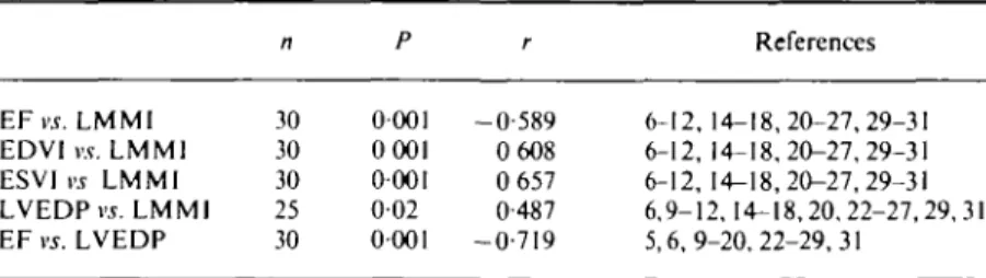
literature n P r References EF vs. LMMl EDVI r t L M M I ESVI vs LMMl LVEDP vs. LMMl EF vs. LVEDP 30 30 30 25 30 0001 0001 0001 0-02 0001 -0-589 0 608 0 657 0-487 -0-719 6-12, 14-18,20-27,29-31 6-12, 14-18,20-27,29-31 6-12, 14-18,20-27,29-31 6,9-12,14-18,20,22-27,29,31 5,6,9-20,22-29,31
EF, left ventricular ejection fraction (%); LM MI, left ventricular angiographic mass index (g m ~2); EDVI, left ventricular end-diastolic volume index (ml m ~2); ESVI, left ventricular
end-systolic volume index (ml m~2); LVEDP, left ventricular end-diastolic pressure
(mmHg): n. number of observations; P. probability (least-square regression analysis);
r, correlation coefficient.
100
200 LMMl (g rrf2)
300 400
Figure I Relationship between left ventricular ejection fraction (EF)
and left ventricular angiographic muscle mass index (LMMl) derived from published mean values. There is a significant inverse correlation. The dashed line is the calculated regression line. r = —0-59. n = 30, /><0-001
and Grossman
117' have considered excess afterload
due to inadequate hypertrophy of normally
func-tioning cardiac muscle to be at the origin of impaired
left ventricular shortening. They described an
excellent (r= —0-96) inverse relationship between
ejection fraction and mean systolic wall stress in 14
patients with aortic stenosis. However, more recent
studies have shown that in aortic stenosis at similar
peak systolic circumferential wall stress, either
normal or increased, patients with depressed
iso-volumic contractile indexes have a significantly
lower ejection fraction than do those with normal
isovolumic contractility'
251and that in the diagram
correlating ejection fraction to peak systolic or
end-systolic wall stress the values of many patients fall
down and to the left of the normal range
128'
31321.
Thus, although afterload mismatch may adversely
affect LV ejection fraction, depression of
contrac-tility associated with advanced LV hypertrophy
appears to be the major determinant of LV pump
dysfunction in aortic stenosis. Another argument
that afterload mismatch is not the main reason for
depressed ejection fraction comes from
obser-vations early after reduction or removal of LV
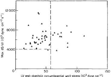
L V systolic function in aortic stenosis 21
12 0 0 0
8000
-4OO0 4 * — —
50 100 150 LV end-diastolic circumferential wall stress (I03 dyne cm"2)
Figure 2 Relationship between maximal rate of rise of left ventncular
circumferential wall stress (max dS/dt) and left ventricular end-diastolic circumferential wall stress in 44 patients with aortic stenosis (AS) and an ejection fraction (EF) ^ 5 7 % . The shaded area encompasses the values found in 23 controls. Thus the upper left quadrant defines the normal relationship between max dS/dt and end-diastolic stress In 12 patients the relationship was shifted to the right. Hence left ventncular myocardial contractility appeared to be mildly depressed in these patients. Mobiliza-tion of preload reserve allowed maintenance of ejecMobiliza-tion fracMobiliza-tion within normal limits
pressure burden. After aortic balloon valvuloplasty
in patients with ejection fraction <55% McKay et
alP
i]reported only a modest immediate increase of
ejection fraction from 40 to 46% and in patients
with aortic stenosis and depressed ejection fraction
(mean preoperative ejection fraction 33%) Schwarz
et a/.'
34' have found an increase to 43% 17 days
after valve replacement, whereas nine months after
surgery ejection fraction had finally increased to
71%.
A LV ejection fraction within the limits of ejection
fractions obtained in control subjects is generally
considered to be indicative of a normal LV
contrac-tility in patients with aortic stenosis except in
situations where afterload is below normal as in
patients with congenital aortic stenosis who show
supranormal LV shortening
1351. Moreover, in
evalu-ating the meaning of a normal ejection fraction in
aortic stenosis it has to be taken into account that
the magnitude of ejection fraction is also influenced
by preload.
To analyse further LV contractility in our 44
patients with a normal ejection fraction, we have
assessed the relationship between maximal rate of
rise of circumferential wall stress (max dS/dt) and
end-diastolic stress (S
cd)
[361. Thirteen patients had
an abnormal relationship in this isovolumic
func-tion diagram (Fig. 2) whereby in 12 preload, as
assessed by S
ed, was increased. Thus in the presence
of mildly reduced contractility, a normal ejection
fraction may be maintained by the use of preload
reserve.
Systolic function and myocardial structure
Myocardial structure was assessed from LV
endomyocardial biopsies in the same 64 patients
with aortic stenosis mentioned above. Muscle fibre
diameter and interstitial fibrosis (IF) in 44 patients
with normal ejection fraction did not differ from the
corresponding values in 20 patients with depressed
ejection fraction. LV fibrous content (FC) was
29-6g m~
2in those with normal and 37-9g m~
2(P < 0025) in those with depressed ejection fraction.
This difference was mainly due to the fact that
LMMI which enters the formula for calculation
of FC (IF x LMMI/100) was higher (/><0-001) in
those with depressed (199 g m ~
2) than in those with
patients with depressed and normal ejection
frac-tion. This observation is at variance with the results
of Schwarz et alP*
]who found VFM to be
signifi-cantly reduced in patients with aortic stenosis and
an ejection fraction < 55%.
Systolic function and prognosis after aortic valve
replacement
In patients with aortic stenosis or combined
aortic valve lesions and an ejection fraction < 50%,
more early deaths in the first month after aortic valve
replacement have been reported than in patients
with an ejection fraction ^50%'
371. However, the
difference was not significant. Subsequent
long-term survival was not affected by the preoperative
ejection fraction. Other studies"
9-
30-
32-
38^
01have
also found no relationship between preoperative
ejection fraction and long-term survival. This lack
of relationship appears to stem from the fact that
postoperative death due to myocardial dysfunction
is uncommon and most deaths are related to
pros-thetic valve complications or co-existent coronary
artery disease
138'. When surgical outcome was
assessed by both postoperative death and
inad-equate restoration of LV function, preoperative
angiographic muscle mass index'
301and end-systolic
volume index'
321were found to be preoperative
predictors of an unsatisfactory postoperative
outcome.
This work was supported by a grant from the Swiss National Science Foundation.
References
[1] Gaasch WH. Left ventricular radius to wall thickness ratio. Am J Cardiol 1979; 43: 1189-94.
[2] Dodge HT, Frimer M, Stewart DK. Functional evalu-ation of the hypertrophied heart in man. Circulevalu-ation Res
1974;35(SupplII): 122-7.
[3] Kreuzcr H, Neuhaus KL. Invasive Untersuchungs-methoden zur Erfassung einer gestorten Kontraktilitat. Verh Dtsch Ges Kreislaufforschg 1976; 42: 31-9. [4] Krayenbuehl HP, Hess OM, Hirzel HO.
Pathophysi-ology of the hypertrophied heart in man. Eur Heart J 1982;3(SupplA): 125-31
[5] Bunnell IL, Grant C, Greene DG Left ventricular func-tion derived from the pressure-volume diagram. Am J Med 1965; 39: 881-94
[6] Kennedy JW, Twiss RD, Blackmon JR, Dodge HT. Quantitative angiocardiography. III. Relationships of left ventricular pressure, volume and mass in aortic valve disease. Circulation 1968; 38: 838-45.
[7] Dodge HT, Baxley WA. Left ventricular volume and mass and their significance in heart disease. Am J Cardiol
1969; 23: 528-37.
Dodge HT. Analysis of wall dynamics and directional components of left ventricular contraction in man. Am J Cardiol 1976,38:322-31.
[9] Trenouth RS, Phelps NC, Neill WA. Determinants of left ventricular hypertrophy and oxygen supply in chronic aortic valve disease. Circulation 1976; 53: 644-50. [10] Kennedy JW, Doces J, Stewart DK. Left ventricular
function before and following aortic valve replacement. Circulation 1977; 56- 944-50.
[11] Johnson LL, Sciacca RR, Ellis K, Weiss MB,Cannon PJ. Reduced left ventricular myocardial blood flow per unit mass in aortic stenosis Circulation 1978; 57: 582-90. [12] Peterson KL, Tsuji J, Johnson A, Di Donna J, LeWinter
M. Diastolic left ventricular pressure-volume and stress-strain relations in patients with valvular aortic stenosis and left ventricular hypertrophy. Circulation
1978; 58: 77-89.
[13] Smith N, McAnulty JH, Rahimtoola SH Severe aortic stenosis with impaired left ventricular function and clinical heart failure: results of valve replacement. Circulation 1978; 58: 255-64.
[14] Pantely G, Morton M, Rahimtoola SH. Effects of suc-cessful, uncomplicated valve replacement on ventricular hypertrophy, volume and performance in aortic stenosis and incompetence. J Thorac Cardiovasc Surg 1978; 75: 383-91.
[15] Schwarz F, Flameng W, Thormann J, Ensslen M, Sesto M, Schlepper M. Cardiac reserve during isoproterenol stress in patients with aortic valve disease before and after corrective surgery. Am Heart J 1978,95: 146-53. [16] Krayenbuehl HP, Turina M, Hess OM, Rothlin ME,
Senning A Pre- and post-operative left ventricular con-tractile function in patients with aortic valve disease. Br Heart J 1979; 41: 204-13.
[17] Gunther S, Grossman W. Determinants of ventricular function in pressure-overload hypertrophy in man. Circulation 1979, 59: 679-88.
[18] Schwarz F, Flameng W, Langebartels F, Sesto M, Walter P, Schlepper M. Impaired left ventricular function in chronic aortic valve disease: survival and function after replacement by Bjork-Shiley prosthesis. Circulation 1979; 60: 48-58.
[19] Thompson R, Yacoub M, Ahmed M, Seabra-Gomes R, Rickards A, Towers M. Influence of preoperative left ventricular function on results of homograft replacement of the aortic valve for aortic stenosis AmJCardiol 1979; 43. 929-38.
[20] Fifer MA, Gunther S, Grossman W, Mirsky I, Carabello B, Barry WH. Myocardial contractile function in aortic stenosis as determined from the rate of stress develop-ment during isovolumic systole. Am J Cardiol 1979; 44:
1318-25
[21] Strauer BE. Myocardial oxygen consumption in chronic heart disease, role of wall stress, hypertrophy and coronary reserve. Am J Cardiol 1979; 44: 730-40. [22] Carabello BA, Green LH, Grossman W, Cohn LH,
Koster JK, Collins JJ. Haemodynamic determinants of prognosis of aortic stenosis and advanced congestive heart failure. Circulation 1980; 62: 42-8.
[23] SpannJF, BoveAA, Natarajan G, KreulcnT Ventricu-lar performance and compensatory mechanisms in patients with aortic stenosis. Circulation 1980; 62: 576-82.
L V systolic function in aortic stenosis 23
[24] Schwarz F, Schaper J, Kittstein D, Flameng W, Walter P, Schaper W Reduced volume fraction of myofibrils in myocardium of patients with decompensated pressure overload. Circulation 1981; 63: 1299-1304.
[25] Huber D, Grimm J, Koch R, Krayenbuehl HP. Determinants of ejection performance in aortic stenosis. Circulation 1981; 64: 126-34.
[26] Eichhom P, Grimm J, Koch R, Hess O, Carroll J, Krayenbuehl HP. Left ventricular relaxation in patients with left ventricular hypertrophy secondary to aortic valve disease. Circulation 1982; 65: 1395-404.
[27] Niemela K, Ikaheimom, Takkunen J. Functional evaluation after aortic valve replacement. Scand J Thor Cardiovasc Surg 1983; 17: 221-5.
[28] De Pace NL, Ren JF, Iskandrian AS, Kotler MN, Hakki AH, Segal BL. Correlation of echocardiographic wall stress and function in aortic stenosis. Circulation 1983; 67- 854-9.
[29] Mehmel HC, Schwarz F, Ruffmann K, Manthey J, von Olshausen K, Kubler W. End-systolic pressure-volume and end-systolic stress-volume relationships in patients with aortic stenosis and with normal valvular function. Basic Res Cardiol 1983; 78: 338-50.
[30] Saoudi NC, Berland J, Senant J, et al. Retrecissement aortique a fraction d'ejection preoperatoire basse. Arch Mai Coeur 1985; 78- 1399-1407.
[31]WisenbaughT,BoothD,deManaA,NissenS,WaltersJ. Relationship of contractile state to ejection performance in patients with chronic aortic valve disease. Circulation
1986;73:47-53.
[32] Carabello BA, Williams H, Gash AK, et al. Hemo-dynamic predictors of outcome in patients undergoing valve replacement. Circulation 1986; 74: 1309—16.
[33] McKay RJ, Safian RD, Lock JE, et al. Assessment of left ventricular and aortic valve function after aortic balloon valvuloplasty in adult patients with critical aortic stenosis. Circulation 1987; 75: 192-203.
[34] Schwarz F, Ehrmann J, Olschewski P, Scheurlen H, Saggan W, Kubler W. Patients with significant aortic incompetence should not be operated on until they are symptomatic. Z Kardiol 1986; 75 (Suppl 2): 133-6. [35] Borow KM, Colan SD, Neumann A. Altered left
ventricular mechanics in patients with valvular aortic stenosis and coarctation of the aorta: effects on systolic performance and late outcome. Circulation 1985; 72: 515-22.
[36] Krayenbuehl HP, Hess OM, Schneider J, Turina M. Physiologic or pathologic hypertrophy. Eur Heart J 1983; 4 (Suppl A): 29-34.
[37] OToole JD, Geiser EA, Reddy PS, Curtiss El, Landfair RM. Effect of preoperative ejection fraction on survival and hemodynamic improvement following aortic valve replacement. Circulation 1978; 58: 1175-84.
[38] Henry WL, Bonow RO, Borer JS, et al. Evaluation of aortic valve replacement in patients with valvular aortic stenosis. Circulation 1980; 61: 814-25.
[39] Forman R, Firth BG, Barnard MS. Prognostic signifi-cance of preoperative left ventricular ejection fraction and valve lesion in patients with aortic valve replace-ment. Am J Cardiol 1980; 45: 1120-25.
[40] Schwarz F, Ruffmann K, Olschweski M, et al. The effect of impaired left ventricular function on the prognosis after isolated aortic valve replacement. Z Kardiol 1986, 75:516-21