TRANSACTIONS OF THE ROYAL SOCIETY OF TROPICAL MEDICINE AND HYGIENE (2002) 96,521-528
Species-specific fieM%%sting of Entamoeba spp. in an area of high endemicity
F. Heckendorn’J, E. K. N’Goran2,3, I. Felger’, P. Vounatsou’, A. Yapis, A. Oettli’, H. P. Mart?, M. Dobler’, M. Traort?, K. L. Lohourignon3 and C. Lengeler’* ‘Swiss Tropical Institute, Department of Public Health and Epidemiology, I? 0. Box, CH-4002 Base& Switzerland; ‘Centre Suisse de Recherches Scientijiques, 01 B.P. 1303, Abidjan 01, C&e d’lvoire; ‘Laboratoire de Biologie Animale, URF Biosciences, Universiti de Cocody, 22 B. I? 582, Abidjan 22, C&e d’lvoire
Abstract
Entamoeba histolytica has been separated in recent years into 2 morphologically identical species: the apathogenic E. dispar and the pathogenic E. histolytica, only the latter being pathogenic. Although various laboratory techniques allow discrimination between the 2 species there is a lack of field data about the suitability of available diagnostic tests for use in epidemiological studies and few epidemiological studies using species-specific diagnosis have been performed at community level in endemic areas, especially in sub-Saharan Africa. We conducted a repeated cross-sectional study of 967 schoolchildren in central Cbte d’Ivoire to compare and evaluate light microscopy, 2 different antigen detection assays, and one polymerase chain reaction (PCR) assay. Microscopy and a non-specific antigen capture Entamoeba enzyme-linked immunosorbent assay (ELISA) were used for the primary screening of all children (time to). The prevalence of the E. histolytica/E. dispar species complex at to was 18.8% by single microscopical examination and 31.4% using the non-specific ELISA. Approximately 2 months after the initial screen- ing, fresh stool specimens were collected on 2 consecutive days (tl and t2) from (i) all the children who were positive by microscopy at to (n = 182) and (ii) 155 randomly selected children who were negative at the primary screening. These samples were tested with a second antigen detection ELISA specific for E. histolytica (n = 238) and with a species-specific PCR assay (n = 193). The second and third examinations (tl and t2) revealed an additional 43 infections with the species complex E. histolytica/E. dispar, so that the cumulative microscopical prevalence for t, and t2 was 27.7%. The overall prevalence of E. histolytica by species-specific ELISA antigen detection was low (0.83%), while the prevalence of E. dispar was 15%. When analysing only microscopically positive samples by PCR (n = 129), the ratio E. histolytica:E. dispar was very low (1:46), suggesting that the vast majority of Entamoeba infections in this area were apathogenic. Both species-specific tests performed well but the ELISA was easier to use for large-scale field screening.
Keywords: amoebiasis, Entamoeba dispar, Entamoeba histolytica, diagnosis, epidemiology, Gte d’Ivoire
Introduction
It is now generally accepted that there are 2 geneti- cally distinct but morphologically indistinguishable spe- cies of Entamoeba, E. histolytica and E. dispar (see DIAMOND & CLARK, 1993). Only E. histolytica has the potential to cause dysentery and extra-intestinal dis- ease; E. dispar is considered to be a harmless commen- Sal. The World Health Organization has reaffirmed the definition of amoebiasis as infection with E. histolytica sensu stricto, with or without clinical manifestation, and recommended that ‘E. histolytica should be specifically identified and, if present, treated; if only E. dispar is identified, treatment is unnecessary’ (ANONYMOUS,
1997).
The diagnosis of E. histolytica has traditionally relied upon microscopical examination of fresh or fixed stool specimens. A number of epidemiological studies had been performed before the clear distinction of the 2 species was established. Because they are microscopi- cally indistinguishable, the validity of studies based on stool surveys only has been questioned. The frequently quoted statistics of 40 million to 50 million people infected and up to 100 000 dying of amoebiasis world- wide are based in large part on these parasitological surveys, and this has led to an unclear picture of the epidemiology of amoebiasis (WALSH, 1986; ANONY- MOUS, 1997).
In recent years, several alternative diagnostic meth- ods have been developed for the differentiation of the 2 species: isoenzyme typing (SARGEAUNT et al., 1978, 1987), deoxyribonucleic acid (DNA) probes for use with the polymerase chain reaction (PCR) and restric- tion endonuclease pattern analysis (TANNICH & BURCHARD, 1991; ROMERO et al., 1992; KATZWIN- KEL-WLADARSCH et al., 1994; TROLL et al., 1997), and enzyme-linked immunosorbent assav (ELISA) antigen- detection systems based on mon&lonal antibo‘hies
*Author for correspondence; phone +41 61 284 8221, fax +41 61 27 1 7951, e-mail [email protected]
(ABD-ALLA et al., 1993; HAQUE et al., 1994). Cultiva- tion of amoebae followed by isoenzyme analysis is a standard method of differentiation of E. histolytica and E. dispar. However, this method is time-consuming, not widely available and essentially impractical for large-scale epidemiological studies. PCR amplification of ribosomal ribonucleic acid (rRNA) genes of Enta- moeba spp., as well as a new ELISA directed against the GaliGalNAc lectin (produced by TechLab Inc., Blacksburg, VA, USA) have been reported to approach the sensitivity and specificity of isoenzyme analysis in detecting E. histolytica directly in stool samples of symptomatic patients (HAQUE et al., 1995, 1998). However, a comparative study using cultured parasites as the source of DNA and antigen showed that the PCR was more sensitive than antigen detection by ELISA (MIRELMAN et al., 1997).
Given the current paucity of reliable data on the worldwide distribution of E. histolytica and E. dispar, it is critically important to arrive at better estimates of the burden of E. histolytica infection by using improved diagnostic tests. In sub-Saharan Africa only a few epi- demiological surveys using species identification by isoenzyme analysis have been conducted, in South Africa (JACKSON et al., 1982; GATHIRAM & JACKSON,
1985, 1987) and Ethiopia (GATTI et al., 1998). Infor- mation is crucial for the clinical and public health management of the disease, at both national and global levels. Moreover, adequate diagnostic methods and epidemiological information are urgently needed in view of the recent developments towards an amoebiasis vaccine (STANLEY, 1997; HUSTON & PETRI, 1998). Obtaining further species-specific prevalence data on amoebiasis and the development of field testing strate- gies are therefore of high priority.
Based on previous wo;k in 68te d’Ivoire revealing a high prevalence of the E. histoZvticalE. disaar comolex (&ZINGER et al., 1999), the 0‘6jectives of the present study were (i) to assess the prevalence of the complex in children in CBte d’Ivoire using a newly available
522
ELISA and repeated microscopy, (ii) to identify E. histolvtica suecificallv usinn an established PCR assav
(TROLL et al., 1997) and in antigen detection ELISA test, and (iii) to compare the diagnostic performance of these tests.
Materials and Methods
Study area and population surveyed
The study was carried out near the town of Agboville in the south of CBte d’Ivoire between March and June 2000. All schoolchildren attending standards 4-6 at 8 randomly selected primary schools in the Agboville school district were enrolled. The objectives of the study were discussed with the school directors and, after obtaining their consent, the sex and age of the children were recorded. The day before the first survey,
Initial sample n = 967 I + 1 1 st microscopical examination F. HECKENDORN ETA..
children were issued with a small plastic container and asked to return the containers with a small portion of their morning stool. After stool collection, children were issued with a new container for the following sample collection. In total 3 stool samples were col- lected, at time to at the beginning of the study, and at times t, and tz, approximately 2 months after to (Fig. 1). Identification of E. histolytica/E. dispar complex infections The stool specimens were taken to the laboratory in Abidjan within 3 h after collection. Before being further processed, each specimen was examined macroscopi- tally for consistency with special emphasis on liquid specimens and on blood visible by eye.
A total of 967 stool samples was collected at time to, and a l-2 g portion was preserved in sodium acetate-
1
9
‘Entamoeba’ antigen detection test
TIME I to r tl = to + 2 months t2 = t, + 1 day
Fig. 1. Screening procedure for 967 schoolchildren in C6te d’Ivoire for detection of the Entamoeba histolytica/E. dtipar complex and specific identification of the parasites. Numbers in circles indicate collection of fresh stool specimens.
SPECIES-SPECIFICDIAGNOSISOF ENTAMOEBASPP
acetic acid-formalin (SAF). The samples were exam- ined after processing according to MARTI & ESCHER (1990) by light microscopy within 6 weeks (Fig. 1). All samples containing E. histolyticalE. dispar and other protists and helminths were recorded semi-quantita- tively by distinguishing between 3 levels: 1+ = 1-5 parasites seen per slide; 2+ = l-2 parasites seen per microscope field; 3+ = more than 2 parasites per mi- croscope field. Microscopical examination was done by one of 3 skilled and experienced laboratory technicians. Slides containing cysts or trophozoites of E. histolyticai E. dispar were systematically checked and confirmed by a second technician.
A single Kato-Katz thick smear was also prepared from the stool samples collected at to, according to KATZ et al. (1972). The slides were examined by light microscopy and all helminth eggs were identified and recorded quantitatively.
Approximately 2 months after the first screening, 155 fresh stool samples were collected from a random selection of previously microscopically negative chil- dren on 2 consecutive days (times t, and tz), processed as described above and examined by light microscopy during the following 3 weeks. These 2 repeat examina- tions allowed the comparison of repeated microscopical examinations with the ‘Entamoeba’ ELISA. The results of examinations at to and t,lt, were not pooled for analysis because of the risk of new infections occurring during the 2 months interval between to and t, lt2. Antigen detection ELBA
A randomly selected sample of 327 specimens of the initial 967 stool samples was tested at to with the ‘Entamoeba’ antigen capture ELISA, to detect the Gal/ GalNAc lectin of both E. histolytica and E. dispar. Additionally, and for comparison with repeated micro- scopical samples, 46 of the initially microscopically negative specimens were also tested at t,/t, by the ‘Entamoeba’ ELISA. The tests were performed accord- ing to the manufacturer’s instructions (TechLab Inc.) and the optical density was measured with an auto- matic photometer at 450 nm.
Species-specific testing
Species-specific diagnosis was done with the aid of (i) a PCR assay developed by TROLL et al. (1997) and (ii) the ‘Histolytica’ ELISA antigen capture test (Tech- Lab Inc.) using stool specimens of children found to be infected with E. histolytica/E, dispar at the first micro- scopical examination (to) and samples from randomly selected microscopically negative children (Fig. 1). Due to problems in tracing children and also some- times to insufficient material in the samples, the num- bers of specimens tested by the 2 species-specific tests differed. The PCR targets rRNA genes of both E. histolytica and E. dispar in 2 separate reactions and therefore allows positive identification of both species. By contrast, the ‘Histolytica’ antigen detection ELISA is specific only for E. histolytica adhesin.
PCR assay
After initial concentration in SAF fixative the result- ing pellets were washed 3 times with distilled water, transferred to 1.5 mL Eppendorf tubes, and suspended in 250 pL digestion buffer (100 mM Tris, pH 8.0; 25 mM ethylenediaminetetraacetic acid). The samples were then frozen and transported to Switzerland for further analysis. Subsequent analysis of the samples was done accordina to TROLL et al. (1993). with the following modificaGons: (i) DNA was ou&ed after extraction using the QIAamp tissue mini-kit (Qiagen AG, Switzerland) in order to eliminate PCR inhibitors: (ii) the purified DNA was used without dilution in the PCR; (iii) deoxythymidine triphosphate was used in- stead of deoxyuridine triphosphate; (iv) restriction en- donuclease (DraI) digests were performed on only a
523
small number of E. dispar samples, in particular those showing faint bands, in order to confirm the identity of the amplified fragment.
‘Histolytica’ antigen detection test
In this assay E. histolytica is specifically identified due to differences in the Gal/GalNAc lectin between E. histolytica and E. dispar (see HAQUE et al., 1995). The tests were performed according to the manufacturer’s instructions (TechLab Inc.).
Chemotherapy
Treatment for intestinal helminth and protozoan parasites was given to all children found to be infected according to the C&e d’Ivoire national guidelines. Medication and doses used were approved by the Chief Medical Officer of the Agboville Health Department and administered by a pharmacist of the local Health Department. E. histolyticalE. dispar and Giardia duode- nalis infections were treated with 3 daily doses of metronidazole (10 mg/kg bodyweight) for 8 d. Infec- tions with Schistosoma mansoni were treated with a single dose of praziquantel (40 mg/kg bodyweight) and all other helminths (i.e. Ascarik Zumbrkoides, Trkhurik trichiuru, hookworms) with a single dose of albenda- zole.
All results of the microscopical examination and all data from the other diagnostic assays were double- entered into the ExcelT” 1997 software package. After transfer to the FoxproT” 2.6 database, frequency and consistency tests were carried out. Analysis of associa- tions and diagnostic performance calculations were carried out with the STATAT” 7.0 statistics package (Stata Corporation, College Station, Texas, USA). Results
Parasitology and non-specific ELISA
Complete results for the first microscopical examina- tion were obtained for 967 children (90% of the en- rolled children). The median age was 12 years with a range from 8 to 16 years. There were significantly more boys (n = 589) than girls (n = 378) (a = 38.8, P < 0.001). Macroscopic stool examination revealed 574 liquid/unformed specimens (59%). Only 5 children presented specimens containing macroscopically de- tectable blood, of which only one was subsequently shown to harbour E. histolvtica/E. disaar bv microsconv. There was no association between liquid/unformed specimens and infection with E. histolyticalE. dispar yclt; ratio = 1.13, 95% confidence interval (CI) 0.82-
The point prevalence of E. histoZytica/E. dispar for the single microscopical examination at tn was 18.8% (1821 967; 95% CI 16.4-21.4). Most of the children infected with E. histolvtica/E. disaar showed infection levels of
1+ (83%). The predomhant protozoan parasite was E. coli with a prevalence of 63.4%. Prevalences for all protozoa are presented in Table 1.
The combined results from the single Kato-Katz thick smear and the first microscopical examination showed that hookworms and S. mansoni were the commonest helminths (Table 1).
The non-specific ‘Entamoeba’ antigen detection ELISA was performed on a total of 327 specimens randomly selected from the initial 967 (Fig. 1). The parasitological prevalence of E. histoZytica/E. dispar in- fections within this sample based on a single micro- scopical examination was 18.7% (61/327), which is close to the prevalence of 18.8% found in the overall sample of 967 children. The ‘Entamoeba’ ELISA de- tected 103 positive cases, giving a higher prevalence rate of 31.5% (103/327; 95% CI 26.5-36.8).
Stool specimens of 155 children who were negative at to were collected again at t, . At tz, one day later, only 103 of them were present. These later examinations revealed an additional 43 infections with the E. histoly-
524 F. HECKENDORN ETAL.
Table 1. Prevalence of intestinal protozoa among 967 schoolchildren in CBte d’Ivoire screened by direct stool microscopy” and intestinal hehninths among 885 children screened by direct microscopy” and a single Kato-Katz thick smear
Intestinal parasite
No. of infected
children Prevalence ( %)b Protozoa
Entamoeba histolyticaiEntamoeba dispar Entamoeba coli 182 18.8 (16.4-21.4) 613 63.4 (60.6-66.81 Giardia duodenalis 8.0 {6.3-9.9) ’ Endolimax nana 4::: 46.2 (43.3-49.7) Chilomastix mesnili 98 10.1 (8.3-12.3) Entamoeba hartmanni 126 13.0 (11.0-15.4) Iodamoeba buetschlii 85 8.8(7.1-10.8) Blastocystis hominis 81 8.4 (6.7-10.4) Helminths Schistosoma mansoni 189 21*4(18*7-24.2) Hookworm 271 30.6(27*6-33.8) Ascark lumbricoides 78 8.8 (7.0-10.9) Trichurik trichiura 85 9.6(7.7-11.7)
BUsing sodium acetate-acetic acid-fonnalin fLvation (MARTI & ESCHER, 1993).
b95% Confidence interval in parentheses.
ticalE. dispar species complex; the cumulative preva- lence for t, and tz was 27.7%.
Species-spec$c testing
152 and 129 samples which were positive at to were tested with the specific ‘Histolytica’ ELISA and the PCR, respectively (Fig. 1). The set of samples tested by the 2 assays did not overlap completely due to problems in sample preparation. The ‘Histolytica test’ detected 3/152 E. histolytica infections (2.0%; 95% CI 0.7-5.6) and the PCR test 2/129 (1.6%; 95% CI 0.4-5.5). E. dispar was detected by the PCR in 93 of the 129 samples tested (72.1%; 95% CI 63.8-79.1). 34 of the 129 samples found to be positive by microscopy were negative by PCR (26.4%; 95% CI 19.5-34.6). Based on PCR results with the microscopically positive sam- ples, the species ratio E. histolytica:E. dispar was c. 1:46. No additional E. histolytica case was detected by the ‘Histolytica’ test or by the PCR when testing the micro- scopically negative samples.
Pe$ormance of diagnostic tests: non-specific tests
Using isoenzyme analysis as the reference test, the ‘Entamoeba’ antigen detection assay has been shown to be more sensitive (80% vs. 60%) and more specific (99% vs. 79%) than microscopy (HAQUE et al., 1995).
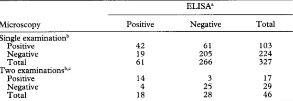
We therefore used the results of the ‘Entamoeba’ test to evaluate single and repeated microscopical examination (Table 2). Single microscopical examination of a stool sample collected at to was moderately sensitive (68.9%; 95% CI 56.4-79.1) and specific (77.1%; 95% CI 71.7-81.7). Repeated microscopical examination on 2 consecutive days at t, and t2 improved sensitivity (77.8%; 95% CI 54.8-91.0) and specificity (89.3%; 95% CI 72*8-96*3), although this was measured on a much smaller sample (n = 46 compared with n= 327), which is reflected in the much wider 95% confidence intervals. If we had taken single microscopy as refer- ence standard, the sensitivity of the ‘Entamoeba’ test would have been only 40.8% (95% CI 31.8-50.4) and the specificity would have been 91.5% (95% CI 87.1- 94.5). With repeated microscopical examination as reference at t, and t2, the sensitivity of the ‘Entamoeba’ test would have been 82.4% (95% CI 59.0-93.8) and th.e specificity would have been 86.2 % (95% CI 69*4- 94*5), again on a much smaller sample.
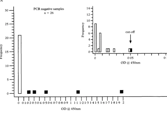
We also compared the ‘Entamoeba’ ELISA with the PCR, combining the PCR results for E. histolytica and E. dispar. In total, 101 specimens were examined by both PCR and the ‘Entamoeba’ test. PCR and the ‘Entamoeba’ test detected 75 and 53 E. histolytica/E. dispar infections, respectively (Fig. 2). The agreement
Table 2. Sensitivity and specificity of one and two consecutive microscopi- cal stool examinations for the detection of the Entamoeba histolyticalE. dispar complex compared with the non-species-specific enzyme-linked immunosorbent assay
ELISA”
Microscopy Positive Negative Total
Single examinationb Positive Negative Total Two examinationsbjc Positive Negative Total 42 61 103 19 205 224 61 266 327 14 3 1: Es ii 46
“Enzyme-linked immunosorbent assay for Emamoeba sp.
Wsing sodium acetate-acetic acid-formalin futation (MARTI L? ESCHER, 1993).
SPECIES-SPECIFIC DIAGNOSIS OF ENTAMOEBA SPP. 525 PCR negative samples n = 26 cut-off 1
mu
q
OD @ 450nm lo- 0; 5- J,,n D
, ,n
, , , , , , ,m,, , , , , , ,y 0 0.1 0.2 0.3 0.4 0.50.6 0.7 0.8 0.9 1 1.1 1.2 1.3 1.4 1.5 1.6 1.7 1.8 1.9 2 OD @ 450nm PCR positive samples n = 75 14-1 12 10 uh 8 8 ’ 6 cut-off 2 41
2 0i A
Io,d 0 II
I
I I I I I I I l I l 0 0.05 0.1 OD @ 450nm B 30 - 25 - 20 - h 8 2 15- i lo- 5- o-n
n D
I I I II I I I I I I I I I I I I I I I I
0 0.1 0.2 0.30.4 0.5 0.6 0.7 0.8 0.9 1 1.1 1.2 1.3 1.4 1.5 1.6 1.7 1.8 1.9 2 OD @ 450nmFig. 2. Optical density (OD) values for the Enrumoeba enzyme-linked immunosorbent assay (ELISA) of stool specimens tested previously by the polymerase chain reaction @‘CR): (A) 26 PCR-negative samples; (B) 75 PCR-positive samples. The small inserts show the same data for a narrower OD range and the OD positivity cut-off value of the Entamoeba ELISA (OD = 0.05); white bars indicate concordance between the PCR and ELISA results, shaded bars indicate discordance.
F. HECKENDORN ETAL. of the 2 tests in detecting positives was only 46.5%
(95% CI 36.5%-56.7%).
Pe$ornaance of diagnostic tests: species-specific tests
185 specimens were tested with both the ‘Histolytica’ antigen detection ELISA and the PCR. The microsco- pical prevalence of the E. histoZytica/E. dispar complex in this sample was 76.8% (142/185; 95% CI 70.2- 82.3). Both specific assays detected only a small num- ber of E. histolytica infections; 3 by the specific antigen detection ELISA and 2 by the PCR (Table 3). Of the 3
E. histoZytica cases detected by ELISA, the PCR result agreed in one case and identified the remaining 2 speci- mens as containing E. dispar. The remaining positive sample detected by PCR was positive for both E. histolytica and E. dispar but was found to be negative by the ‘Histolytica’ antigen detection ELISA. As a quality control measure, the samples found to be positive for
E. histolytica by either PCR (n = 2) or ‘Histolytica’ ELISA (n = 3) were re-examined by PCR by an inde- pendent investigator. The second examination con- firmed all the results. Using the PCR as reference standard, the specific antigen detection ELISA exhib- ited an overall test efficiency (proportion of true posi- tives and true negatives) of 98.4% (95% CI 95.3- 99.4).
Discussion
The high endemicity of E. histolyticalE. dispar in Cote d’Ivoire was confirmed by the present study with schoolchildren, who had an infection prevalence of 18.8% derived from a single microscopical examination and one of 31.4% with the ‘Entamoeba’ ELISA. The second and third microscopical examinations per- formed revealed a considerable number of additional infections but we did not consider these cumulatively because we could not exclude reinfections in the period of 2 months between the first and the second specimen collections. These prevalence estimates are conserva- tive because of the probability of missing cases with a single examination.
However, the difference between the prevalence rates at t, and t2 exemplifies the problem of lack of sensitivity of a single microscopical examination. It has been shown by other workers that a single examination of formalin-fixed stool fails to identifv all infections. In a laboratory-based study (MARTI & +KOELLA, 1993), ex- amination of 3 stool samples fixed in SAF resulted in 92% sensitivity when identifying E. histolyticalE. dispar;
98% sensitivity was achieved only after >4 examina- tions. Other reports have highlighted the need to exam- ine 3 or more formalin-fixed stool specimens on separate days to detect >80-90% of infections because of intermittent shedding of cysts (KNIGHT, 1974; JUNI- PER, 1978).
By using the SAF sedimentation technique we re- duced the risk of false positive diagnoses due to confu- sion of leucocytes and macrophages with amoebic cysts and trophozoites, as previously reported with examina-
tion of direct smears (KNIGHT, 1974). However, mis- identification of other protozoa as E. histolyticalE. dispar cysts may have influenced the specificity of our detection. The risk of confounding immature E. coli
cysts with those of E. histolyticalE. dispar is real, espe- cially when differential morphological features such as chromatoid bodies are absent. This issue is of particular importance since both E. colt’ and E. histolyticalE, dispar
are generally found in the same areas with poor hygie- nic conditions, and E. coli is usually the more prevalent species.
Other field-based studies in West Africa, relying on single examinations of formalin-ether concentrated stool specimens, also found high prevalences of E. histolytica/E. dispar (52.3% and 37%; BRAY & HARRIS, 1977 and UTZINGER et aZ., 1999, respectively). In contrast, community-based surveys in other endemic areas, using similar diagnostic methods, reported sub- stantially lower prevalence rates. A study in South Africa examined 800 specimens and found the preva- lence of the species complex to be only 28% (GATHIR- AM & JACKSON, 1985). In south-east Asia and in Mexico, E. histolytica/E. dispar infection rates of 12.4% and 8.1% have been reported (ACUNA-SOTO et al.,
1993; RIVERA et al., 1998). In numerous other studies, prevalences of E. histolytica/E. dispar were mostly based on examination of direct smears and’or the surveys were hospital-based rather than community-based, and therefore cannot be compared easily with our findings.
This is the first community-based study looking at soecies-snecific nrevalence of E. histolvtica and E. disaar
in sub-Saharan-Africa using diagno&c methods other than isoenzyme analysis. E. histolytica infections were assessed separately by a PCR assay and the ‘Histolytica’ ELISA antigen detection test developed by Techlab Inc. Among specimens shown microscopically to con- tain E. histolyticalE. dispar, both species-specific tests detected a surprisingly low number of E. histolytica
infections (2 by the PCR assay, including one sample with both E. histolytica and E. dispar, and 3 by the non- specific ELISA antigen detection test). As expected, PCR identified most of the remaining samples as E. dispar, leading to a low species ratio of E. histolytica to
E. dispar of 1:46.
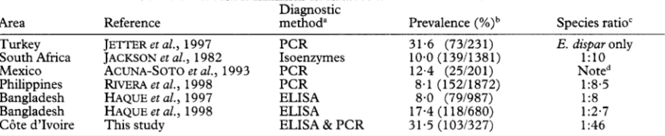
It could be argued that by testing only the subjects found to be infected at the first microscopical examina- tion, the prevalence of infection might not be properly reflected. However, given the fact that no further E. histolytica case was discovered in a sample of 86 micro- scopically negative samples tested by ELISA and 64 microscopically negative samples tested by PCR, we are reasonably confident that our ratio of 1:46 is accu- rate. This finding contrasts with most other surveys using the approach of pre-selecting E. histoZytica/E. dispar infections by a non-specific test followed by the differentiation of the species, which found considerably lower proportions of E. dispar (Table 4).
On the basis of the small number of community- based studies which involved species-specific diagnosis,
Table 3. Comparison of species-specific Entamoeba histolytica antigen detection enzyme-linked immunosorbent assay and the polymerase chain reaction among 185 infected schoolchildren in C6te d’Ivolre
Polymerase chain reaction Positive Negative Species-specific ELISA”
Positive 1 2
Negative lb 181
Total 2 183
“Enzyme-linked immunosorbent assay.
bSpecimen positive for both Entamoeba histolytica and E. dispar.
Total 3 182 185
SPECIES-SPECIFIC DIAGNOSIS OF ENTAMOEBA SPP. 527
Table 4. Reported prevalence rates and ratios for Entamoeba histolytica and E. dispar in amoebiasis endemic areas
Area Reference
Turkey JETTER et al., 1997
South Africa JACKSON et al., 1982
Mexico ACUNA-SOTO et al., 1993 Philippines RIVERA et al., 1998 Bangladesh HAQUE et al., 1997
Bangladesh HAQUE et al., 1998 Cote d’Ivoire This study
Diagnostic method” PCR Isoenzymes PCR PCR ELISA ELISA ELISA & PCR Prevalence (%)b 31.6 (73/231) 10.0 (13911381) 12.4 (25/201) 8.1 (152/1872) 8.0 (79/987) 17.4 (1181680) 31.5 (103/327) Species ratio’ E. dispar only 1:lO Noted 1:8.5 1:8 1:2*7 I:46 “ELISA = enzyme-linked immunosorbent assay; PCR = polymerase chain reaction.
bE. histolyticalE. dispar complex. ‘E. histolytiia:E. dispar.
d56% of all individuals infected harboured mixed infections with E. histolytica and E. dispar.
it appears that the species ratio E. histoZytica:E. dispar
varies considerablv. Whether this observation reflects small-scale rather -than large-scale geographical differ- ences in the distribution of E. histolytica cannot be answered at this time. In order to increase the epi- demiological understanding of amoebiasis, this issue needs urgently to be further investigated.
The ‘Entamoeba’ test and the PCR assay showed only limited agreement in detecting infections (46.5%). PCR detected a substantially higher number of E. histoZytica/E. dispar infections than the ‘Entamoeba test’. Possibly the additional cases detected by PCR reflected the low sensitivity of a single examination using the other test. However, technical problems with the PCR technique cannot simply be excluded. This technique is potentially prone to cross-contamination due to its power to amplify minute traces of target DNA. To test for possible contamination, our samples were retested twice by PCR by an independent investi- gator using additional controls, but we could not find a problem with the assay. However, contamination can occur between the field and the laboratory during sample handling or DNA preparation and this cannot be traced later.
When the PCR assay was taken as the standard, the ‘Histolytica’ ELISA antigen detection test showed high efficiency (98.4%). However, the 2 tests disagreed with 3 samples which were positive for E. histolytica by either PCR or ELISA (Table 3). In contrast, another study also using the TechLab specific ELISA and a PCR assay for the diagnosis of E. histolytica in stool samples, found a correlation of 84% between the 2 tests (HAQUE et al., 1998). Our findings concerning E. histolytica
infections are difficult to interpret because of the small number of infections. Despite a rather large initial sample size we failed to detect many such infections because of the unexpectedly low E. histoZyticalE. dispar
ratio.
In conclusion, this study produced valuable data in relation to the occurrence of E. disaar and E. histolvtica
in communities in sub-Saharan At&a. While the pre- valence of the species complex was high, the prevalence of E. histolytica was low. This might explain the paradox of the low amount of amoebiasis morbidity reported by health services on the African continent despite a high apparent microscopical infection rate. Field testing with the new generation of ELISA kits proved to be clearly superior to microscopy. Because of the small number of E. histolytica cases in our sample the in- tended comparison of test performance between the ‘Histolytica’ ELISA and the PCR did not result is a clear conclusion about which test is superior. However, unlike the PCR, the ELISA proved to be easy to use in the field and, if its high diagnostic performance is confirmed, it might well become the standard tech- nique for large-scale field screening.
Acknowledgements
The present work was mainly supported by funds from the Swiss Tropical Institute in Basel. Additional support was provided by TechLab Inc. through supplying free kits for
ELISA and PCR testing. The support of the Centre Suisse de
Recherche Scientifiaue en CBte d’Ivoire. and esneciallv its Director Dr Olivier Girardin, is gratefully acknowlehged. Spe- cial thanks also go to the Agboville school district officer and all the teachers and pupils who participated in the study. The contribution of Mr Silue Kouna, who helped in the laboratory, is gratefully acknowledged.
References
Abd-Alla, M. D., Jackson, T. F., Gathiram, V., El Hawey, A. M. & Ravdin, 1. I. (1993). Differentiation of nathoeenic Entamoeba h&o&a infections from non-pathogenic infec- tions by detection of galactose-inhibitable adherence protein antigen in sera and faeces. Journal of Clinical Microbiology, 31,2845-2850.
Acuna-Soto, R., Samuelson, J., De Girolami, P., Zarate, L., Millan-Velasco, F., Schoolnick, G. & Wirth, D. (1993). Application of the polymerase chain reaction to the epidemi-
ology of pathogenic and non-pathogenic Entamoeba histolyti- ca. American Journal of Tropical Medicine and Hygiene, 48, 58-70.
Anonymous (1997). A consultation with experts on amoebia- sis. Mexico City, Mexico, 28-29 January. WHOIPAHOI UNESCO report. Epidemiological Bulletin, 18, 13- 14. Bray, R. S. & Harris, W. G. (1977). The epidemiology of
infection with Entamoeba histolytica in The Gambia, West Africa. Transactions of the Royal Society of Tropical Medicine and Hygiene, 71,401-407.
Diamond, L. S. & Clark, C. G. (1993). A redescription of Entamoeba histolytica Schaudinn, 1903 (emended Walker,
1911) separating it from Entamoeba dispar Brumpt, 1925. Journal of Eukayotic Microbiology, 40, 340-344.
Gathiram, V. & Jackson, T. F. (1985). Frequency distribution of Entamoeba histolytica zymodemes in a rural South African population. Lancet, i, 719-721.
Gathiram, V. & Jackson, T. F. (1987). A longitudinal study of asymptomatic carriers of pathogenic zymodemes of Entamoeba histolytica. South African Medical Townal. 72.
669-672. - I _
Gani, 8, Mahdi, R., Bruno, A., Cevini, C. & Scaglia, M.
(1998). A survey of amoebic infection in the Wonii area of central Ethiopia. Annals of Tropical Medicine and i>arasitol- OFV. 92.173-179.
Ha&e, R., Neville, L. M., Wood, S. & Petri, W. A., jr (1994).
Detection of Entamoeba histolyrica and Entamoeba disaar directly in stool. American Jo&al of Tropical Medicine and HvPiene. 50.595-596.
Ha<&, R, Neville, L. M., Hahn, I’. & Petri, W. A., jr (1995). Rapid diagnosis of Entamoeba infection bv using Entamoeba and Entamoeba hiswlytica stool antigen detection kits. Jour- nal of Clinical Microbiology, 33, 2558-2561.
Haque, R., Faruque, A. S. & Petri, W. A. (1997). Entamoeba histolytica and Entamoeba dtipar infection in children in Bangladesh. Archives of Medical Research, 28, special num- ber, 317-318.
Haque, R., Ali, I. K., Anther, S. & Petri, W. A., jr (1998). Comparison of PCR, isoenzyme analysis, and antigen detec- tion for diagnosis of Entamoeba histolytica infection. Journal of Clinical Microbiology, 36, 449-452.
528 F. HECKENDORN ETAL.
Huston, C. D. & Petri, W. A., jr (1998). Host-pathogen interaction in amoebiasis and progress in vaccine develop- ment. European Journal of Clinical Microbiology and Infectious Diseases, 17,601-614.
Tackson. T. F.. Sareeaunt, P. G.. Williams, 1. E. & Simiee. A. E. (1982). dbse&ationi on &nodeme &dies of Entamdeba histolytica in Durban, South Africa. Archives of Investigational
Medicine (Mexico), 13, supplement 3, 83-88.
Jetter, A., Walderich, B., Britten, D., Mete, O., Goral, V., Burchard, G. D. & Ackers, J. (1997). An epidemiological study of Entamoeba histolytica and Entamoeba dispar infection in eastern Turkey using a calorimetric polymerase chain reaction. Archives of Medical Research, 28, special number, 319-321.
Juy~;; K. (1978). Amoebiasis. Clinical Gastroenterology, 7, Katz, N., Chaves, A. & Pellegrino, J. (1972). A simple device
for quantitative stool thick-smear technique in schistosomia- sis mansoni. Revista do Instiruto de Medicina Tropical de SZo Paulo, 14,397-400.
Katzwinkel-Wladarsch. S.. Loscher, T. & Rinder, H. (1994). Direct amplification- anh differentiation of pathogenic aid non-pathogenic Entamoeba histolytica DNA from stool speci- mens. American Journal of Tropical Medicine and Hygiene, 51,
115-118.
Knight, R. (1974). Amoebiasis. Tropical Doctor, 4, 6- 11. Marti, H. P. & Escher, E. (1990). SAF-an alternative fixation
solution for parasitological stool specimens. Schweizeriiche Medizinische Wochenschrift, 120, 1473-1476.
Marti, H. & Koella, J. C. (1993). Multiple stool examinations for ova and parasites and rate of false-negative results. Journal of Clinical Microbiology, 31, 3044-3045.
Mirelman, D., Nuchamowitz, Y. & Stolarsky, T. (1997). Comparison of use of enzyme-linked immunosorbent assay- based kits and PCR amplification of rRNA genes for simul- taneous detection of Entamoeba histolytica and Entamoeba dispar. Journal of Clinical Microbiology, 35, 2405-2407. Rivera, W. L., Tachibana, H. & Kanbara, K. (1998). Applica-
tion of the polymerase chain reaction (PCR) in the epidemi-
ology of Emamoeba histolytica and Emamoeba dispar infections. Tokai Journal of Experimental and Clinical Medi- cine, 23,413-415.
Romero, J. L., Descoteaux, S., Reed, S., Orozco, E., Santos, J. & Samuelson, J. (1992). Use of polymerase chain reaction and nonradioactive DNA probes to diagnose Entamoeba histolytica in clinical samples. Archives of Medical Research, 32,277-279.
Sargeaunt, P. G., Williams, J. E. & Grene, J. D. (1978). The differentiation of invasive and non-invasive Entamoeba histo- lytica by isoenzyme electrophoresis. Transactions of the Royal Socie y of Tropical Medicine and Hygiene, 72, 5 19-52 1.
Sargeaunt, P. G., Jackson, T. F., Wiffen, S., Bhojnani, R.,
Williams, J. E., Felmingham, D., Goldmeir, D., Allason- Jones, E., Mindel, A. & Phillips, E. (1987). The reliability of Entamoeba histolytica zymodemes in clinical laboratory
diagnosis. Archives of Investigational Medicine (Mexico), 18, 69-75.
Stanley, S. L. (1997). Progress towards development of a vaccine for amebiasis. Clinical Microbiological Reviews, 10, 637-649.
Tannich, E. & Burchard, G. D. (1991). Differentiation of pathogenic from non-pathogenic Entamoeba histolytica by restriction fragment analysis of a single gene amplified in
vitro. Journal of Clinical Microbiology, 29, 250-255.
Troll, H., Marti, H. P. & Weiss, N. (1997). Simple differential detection of Entamoeba histolytica and Entamoeba dispar in
fresh stool specimens by sodium acetate-acetic acid-forma-
lin concentration and PCR. Journal of Clinical Microbiology, 35,1701-1705.
Utzinger, J., N’Goran, E. K., Marti, H. P, Tanner, M. & Lengeler, C. (1999). Intestinal amoebiasis, giardiasis and geohelminthiases: their association with other intestinal parasites and reported intestinal symptoms. Transactions
of the Royal Society of Tropical Medicine and Hygiene, 93, 137-141.
Walsh, J. A. (1986). Problems in recognition and diagnosis of amebiasis: estimation of the global magnitude of morbidity and mortality. Reviews of Infectious Diseases, 8, 228-238.
Received 14 November 2001; accepted for publication 1 March 2002
/ Announcement
1
ROYAL SOCIETY OF TROPICAL MEDICINE AND HYGIENE Garnham Fellowships
Professor Cyril Garnham was one of the UK’s leading parasitologists in the 20th century and his work was characterized by outstanding achievement as both laboratory scientist and field worker in the tropics. The special place that Garnham occupies among his colleagues is recognized by the Fund set up in his memory to establish research fellowships for young scientists.
The aim of the Garnham Fellowship is to encourage young scientists to carry out short-term field projects. Suitable applicants are invited to apply to the Fund, which is administered by the Royal Society of Tropical Medicine and Hygiene.
There are no restrictions by nationality or age, and fellowship of the Royal Society of Tropical Medicine and Hygiene is not a requirement. Applications from non-Fellows should be supported by a Fellow who can attest to the value of the project and to the competence of the applicant to carry out the work.
l One Garnham Fellowship of up to E2000 will be awarded annually
l The Garnham Fellowship is to be used for short-term field projects of up to 2 years’ duration
l Preference will be given to topics in parasitology or medical entomology and to applicants with less than 5
years’ postdoctoral experience
l Applicants are required to submit a detailed project, with costing of the work proposed, and a supporting
statement from their head of department or supervisor, at least 6 months before the date of commencement
l A short report should be submitted within 3 months of completion of the study
Application forms may be obtained from the Administrator, Royal Society of Tropical Medicine and Hygiene, Manson House, 26 Portland Place, London, WlB lEY, UK; fax +44 (0)20 7436 1389, e-mail [email protected]