Cyclic Peptide Mimetic of Damaged Collagen
The MIT Faculty has made this article openly available.
Please share
how this access benefits you. Your story matters.
Citation
Aubrey J. Ellison, I. Caglar Tanrikulu, Jesús M. Dones, and
Ronald T. Raines. "Cyclic Peptide Mimetic of Damaged Collagen."
Biomacromolecules 2020 21 (4), 1539-1547. DOI: 10.1021/
acs.biomac.0c00103.
As Published
10.1021/acs.biomac.0c00103
Publisher
American Chemical Society (ACS)
Version
Final published version
Citable link
https://hdl.handle.net/1721.1/125525
Terms of Use
Creative Commons Attribution 4.0 International license
Cyclic Peptide Mimetic of Damaged Collagen
Aubrey J. Ellison, I. Caglar Tanrikulu, Jesús M. Dones, and Ronald T. Raines*
Cite This:Biomacromolecules 2020, 21, 1539−1547 Read Online
ACCESS
Metrics & More Article Recommendations*
sı Supporting InformationABSTRACT:
Collagen is the most abundant protein in humans and
the major component of human skin. Collagen mimetic peptides
(CMPs) can anneal to damaged collagen in vitro and in vivo. A duplex
of CMPs was envisioned as a macromolecular mimic for damaged
collagen. The duplex was synthesized on a solid support from the
amino groups of a lysine residue and by using ole
fin metathesis to link
the N termini. The resulting cyclic peptide, which is a monomer in
solution, binds to CMPs to form a triple helix. Among these, CMPs
that are engineered to avoid the formation of homotrimers but
preorganized to adopt the conformation of a collagen strand exhibit
enhanced association. Thus, this cyclic peptide enables the assessment
of CMPs for utility in annealing to damaged collagen. Such CMPs have
potential use in the diagnosis and treatment of
fibrotic diseases and
wounds.
■
INTRODUCTION
The collagen triple helix is the most abundant structure
adopted by a biopolymer within the human body. There,
collagen comprises one-third of the total protein, accounts for
three-fourths of the dry weight of skin, and is the most
prevalent component of the extracellular matrix.
1Collagen is damaged in
fibrotic and other diseases and in
wounds. Collagen mimetic peptides (CMPs) can anneal to
damaged collagen.
2Such annealing could allow for the delivery
of diagnostic or therapeutic agents that are conjugated to the
CMP. Indeed, we have used such CMP conjugates to anneal
fluorescent dyes,
3a growth factor,
4and even a sunscreen
5to
natural collagen. Damaged collagen in a human body is,
however, a complex target that confounds physicochemical
analyses, complicating the assessment of therapeutic potential
for new CMP designs.
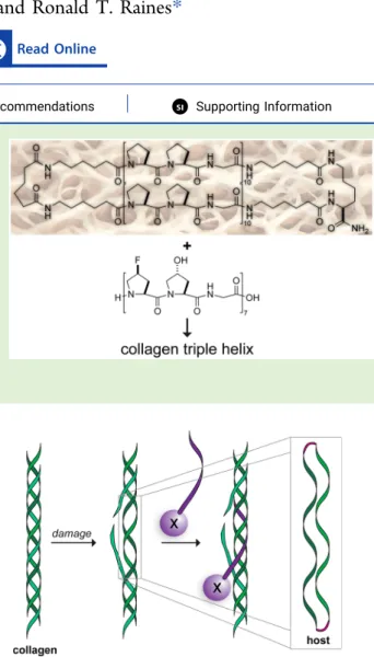
We sought to develop a molecular mimic of damaged
collagen. We envisioned doing so with a collagen
“duplex”, that
is, two cross-linked CMPs. Several double-stranded duplexes
have been synthesized and used to form collagen triple
helices.
6In most of these precedents, one or both ends of the
peptides are free, allowing for the assembly of many possible
complexes. To make a duplex more amenable to rigorous
analyses and more biomimetic, we sought to tether two parallel
strands at both termini (
Figure 1
). This design not only
minimizes the conformational entropy of the duplex, but also
mimics natural collagen
fibers that have a disrupted triple helix,
as might be found in damaged collagen. Here, we describe the
creation of a
“host” and report on its interaction with “guest”
strands, that is, CMPs.
■
EXPERIMENTAL METHODS
General. Boc-flp−OH and Fmoc-flp−OH were from Omega-Chem (Lévis, Québec). (Here, “flp” refers to (2S,4S)-4-fluoropro-line.) Boc-Flp−OH, Fmoc-Hyp−OH, and other amino acid derivatives, resins, Fmoc-OSu, and HOBt were from Chem-Impex
Received: January 21, 2020
Revised: March 10, 2020
Published: March 11, 2020
Figure 1. Scheme showing the molecular mimicry of damaged collagen by a cyclic“host” of two parallel collagen strands. A collagen mimetic peptide with a conjugated pendant“X” (purple) is shown annealing to the damaged collagen.
Article pubs.acs.org/Biomac
License, which permits unrestricted use, distribution and reproduction in any medium, provided the author and source are cited.
International (Wood Dale, IL). (Here, “Flp” refers to (2S,4R)-4-fluoroproline and “Hyp” refers to (2S,4R)-4-hydroxyproline.) DIC and 4-methylpiperidine were from Oakwood Chemical (Estill, SC). Streptavidin-coated fluorescent blue particles were product SVFP-106805 from Spherotech (Lake Forest, IL). 6-Aminohexanoic acid and all other reagents were from Sigma−Aldrich (St. Louis, MO) and were used without further purification.
DMF was dried with a Glass Contour system from Pure Process Technology (Nashua, NH). In addition, DMF was passed through an associated isocyanate “scrubbing” column to remove any amines. Water was purified with an Arium Pro system from Sartorius (Göttingen, Germany).
The phrase “concentrated under reduced pressure” refers to the removal of solvents and other volatile materials with a rotary evaporator at water-aspirator pressure (<20 Torr) while maintaining a water bath below 40°C. Residual solvent was removed from samples with a high vacuum (<0.1 Torr).
All procedures were performed in air at ambient temperature (∼22 °C) and pressure (1.0 atm) unless indicated otherwise.
Instrumentation. Solid-phase peptide synthesis was performed with a Liberty Blue Peptide Synthesizer from CEM (Matthews, NC). Synthetic peptides were purified by HPLC with a Prominence instrument from Shimadzu (Kyoto, Japan) equipped with a VarioPrep 250/21 C18 column from Macherey−Nagel (Düren, Germany). Molecular mass was determined by matrix-assisted laser desorption/ ionization−time-of-flight (MALDI−TOF) mass spectrometry on an α-cyano-4-hydroxycinnamic acid or sinapic acid matrix with a microflex LRF instrument from Bruker (Billerica, MA). Purity analyses were performed with an Acquity UPLC H-Class system from Waters that was equipped with an Acquity photodiode array detector, Acquity quaternary solvent manager, Acquity sample manager with a flow-through needle, Acquity UPLC BEH C18 column (2.1 × 50 mm, 1.7 μm particle size), and Empower 3 software.1H and13C NMR spectra were acquired with an Avance III
400 spectrometer from Bruker. Sedimentation equilibrium experi-ments were performed with an XL-A analytical ultracentrifuge and An-60 Ti rotor from Beckman Coulter (Brea, CA) at the Biophysics Instrumentation Facility of the University of Wisconsin−Madison (UW BIF). Beads were imaged using a Eclipse Ti inverted confocal microscope from Nikon (Melville, NY) at the Biochemistry Optical Core of the University of Wisconsin−Madison. Flow cytometry was performed with an Accuri Flow Cytometer with C-Sampler from BD (San Jose, CA) at the UW BIF. CD data were acquired with a Model 420 CD spectrophotometer from Aviv Biomedical (Lakewood, NJ) at the UW BIF.
Small-Molecule Synthesis. Fmoc-aminohexanoic Acid. 6-Aminohexanoic acid (1.00 g, 7.62 mmol) was dissolved in a saturated aqueous solution of NaHCO3(50 mL). In a separateflask, Fmoc-OSu
(2.82 g, 8.38 mmol) was dissolved in dioxane (50 mL). The two solutions were combined, and the reaction mixture became cloudy and was stirred for 16 h. The mixture was then concentrated under reduced pressure. The residue was dissolved in EtOAc and washed with aqueous 1.0 M HCl and brine. The organic layer was dried over Na2SO4(s), decanted, and concentrated under reduced pressure.
Crude product was purified by chromatography on silica-gel, eluting with EtOAc (40% v/v) and acetic acid (1% v/v) in hexanes to yield Fmoc-6-aminohexanoic acid (2.56 g, 95%) as a white solid. HRMS− ESI (m/z): [M + H]+calcd, 354.17; found, 354.17.1H NMR (400
MHz, MeOD,δ): 7.78 (d, J = 7.5 Hz, 2H), 7.63 (d, J = 7.5 Hz, 2H), 7.37 (t, J = 7.4 Hz, 2H), 7.33−7.26 (m, 2H), 4.33 (d, J = 6.8 Hz, 2H), 4.18 (t, J = 6.9 Hz, 1H), 3.08 (t, J = 7.0 Hz, 2H), 2.27 (t, J = 7.4 Hz, 2H), 1.60 (p, J = 7.5 Hz, 2H), 1.49 (p, J = 7.1 Hz, 2H), 1.33 (p, J = 10.1, 6.0 Hz, 2H).13C NMR (101 MHz, MeOD,δ): 157.49, 143.95, 141.20, 127.34, 126.71, 124.75, 119.50, 66.11, 47.13, 40.17, 33.71, 29.18, 25.97, 24.45.
Boc-Flp-OBn. Boc-Flp−OH (2.5 g, 10.7 mmol) was dissolved in DMF. Solid Cs2CO3(1.5 g, 10.7 mmol) was added, and the reaction
mixture was stirred for 10 min. Benzyl bromide (1.27 mL, 10.7 mmol) was added dropwise, and the mixture was stirred for 16 h. The mixture was then concentrated under reduced pressure. Crude
product was purified by chromatography on silica gel, eluting with EtOAc (10% v/v) in hexanes to yield product (2.26 g, 65%). HRMS− ESI (m/z): [M + H]+calcd, 324.15; found, 324.16.1H NMR (400
MHz, CDCl3,δ): 7.35 (d, J = 4.5 Hz, 5H), 5.32−5.19 (m, 1H), 5.19−
5.14 (m, 1H), 5.14−5.04 (m, 1H), 4.58−4.38 (m, 1H), 3.99−3.75 (m, 1H), 3.61 (ddt, J = 36.1, 13.0, 3.9 Hz, 1H), 2.70−2.47 (m, 1H), 2.20−1.97 (m, 1H), 1.53−1.31 (m, 9H), which are consistent with literature values for this known compound.7
Boc-flp-Flp-OBn. Boc-Flp-OBn (2.26 g, 6.99 mmol) was dissolved in 4 N HCl (8.0 mL), and the reaction mixture was stirred for 30 min. The reaction mixture was then concentrated under reduced pressure. The residue was dissolved in DMF. DIEA (4.87 mL, 27.96 mmol) was added dropwise. Solid PyBrOP (3.91 g, 8.39 mmol) and Boc-flp−OH (1.79 g, 7.69 mmol) were added, and the reaction mixture was stirred for 16 h. The residue was concentrated under reduced pressure, taken up in EtOAc, and washed successively with 1.0 M HCl (2×), saturated aqueous NaHCO3(2×), and brine (2×). The organic layer
was dried over Na2SO4(s),filtered, and concentrated under reduced
pressure to yield crude product (3.96 g), which was carried forward without further purification. HRMS−ESI (m/z): [M + H]+ calcd,
439.20; found, 439.20.1H NMR (400 MHz, CDCl 3,δ): 7.35 (d, J = 4.0 Hz, 6H), 5.32−5.10 (m, 4H), 4.55 (dt, J = 20.6, 8.4 Hz, 1H), 4.06−3.77 (m, 4H), 3.66 (ddt, J = 36.6, 13.0, 3.5 Hz, 1H), 2.62 (dddt, J = 28.3, 16.6, 8.0, 1.8 Hz, 1H), 2.22−1.99 (m, 2H), 1.85 (ddt, J = 71.3, 13.3, 6.7 Hz, 1H), 0.94 (d, J = 6.7 Hz, 3H), 0.85 (dd, J = 6.7, 2.3 Hz, 4H).13C NMR (101 MHz, CDCl 3,δ): 172.16, 172.04, 155.00, 154.56, 135.47, 135.28, 128.65, 128.58, 128.52, 128.42, 128.33, 128.16, 92.65, 91.86, 90.86, 90.08, 77.25, 71.88, 71.83, 67.09, 67.02, 60.40, 57.72, 57.48, 53.58, 53.35, 53.15, 52.93, 37.77, 37.54, 36.71, 36.48, 28.36, 28.17, 27.98, 27.86, 19.03, 18.96, 18.92, 14.22.
Fmoc-Gly-flp-Flp-OBn. Crude Boc-flp-Flp-OBn (3.96 g) was dissolved in 4 N HCl (8.0 mL), and the reaction mixture was stirred for 30 min. The mixture was then concentrated under reduced pressure. The residue was dissolved in DCM. DIEA (3.65 mL, 20.97 mmol) was added dropwise to the resulting solution. Solid Fmoc-Gly-OPfp (3.24 g, 6.99 mmol) was added, and the reaction mixture was stirred for 16 h. The residue was concentrated under reduced pressure, taken up in EtOAc, and washed successively with 1.0 M HCl (2×), saturated aqueous NaHCO3(2×), and brine (2×). The organic
layer was dried over Na2SO4(s), filtered, and concentrated under
reduced pressure. Crude product was purified by chromatography on silica gel, eluting with dichloromethane, followed by a methanolflush to yield product (3.70 g, 83%). HRMS−ESI (m/z): [M + NH4]+
calcd, 635.23; found, 635.27.1H NMR (400 MHz, CDCl3,δ): 7.75 (d, J = 7.5 Hz, 2H), 7.59 (dd, J = 7.5, 3.2 Hz, 2H), 7.43−7.26 (m, 9H), 5.86−5.48 (m, 1H), 5.44−5.02 (m, 4H), 4.85−4.67 (m, 2H), 4.45−4.02 (m, 4H), 4.03−3.60 (m, 3H), 2.62 (ddd, J = 20.3, 14.1, 8.1 Hz, 1H), 2.56−2.22 (m, 2H), 2.08 (dddd, J = 40.9, 18.3, 9.1, 4.8 Hz, 1H), 1.82 (s, 2H).13C NMR (101 MHz, CDCl 3,δ): 171.14, 168.75, 167.23, 156.27, 143.88, 141.26, 135.40, 128.58, 128.39, 128.29, 127.69, 127.10, 125.21, 119.95, 91.95, 77.25, 67.21, 57.96, 57.13, 47.08, 43.41, 35.31, 34.48, 34.26. Fmoc-Gly-flp-Flp−OH. Fmoc-Gly-flp-Flp−OH (3.70 g, 5.98 mmol) was dissolved in methanol (25 mL). The head space was purged with N2(g). Pd/C (10% w/w, 0.64 g) was added, and theflask
was capped with a septum. H2(g) was added via a balloon. The
reaction was monitored by thin-layer chromatography and observed to be complete at 6 h. The reaction mixture was filtered through diatomaceous earth and concentrated under reduced pressure. Crude product was purified by chromatography on silica gel, eluting with 1% v/v acetic acid and 20% v/v methanol in EtOAc to yield product (2.95 g, 93%). HRMS−ESI (m/z): [M − H]−calcd, 526.52; found,
526.18.1H NMR (400 MHz, MeOD,δ): 7.81 (d, J = 7.6 Hz, 2H), 7.74−7.64 (m, 2H), 7.48−7.28 (m, 4H), 5.49−5.03 (m, 3H), 4.72− 4.20 (m, 4H), 4.20−3.43 (m, 5H), 2.85−2.27 (m, 3H), 2.26−2.06 (m, 1H).13C NMR (101 MHz, CDCl 3,δ): 177.16, 173.49, 172.00, 160.94, 147.71, 145.13, 132.21, 131.59, 131.14, 130.97, 130.78, 128.99, 123.79, 96.63, 96.29, 94.84, 94.48, 71.07, 68.30, 61.94, 61.35, 50.96, 46.99, 39.48, 39.26, 38.24, 38.03.
Biomacromolecules
pubs.acs.org/Biomac Articlehttps://dx.doi.org/10.1021/acs.biomac.0c00103
Biomacromolecules 2020, 21, 1539−1547
Peptide Synthesis. Peptides were prepared by automated solid-phase peptide synthesis. Fmoc-deprotection was achieved by treatment with 4-methylpiperidine (20% v/v) in DMF. Tripeptides, amino acids, and small-molecule carboxylic acids (5 equiv) were activated by using DIC and HOBt. Peptides were cleaved from the resin with 96.5:2.5:1.0 TFA/H2O/TIPSH (5 mL), precipitated from
diethyl ether at−80 °C, and isolated by centrifugation. Peptides were purified by preparative HPLC using a gradient of 10−50% B (single strands) or 40−65% (hosts) over 50 min (A: H2O containing 0.1% v/
v TFA; B: acetonitrile containing 0.1% v/v TFA). The purity of each peptide was assessed to be >95% by UPLC.
Host-o. The open host was synthesized by first doing a 0.05 mmol coupling of Fmoc-Lys(Fmoc)−OH to TGT S RAM resin (0.22 mmol/g). Next, Fmoc-6-aminohexanoic acid was single-coupled at a 0.10 mmol scale. Additional amino acid and small-molecule carboxylic acid additions were done at a 0.10 mmol scale and double-coupled. MALDI (m/z): [M + H]+ calcd, 5757.05; found, 5757.07. A 0.05
mmol scale synthesis afforded 7.6 mg (2.7%) of host-o after purification.
Host-c. The closed host was synthesized by olefin metathesis on the N-terminal 4-butenoic acids of the open next on resin, following a procedure similar to that for the stapling of peptide side chains.8 Peptide bound to resin was added to a Schlenkflask. The resin was then dried for at least 3 h on a high-vacuum manifold. Then, theflask was purged rigorously with N2(g). While under N2(g), the resin was
preswelled in 2.5 mL of dry CH2Cl2for at least 15 min prior to the
addition 0.5 mL of a 2.5 mM solution of the Grubbs G2 catalyst (C46H65Cl2N2PRu)9 in dry CH2Cl2 using standard Schlenk
techniques. The reactionflask was equipped quickly with an oven-dried reflux condenser, purged with N2(g), and heated in an oil bath
at 40°C for 36 h under N2(g). After 36 h, another 0.5 mL aliquot of
the Grubbs G2 catalyst solution was added, and theflask was heated at 40°C for another 36 h under N2(g). Dry solvent was added over
the course of the reaction to maintain at least 3 mL of CH2Cl2. The
reaction mixture was then allowed to cool to room temperature and filtered. The resin was washed with DCM to remove any remaining catalyst. MALDI (m/z): [M + H]+calcd, 5729.02; found, 5728.55. A
0.05 mmol scale synthesis afforded 7.0 mg (2.4%) of host-c after purification.
Host-r. Following olefin metathesis, the resin was filtered to remove catalyst and a small sample was taken to confirm the generation of host-c by MALDI mass spectrometry. The resin was returned to a Schlenk flask and suspended in DCE. Again, following a literature precedent,10Umicore M2 (46.5 mg, 0.05 mmol) and Et3SiH (0.80
mL, 9.4 mmol) were added to theflask. The vessel was capped with a septum, and the reaction mixture was heated to 60°C for 72 h. The resin was washed with DCM to remove any remaining catalyst. MALDI (m/z): [M + H]+ calcd, 5731.03; found, 5731.67. A 0.05
mmol scale synthesis afforded 9.8 mg (3.4%) of host-r after purification.
Host-o−Biotin and Host-r−Biotin. Biotin was conjugated to the Nε-amino group of a lysine residue installed near the C-terminus of host-o and host-r. Specifically, Fmoc-Lys(Boc)−OH was coupled to TGT S RAM resin (0.22 mmol/g) followed by a (Gly-Ser)3sequence
synthesized by the addition of Fmoc-protected amino acids. The remainder of the host-o and host-r syntheses followed as described above.
After cleavage from the resin, crude host-o (5.3 mg, 0.84μmol) was dissolved in 500μL of DMSO. A solution of biotin−NHS ester (4.0 mg, 11.7μmol) and DIEA (0.1 mL, 0.57 mmol) in 500 μL of DMSO was added, and the mixture was allowed to react for 12 h. The host-o−biotin conjugate was purified by HPLC. MALDI (m/z): [M + H]+
calcd, 6543.38; found, 6541.63. A 0.05 mmol scale synthesis afforded 0.4 mg (0.52%) of host-o−biotin after purification.
After cleavage from the resin, crude host-r (4.3 mg, 0.68 mmol) was dissolved in 500μL of DMSO. A solution of biotin−NHS ester (6.7 g, 19.6 μmol) and DIEA (0.1 mL, 0.57 mmol) in 500 μL of DMSO was added, and the mixture was allowed to react for 12 h. The host-r−biotin conjugate was purified by HPLC. MALDI (m/z): [M +
H]+ calcd, 6517.37; found, 6519.48. A 0.05 mmol scale synthesis
afforded 0.2 mg (0.14%) of host-r−biotin after purification. (flp-Hyp-Gly)7. (flp-Hyp-Gly)7 was synthesized by the addition
(double-coupling) of Fmoc-protected amino acids to preloaded Fmoc-Gly-Wang resin (0.65 mmol/g). MALDI (m/z): [M + H]+ calcd, 2015.80; found, 2015.10. A 0.05 mmol scale synthesis afforded 6.4 mg (6.4%) of (flp-Hyp-Gly)7after purification.
(flp-Flp-Gly)7. (flp-Flp-Gly)7was synthesized by using
Fmoc-Gly-flp-Flp−OH tripeptide and Fmoc-flp−OH and Fmoc-Flp−OH monomers. Six segment condensations of tripeptide were followed by the addition of each monomer, with each addition being double-coupled on preloaded Fmoc-Gly-Wang resin (0.65 mmol/g). The peptide was then cleaved from the resin and purified by HPLC. MALDI (m/z): [M + H]+ calcd, 2028.77; found, 2028.75. A 0.05 mmol scale synthesis afforded 8.3 mg (8.1%) of (flp-Flp-Gly)7 after
purification.
(Pro-Pro-Gly)7. (Pro-Pro-Gly)7was synthesized by the addition of
Fmoc-protected amino acids to preloaded Fmoc-Gly-Wang resin (0.65 mmol/g). MALDI (m/z): [M + H]+ calcd, 1777.02; found,
1776.87. A 0.05 mmol scale synthesis afforded 18.0 mg (20.2%) of (Pro-Pro-Gly)7after purification.
(Pro-Ile-Gly)7. (Pro-Ile-Gly)7 was synthesized by the addition of
Fmoc-protected amino acids to preloaded Fmoc-Gly-Wang resin (0.65 mmol/g). MALDI (m/z): [M + H]+ calcd, 1890.31; found,
1890.30. A 0.05 mmol scale synthesis afforded 9.6 mg (10.2%) of (Pro-Ile-Gly)7after purification.
Fluorescein−CMP. Ac-Lys-(Ser-Gly)3-(Pro-Pro-Gly)7 was
synthe-sized by the addition of Fmoc-protected amino acids to preloaded Fmoc-Gly-Wang resin (0.65 mmol/g) and then cleaved from the resin. 5(6)-Carboxyfluorescein (112.4 mg, 0.30 mmol), HATU (104.8 mg, 0.28 mmol), and DIEA (100μL, 0.57 mmol) were incubated for 15 min in 500μL of DMSO. This solution was added to a solution of crude Ac-Lys-(Ser-Gly)3-(Pro-Pro-Gly)7 (74.2 mg) in 500 μL of
DMSO, and the mixture was allowed to react for 12 h. MALDI (m/ z): [M + Na]+ calcd, 2759.20; found, 2760.47. A 0.05 mmol scale
synthesis afforded 3.5 mg (2.5%) of Ac-Lys(fluorescein)-(Ser-Gly)3
-(Pro-Pro-Gly)7after purification.
Fluorescein−D-CMP. Ac-Lys-(Ser-Gly)3-(D-Pro-D-Pro-Gly)7 was
synthesized by amino acid addition on preloaded Fmoc-Gly-Wang resin (0.65 mmol/g). Fmoc-deprotection was achieved by treatment with 4-methylpiperidine (20% v/v) in DMF. The amino acid monomer (5 equiv) was converted to an active ester by using DIC and HOBt. The peptide was then cleaved from the resin. A solution of 5(6)-carboxyfluorescein (9.7 mg, 25.7 μmol), HATU (9.1 mg, 23.9 μmol), and DIEA (100 μL, 0.57 mmol) in 500 μL of DMSO was allowed to react for 15 min. A solution of crude Ac-Lys-(Ser-Gly)3-(D
-Pro-D-Pro-Gly)7(36.0 mg) dissolved in 500μL of DMSO was added
to the solution and allowed to react for 12 h. The peptide was then purified by HPLC. MALDI (m/z): [M + Na]+calcd, 2759.20; found,
2760.47. A 0.05 mmol synthesis afforded 2.5 mg (1.8%) of Ac-Lys(fluorescein)-(Ser-Gly)3-(D-Pro-D-Pro-Gly)7after purification.
Biotin−Fluorescein. Fmoc-Lys(Boc)−OH was coupled first to TGT S RAM resin (0.22 mmol/g) followed by a (Gly-Ser)3sequence
that was synthesized by the addition of Fmoc-protected amino acids. The resulting decapeptide was treated on-resin with biotin−NHS ester (5 equiv). The peptide was then cleaved from the resin and deprotected. A solution of 5(6)-carboxyfluorescein (9.5 mg, 25.2 μmol), HATU (8.7 mg, 22.8 μmol), and DIEA (100 μL, 0.57 mmol) in 500μL DMSO were incubated for 15 min. This solution was added to a solution of crude biotin-(Gly-Ser)3-Lys-NH2(12.3 mg) in 500μL
of DMSO, and the mixture was allowed to react for 12 h. The biotin− fluorescein conjugate was then purified by HPLC. MALDI (m/z): [M + H]+calcd, 1162.41; found, 1162.52. A 0.05 mmol synthesis afforded
1.5 mg (2.5%) of biotin-(Gly-Ser)3-Lys(fluorescein)-NH2 after
purification.
Analytical Ultracentrifugation. A host (100 μL of a 45 μM solution) and matching buffer (110 μL) were placed in a cell with an Epon 12 mm double-sector charcoal-filled centerpiece from Beckman Coulter. Experiments were run at 4°C for more than 7 days at speeds of 20000, 26000, 34000, and 42000 rpm, and gradients recorded at 1541
235 nm were monitored until superimposable 4 h apart. Equilibrium gradients at 4°C were modeled as single and multiple noninteracting species through nonlinear least-squaresfits to the gradient data, using a buffer density of 1.00037 g/mL and partial specific volumes of 0.7471 and 0.7460 mL/g calculated based on amino acid and functional group content for host-o and host-r, respectively.11 Nonsedimenting baseline-attenuance was applied during data analysis, which was performed with programs written by D. R. McCaslin (UW BIF) for IGOR PRO software from WaveMetrics (Lake Oswego, OR).
Confocal Microscopy and Flow Cytometry. A 200 μL suspension of streptavidin-coated fluorescent blue particles was added to 200 μL of a 58 μM solution of biotinylated host-o or host-r in H2O, and the mixture was agitated for 9 h. Beads were
pelleted by centrifugation at 12000 rpm on a benchtop centrifuge for 2 min. The supernatant was removed, and the beads were resuspended in 200μL of H2O and stored at 4°C.
A 10 μL aliquot of suspended beads was treated with Ac-Lys(fluorescein)-(Ser-Gly)3-(Pro-Pro-Gly)7 such that the final
con-centration of added peptide was 60 μM. Mixtures were allowed to anneal by heating the samples to 65°C and cooling slowly to 4 °C at a rate of −12 °C per hour. At 4 °C, beads were pelleted, the supernatant was removed, and the beads were resuspended in water. This process was repeated three times to wash the beads. Finally, beads were resuspended in 300 μL of water. For confocal images, beads (3μL) were spotted on a microscope slide and allowed to dry. Forflow cytometry, resuspended beads were imaged with 488 and 640 nm lasers by using 530/30 and 675/25 nmfilters, respectively.
Circular Dichroism Spectroscopy. Solutions of peptide were prepared in 50 mM HOAc. Many experiments in this study involve mixtures of hosts and guest peptides, and preparation of equimolar mixtures of the components is of vital importance. To ensure equimolar mixtures, relative concentrations were determined by integrating the absorbance of peptides at 218 nm during UPLC, and concentrations of each peptide were calculated based on the absorbance of 180μM (Pro-Pro-Gly)10and the assumption that the
extinction coefficient of all Xaa-Yaa-Gly repeats were identical at 218 nm. Analyte solutions were prepared such that the concentration of individual collagen strands was 180 μM. For example, solutions contained 60μM of a double-stranded host and 60 μM of a (Xaa-Yaa-Gly)7 peptide. To facilitate formation of the most stable complex,
analyte solutions were heated to 65°C, then cooled to 4 °C at a rate of−12 °C per hour. Samples were left at 4 °C for at least 48 h before data acquisition.
CD spectra of peptides at 180 μM strand concentration were recorded at 4°C with a 1 nm band-pass filter and an averaging time of 3 s in a 0.1 cm path-length quartz cuvette. For thermal denaturation experiments, the CD signal was monitored at 226 nm as the sample was heated at a rate of 12°C/h in 3 °C steps. The value of Tm, which
is the temperature at the midpoint of the thermal transition, was calculated byfitting the data to a Boltzmann sigmoidal curve with the program Prism from GraphPad (La Jolla, CA).
CD spectra of equimolar noninteracting host and guest mixtures, [θ]host+guest, were calculated based on the spectra of individual
components by using the following equation:
n n
n n
( ) ( )
host guest
host res,host guest res,guest res,host res,guest θ θ θ [ ] = [ ] × + [ ] × + + (1) where [θ]hostand [θ]guestare the respective mean residue ellipticities
for a host with nres,hostresidues and a guest with nres,guestresidues.
■
RESULTS
Design and Synthesis of a Host. We sought to design a
host in which two parallel CMPs are tethered together at both
ends. Because lysine has two amino groups that could serve to
initiate synthesis, we began with lysine immobilized via its
carboxyl group. The collagen triple helix forms with a
one-residue stagger between its strands, which the distance
between the
α and ε amino groups of lysine helps to recreate.
We reasoned that the linkers on both ends must be
flexible to
allow for proper folding. Accordingly, we condensed
Fmoc-6-aminohexanoic acid spacer to both of the amino groups of
lysine. Then, we used standard solid-phase peptide synthesis
methods to add a (Pro-Pro-Gly)
10sequence to each amino
group.
We considered
flexibility as well in the design of the
N-terminal linker, which would close the macrocycle.
6-Aminohexanoic acid spacers were attached at the N-termini
of the two (Pro-Pro-Gly)
10peptides while still on the solid
support. Initial attempts to close the macrocycle enlisted small
molecules. Glutaric acid resulted in a mixture of products
containing one or two amides but no detectable macrocycle.
Similarly, adding a cysteine to each end and treating with
dibromobimane
12provided a mixture of products containing
one or two thioethers but no macrocycle. A cyclic duplex that
has been optimized to anneal to endogenous single strands of
collagen has been closed by forming a disul
fide bond,
13but
that methodology was unsuccessful in our hands. Finally, we
sought to deploy peptide
“stapling”
8,14by condensing
3-butenoic acid to the N termini (generating the open host) and
using ole
fin metathesis on-resin with Grubbs G2 catalyst to
form a macrocycle (the closed host). Mass spectrometry
revealed a decrease of 28 amu, consistent with the loss of
ethylene and successful tethering of the two ends (
Figure S1
).
We reduced the nascent alkene to endow greater
flexibility (the
reduced host). The open, closed, and reduced hosts (host-o,
host-c, and host-r, respectively) are depicted in
Figure 2
.
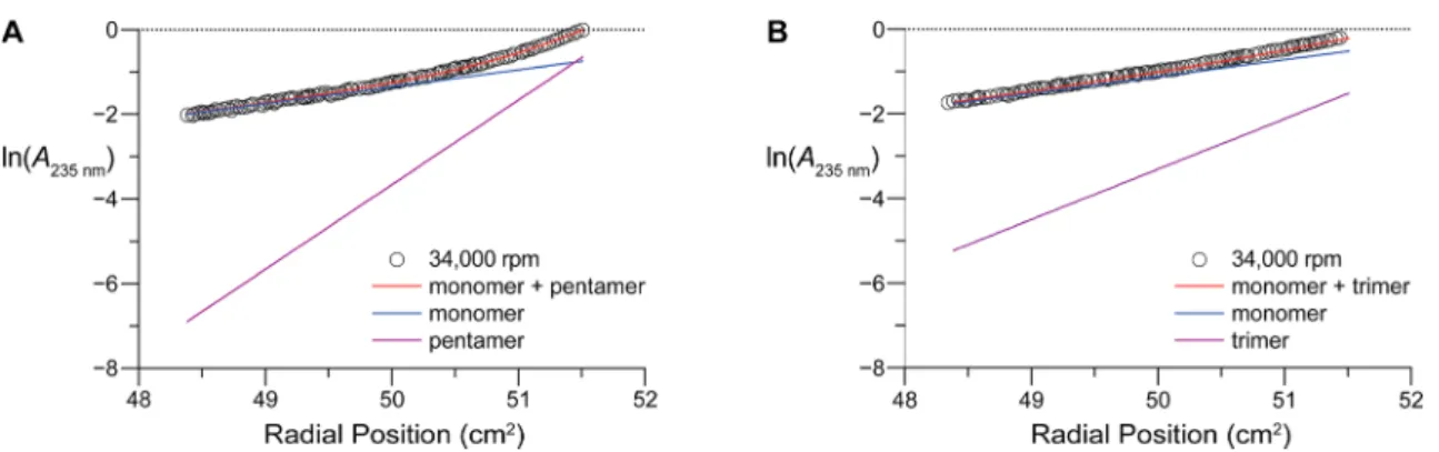
Analysis of Host Self-Association with
Ultracentrifu-gation. To assess the aggregation state of host-o and host-r in
solution, we subjected their solutions to analytical
ultra-centrifugation (AUC). Although sedimentation-equilibrium
data
fitted to a single-species model presents host-o as a
dimer and host-r as a monomer in solution, introduction of a
multimer in the models improves
fits significantly by better
representing the contributions from high-molecular weight
Figure 2.Chemical structures of the open host (host-o), closed host (host-c), and closed, reduced host (host-r). The macrocyclic rings of host-c and host-r contain 221 atoms.Biomacromolecules
pubs.acs.org/Biomac Articlehttps://dx.doi.org/10.1021/acs.biomac.0c00103
Biomacromolecules 2020, 21, 1539−1547
(MW) species. Monomer
−pentamer and monomer−trimer
models best explain the high-MW species observed in host-o
and host-r gradients, respectively (
Figure S2
). Monomer
−
multimer models for host-o and host-r data collected at 34,000
rpm exempli
fies the abundance of each species across the
gradients. The presence of high-MW species in the host-o
sample results in an appreciable deviation from linearity
(
Figure 3
A), while the host-r gradient remains linear with only
minor deviation from sedimentation expected of a monomer
(
Figure 3
B). Thus, AUC indicates multimerization of host-o,
whereas host-r remains essentially in a monomeric state.
Analysis of Host
·CMP Complex Formation with
Fluorescence Spectroscopy. To reveal whether a triple
helix forms between the hosts and a CMP, we employed
fluorescence spectroscopy. Host-o and host-r were conjugated
to biotin and then complexed to
fluorescent beads that were
coated with streptavidin. A
fluorescein probe was tethered to a
CMP, (Pro-Pro-Gly)
7. Upon mixing, coinciding
fluorescence
would indicate association and, presumably, triple-helix
formation (
Figure 4
A). Application of the positive control
biotin
−fluorescein, (Ser-Gly)
3conjugated to both biotin and
fluorescein, reveals a green halo upon the red fluorescence of
beads under confocal microscopy where the focal plane bisects
the bead (
Figure 4
B). No
fluorescent quenching was observed
upon bead-labeling. The same pattern was apparent for host-o
and host-r coated beads when mixed with the
fluorescein−
CMP conjugate. In contrast, treatment with
fluorescein−CMP
alone did not lead to green
fluorescence (data not shown).
Host-coated beads mixed with
fluorescein−
D-CMP, the
enantiomeric peptide incapable of forming a triple helix with
either host, showed a substantial reduction in bead-labeling
(
Figure S3
). This reduction is indicative of the speci
fic binding
of
fluorescein−CMP to hosts on the bead surface.
The qualitative results obtained with microscopy were
quanti
fied by examining beads with flow cytometry, where
10000 events were evaluated for each sample in a single run.
Again, beads coated with both host-o and host-r showed
substantial labeling upon incubation with the
fluorescein−
CMP conjugate (
Figure 5
). The
fluorescence with host-r was
greater than that with host-o, which is not closed on its N
terminus. Moreover, the triple helix formed by host-r with a
CMP has greater kinetic stability than does that with host-o,
where agitation for 9 h diminished
fluorescence.
Analysis of Host
·CMP Complex Formation with
Circular Dichroism Spectroscopy. The collagen triple
helix generates a distinct circular dichroism (CD) spectrum
with maximum ellipticity near 226 nm.
15In our host
·guest
system, however, that diagnostic method is complicated by two
factors. First, the hosts themselves have CD spectra like that of
a triple helix (
Figure 6
A). Second, guest strands alone can form
a homotrimeric helix as well as a host
·guest complex, and those
Figure 3.Graphs of analytical ultracentrifugation data obtained at 34000 rpm for host-o and host-r. (A) Data for host-ofitted to a monomer + pentamer mode. (B) Data for host-rfitted to a monomer + trimer mode.Figure 4.Binding of immobilized host-o and host-r to afluorescent CMP. (A) Scheme showing the experimental design. (B) Representative confocal microscopy images. Streptavidin-coated fluorescent beads (red) were treated with biotin-conjugated host-o or host-r and then incubated withfluorescein−CMP (green), which is Ac-Lys(fluorescein)-(Ser-Gly)3-(Pro-Pro-Gly)7. Also shown are
im-ages from a negative control of untreated beads and a positive control of beads treated with a biotin−fluorescein conjugate (green), which is biotin-(Gly-Ser)3-Lys(fluorescein)-NH2. Scale bar: 10μm.
two triple helices are likely to have indistinguishable CD
spectra. Nonetheless, interactions with a monomeric host (e.g.,
host-r) could reveal trends, especially if analyses are restricted
to strands that do not form homotrimers. Accordingly, changes
in CD signal upon mixing such strands with a host could be
attributed to the formation of a host
·guest triple helix.
The CMPs (
flp-Hyp-Gly)
7,
3b,16
(
flp-Flp-Gly)
7,
17
(Pro-Pro-Gly)
7,
17and (Pro-Ile-Gly)
718cannot form stable homotrimeric
triple helices, exhibit low CD signal, and are ideal for testing
the utility of host-r as a mimic for damaged collagen (
Figure
6
B). In contrast to their spectra alone, a collagen signature is
apparent in their mixtures with host-r (
Figure 6
C). To
determine the extent of interaction between host-r and each
guest, we used data on individual species to calculate the CD
spectra where the components of the mixture do not interact.
The calculated spectra explain the CD signal observed for
mixtures of host-r with (Pro-Pro-Gly)
7and (Pro-Ile-Gly)
7(
Figure 6
C). In contrast, (
flp-Hyp-Gly)
7and (
flp-Flp-Gly)
7interact cooperatively with host-r, producing signi
ficantly
higher triple-helix signal than each species can independently
contribute. In comparison, host-o
·guest complexes do not yield
similar levels of cooperativity (
Figure S4
).
The results from temperature-denaturation experiments are
consistent with the spectral analysis above, and also highlight
the impact of macrocycle formation on host structure.
Although similar folded states can be imagined for all hosts,
macrocycle formation and
flexibility limit their conformations
upon denaturation. This is clearly re
flected in their
denaturation pro
files (
Figure S5A
). Whereas a distinct
transition is apparent for host-o (T
m= 45.6
°C), an extremely
shallow transition is apparent for the highly constrained host-c
and increases after reduction to host-r. This trend is consistent
with that observed in CD spectra (
Figure 6
A). Furthermore,
the thermal transitions for host-r
·guest complexes become
increasingly recognizable when a more cooperative guest is
selected (
Figure S5B
), consistent with our analysis of host
·
guest cooperativity (
Figure 6
C).
The peptides (
flp-Hyp-Gly)
7, (
flp-Flp-Gly)
7, and
(Pro-Pro-Gly)
7were used previously as invasive strands to deliver cargo
to damaged collagen.
3−5(Pro-Hyp-Gly)
7has also been
employed for this purpose.
19Unlike the other peptides,
however, (Pro-Hyp-Gly)
7readily forms homotrimers at room
temperature, complicating its therapeutic use. As anticipated,
application of (Pro-Hyp-Gly)
7to host-r does not enhance
triple-helical content (
Figure S6
).
■
DISCUSSION
A collagen duplex could be an e
ffective mimic of damaged
collagen (
Figure 1
). To optimize a duplex for this purpose, we
synthesized two parallel strands of (Pro-Pro-Gly)
10from the
amino groups of an immobilized lysine residue. The
N-terminal closing of this acyclic duplex, host-o, was achieved by
ole
fin metathesis using the Grubbs G2 catalyst. Reduction of
the ensuing alkene a
fforded a cyclic duplex, host-r (
Figure 2
).
This simple modi
fication appears to be critical, as it results in a
dramatic increase in CD signal (
Figure 6
A).
Figure 5.Quantification of binding of immobilized host-o and host-r to a CMP. Streptavidin-coatedfluorescent beads were treated with host-o−biotin or host-r−biotin, and then with fluorescein−CMP, which is Ac-Lys(fluorescein)-(Ser-Gly)3-(Pro-Pro-Gly)7. Beads were
also treated with biotin−fluorescein conjugate (which is biotin-(Gly-Ser)3-Lys(fluorescein)-NH2), host-o alone, or host-r alone. Values
represent the percent of the sample population with fluorescein-labeling relative to that from treatment with biotin−fluorescein.
Figure 6.Circular dichroism spectra of hosts (A), guest CMPs (B), and host·CMP complexes (C). Calculated spectra for noninteracting mixtures of host-r and CMPs are shown (dashed gray lines) together with acquired spectra for host-r·CMP complexes (red lines). Spectra were obtained in 50 mM HOAc at 4°C.
Biomacromolecules
pubs.acs.org/Biomac Articlehttps://dx.doi.org/10.1021/acs.biomac.0c00103
Biomacromolecules 2020, 21, 1539−1547
Analytical ultracentrifugation revealed that host-r exists
primarily as a monomer in solution (
Figure 3
B). Yet, it
exhibits triple-helical character by CD spectroscopy, despite its
topological inability to form a triple helix. This
finding is
consistent with our recent discovery that two singly
cross-linked CMPs adopt a collagen-like structure even in the
absence of a third strand.
20Although host-r exhibits a lower triple-helix signal than does
host-o (
Figure 6
A), the bene
fits of macrocycle formation
outweigh any accompanying structural penalty. Closing the
macrocycle reduces multimerization (
Figure 3
), improves the
cooperativity of host
·guest association (cf.:
Figures 6
C and
S4
), and enables better retention of a CMP on a host-coated
bead surface (
Figure 5
). The kinetically stable interaction of
such beads with (Pro-Pro-Gly)
7is especially interesting, as this
CMP displays little cooperativity with host-r. CD spectra
calculated for noninteracting species are likely overestimations
due to higher concentrations used to obtain data for individual
species. Perhaps more importantly, the bead-retention
experi-ment is unique in being a measureexperi-ment done on a solid
support, which can enhance ligand association as compared to
solution-state measurements.
21Interestingly, (Pro-Pro-Gly)
7has been used as an invasive strand to deliver cargo to
wound beds, which similarly display damaged collagen on a
3-D surface.
3a,4,5Di
fferent CMPs were examined as “guests”−third strands.
Of those, (
flp-Hyp-Gly)
7and (
flp-Flp-Gly)
7are superior at
promoting the formation of a triple helix with host-r (
Figure
6
C). Their ability to form a stable triple helix can be attributed
to the preorganization endowed by their
flp residues (which
adopt a requisite C
γ-endo ring pucker and
ϕ ∼ −75° dihedral
angle) and Hyp and Flp residues (which adopt a requisite C
γ-exo ring pucker and smaller
ϕ ∼ −60° dihedral angle).
1Interestingly, the interaction of host-r with (
flp-Hyp-Gly)
7produces a greater rise in CD signal than that with (
flp-Flp-Gly)
7, even though Flp-to-Hyp substitutions at the Yaa
position of a (Pro-Yaa-Gly)
7peptide lowers thermostability.
22This
finding is consistent with our recent discovery that
(flp-Hyp-Gly)
7is an optimal strand for annealing to damaged
collagen in vitro and ex vivo.
3bHence, we conclude that host-r
provides a reliable mimic of damaged collagen.
Although all CMPs tested are de
ficient in homotrimer
formation, they are so due to di
fferent factors. The peptides,
(
flp-Hyp-Gly)
7and (
flp-Flp-Gly)
7, feature residues with high
triple-helical propensity, but cannot form triple helices due to
severe steric hindrance between Xaa =
flp and Yaa = Flp or
Hyp residues on neighboring strands.
3b,7In contrast,
(Pro-Pro-Gly)
7and (Pro-Ile-Gly)
7lack the preorganization of their
counterparts, and their triple helices are not stable above 4
°C
despite being unhindered by sterics. Thus, (Pro-Pro-Gly)
7and
(Pro-Ile-Gly)
7are not well-positioned for cooperative
inter-actions with host-r, which is corroborated by our
findings
(
Figure 6
C). Interestingly, we also observe similar
uncooper-ative behavior in mixtures of host-r and (Pro-Hyp-Gly)
7, a
peptide that forms stable homotrimers up to 36
°C (
Figure
S5
).
23Together, our results point to engineered guest peptides,
and especially to (
flp-Hyp-Gly)
7, as the optimal CMPs for
targeting collagen damage.
■
CONCLUSIONS
Host-r is a macrocycle that contains two collagen-mimetic
peptides and forms a collagen triple helix with a third
collagen-mimetic peptide. Its development provides opportunities for
new types of analyses. Historically, the rigorous analysis of
triple-helix formation has been complicated by the process
being termolecular. For example, hysteresis often confounds
analyses of the unfolding
−refolding transition with three
strands.
24As the basis of a bimolecular rather than a
termolecular system, host-r could provide clarity as well as
new insight.
In addition, we note that human type-I collagen, which is the
most abundant protein in the extracellular matrix and
connective tissue,
1is composed of two
α1[I] strands and
one
α2[I] strand. In collagen fibrils, a
Gly-Phe-Hyp-Gly-Glu-Arg sequence clusters integrins on the surface of endothelial
cells and promotes wound healing.
25Disruption of the triple
helix in this region could be especially deleterious to wound
repair. Host-r variants composed of two copies of the
GAOGPSGARGERGFOGERGVQGPOGPAGPR sequence
of human
α1[I] strands (where “O” refers to Hyp) could
lead to the discovery of CMPs that enable new therapeutic
interventions.
13b■
ASSOCIATED CONTENT
*
sı Supporting InformationThe Supporting Information is available free of charge at
https://pubs.acs.org/doi/10.1021/acs.biomac.0c00103
.
Figures S1−S6, NMR spectra, and UPLC traces (
)
■
AUTHOR INFORMATION
Corresponding AuthorRonald T. Raines
− Department of Chemistry and Department
of Biochemistry, University of Wisconsin
−Madison, Madison,
Wisconsin 53706, United States; Department of Chemistry,
Massachusetts Institute of Technology, Cambridge,
Massachusetts 02139, United States;
orcid.org/0000-0001-7164-1719
; Phone: (617) 253-1470; Email:
rtraines@
mit.edu
Authors
Aubrey J. Ellison
− Department of Chemistry, University of
Wisconsin
−Madison, Madison, Wisconsin 53706, United States
I. Caglar Tanrikulu
− Department of Biochemistry, University of
Wisconsin
−Madison, Madison, Wisconsin 53706, United
States; Department of Chemistry, Massachusetts Institute of
Technology, Cambridge, Massachusetts 02139, United States;
orcid.org/0000-0002-7165-0399
Jesu
́s M. Dones − Department of Chemistry, University of
Wisconsin
−Madison, Madison, Wisconsin 53706, United
States; Department of Chemistry, Massachusetts Institute of
Technology, Cambridge, Massachusetts 02139, United States
Complete contact information is available at:
https://pubs.acs.org/10.1021/acs.biomac.0c00103
Notes
The authors declare no competing
financial interest.
■
ACKNOWLEDGMENTS
We are grateful to Dr. Darrell R. McCaslin and Dr. Martha M.
Vestling for facility support and contributive discussions and to
Prof. Clark R. Landis for use of his glovebox. A.J.E. was
supported by a Chemistry
−Biology Interface (CBI) Training
Grant T32 GM008505 (NIH). This work was supported by
Grant R56 AR044276 (NIH).
■
REFERENCES
(1) Shoulders, M. D.; Raines, R. T. Collagen structure and stability. Annu. Rev. Biochem. 2009, 78, 929−958.
(2) (a) Siebler, C.; Erdmann, R. S.; Wennemers, H. From azidoproline to functionalizable collagen. Chimia 2013, 67, 891− 895. (b) Chattopadhyay, S.; Raines, R. T. Collagen-based biomaterials for wound healing. Biopolymers 2014, 101, 821−833. (c) Wahyudi, H.; Reynolds, A. A.; Li, Y.; Owen, S. C.; Yu, S. M. Targeting collagen for diagnostic imaging and therapeutic delivery. J. Controlled Release 2016, 240, 323−331. (d) Shekhter, A. B.; Fayzullin, A. L.; Vukolova, M. N.; Rudenko, T. G.; Osipycheva, V. D.; Litvitsky, P. F. Medical applications of collagen and collagen-based materials. Curr. Med. Chem. 2019, 26, 506−516.
(3) (a) Chattopadhyay, S.; Murphy, C. J.; McAnulty, J. F.; Raines, R. T. Peptides that anneal to natural collagen in vitro and ex vivo. Org. Biomol. Chem. 2012, 10, 5892−5897. (b) Dones, J. M.; Tanrikulu, I. C.; Chacko, J. V.; Schroeder, A. B.; Hoang, T. T.; Gibson, A. L. F.; Eliceiri, K. W.; Raines, R. T. Optimization of interstrand interactions enables burn detection with a collagen-mimetic peptide. Org. Biomol. Chem. 2019, 17, 9906−9912. (c) Song, J. Y.; Pineault, K. M.; Dones, J. M.; Raines, R. T.; Wellik, D. M. Hox genes maintain critical roles in the adult skeleton. Proc. Natl. Acad. Sci. U. S. A., DOI: 10.1073/ pnas.1920860117.
(4) Chattopadhyay, S.; Guthrie, K. M.; Teixeira, L.; Murphy, C. J.; Dubielzig, R. R.; McAnulty, J. F.; Raines, R. T. Anchoring a cytoactive factor in a wound bed promotes healing. J. Tissue Eng. Regener. Med. 2016, 10, 1012−1020.
(5) Ellison, A. J.; Raines, R. T. A pendant peptide endows a sunscreen with water-resistance. Org. Biomol. Chem. 2018, 16, 7139− 7142.
(6) (a) Koide, T.; Homma, D. L.; Asada, S.; Kitagawa, K. Self-complementary peptides for the formation of collagen-like triple helical supramolecules. Bioorg. Med. Chem. Lett. 2005, 15, 5230− 5233. (b) Kotch, F. W.; Raines, R. T. Self-assembly of synthetic collagen triple helices. Proc. Natl. Acad. Sci. U. S. A. 2006, 103, 3028− 3033. (c) Yamazaki, C. M.; Asada, S.; Kitagawa, K.; Koide, T. Artificial collagen gels via self-assembly of de novo designed peptides. Biopolymers 2008, 90, 816−823. (d) Yamazaki, C. M.; Kadoya, Y.; Hozumi, K.; Okano-Kosugi, H.; Asada, S.; Kitagawa, K.; Nomizu, M.; Koide, T. A collagen-mimetic triple helical supramolecule that evokes integrin-dependent cell responses. Biomaterials 2010, 31, 1925−1934. (e) Tanrikulu, I. C.; Raines, R. T. Optimal interstrand bridges for collagen-like biomaterials. J. Am. Chem. Soc. 2014, 136, 13490−13493. (f) Delsuc, N.; Uchinomiya, S.; Ojida, A.; Hamachi, I. A host-guest system based on collagen-like triple-helix hybridization. Chem. Commun. 2017, 53, 6856−6859.
(7) Hodges, J. A.; Raines, R. T. Stereoelectronic and steric effects in the collagen triple helix: Toward a code for strand association. J. Am. Chem. Soc. 2005, 127, 15923−15932.
(8) Blackwell, H. E.; Clemons, P. A.; Schreiber, S. L. Exploiting site-site interactions on solid support to generate dimeric molecules. Org. Lett. 2001, 3, 1185−1188.
(9) Scholl, M.; Ding, S.; Lee, C. W.; Grubbs, R. H. Synthesis and activity of a new generation of ruthenium-based olefin metathesis catalysts coordinated with 1,3-dimesityl-4,5-dihydroimidazol-2-yli-dene ligands. Org. Lett. 1999, 1, 953−956.
(10) Jida, M.; Betti, C.; Schiller, P. W.; Tourwe, D.; Ballet, S. One-pot isomerization-cross metathesis-reduction (ICMR) synthesis of lipophilic tetrapeptides. ACS Comb. Sci. 2014, 16, 342−351.
(11) Durchschlag, H.; Zipper, P. Calculation of the partial volume of organic compounds and polymers. Ultracentrifucation 1994, 94, 20− 39.
(12) (a) Kim, J.-S.; Raines, R. T. Dibromobimane as a fluorescent crosslinking reagent. Anal. Biochem. 1995, 225, 174−176. (b) Sardi, F.; Manta, B.; Portillo-Ledesma, S.; Knoops, B.; Comini, M. A.; Ferrer-Sueta, G. Determination of acidity and nucleophilicity in thiols by reaction with monobromobimane and fluorescence detection. Anal. Biochem. 2013, 435, 74−82.
(13) (a) Takita, K. K.; Fujii, K. K.; Kadonosono, T.; Masuda, R.; Koide, T. Cyclic peptides for efficient detection of collagen. ChemBioChem 2018, 19, 1613−1617. (b) Takita, K. K.; Fujii, K. K.; Ishii, K.; Koide, T. Structural optimization of cyclic peptides that efficiently detect denatured collagen. Org. Biomol. Chem. 2019, 17, 7380−7387.
(14) Blackwell, H. E.; Sadowsky, J. D.; Howard, R. J.; Sampson, J. N.; Chao, J. A.; Steinmetz, W. E.; O’Leary, D. J.; Grubbs, R. H. Ring-closing metathesis of olefinic peptides: Design, synthesis, and structural characterization of macrocyclic helical peptides. J. Org. Chem. 2001, 66, 5291−5302.
(15) Fields, G. B.; Prockop, D. J. Perspective on the synthesis and application of triple-helical collagen-model peptides. Biopolymers 1996, 40, 345−357.
(16) Barth, D.; Milbradt, A. G.; Renner, C.; Moroder, L. A (4R)- or a (4S)-fluoroproline residue in position Xaa of the (Xaa-Yaa-Gly) collagen repeat severely affects triple-helix formation. ChemBioChem 2004, 5, 79−86.
(17) Hodges, J. A.; Raines, R. T. Stereoelectronic effects on collagen stability: The dichotomy of 4-fluoroproline diastereomers. J. Am. Chem. Soc. 2003, 125, 9262−9263.
(18) (a) Tamburro, A. M.; Guantieri, V.; Cabrol, D.; Broch, H.; Vasilescu, D. Experimental and theoretical conformational studies on polypeptide models of collagen. Int. J. Pept. Protein Res. 1984, 24, 627−635. (b) Persikov, A. V.; Ramshaw, J. A. M.; Kirkpatrick, A.; Brodsky, B. Amino acid propensities for the collagen triple-helix. Biochemistry 2000, 39, 14960−14967.
(19) (a) Wang, A. Y.; Mo, X.; Chen, C. S.; Yu, S. M. Facile modification of collagen directed by collagen mimetic peptides. J. Am. Chem. Soc. 2005, 127, 4130−4131. (b) Hwang, J.; Huang, Y.; Burwell, T. J.; Peterson, N. C.; Connor, J.; Weiss, S. J.; Yu, S. M.; Li, Y. In situ imaging of tissue remodeling with collagen hybridizing peptides. ACS Nano 2017, 11, 9825−9835.
(20) Tanrikulu, I. C.; Westler, W. M.; Ellison, A. J.; Markley, J. L.; Raines, R. T. Templated collagen “double helices” maintain their structure. J. Am. Chem. Soc. 2020, 142, 1137−1141.
(21) (a) Hintersteiner, M.; Buehler, C.; Auer, M. On-bead screens sample narrower affinity ranges of protein-ligand interactions compared to equivalent solution assays. ChemPhysChem 2012, 13, 3472−3480. (b) Daniel, C.; Roupioz, Y.; Gasparutto, D.; Livache, T.; Buhot, A. Solution-phase vs surface-phase aptamer-protein affinity from a label-free kinetic biosensor. PLoS One 2013, 8, No. e75419.
(22) (a) Holmgren, S.; Taylor, K.; Bretscher, L.; Raines, R. T. Code for collagen’s stability deciphered. Nature 1998, 392, 666−667. (b) Holmgren, S.; Bretscher, L. E.; Taylor, K. M.; Raines, R. T. A hyperstable collagen mimic. Chem. Biol. 1999, 6, 63−70.
(23) Bretscher, L. E.; Jenkins, C. L.; Taylor, K. M.; DeRider, M. L.; Raines, R. T. Conformational stability of collagen relies on a stereoelectronic effect. J. Am. Chem. Soc. 2001, 123, 777−778.
(24) (a) Davis, J. M.; Bächinger, H. P. Hysteresis in the triple helix-coil transition of type III collagen. J. Biol. Chem. 1993, 268, 25965− 25972. (b) Fischer, G. Chemical aspects of peptide bond isomer-isation. Chem. Soc. Rev. 2000, 29, 119−127. (c) Mizuno, K.; Boudko, S. P.; Engel, J.; Bächinger, H. P. Kinetic hysteresis in collagen folding. Biophys. J. 2010, 98, 3004−3014. (d) Egli, J.; Siebler, C.; Köhler, M.; Zenobi, R.; Wennemers, H. Hydrophobic moieties bestow fast-folding and hyperstability on collagen triple helices. J. Am. Chem. Soc. 2019, 141, 5607−5611.
(25) (a) Knight, C. G.; Morton, L. F.; Onley, D. J.; Peachey, A. R.; Messent, A.; Smethurst, P. A.; Tuckwell, D. S.; Farndale, R. W.; Barnes, M. J. Identification in collagen type I of an integrin α2β1
-binding site containing an essential GER sequence. J. Biol. Chem. 1998, 273, 33287−33294. (b) Sweeney, S. M.; DiLullo, G.; Slater, S. J.; Martinez, J.; Iozzo, R. V.; Lauer-Fields, J. L.; Fields, G. B.; Antonio, J. D. S. Angiogenesis in collagen I requires α2β1 ligation of a
GFP*GER sequence and possibly p38 MAPK activation and focal adhesion disassembly. J. Biol. Chem. 2003, 278, 30516−30524. (c) Seo, N.; Russell, B. H.; Rivera, J. J.; Liang, X.; Xu, X.; Afshar-Kharghan, V.; Höök, M. An engineered α1 integrin-binding
Biomacromolecules
pubs.acs.org/Biomac Articlehttps://dx.doi.org/10.1021/acs.biomac.0c00103
Biomacromolecules 2020, 21, 1539−1547
collagenous sequence. J. Biol. Chem. 2010, 285, 31046−31054. (d) Sipilä, K. H.; Drushinin, K.; Rappu, P.; Jokinen, J.; Salminen, T. A.; Salo, A. M.; Käpylä, J.; Myllyharju, J.; Heino, J. Proline hydroxylation in collagen supports integrin binding by two distinct mechanisms. J. Biol. Chem. 2018, 293, 7645−7658.