HAL Id: tel-01868324
https://tel.archives-ouvertes.fr/tel-01868324
Submitted on 5 Sep 2018
HAL is a multi-disciplinary open access
archive for the deposit and dissemination of sci-entific research documents, whether they are pub-lished or not. The documents may come from teaching and research institutions in France or abroad, or from public or private research centers.
L’archive ouverte pluridisciplinaire HAL, est destinée au dépôt et à la diffusion de documents scientifiques de niveau recherche, publiés ou non, émanant des établissements d’enseignement et de recherche français ou étrangers, des laboratoires publics ou privés.
data
Agustina Razetti
To cite this version:
Agustina Razetti. Modelling and characterizing axon growth from in vivo data. Signal and Image processing. COMUE Université Côte d’Azur (2015 - 2019), 2018. English. �NNT : 2018AZUR4016�. �tel-01868324�
Agustina RAZETTI
Équipe MORPHEME – INRIA Sophia Antipolis/I3S/iBV
Présentée en vue de l’obtention
du grade de docteur en Automatique, traitement
du signal et des images
d’Université Côte d’Azur Dirigée par : Xavier Descombes Soutenue le : 13 Avril 2018
Devant le jury, composé de :
Alin Achim, PhD, Bristol University
Florence Besse, PhD, Institute Biology Valrose Kristian Franze, PhD, Cambridge Univeristy Caroline Medioni, PhD, Institute Biology Valrose Jonas Ranft, PhD, École Normale Supérieur Anuj Srivastava, PhD, Florida State University Michèle Studer, PhD, Institute Biology Valrose
DOCTORAL SCHOOL STIC
SCIENCES ET TECHNOLOGIES DE L’INFORMATION ET DE LA COMMUNICATION
P H D T H E S I S
to obtain the title of
PhD of Science
of the University Côte d’Azur
Specialty : C
ONTROL SYSTEMS,
SIGNAL AND IMAGE PROCESSINGDefended by:
Agustina RAZETTI
Modelling and characterizing axon
growth from in vivo data
Thesis advisor: Xavier DESCOMBES
Prepared at MORPHEME Team, INRIA Sophia Antipolis/I3S/iBV
Defended on April 13, 2018
Jury :
Reporters :
Alin A
CHIM— Bristol University
Kristian F
RANZE— Cambridge University
Anuj S
RIVASTAVA— Florida State University
Director :
Xavier D
ESCOMBES— Inria Sophia Antipolis Méditerranée
Examinators :
Florence B
ESSE— Institute Biologie Valrose
Caroline M
EDIONI— Institute Biologie Valrose
Jonas R
ANFT— École Normale Supérieure
DES IMAGES
présentée et soutenue par:
Agustina RAZETTI
Modélisation et caractérisation de la
croissance des axones à partir de
données in vivo
Thèse dirigée par: Xavier DESCOMBES
préparée dans l’Équipe MORPHEME, INRIA Sophia Antipolis/I3S/iBV
Prévu pour la défense le 13 Avril, 2018
Jury :
Rapporteurs :
Alin A
CHIM— Bristol University
Kristian F
RANZE— Cambridge University
Anuj S
RIVASTAVA— Florida State University
Directeur :
Xavier D
ESCOMBES— Inria Sophia Antipolis Méditerranée
Examinateurs :
Florence B
ESSE— Institute Biologie Valrose
Caroline M
EDIONI— Institute Biologie Valrose
Jonas R
ANFT— École Normale Supérieure
How the brain wires up during development remains an open question in the scientific community across disciplines. Fruitful efforts have been made to elucidate the mechanisms of axonal growth, such as pathfinding and guiding molecules. However, recent evidence suggests other actors to be involved in neuron growth in vivo. Notably, axons develop in populations and embedded in mechanically constrained environments. Thus, to fully understand this dynamic process, one must take into account collective mechanisms and mechanical interactions within the axonal populations. However, techniques to directly measure this from living brains are today lacking or heavy to implement.
This thesis emerges from a multidisciplinary collaboration, to shed light on axonal development in vivo and how adult complex axonal morphologies are attained. Our work is inspired and validated from images of single wild type and mutated Drosophila γ axons, which we have segmented and normalized.
We first proposed a mathematical framework for the morphological study and classifica-tion of axonal groups. From this analysis we hypothesized that axon growth derives from a stochastic process, and that the variability and complexity of axonal trees result from its intrinsic nature, as well as from elongation strategies developed to overcome the mechanical constraints of the developing brain. We designed a mathematical model of single axon growth based on Gaussian Markov Chains with two parameters, accounting for axon rigidity and attraction to the target field. We estimated the model parameters from data, and simulated the growing axons embedded in spatially constraint populations to test our hypothesis.
We dealt with themes from applied mathematics as well as from biology, and unveiled unexplored effects of collective growth on axonal development in vivo.
Key words: axon growth, morphogenesis, stochastic modelling, Gaussian Markov chains, mechanical interactions, axon branching, elongation strategies
à partir de cerveaux vivants sont aujourd’hui insuffisantes ou lourdes à mettre en œuvre. Cette thèse résulte d’une collaboration multidisciplinaire, pour faire la lumière sur le développement axonal in vivo et les morphologies complexes des axones adultes. Notre travail a été inspiré et validé à partir d’images d’axones γ individuels chez la drosophile, de type sauvage et modifiés génétiquement, que nous avons segmentés et normalisés.
Nous avons d’abord proposé un cadre mathématique pour l’étude morphologique et la classification des groupes axonaux. A partir de cette analyse, nous avons émis l’hypothèse que la croissance axonale dérive d’un processus stochastique et que la variabilité et la complexité des arbres axonaux résultent de sa nature intrinsèque, ainsi que des stratégies d’élongation développées pour surmonter les contraintes mécaniques du cerveau en développement. Nous avons conçu un modèle mathématique de la croissance d’un axone isolé fondé sur des chaînes de Markov gaussiennes avec deux paramètres, représentant la rigidité axonale et l’attraction du champ cible. Nous avons estimé les paramètres de ce modèle à partir de données réelles et simulé la croissance des axones à l’échelle de populations et avec des contraintes spatiales pour tester notre hypothèse.
Nous avons abordé des thèmes de mathématiques appliquées ainsi que de la biologie, et dévoilé des effets inexplorés de la croissance collective sur le développement axonal in vivo.
Mots clés: croissance axonale, morphogenèse, modélisation stochastique, chaînes de Markov gaussiennes, interactions mécaniques, ramification axonale, stratégies d’élongation
some random meaning to symbols.
Jonas Ranft. Thank you a lot for your time when reading it, your valuable comments and corrections and interesting questions.
My everyday life during this thesis would have not been the same without the company of the amazing people that constitute the Morpheme team. Thank you for all the shared lunchtimes, coffee breaks, jokes and good moments, but also for your support and help when I needed it. A special mention for my fellow PhD students during these years, Alejandro Mottini, Alexis Zubiolo, Emmanuel Soubies, Gael Michelin, Lola Bautista, Arnauld Bletterer and Anca Grapa. Also, for the permanent members of Morpheme, thank you for helping to create and transmit this very special friendly environment in the team.
Last -but not least- I want to thank the people who stood besides me through all this process, since I decided to do my PhD in France to the very last day of my defence, in particular my mother, father and brother, as well as my closest friends and Flo, for the everyday encouragement.
1.4 Manuscript Organization . . . 4
1.5 List of Publications . . . 5
2 Background 7 2.1 Axon growth: state of the art . . . 7
2.2 Axon growth models: state of the art . . . 14
2.3 Biological model: Drosophila γ neurons . . . 19
2.3.1 Imp as a regulator of axon regrowth and branching . . . 20
2.4 Database Description . . . 22
2.4.1 Adult single cell confocal in vivo images . . . 23
2.4.2 4D image sequences of the developing brain . . . 23
3 Morphological Study 27 3.1 Digital reconstruction of the axonal trees . . . 28
3.1.1 Segmentation . . . 30
3.1.2 Tree hierarchy . . . 30
3.2 Stochastic modelling of the main morphological features . . . 34
3.2.1 Main axon length . . . 34
3.2.2 Main axon morphology . . . 35
3.2.3 Branching point occurrence . . . 38
3.3 Likelihood Analysis . . . 43
3.4 Simulation . . . 48
3.5 Discussion and Contributions . . . 50
3.5.1 Axon growing rate and branch formation . . . 50
3.5.2 Wild-type axons are mostly differentiated by their branch length distribution . . . 51
3.5.3 impmutants present two different phenotypes . . . 51
3.5.4 Profilin overexpression partially rescues the main axon length as well as the branch length distribution . . . 52
3.5.5 Conclusion . . . 53
4 Space-Embedded Axon Growth Model 55 4.1 Data treatment . . . 56
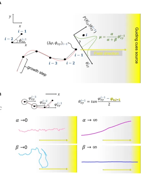
4.2 Mathematical model formulation . . . 57
4.3 Parameter estimation . . . 64
4.3.1 Parameter estimation of Markov chains . . . 65
4.3.2 Parameter estimation from a population of real axons . . . 71
4.4 Space-embedded simulations . . . 93
4.4.1 Dynamic simulation framework . . . 93
4.4.2 Medial lobe morphology reconstruction . . . 98
4.4.3 External Field determination . . . 98
4.4.4 Volume exclusion . . . 104
4.4.5 Resulting trajectories . . . 108
4.5 Branching mechanisms . . . 112
4.5.1 γ neurons present two types of branches . . . 114
4.5.2 Modelling the branch process . . . 122
4.6 Mutant phenotype predictions . . . 136
4.7 Parameter value significance and other considerations . . . 146
4.8 Discussion and contributions . . . 156
5 General conclusion, contributions and perspectives 173 5.1 General conclusion . . . 173
5.2 Perspectives . . . 175
3.2 Scheme of the proposed stochastic framework for the comparison of axonal
morphologies between groups . . . 29
3.3 Extraction of neuron morphology from confocal 3D images . . . 31
3.4 Alignment of the medial lobe to the horizontal axis . . . 31
3.5 Scheme of the three-hierarchy algorithm . . . 33
3.6 Results of the hierarchy algorithm . . . 33
3.7 Axon path scheme . . . 34
3.8 Main axon length distributions for each biological sample. . . 35
3.9 Second order Markov model for axon morphology. . . 37
3.10 Interstitial branch formation scheme. . . 39
3.11 Bernoulli model for branch occurrence. . . 41
3.12 Real vs. Simulated axons . . . 49
3.13 Real vs. Simulated axons . . . 50
4.1 γ axon normalization to the same standard medial lobe (SML) . . . 58
4.2 γ axon normalization: results . . . 59
4.3 Normalized γ axon populations. . . 60
4.4 3D Mathematical model of individual axon growth. . . 63
4.5 Concept of model renormalization. . . 65
4.7 Data reconstruction and simulation with ∆ρ = 0.09µm (pixel size) . . . 67
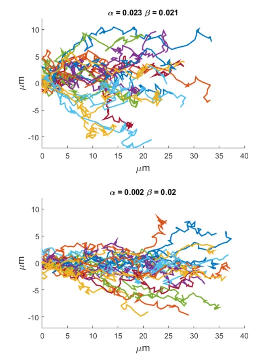
4.8 Parameter estimation method performance (α) . . . 72
4.9 Parameter estimation method performance (β ) . . . 73
4.10 Simulations using parameters estimated from data considering the axons altogether . . . 74
4.11 Distributions of the values of α and β estimated axon per axon from real data 75 4.12 Axon by axon parameter estimation - Real wild-type and simulated axons . 78 4.13 Axon by axon parameter estimation - Embedded-simulated and simulated axons, no branching . . . 79
4.14 Axon by axon parameter estimation - Embedded-simulated and simulated axons, with branches . . . 80
4.15 Parameter estimation results using the histogram comparison method . . . . 81
4.16 Design of a statistical test of hypothesis . . . 85
4.17 Validation of the bayesian and statistical approaches . . . 88
4.18 Independence on initial values . . . 89
4.19 Effect of the breakdown take-out algorithm on the parameter histograms . . 92
4.20 Jump point detection on real axons . . . 94
4.21 Simulation of γ axon growth in the context of a population of physically interacting axons. . . 97
4.22 Medial lobe geometry reconstruction . . . 99
4.23 γ main axon directionality . . . 101
4.24 Alternative attractive field configurations and chosen field . . . 102
4.25 Attractive field calibration. . . 103
4.26 Influence of the chosen field configuration on axon trajectories in function of α and β values . . . 104
4.27 Neurite 3D geometry and volume exclusion . . . 105
4.28 Diameter determination . . . 106
4.29 Error (%) (percentage of non-elongated axons) in function of the neurite diameter: Study . . . 107
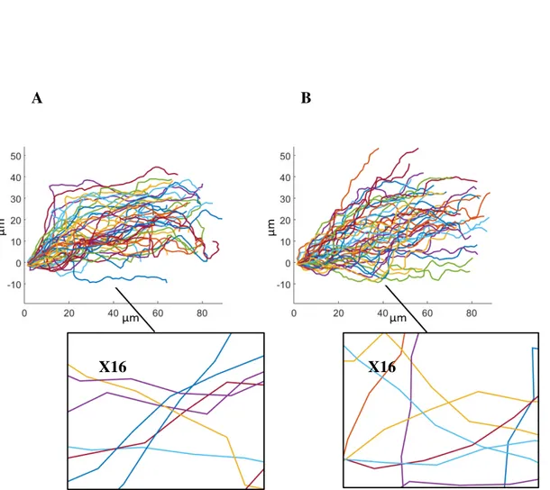
4.30 Space-embedded simulated vs. real axon trajectories . . . 108
4.31 Space-embedded simulated vs. real axon lengths and travelled distances . . 110
4.32 Space-embedded simulated vs. real axon lengths and travelled distances: 3D vs. 2D projections . . . 111
4.33 Emergent asynchronicity within the γ population . . . 113
4.34 Adult γ branch length distribution . . . 115
4.46 Modelling of the branch process - Branching upon contact . . . 131
4.47 γ population with mechanical branching occurrence . . . 132
4.48 Real and simulated axons are morphologically alike . . . 134
4.49 Branch length distribution and linear density of simulated γ axons with mechanical branching . . . 135
4.50 Relevant branch angle distributions from simulations . . . 137
4.51 Relevant branch angle distributions from simulations - bis . . . 138
4.52 Single non-branching axon in a normal population . . . 140
4.53 imp mutant γ axons . . . 141
4.54 Type I branch proportion influences axonal elongation . . . 142
4.55 un f mutant recued by TOR overexpression . . . 144
4.56 Misguided axons in a normal population . . . 145
4.57 Variance of θiconditioned by θi−1 (σ02) and of θi(σθ2), in function of α and β 147 4.58 Axon elongation in function of varying single parameter values . . . 148
4.59 Axon elongation in function of varying parameter values in the medial lobe and in a tube . . . 149
4.60 Morphologies of axons simulated with parameters optimal for elongation . 151 4.61 Anti-mechanic branching . . . 153
4.62 Implementation of random pauses during the neurite growth . . . 154
4.63 Percentage of non-elongated axons for different strategies of growth stop . . 155
4.64 Neurite tip growth speed distribution in the medial lobe . . . 157
4.65 Non-elongated axon percentage for different studied cases . . . 162
4.66 Parameter variation analysis . . . 166
4.67 Axon entropy calculation . . . 167
4.68 Breakdown and energetic analysis for real vs. simulated axons . . . 169
B.1 Iterative method algorithm: Inizialisation . . . 191
B.2 Iterative method algorithm: Application - Number or iterations . . . 192
B.3 Axon by axon parameter estimation - Long simulated axons . . . 192
B.4 Error percentage in function of different nmax and nR . . . 193
B.5 Emergent asynchronicity within the γ population (second example) . . . 194
B.6 Emergent asynchronicity within the γ population (third example) . . . 195
B.7 Analysis of the distances between branches in γ axons . . . 196
B.8 Wild type γ axons reconstructed from data . . . 197
B.9 Simulated wild type γ axons . . . 198
B.10 Simulated wild type γ axons - bis . . . 199
B.11 Branching deficiency phenotypes . . . 200
B.12 Simulated wild type γ axons with αbp and βbp . . . 201
B.13 Number of branches in function of branching mechanisms . . . 202
3.4 Value of the parameters A and p describing the branching point distribution. 41
3.5 Branch length distribution by length category per group (%). . . 42
3.6 pvalues from the non-parametric Kruskal Wallis test comparing the branch length distribution in L2between the studied groups. Significant differences appear in blue. . . 42
3.7 pvalues from the non-parametric Kruskal Wallis test comparing the branch length distribution in L4between the studied groups. Significant differences appear in blue. . . 42
3.8 Global likelihood analysis considering the four features. . . 45
3.9 Likelihood analysis according to the main axon length feature. . . 46
3.10 Likelihood analysis according to the main axon shape feature. . . 46
3.11 Likelihood analysis according to the branching point feature. . . 46
3.12 Likelihood analysis according to the branch length distribution feature. . 47
3.13 Global likelihood analysis considering the four features. Imp includes Imp Sh and Imp L. . . 47
3.14 Global likelihood analysis considering the four features. Imp is split between L and Sh for possible classification groups. . . 47
4.1 Values of the parameters estimated for different groups in our database using the histogram comparison method. . . 79
4.2 Values of the parameters and jump probability Pj estimated for different
groups in our database using the breakdown take-out method, with H0:α0=0,β0=βm and initial values (αm= βm= 20, λ = 0.1). Bayesian approach. . . 89
4.3 Values of the parameters and jump probability Pj estimated for different
groups in our database using the breakdown take-out method, with H0:α0=0,β0=βm and initial values (αm= βm= 20). Statistical approach - p = 0.001. . . 90
4.4 Values of the parameters and jump probability Pj estimated for different
groups in our database using the breakdown take-out method, with H0:α0=0,β0=βm and initial values (αm= βm= 20). Statistical approach - p = 0.0001. . . 90
4.5 Values of the parameters and jump probability Pj estimated for different
groups in our database using the breakdown take-out method, with H0:α0=0,β0=βm and initial values (αm= βm= 20). Statistical approach - p = 0.01. . . 90
4.6 Values of the parameters and jump probability Pj estimated for different
groups in our database using the breakdown take-out method, with H0:α0=αm,β0=0 and initial values (αm= βm= 20, λ = 0.1). Bayesian approach . . . 90
4.7 Values of the parameters and jump probability Pj estimated for different
groups in our database using the breakdown take-out method, with H0:α0=αm,β0=0 and initial values (αm= βm= 20). Statistical approach - p = 0.001. . . 91
4.8 Values of the parameters and jump probability Pj estimated for different
groups in our database using the breakdown take-out method, with H0:α0=αm,β0=0 and initial values (αm= βm= 20). Statistical approach - p = 0.0001. . . 91
4.9 Values of the parameters and jump probability Pj estimated for different
groups in our database using the breakdown take-out method, with H0:α0=αm,β0=0 and initial values (αm= βm= 20). Statistical approach - p = 0.01. . . 91
development and axonal morphogenesis in vivo, using diverse domains such as applied mathematics, probability theory, modelling and informatics. Intrinsically interdisciplinary, this work proposes new hypotheses and answers on this fundamental subject, and intends to close the existent gap between plausible experimentation and the complex reality of biological mechanisms.
During the last decades, important advances have been achieved in recognizing the main factors implicated in neuron development. External guiding cues involved in axonal pathfind-ing have been largely described, as well as the internal molecular machinery that allows axonal elongation, including many of the biochemical pathways regulating these processes. Interestingly, recent work shows that axons are not only capable of sensing and responding to external chemical signals, but are also guided by local changes in the mechanical properties of their surroundings. While most of these studies consider as mechanical environment mainly the extracellular matrix and the general mechanical properties of the tissue, little is known about intra-population axon-axon mechanical interactions and space confinement. In consequence, the effect of these physical phenomena on axon development and how they influence their final morphologies are still unknown. Furthermore, to measure mechanical interactions between individual growing neurons in the developing brain is experimentally not trivial, as tools to reproducibly visualize and manipulate axon-axon interactions in vivo in populations of growing neurons are either lacking or heavy to implement.
In this work we aim to better understand the origin of the complexity and variability of adult axonal morphologies as a consequence of their developmental process in vivo. Thus,
we consider and integrate the mentioned intra-population mechanical interactions in the otherwise better known individual axonal growth process. To do so, we propose in silico approaches validated on available in vivo data to expand the information given by the latter. As a result, we show the importance of considering axonal development as a dynamic and complex process of collective morphogenesis, rather than the sum of the growth of isolated individual axons.
Specifically, we first proposed an original classification framework to better characterize axonal morphologies and understand the similarities and differences between genotypically different axon groups. The framework consists of the stochastic modelling of relevant mor-phological features, the estimation of the model parameters from data and the classification of axons using likelihood analysis, enriched with statistics for a global description of ax-onal groups. Next, we designed a mechanistic individual axon elongation model based on Gaussian Markov chains with biologically related parameters, capable of generating realistic axonal morphologies. The estimation of the model parameters from real data happened to be not trivial, so we explored different approaches to solve this question. Using this individual axon growth model, we simulated the development of entire axonal populations considering external constraints such as an attractive guiding source, realistic geometrical limits and volume exclusion between the growing axons. These simulations allowed us to analyse the emergent effects of considering a population of axons represented as interacting self propelled elements. The validation of the simulation results with real data showed that our model is able to reproduce realistic morphologies. Interestingly, our simulations reproduced morphological characteristics of the axonal population that had not been included in the model, but rather emerged from its rules. Because our framework is based on the real biological process, it allowed us to better understand some aspects of axonal development in vivo. Finally, we could reproduce different mutation phenotypes by changing related aspects in our model. In consequence, our approach nicely assembles the three desirable aspects of a model: to be generative, explicative and predictive.
This thesis is the result of a solid collaboration with biologists from the Besse team at the Institute de Biologie de Valrose, Nice, France. They have provided us with rich image datasets from which this work was built, as well as biological expertise and advice. The stress of this thesis has been set on the inter collaboration and continuous feedback between mathematical modelling techniques and biological pertinence. Our dataset, composed of 3D confocal images of fixed in vivo single Drosophila γ axons at adult stage, as well as live 4D sequences of single or a group of Drosophila γ axons growing during metamorphosis, not only inspired our model, but was also used to estimate its parameters, as well as to validate
axons.
• To propose a model for axonal development that considers the stochastic path genera-tion and branching mechanisms with parameters that can be estimated from data; and that allows a straightforward implementation of population growth considering exter-nal constraints such as an attractive/repellent source, geometrical limits and volume exclusion with other axons. Ideally, this model should be generative, explicative and predictive of different real axon genotypes.
1.3
Main Contributions
The major contributions of this work are: In modelling
• Tree-like structures classification method based on the stochastic modelling of mor-phological features.
• Two-parameter stochastic model to represent trajectories in space that allows renormal-ization (equivalence between different spatial scales) and parameter estimation from data.
• General framework with two opposite approaches to detect jump points and estimate the parameters from piecewise homogeneous Markov chains.
In biology
• Deep morphological analysis of wild-type and mutant γ axons which resulted in new observations.
• Showcase the importance of mechanical interactions between growing axons and confinement on in vivo neuronal development.
• Proposal of a novel elongation mechanism where branching is intrinsic to the axonal growth process.
• Original interpretation of the imp mutant phenotype.
1.4
Manuscript Organization
This manuscript is organised in three sequential blocks: i-general axon growth biological and modelling background, ii- stochastic framework for the study of axonal morphology and iii- the development of the individual axon growth model and the simulation of developing populations.
Chapter 2 presents the background of this work (i). We first summarize the state of the art on axonal growth as well as on axonal growth mathematical and computational models, highlighting the aspects relevant for this thesis. We then describe the used biological model: the Drosophila mushroom body γ neurons (wild-type and mutant) and the image database used throughout this work.
We start our contributions in Chapter 3, where we mainly aim at a deep understanding of the axon morphologies in our database (ii). We first describe the data treatment scheme that allowed us to obtain 3D axonal reconstructions from the images, and present a general stochastic framework for neuronal morphological comparison. In particular, it consists in applying stochastic models for each axon morphological feature, estimate their parameters from data, to finally contrast and compare the axons using likelihood analysis and statistics (Razetti et al., 2016, 2017).
Chapter 4 represents the main part of this thesis. It consists of the development of a mech-anistic and dynamic mathematical model of individual axon growth, and its implementation in a realistic environment considering mechanical interactions between the growing axons (iii). The chapter starts by describing the data treatment that allows considering isolated axons as a population. Then we describe the proposed mathematical model, and present different approaches to estimate its parameters from data. Further, we perform simulations and analyse the results, comparing and contrasting them with data. We propose different branching occurrence hypothesis, and unveil new plausible elongation mechanisms. Finally, we further study the significance of the parameter values obtained from data and their implications on the collective growth of space-constraint tubular structures.
Conference on Biomedical Engineering Systems and Technologies. Book chapter
• Razetti, A., Descombes, X., Medioni, C., & Besse, F. (2017). A Stochastic Framework for Neuronal Morphological Comparison: Application to the Study of imp Knockdown Effects in Drosophila Gamma Neurons. In: Fred A., Gamboa H. (eds) Biomedical En-gineering Systems and Technologies. BIOSTEC 2016. Communications in Computer and Information Science, vol 690. Springer, Cham
Conference with abstract
• Razetti, A., Medioni, C., Malandain, G., Besse, F., & Descombes, X. (2017, May). Modelling collective axon growth from in vivo data reveals the importance of phys-ical axon-axon interactions. In Cell biology of the neuron: Polarity, plasticity and regeneration.
• Razetti, A., Medioni, C., Malandain, G., Besse, F., & Descombes, X. (2018). Un-derstanding in vivo axonal development as an inherently asymmetric process. First European Asymmetry Symposium.
2.1
Axon growth: state of the art
The first historic known mention of the human brain, «skull-offal», was found in a series of Egyptian documents on medicine and surgery called the Edwin Smith Papyrus, written in 1600 BC (but probably copied from earlier texts, from 3000 BC). Even though bounds in the head were at that time actually associated with different serious pathologies, like speech or walk loss, ancient Egyptians believed that intelligence and sensation were driven by the heart, and not brain, and thus despised the importance of this organ. Almost 5000 years later, Ramon y Cajal (1852-1934) first described the neuron as the functional unit of the brain. Since then, the complexity of this marvellous organ has not stopped intriguing and challenging scientists of different disciplines across the world. In this thesis, we explore one of the most captivating open questions in neurobiology: «How» is the brain created. In particular, we focus on neuron growth and how its complex final morphology is attained. Specifically, we address the question about how the physical presence of other neurons as well as spatial confinement are involved in the growth process.
During development, neurons in our nervous system extend their axonal trees in order to reach target neurons or structures, and establish complex networks that allow the correct functioning of the body and the mind (see Fig. 2.1 for a general scheme of the neuron). To do so, they need to recognize the spatial position of their final target as well as the path they should follow until they reach it; and complete the growth process in a limited time.
Furthermore, axons can also create branches that emerge from their shafts, enhancing the number of possible connections. However, when and where these branches are formed, as well as how long each of them should grow is still poorly understood.
When neurons somehow fail to decode or correctly follow these instructions, they develop corrupted adult morphologies that give rise to a large spectrum of diseases. Engle (2010) reviews human syndromes known to be caused by aberrant axon connectivity. Corpus Callosum Dysgenesis, L1 Syndrome and Joubert Syndrome and Related Disorders arise when axons fail to cross the brain mid-line. They can cause a variety of symptoms, such as severe mental retardation. Other serious syndromes are caused by guidance defects of cranial nerves. For example, the Kallmann Syndrome is believed to be caused by errors in growth and guidance of olfactory axons. Another example is Albinism, in which visual deficiencies arise from altered pattern of axonal projections in the visual system, due to axonal misguidance.
Thus, both to understand how the brain achieves its complex wiring to function in normal conditions, as well as to unveil the causes of severe syndromes, the study of axonal growth during development is highly relevant. During this section, we introduce the basic mechanisms of axonal development that motivated this work. We first explain how axons find their paths guided by chemical gradients to achieve elongation. Then, we explore different new findings on the role of the mechanical environment of the developing brain and how it also constitutes an important cue on axonal growth, as well as refines axonal final morphologies. Finally, we stress the importance of considering axonal elongation as a collective process, where axons do not grow isolated, but within populations, and explain main aspects of axonal branching.
Axon elongation is possible thanks to the structure present in their tip during development named the growth cone (GC). GCs present highly dynamic behaviours, and are subdivided into three main regions: i) a proximal core region enriched in microtubules ii) a flat area called lamellipodia made of inter-crossed actin fibres, and iii) finger-like protrusions named filopodia, which are held by elongated actin fibres (Fig. 2.2). The growth cone senses its chemical environment and decides the direction of the neurite growth. Many signalling molecules have been described to interact with the GC during development. They can be found on other cells surfaces, attached to the extra cellular matrix (short-rate cues) or diffusing in the extracellular domain (long-rate cues). Directional guiding cues set gradients that indicate the GC preferred paths or directionality of growth, while positional cues indicate when to stop. These molecules determine the outgrowth as well as the guidance of the axon shaft, and can be attractive or repellent, providing the essential information to guide the axon towards their specific target, even during long trajectories (Bixby and Harris, 1991; Guan and
Fig. 2.1 Scheme of a unipolar neuron. Input from one or more neurons is received by the dendrites, and the information travels along the axon shaft towards its branches. These terminals form synapses with other neurons to which the information is transmitted. The morphology of the axonal tree determines the neurons connectivity patterns.
Rao, 2003; Kolodkin and Tessier-Lavigne, 2011; Plachez and Richards, 2005; Song and Poo, 2001; Tessier-Lavigne et al., 1996). Interestingly, guiding cues are multifunctional, meaning that the same molecule can be attractive or repellent, as well as act in a short or long rate manner. In addition, GCs respond not only to extrinsic but also to intrinsic factors, and the combination of these mechanisms may be at the origin of the diversity and complexity of GC behaviour that wires up the brain (Dickson, 2002).
Growth cone chemotaxis (i.e. movement in response to the influence of chemical stimula-tion) occurs in two basic actions: «directional sensing» and «motility», which together occur in the context of «polarization». «Directional sensing» means that the growth cone is capable of sensing and amplifying noisy gradients of molecules to decode the spatial information of growth directionality (Fig. 2.2A). «Motility» is achieved thanks to the coordinated dynamics of the cytoskeleton components (actin and tubulin) in the growth cone, and it means that the GC can reshape itself to be oriented towards the maximum attraction gradient. This remodelling combines two actions: i) the extension of protrusions towards the gradient directionality and ii) the retraction of the rest of the structure (Fig. 2.2B). These actions lead to the GC polarization, meaning that it maintains a leading edge, or front, and a tail. Interestingly, GCs can present different degrees of polarization. For example, "paused" GCs were observed to present varied and complex shapes, to maximize their sensitivity, while CGs growing along a well defined gradient adopt simpler bullet-shapes, related with less sensitivity and faster movement (Dent et al., 2011; Dickson, 2002; Mortimer et al., 2008; Plachez and Richards, 2005). Once the direction of the movement has been sensed, the shaft has to elongate (Fig. 2.2C). The most accepted theory of how this elongation is achieved is called "the clutch" hypothesis (Mitchison and Kirschner, 1988). It suggests that the adhesion of the growth cone to adhesive substrates in other cells surfaces or in the extracellular matrix creates a "clutch", or anchor, that prevents retrograde flow and thus promotes actin-based outgrowth of the growth cone (Lowery and Van Vactor, 2009).
Interestingly, even though bibliography on axon outgrowth is vast and chemotaxis has been largely described, recent work shows that axons are not only capable of sensing and responding to external chemical signals, but are also guided by local changes in the mechanical properties of their surrounding (Francisco et al., 2007; Franze, 2013; Kerstein et al., 2015; Koser et al., 2016; Sagasti et al., 2005). Francisco et al. (2007) study the growth of embryonic chicken primary sensory neurons both in vitro and in vivo. Their results highlight the importance of the 3D mechanical environment in axonal elongation, defined by the extracellular matrix and spatial confinement. They show in vivo that growth cones have mechanisms to sense mechanical constraints and respond by the redirection of the growth, and thus propose that mechanical constraints may act complementary to chemical repellent
A B C
Fig. 2.2 Scheme of growth cone chemotaxis. (A) The growth cone senses a gradient set by external guiding cues. (B) It modifies its shape in order to direct its front toward the direction of the attraction field. (C) The axon shaft grows towards the established direction.
cues. Moreover, in vitro, they observe that the number of neurons establishing an axon was affected by the constrained space, proposing a direct link between available space and net axon growth. From dynamic observations, they report that axons encountering mechanical barriers spend time interacting with them, and propose this is the reason they stay shorter compared to axons without or encountering less mechanical constraints during their growth. This last conclusion gives rise to an interesting proposal, specifically when considering the developing brain, where the growth and developmental programs are time-limited. Kerstein et al. (2015) review the last findings on how the mechanical micro-environment of the GCs control their trajectory. They highlight the role of mechanical signals sent by immobilized molecules attached to the 3D extracellular matrix and of forces generated between the growth cone and its environment. They also propose that mechanical barriers may influence axonal elongation, as it is observed for the loss of regeneration capacity of peripheral axon with age, which is mainly caused by the mechanical barriers imposed by glial and nerve debris. Finally, they highlight the importance of durotaxis (i.e. movement in response to the influence of mechanical gradients), also largely studied by Koser et al. (2016). Through in vitro and in vivo experiments, this work shows the importance of local tissue stiffness as well as the role of mechanosensitive ion channels during neuron growth in the Xenopus retinal ganglion cells. They find out that axons exhibit a faster and directionally persistent net growth in stiffer tissues, while in soft tissues the growth cone moves locally faster exploring its surrounding but the net axonal elongation is slower. Thus, in general, axons growing in softer tissues grow less coherently and cross each other more frequently. Also, they have observed axons extending from stiffer to softer regions which were guided mainly by this gradient of density. As well as Francisco et al. (2007), Koser et al. (2016) mention the importance of space availability on the mechanical control of axon growth. All this works underline the importance of including physical interactions to our understanding of neuron growth and conclude that a major challenge and open question is to understand how the mechanical environment of the developing neurons affects their growth and final morphology in vivo. While most of them consider as mechanical environment mainly the extracellular matrix, spatial confinement and other tissue components, Sagasti et al. (2005) interestingly highlights the importance of inter-axon contact interactions for the morphogenesis of axonal trees. Through live imaging of Zebrafish peripheral sensory axons, they conclude that direct repulsion caused upon axon-axon contact is the predominant force sculpting the final trees. As mentioned in the Introduction, inter-axon mechanical interactions, as well as space confinement, are main topics of study of this thesis.
Taking into account inter-axon interactions during development carries wider considera-tions. In particular, it means that we do not further consider neurons growing and relating
displacements autonomously, but still rely on cell-cell contacts for overall group navigation. This behaviour is called multicellular streaming or loose collective cell migration (Friedl et al., 2012; Rørth, 2012; Schumacher et al., 2016), and it is similar to the mechanism described by Sagasti et al. (2005) to shape the final axonal trees in Zebra f ish peripheral sensory axons. Here, neurons respond to internal thus individual developmental programs, but rely on the contact with others to adopt their adult morphology. Furthermore, other important forces than attractive and repulsive local interactions are described to be needed to order and give directionality to the collective system; such as space confinement and external chemoattractants (Haeger et al., 2015; Theveneau and Mayor, 2012; Vedula et al., 2013).
Different strategies to optimize growth in the context of increasing numbers of neurons competing for space have been developed across evolution. These include organization of neurons into populations that coordinate to grow and innervate target territories. Knowing the relative position of neighbour growing axons enormously helps populations of neurons to find they targets. Similarly, axon-axon interactions can be helpful to refine target innervation previously indicated by chemical cues. For example, in the fly, repulsive axon-axon interac-tions between the olfactory receptor neurons assure them to innervate the correct area of the antennal lobe (Hong and Luo, 2014; Luo and Flanagan, 2007). Similar observations were done also in vertebrates (Petrovic and Schmucker, 2015). This kind of target independent interactions play a fundamental role in axonal development and organization. On its side, also branching can be considered as an axonal strategy to increase the number of targets and thus the circuit complexity (Schmidt and Rathjen, 2010). However, even though in vitro studies have already identified branch-inducing factors, the mechanisms governing axonal branching in vivo remain an open question (Petrovic and Schmucker, 2015).
On their side, branches are an essential aspect of axonal morphology and functionality. During development, they can emerge by two distinct mechanisms: splitting of the terminal GC, or extend interstitially. In the latter case, branches develop from growth cone remnants
that accumulate on the axon shaft in regions where GC paused (Kalil and Dent, 2014; Schmidt and Rathjen, 2010), and was reported to be the most frequent branch occurrence mechanism in vivo(Schmidt and Rathjen, 2010). Different signalling pathways exist that regulate the different branching triggering mechanisms, even in the same type of axon, and it is the role of the GC to interpret and integrate simultaneously several extracellular cues that activate different internal cascades and may indicate to start a branching event. In addition, neuronal activity can also trigger or regulate branch occurrence, by generating transient fluctuations on intracellular calcium, which is an essential second messenger involved in the process (Gibson and Ma, 2011; Kalil and Dent, 2014). The question of axon branching in vivo is a relevant subject of this thesis, and is further studied on Chapter 4.
In summary, recent studies have highlighted the importance of the mechanical environ-ment of the developing brain, proposing that physical cues and axon-axon contact need to be equally considered as chemical guidance in the neuron growth process. However, up to now, most attention has been paid to the mechanical interaction with the extracellular matrix and other neighbouring tissues, rather than direct contact between growing neurons, which has already been described for other cell types as loose collective phenomena (Friedl et al., 2012; Rørth, 2012; Schumacher et al., 2016). Although studies have revealed that inter-neuron coordination and interactions are particularly important in the context of a developing population (Demyanenko and Maness, 2003; Goodhill and Richards, 1999; Hua et al., 2005; Luo and Flanagan, 2007; Sagasti et al., 2005; Wang and Marquardt, 2013), axon growth has so far been mainly studied in vitro on isolated neurons, or in vivo on entire neuronal populations. However, to understand population growth as a whole, one needs to understand the behaviour of single constituent neurons, and how they interact and influence themselves to produce global growth. In this thesis we propose to consider axons growing as a population, where elongation and tree-shaping emerge from internal growth rules as well as from external interactions. In particular, we study the mechanical interactions between developing axons within a dense growing population, and explore their influence on adult morphology.
2.2
Axon growth models: state of the art
The bibliography regarding mathematical modelling of individual axon growth is vast. It is beyond the scope of this introduction to review in detail on the subject, but rather to mention its main areas and advances during the last years. Simpson et al. (2009) discuss how the field of axon growth models is fragmented -i.e. lacks of a unified framework- and propose a classification scheme into phenomenological, mechanistic and abstract. Phenomenological
would not only be heavy to implement, but also difficult to analyse and tune its parameters. Here, we present and briefly discuss a different way of classifying axonal growth models, better suited to this work. We consider four main types of models: i) in vitro-inspired, ii) 3D realistic, iii) inter-neuron interaction/competition and iv) 3D space-embedded models. We describe and cite relevant bibliography on groups i) to iii), and then go into more detail on group iv, which results the most pertinent for this work. Finally, we discuss models that take into account branch generation and their particular approaches and limits.
i) In their origin, axonal growth models have been mainly concerned by the study of GC guidance and neurite (axon/dendrite) elongation, and were mostly based on in vitro experiments, proposing isolated neurons growing in 2D environments. They include models describing overall morphology generation based on the diffusion of chemical substances in the environment and axon/branch shafts (Graham and Van Ooyen, 2001), on stochastic rules regarding branch occurrence probabilities for different neuron types (van Pelt and Schierwagen, 2004), or based on the specific role of molecules regulating internal calcium concentration (Hely et al., 2001). Others focused on microtubule or actin dynamics (De Rooij et al., 2017; McLean et al., 2004), as well as growth cone guidance, path-finding and elongation (Aeschlimann and Tettoni, 2001; Kobayashi et al., 2010; Mortimer et al., 2009; Nguyen et al., 2016; Segev and Ben-Jacob, 2000). For example, Nguyen et al. (2016) describes a stochatic model of axonal guidance based on persistence, bias and noise to better understand how GCs react by turning to different molecular gradients, in vitro and in vivo. These models are helpful to understand axonal growth and path-finding mostly at the molecular level, to investigate the role of the specific mechanism under study.
ii) Another important kind of models are those that aim at generating 3D realistic neurons (Ascoli and Krichmar, 2000; Eberhard et al., 2006; Koene et al., 2009). Nevertheless, their approach is mainly generative and thus generally based on descriptive and phenomenological rules from deep data analysis and their goal is to generate morphologically realistic neurons
and neuronal networks and do not intend to explain or better understand the generative process itself.
iii) Models taking into account inter-neuron interactions as well as different aspects of axon competition have been also largely described in the literature (Deppmann et al., 2008; Goodhill, 2007; Triplett et al., 2011; Van Ooyen, 2011; van Ooyen and Ribchester, 2003). Goodhill (2007) and Van Ooyen (2011) review models of competition for post target sites and neurotrophins (a guidance molecule) during neural map formation, largely called synaptic competition. van Ooyen and Ribchester (2003), for example, focus on how synaptic competition lead to the reduction of synaptic connections of target neurons. We should notice that these types of competitions mainly consider chemical resources, or space limitation but linked directly with innervation of targets and not during the whole process of axonal elongation and morphogenesis.
iv) Big advances have been done regarding the mathematical and computational modelling of neuron development, from the first mostly simple models, in 2D and focused to explore a single or a few mechanisms (i), to more complex and general ones, reproducing entire realistic neuronal 3D networks (ii) or taking into account some aspects of neuron-neuron competition (iii). However, up to date, 3D models considering spatial constraints and mechanical neuron-neuron interactions -3D physically space-embedded- are so far rare (Luczak, 2006; Torben-Nielsen and De Schutter, 2014; Vanherpe et al., 2016; Zubler and Douglas, 2009; Zubler et al., 2013). Luczak (2006) proposes a generative approach based on diffusion-aggregation process for dendritric tree generation. He early mentions the idea and tries to quantify the consequences of resource and space competition between developing neurons in confinement. Even though this work presents novel and interesting concepts, the mathematical approach is quite abstract, thus difficult to relate with the biological process beyond. Some years later, Zubler and Douglas (2009) proposed a model of neuron growth based on physical forces between objects and diffusion of substances through the extracellular domain. Their approach is very complex, as they model in detail the physical forces acting on the axons. They also take the neurons soma into account, modelled as spheric structures, which occupy most of the free space and thus represent the main mechanical obstacle for the growing tips. In addition, this work does not consider an internal intrinsic growth program, thus elongation depends exclusively on the exerted forces, without considering any random component in the mathematical formulation. Zubler et al. (2013) use this platform and presents a complete model of cortical neuron generation. They highlight the importance of considering the physical environment in morphogenesis, for example to correctly reproduce branch tortuosity. They also test and comment different plausible branch generation mechanisms. However, their work is exclusively cortex-oriented, thus their results are difficult to translate to other
intrinsic as well as extrinsic rules. Finally, Vanherpe et al. (2016) features the problem of axon-like structures growing in limited spaces considering volume exclusion, and develops an interesting framework for the development of non-intersecting tubular-like structures in confined spaces. Their neurons are modelled by a spherical soma and branches that consist on sequences of tubes, and consider structures of different complexity: from single random walks to trees. Regarding volume exclusion, they study soft and hard boundaries, as well as different levels of intersection avoidance. The mathematical formulation of the walks takes into account the axon rigidity, as well as random noise and attraction-repulsion rules. Their work highlights the dependence of axon elongation and final morphology on the particular spatial boundaries (soft/hard), as well as on neurite density and the exact mathematical growth model, which depends on the particular parameter tuning (and is relative to the walk noise). This last work proposes relevant measurements and physic results, relating elongation with volume exclusion. However, their scope and experiments are still far from the real biological process, as they do not compare their results with, or estimate their parameters from, real data.
Even if recent bibliography has already started to address the problem of more realistic 3D space-embedded neuron development models, most of them still under-consider the question of branch formation mechanisms. In these models, branch occurrence is considered abstractly (Luczak, 2006) or, as in most cases, in a phenomenological way (Ganguly et al., 2016; Koene et al., 2009; Torben-Nielsen and De Schutter, 2014). Vanherpe et al. (2016) proposes branch formation to be space-related, but in a cortex-related scenario, where the (otherwise phenomenological) generation rules change layer by layer. Zubler et al. (2013) and Zubler and Douglas (2009) are also focused on cortical neurons, and take into account external signals as well, which are chemical gradients. Interestingly, Torben-Nielsen and De Schutter (2014) proposed simple branching origin and ending mechanisms, and highlighted that they are not sufficient to explain axonal development and morphological variability, and
suggest that branch probability should depend on extrinsic as well as intrinsic rules. On the other hand, Suleymanov et al. (2013) presented a model oriented to simulate branching point creation along the shaft of a developing axon, where the guiding cues that promote branching come from neighbour axons. However, the approach was basically deterministic, the population numbers are quite small (≤ 20) and they did not consider mechanical but only chemical interactions. Cuntz et al. (2010) presented a model that generates dendritic arbours based on Ramón y Cajal′s laws of conservation of cytoplasm and conduction time in neural circuitry. Their model allows to generate realistic morphologies of different tested neuron types, as well as neuron networks. Remarkably, they highlight the importance of sharp physical boundaries of the tissue, as well as of the competition for inputs between neighbouring neurons. In addition, they showed that a direct consequence of growing under competitive conditions was spatial tiling, which might determine the variability in neuronal branching observed in data. Similar comments on morphological observed variability were done in Torben-Nielsen and De Schutter (2014). Hannezo et al. (2017) study branching morphogenesis in different organs (mouse mammary gland, kidney, and human prostate), and propose that branching morphogenesis emerges from the spatial competition between growing tips, which themselves grow and expand following stochastic laws. This work underlines that, if the same process occurred without competition for space, branched organs would be characterized by stereotypical rounds of purely symmetric branching, with the number of branches increasing exponentially with the branch level. Thus, that complex branched structures develop as a self-organized process, based on simple rules, without the need of a deterministic system of genetically programmed events.
In conclusion, models have evolved in complexity and in realism, reaching the category we called 3D embedded models, which takes into account not only the path-finding but also the environment of the growing axons. These works have all stressed the importance of considering space-embedded processes and interactions with the cellular environment when studying neuronal morphologies. However, they have not, or could not, use parameters estimated from real data. Large number of parameters make them difficult to estimate, so the models somehow loose contact with data. Another observation is that they rarely showcase explicative or predictive aspects of their approaches. Regarding branching, the occurrence mechanisms remain quite simple and mostly phenomenological rather than mechanistic. In this work (Chapter 4), we develop a model that is simple enough to allow biologically meaningful parameters that can be estimated from data, but that is still mechanistic, taking into account the main aspects of axonal development in vivo at the cellular scale. Our model can be also easily embedded in realistic environments, considering other axons, external physical limits and chemical gradients. Also, we test branching mechanisms that are not
fluorescently-labelled individual axons in fixed tissue or in real time can easily be combined with genetic manipulations, to obtain images of single (or a few) axons that can be wild-type or present mutations.
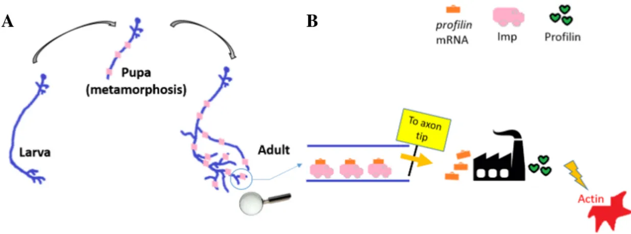
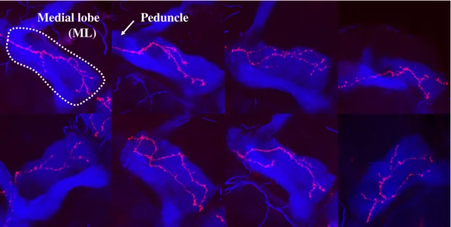
Specifically, we study the γ neurons, a population forming with companion populations -the αβ and α′β′neurons - bilateral structures located in the central Drosophila brain, called the Mushroom Bodies (MBs) (Fig. 2.3A and B). These structures, each composed of roughly 2,000 cells (Aso et al., 2009), are high-order integration centres involved in olfactory learning and memory functions (Heisenberg, 2003). The population of MB γ neurons consists of about 650 cells, and constitutes a model particularly suitable to study axon growth in a population context, as it is the only one in the Mushroom body that goes through the process of remodelling (pruning followed by regrowth). This means that, after an initial growth at larval stages, distal γ axons degenerate during metamorphosis and regrow synchronously, in a short time frame, to establish adult axonal projection patterns (Rabinovich et al., 2016; Watts et al., 2003; Yaniv and Schuldiner, 2016) (Fig. 2.4 C). At this stage, axons regrow in a complex and dense environment constituted of surrounding neuronal populations and glial cells (Aso et al., 2014; Hakim et al., 2014). Furthermore, axon growth is associated with branching, such that adult γ axons typically exhibit a varied number of side branches (Lee et al., 1999). Axon arborization patterns are however not completely stereotypic, as an interesting but unexplained wide range of morphologies is observed within populations of γ neurons (see Fig. 2.5).
As many invertebrate neurons, γ neurons are unipolar (Fig. 2.1). Their cell bodies are clustered at the dorsal posterior surface of the brain, and their dendrites project just beneath, in a structure termed calyx (Fig. 2.3B). Proximally, MB axons fasciculate to form a dense fiber projecting ventrally: the peduncle (Fig. 2.3B). More distally, axons de-fasciculate to innervate the so-called medial lobe (ML), following either straight paths or more tortuous trajectories (Fig. 2.3C). In the medial lobe, they establish a dense network of branches (Figs. 2.3B and C and 2.5).
As described, γ axons grow through two very different physical environments. Firstly in the peduncle, where the axon shafts elongate rigidly in a tight and ordered parallel bundle. Secondly, they enter the ML. In contrast, the ML is a large structure, where the neurites are physically less constrained and develop long terminal branches, giving birth to a big variety of morphologies. This particular conditioning of the neuronal morphology by the physical environment through different stages of neural growth is explored in Koser et al. (2016) for diverse neuron types.
In conclusion, γ neurons represent an accurate biological model to study mechanical constraints and inter-axon interactions during axon development, as they grow as a dense population, forming unstereotypical morphologies in a limited brain area.
2.3.1
Imp as a regulator of axon regrowth and branching
Actin is a fibrous elongated protein composed by smaller subunits (G-actin) that have the ability to assemble and disassemble dynamically. As mentioned in Section 2.1, it is found on the axonal extremities during development and its role is to guide the axon tip through the correct path following internal and external cues, as well as to provide with the force to make the axon extend. There are many actin regulators which, depending on the particular context, can favour or prevent actin fibers assembly or dismantling.
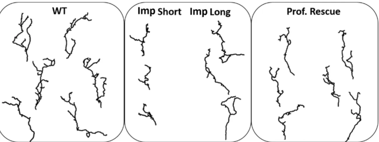
In this study we consider, in addition to wild-type individuals, axons that are mutant for the conserved mRNA transport protein Imp. Molecular and genetic analysis performed in the Besse laboratory have shown that Profilin mRNA, which encodes an actin cytoskeleton regulator (Luo, 2002; Schlüter et al., 1997; Verheyen and Cooley, 1994), is a direct and functional target of Imp and both are key regulators of the Drosophila γ neuron axonal remodelling process, acting on the same molecular pathway. Imp carries pro f ilin mRNA along the axon shaft towards the extremities where the regrowth occurs, enabling Profilin proteins to be synthetized in situ. Fig. 2.4 A and B present a scheme of Imp and Profilin implication during metamorphosis. Medioni et al. (2014) have shown that Imp is essential to allow axonal regrowth of γ neurons during metamorphosis. Our database (Section 2.4) includes imp mutant γ axons, which we used to validate the morphological analysis and different growth hypothesis in this thesis. Furthermore, in Section 4.6 we propose a novel interpretation of the role of Imp in the remodelling process, and its impact on adult γ axon morphology.
Regarding the imp mutant phenotype of a single mutated axon in an otherwise wild-type environment, Medioni et al. (2014) reported that, in adults, about 50% of imp mutants displayed shorter axons than wild-types and failed to reach their target. imp mutants also exhibit an overall loss of branch number and complexity. Interestingly, they observed that
C
Fig. 2.3 Characteristics of adult Mushroom Body γ neurons. (A) Wild-type adult Drosophila brain expressing the membrane-tagged CD8-GFP construct in γ neurons, under the control of the MB009B-Gal4. Nuclei are labelled in white with DAPI. The dotted line on the top view image corresponds to the midline. This images were obtained using the 10x 0.6 NA air objective of a LSM780 NLO Zeiss microscope. Z sections were taken every 0.5 µm, with a xy pixel size of 0.57 µm. 3D projections have been created using FiJi (Schindelin et al., 2012).(B) 3D reconstruction of one Mushroom Body where γ neurons only were labelled. Genotype: MB009B-Gal4/UAS CD8-GFP. This image was obtained using the 40X 1.2 NA water objective and the pulsed laser of a LSM780 NLO Zeiss microscope. Z sections were taken every 0.4 µm, with a xy pixel size of 0.11 µm. The 3D reconstruction was generated using the surface mode of Imaris 8.1 Bitplane software. (C) Axonal arborizations of two individual adult γ neurons labelled by GFP using the MARCM technique. Confocal images taken along the z axis were projected (Maximum Intensity Projection). Dotted lines delimit the shape of the medial lobe. Scale bars: 20 µm in A, 50 µm in B and 10 µm in C.
the over-expression of Profilin in imp mutant backgrounds partially restored the main axon length, but not the branch complexity. imp mutant axons rescued by Profilin over-expression are also included in our database (Section 2.4). These results suggest that Imp controls axonal extension during remodelling at least partly by regulating pro f ilin mRNA expression. However they also suggest that the branching process may be dependent on the regulation of other Imp mRNA targets, yet to be identified. In Section 4.6 we further investigate the role of Imp in γ neuron remodelling. We propose a novel hypothesis on the origin of the short phenotype, and explore the mechanistic reasons for this partial rescue observed after Profilin over-expression.
A B
Fig. 2.4 The role of Imp and Profilin during remodelling. (A) Scheme of a γ neuron remodelling. Imp (pink) is transported to axons during metamorphosis. (B) Schematic representation of the role of Imp (pink) as a pro f ilin mRNA (orange) transport protein. Imp carries pro f ilin mRNA from the neuron body towards its extremities. This allows the production of Profilin proteins in situ, for a rapid control of actin polymerization in the growing tip.
2.4
Database Description
The development and validation of models proposed in this thesis rely on two databases, provided by Florence Besse and Caroline Medioni in the Besse Team at IBV (France). The first one consists of 3D confocal images of individually-labelled γ neurons from adult Drosophilabrains, and the second one of 3D+t image sequences of growing γ axons in the Drosophiladeveloping brain.
(Chapter 4).
The database consists of: 43 wild-type (WT), 45 imp mutants (Imp), 15 imp mutants rescued with Profilin over-expression (Prof Rescue), 15 profilin mutants (Prof), 42 imp mutants rescued by Imp over-expression (Imp Rescue) and 27 un f mutants rescued by T OR (Unf). Both imp and pro f ilin mutations used here are protein-null mutations. Wild-type and impdatabases are used through all this work, while the others served for particular studies. For all the studied mutants, the labelled axon(s) in the images is (are) the only one(s) of the γ population that present the mutation. Thus, we observe mutant axons that have developed within an otherwise wild-type γ neuron environment.
MARCM clones were generated as described in Wu and Luo (2006), using the following fly stocks: hsp-flp, tub-Gal80, FRT19A; 201YGal4,UAScGFP; FRT19A + and FRT19A imp7. Brains were dissected at the adult stage, and stained with anti-GFP (molecular probes life technology; ref A11122) and anti-FasciclinII (1D4, DSHB) primary antibodies, revealed by respectively anti-rabbit Alexa 546 and anti-mouse Cy5 secondary antibodies (see Medioni et al. (2014) for a detailed procedure). Brains were mounted in propyl-galate mounting medium, and imaged with an inverted Zeiss LSM 710 confocal microscope equipped with a 40X/1.1 NA water objective. Z sections were taken every 0.6 to 0.9 µm, with a xy pixel size of 0.09 µm. Fig. 2.5 shows the maximum intensity Z projection of both channels of different images in this database.
2.4.2
4D image sequences of the developing brain
This database consists of movies of the living developing brain, where a single γ axon or a group of axons are visualized via targeted expression of the GFP marker during metamorpho-sis (examples in Fig. 2.6). These data have been used for the branch classification regarding formation and functionality that we present on Chapter 4, as well as to analyse the dynamic aspects of axonal growth as a population.
Medial lobe (ML)
Peduncle
Fig. 2.5 Adult single cell confocal in vivo images. Examples of our database, exhibiting a single adult wild-type γ axon (red) and the dorsal and medial lobes of the MB (blue). The maximal intensity projection in Z is shown for simplicity. The white dotted line in the first image indicates the medial lobe.
To create these movies, brains were dissected out of pupae 24-30 APF (After Pupa Formation) and mounted in a Labtek II chambered coverglass (#155378, Fisher Scientific) in culture medium (Schneider medium, 10% FCS, 1% Antibiotic Antimycotic Solution (Sigma), 200µg/ml insulin (Sigma), 1µg/ml ecdysone (20HE; Sigma) (see Medioni et al. (2015b) for a detailed protocol). Brains were imaged using a Zeiss LSM780 NLO inverted two-photon microscope and a 40X/1.2 NA water objective. Medial lobe regions were imaged. Z sections were taken every 0.8µm and covered the entire medial lobe volume. Z stacks were acquired every 5 minutes over up to 15 hours. Pixel size is 0.13µm.
Drift correction was carried out by Gregoire Malandain (see Medioni et al. (2015a)). For the single axon cases, a two-fold drift compensation was performed. First, maximum intensity (MIP) projections of the acquired images were computed, couple of successive MIPs (that are 2D images) were co-registered with 2D affine transformations, and these transformations were compounded to express all the transformations with respect to a reference image (say the first one). This first step allowed an in-plane drift compensation, but a motion along the Z direction may still exist. A residual translation along the Z direction was then estimated using the multiple transformation strategy described in Medioni et al. (2015a). The compounding of the 2D affine transformation and the Z translation yielded the final 3D drift compensation.
The segmentation of the single axon movies used to quantify the length distribution of dynamic branches (Fig. 4.35 in Section 4.5.1) was also carried out by Gregoire Malandain,
A
B
Fig. 2.6 Video of the developing Drosophila brain. (A) Image sequence extracted from one of the movies in our database, where a single γ neuron from a wild-type brain undergoing metamorphosis is followed, during the regrowth phase. Red arrows point at a branch that shortens with time, while yellow arrows point at a branch that is growing. (B) Detail from another movie. The first two frames show an elongation step, the third one retraction and branching event, and the last one growth again. Arrows shows axonal tips and asterisks, the formation of branches. Scale Bars: 10µm.
In this chapter we propose a deep axonal morphological analysis, and apply it to the study of adult γ axons to understand the behaviour and relevance of main shape features; as well as to further understand the role of Imp and the importance of pro f ilin mRNA expression regulation during remodelling.
To do so, we developed a stochastic framework to exhaustively compare the adult γ axon morphologies between the WT, Imp and Prof Rescue groups (Fig. 3.1). Our framework can be summarized in three steps: i) selection of relevant morphological features describing the axons, ii) stochastically model the behaviour followed by each of the chosen morphological features and estimate the parameters associated to each model from the data (for WT and mutant groups) and iii) classify each individual axon to a group (WT, Imp or Prof Rescue), considering the features separately and altogether using the maximum likelihood of the applied models. The classification results give a quantitative measure of the global similarity or difference between groups (through its confusion matrix). We also apply statistical tests under null hypothesis between the neuron groups for each morphological feature to enrich the analysis. This approach provides both a biological interpretation and a quantification of the resemblance between biological samples, detecting differences as well as similarities between the groups. This framework is general and can be applied to model and characterize different axon types (Fig. 3.2).
Neuron morphological automatic classification has already been addressed in the bibliog-raphy. Kong et al. (2005) proposed an unsupervised clustering of ganglion cells in the mouse retina by the k-means algorithm in order to define cell types. They initially disposed of 26