HAL Id: inserm-00136072
https://www.hal.inserm.fr/inserm-00136072
Submitted on 19 Mar 2007
HAL is a multi-disciplinary open access archive for the deposit and dissemination of sci-entific research documents, whether they are pub-lished or not. The documents may come from teaching and research institutions in France or abroad, or from public or private research centers.
L’archive ouverte pluridisciplinaire HAL, est destinée au dépôt et à la diffusion de documents scientifiques de niveau recherche, publiés ou non, émanant des établissements d’enseignement et de recherche français ou étrangers, des laboratoires publics ou privés.
antibody with dexamethasone and 140 mg/m2 of
melphalan in multiple myeloma: results of a pilot study
including biological aspects.
Jean-François Rossi, Nathalie Fegueux, Zhao Yang Lu, Eric Legouffe, Carole
Exbrayat, Marie-Cécile Bozonnat, Robert Navarro, Ernesto Lopez, Philippe
Quittet, Jean-Pierre Daures, et al.
To cite this version:
Jean-François Rossi, Nathalie Fegueux, Zhao Yang Lu, Eric Legouffe, Carole Exbrayat, et al.. Op-timizing the use of anti-interleukin-6 monoclonal antibody with dexamethasone and 140 mg/m2 of melphalan in multiple myeloma: results of a pilot study including biological aspects.. Bone Mar-row Transplantation, Nature Publishing Group, 2005, 36 (9), pp.771-9. �10.1038/sj.bmt.1705138�. �inserm-00136072�
O
PTIMIZING THE USE OF ANTI-
INTERLEUKIN-6
MONOCLONAL ANTIBODY WITH DEXAMETHASONE AND140
MG/
M²
OF MELPHALANIN MULTIPLE MYELOMA
:
RESULTS OF A
P
ILOTS
TUDYI
NCLUDINGB
IOLOGICALA
SPECTSJean-François Rossi M.D.Ph.D.1*, Nathalie Fegueux M.D. 1, Zhao Yang Lu M.D.Ph.D. 2,Eric Legouffe M.D. 1, Carole Exbrayat M.D. 1, Marie-Cécile Bozonnat3, Robert Navarro M.D. 1, Ernesto Lopez M.D. 1, Philippe Quittet M.D.1, Jean-Pierre Daures M.D.Ph.D.3, Valérie Rouillé1,
Tarik Kanouni1, John Widjenes Ph.D.4, Bernard Klein Ph.D. 2
1-Service d’Hématologie et d’Oncologie Médicale CHU Montpellier, 375, avenue du Doyen Giraud 34295 Montpellier Cédex 05 France
2-Unité de Thérapie Cellulaire et Génique, Hôpital Saint-Eloi, CHU Montpellier Avenue Augustin Fliche, 34056 Montpellier Cédex
3-Institut de Recherche clinique. Unité de Biostatistique, d’épidémiologie et de recherche clinique, 641, avenue du Doyen Giraud, 34093 Montpellier Cédex
4-Diaclone Research, 1, boulevard Alexandre Flemming, BP1985, 25020 Besançon Cédex
*To whom correspondence should be sent E-mail: jf-rossi@chu-montpellier.fr
Summary
Interleukin-6 (IL-6) is a major survival factor for multiple myeloma (MM) cells preventing apoptosis induced by dexamethasone (DEX) or chemotherapy. 24 consecutive patients with MM in first-line therapy received DEX for 4 days, followed by melphalan (HDM: 140mg/m²) and autologous stem cell transplantation (ASCT). The anti-IL-6 monoclonal antibody (mAb) (B-E8) was given till haematological recovery, starting 1 day before DEX. Results were historically compared to MM patients treated with HDM 140mg/m² and 200mg/m². Our results show 1) that B-E8 was able to fully neutralize IL-6 activity in vivo before and after HDM as shown by inhibition of CRP production; 2) no haematological toxicity; 3) a significant reduction of mucositis and fever; 4) a median event-free survival of 35 months and an overall survival 68.2% at 5 years with a median follow-up of 72 months; 5) the overall daily IL-6 production progressively increased on and after 7 days post-HDM, with the increased serum CRP levels. In the 5/24 patients with uncontrolled CRP production, a large IL-6 production was detected (320 µg/day) that could not likely be neutralized by B-E8. These data show the feasibility to neutralize IL-6 in vivo with anti-IL-6 mAb in the context of HDM.
Key words: anti-interleukin-6, multiple myeloma, autologous transplantation Running title: anti-interleukin-6 and high dose melphalan in multiple myeloma.
INTRODUCTION
Multiple myeloma (MM) is still an incurable disease despite the fact that high dose melphalan (HDM) increases five to six-fold the complete response rate leading to a significant increase of the median survival, from 44 months with conventional chemotherapy to 57 months with HDM (1-4). The standard HDM is a combination of melphalan 140 mg/m² plus total body irradiation or melphalan 200 mg/m² (2, 5), both associated with increased toxicity for older patients (6). Tandem HDM has been tested with major improvement of response rate in Phase II studies (7-9) and potent survival benefit in a randomized Phase III study (4). A reduction of tumor contamination of the autograft, using purification of CD34 peripheral blood stem cells (PBSC) is feasible (10, 11), but did not improve survival in a randomized study (12).
Interleukin-6 (IL-6) is a major myeloma survival factor in vitro (13, 14). In particular, anti-IL-6 monoclonal antibodies (mAb) can block the proliferation of primary myeloma cells in short-term culture (15, 16) and IL-6-dependent myeloma cell lines can be reproducibly obtained from MM patients with extramedullary proliferation (17). In vivo, IL-6 is overproduced by the tumor environment in MM patients (18) and serum levels of IL-6 and soluble IL-6 receptors are increased in association with a poor prognosis (19, 20). Thus, anti-IL-6 mAb can be useful to induce myeloma cell apoptosis.
We have previously found that treatments of patients with terminal disease with anti-IL-6 mAb can block the in vivo proliferation of myeloma cells and reduce IL-6-related toxicities (i.e. fever, cachexia) (21-23). A limitation of the anti-IL-6 treatments was the very large production of endogenous IL-6 in some patients with MM that could not be neutralized by the anti-IL-6 mAb (24, 25).
As blocking IL-6 can increase the apoptosis of myeloma cells induced by dexamethasone (DEX) (26) and the drug sensitivity of tumor cells (27, 28), we have investigated the possibility to treat patients having MM with the B-E8 anti-IL-6 mAb, high dose of DEX (40 mg/day for 4 days), and
high dose of melphalan (140 mg/m²) supported by autologous stem cell transplantation (ASCT). The anti-IL-6 mAb was given before and during DEX-HDM to potentiate the apoptotic effect of DEX and HDM. It was also administered for 15 days after HDM to block the production of IL-6 occurring after HDM in association with the haematopoietic recovery. Indeed, the production of IL-6 which occurs after HDM might favour the survival, reparation and growth of the minor fraction of myeloma cells that are not destroyed by HDM. We found that prolonged anti-IL-6 treatment in association with DEX and HDM was feasible, did not affect haematopoietic recovery but reduced HDM-related toxicities. In addition, using the original methodology developed by our group (24, 29, 30), we demonstrated for the first time a dramatic increase in IL-6 production after HDM. These data confirm the potential importance to block IL-6 that is a main myeloma cell survival factor, to avoid the rescue of some myeloma cells after HDM.
PATIENTS AND METHODS
Patients treated with anti-IL-6 mAb
Twenty-four consecutive patients with newly diagnosed MM and justifying a high dose therapy were included in this study, after written informed consent and the agreement of the ethical committee of Montpellier University Hospital. All of them had standard evaluation, including MRI particularly for defining high-risk patients with stage I DS, and having multifocal lesions. These patients had 4 courses of standard VAD (vincristine 0.4 mg/day and doxorubicin 9 mg/m²/day for 4 days by continuous I.V. route and dexamethasone 40 mg/day at days 1-4, 9-12 and 17-20) after diagnosis. Then, patients received cyclophosphamide (4 g/m²) followed by administration of granulocyte colony stimulating factor (Neupogen*, Amgen-Roche, Paris France) at 5µg/kg/day, starting three days after cyclophosphamide until collection of peripheral blood stem cells (PBSC).
The 24 patients received B-E8, an IgG1 murine monoclonal directed against IL-6, prepared by Diaclone Besançon, France, as previously described (21, 22). The patients received DEX (40mg/day from day-5 to day-2), high dose melphalan (140mg/m², at day-2), followed by transplantation of autologous PBSC at day 0. They received 200 mg of anti-IL-6 at day –6 followed by 20 mg/day from day -5 until haematological recovery, corresponding to white blood cell count superior to 106/L. B-E8 was diluted in 100 mL saline serum with 0.5 % human albumin (LFB, Les Ullis, France) and then administered as 1-hour infusion every day at the same time. The dose was chosen according to previous clinical results and pharmacological data (21-25).
From day -6 until haematological recovery corresponding to the last B-E8 injection, patients were hospitalized in the same transplantation unit with standard treatment used in aplasia, including IV antibiotics (cefpirome 2 g x 2 and netilmicine 150 mg x 3/day, plus vancomycin 500 mg x 6/day in case of infection and/or appearance or persistence of fever after 48 h of standard antibiotics, ceftazidim or imipenem-cilastine and/or amphotericin B, being the third line therapy) started as soon
as aplasia (less than 500/mm3 neutrophils) occurred which corresponds to day 3 or 4 after autologous transplantation.
Patient’s population with matched prognostic factors.
From 01/1995 to 05/2001, 167 consecutive MM patients were treated by HDM supported by ASCT in our department (Haematology-Oncology, University Hospital, Montpellier, France). HDM included 140 mg/m² melphalan for older patients, 200 mg/m² melphalan or 140 mg/m² melphalan plus total body irradiation (TBI) for younger patients, or B-E8 anti-IL-6 mAb, DEX and 140 mg/m² melphalan in the current study (anti-IL-6-DEX-HDM140 group). We selected two groups of MM patients with matched prognostic factors as compared with the anti-IL-6-DEX-HDM140 group included in this study, one group of 25 patients treated with 140 mg/m² melphalan (HDM140 group) and one group of 24 patients treated with 200 mg/m² melphalan (HDM200 group). Comparisons were made for response and tolerance.
C Reactive Protein (CRP) evaluation after HDM in patients with MM.
In order to evaluate the duration of IL-6 overproduction in MM patients treated with HDM (200mg/m²), serum CRP levels were measured in 27 MM patients for 15 days after HDM. These patients had no major infection during aplasia. 20 of these 27 patients presented fever ≥ 38°C for less than 4 days. The clinical parameters of these 27 patients were similar to those treated with anti-IL-6 mAb, in term of age (median 62 years, range: 35-68) and haematological recovery (mean duration of neutropenia 12 2 days, for neutrophils < 0.5 x 10⁶/L and thrombopenia 13 2 days, for platelet counts < 20 x 106/L).
Evaluation of clinical and standard biological response
All the patients had standard clinical and biological follow up, including haematological parameters with blood cell counts and measurements of CRP levels every day, and creatinin, liver tests, checked every two days in the serum. Blood, urine and other types of samples were done for identification of infectious organism if necessary. All the patients had an evaluation of the disease including bone marrow aspirate, electrophoresis and immuno-electrofixation of proteins in serum and urine, creatinin, 2microglobulin, calcium, white blood cell count, CRP and Ig measurement in the serum, at diagnosis, at the inclusion, 90 days after ASCT, every 6 months or when clinical events were observed. X-ray evaluation was performed at diagnosis, at the inclusion and 3 months after the end of treatment. MRI imaging of the spine and iliac bones was done at the same periods and every year or in case of clinical progression.
Response was evaluated 90 days after autologous transplantation by using standard criteria (31) and additional definition of very good partial response (VGPR) as previously mentioned (2, 4).
Determination of plasma IL-6 and B-E8 mAb concentrations
Two anti-IL-6 mAb (AH-65 as a capture mAb and B-E4 as a detector mAb) were used in ELISA to determine the concentration of plasma IL-6 as previously described (23, 24). These anti-IL-6 mAb recognized different epitopes on the IL-6 molecule than the epitope recognized by the B-E8 mAb injected to the patients. B-E8 mAb concentrations were also assayed by an ELISA (32).
Calculation of daily IL-6 production in vivo
We used a mathematical model previously developed to calculate total IL-6 production in patients receiving anti-IL-6 mAb (24). In brief, at the concentration of anti-IL-6 mAb reached in the patients’ plasma, all circulating IL-6 is bound to the anti-IL-6 mAb in the form of monomeric complex with a half-life of 3-4 days similar to that of the free antibody. In these patients treated with
anti-IL-6 mAb, IL-6 is no longer eliminated by kidneys or consumed by cells in vivo due to the blockage of its binding to IL-6R. Thus, knowing the amount of anti-IL-6 mAb injected daily and the fluctuations of the concentrations of the free anti-IL-6 mAb and of the IL-6/anti-IL-6 mAb immune complexes, the overall production of IL-6 on day “n” in the whole organism can be estimated by the formula: IL-6 production on day “n” = 2 * M * b ( [IL6n] – b * [IL6n-1] ) / { ( [Mn] – b * [Mn-1] ) * (1+b) } as indicated before (23). In this formula, M, [IL6n], [IL6n-1], [Mn] and [Mn-1] are respectively the daily amount of injected mAb, the IL-6 concentration on day n, the IL-6 concentration on day n-1, the mAb concentration on day n and the mAb concentration on day n-1 respectively. “b” is a coefficient dependent on the half-life of the mAb (b = e -(ln2) / (half-life of MAb) ). If
IL-6 biological activity is not completely blocked in an anti-IL-6-treated patient (in the case of partial blockage of CRP for example), the true overall IL-6 production in vivo is superior to the production estimated by this method.
Statistical methods
Comparisons between the groups of patients (anti-IL-6-DEX-HDM140 group and HDM140 and HDM200 historical control groups) in terms of demographic, efficacy, and safety data were performed using the two-sample t-test or Wilcoxon rank sum test. Overall survival (OS) and event-free survival (EFS) were calculated from the date of diagnosis and from the date of transplant and summarized using Kaplan-Meier curves (33).
RESULTS
Serum CRP levels in MM patients treated with HDM
Figure 1 shows the mean serum CRP levels of 27 patients with MM treated with HDM. The mean CRP levels progressively increased following HDM with a peak observed on day 12 after the ASCT. 21/27 patients had fever superior to 38°5 during aplasia with a correlation between CRP and fever when it was superior to this level. The level of CRP was associated with fever and additional signs including mucositis or other symptoms (data not shown). As CRP production is induced by IL-6 in vivo (15), these data suggest an increase in IL-IL-6 production after HDM in these patients with MM.
Patients
In Table I, is presented a comparison of the clinical characteristics of the group of 24 MM patients treated with anti-IL-6-DEX-HDM140 and two control groups of MM patients with matched prognostic factors treated with either HDM140 or HDM200. Patients’ characteristics were identical between the three groups, except a higher age of patients receiving HDM140 as mentioned in Table I. 13/24 patients had detectable CRP serum level at the inclusion (15.5 6.9 mg/L). The 24 patients received 5.16 1.64 x 10⁶ CD34+ cells/kg (Table II). Patients in the two control groups received ASCT with similar number of CD34+ cells x106 /kg (respectively 4.24 1.30, and 4.40 1.57, respectively in the HDM140 group and HDM200 groups). Duration of anti-IL-6 mAb treatment was dependent on haematological recovery, i.e. neutrophil count > 0.5 x 10⁶/L, platelet count > 20 x 106/Land transfusion independency. The median duration of anti-IL-6 treatment was 17 days (range 18-26).
Clinical evaluation
Feasibility and toxicity
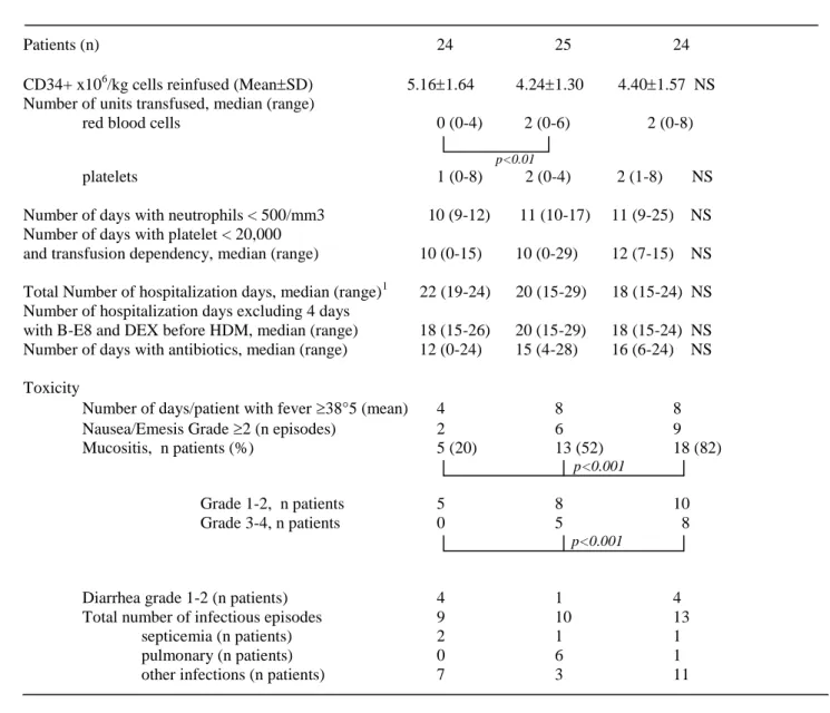
No toxic death, toxicity, or allergic incidents were observed in patients treated with anti-IL-6 mAb, DEX and HDM140. As mentioned in Table II, the number of infectious episodes was not different from those observed in the two control groups of patients who did not receive anti-IL-6 mAb. We observed 2 septicemia (that was rapidly controlled) as compared to 1 septicemia for both control groups. No pulmonary infections were observed in the group of patients treated with anti-IL-6 mAb as compared to respectively anti-IL-6 and 1 episodes in the 2 control groups. No delay for diagnosing infectious episodes was observed, with no difference of duration for antibiotics administration between anti-IL-6 mAb-treated patients and control patients. Of interest, the anti-IL-6 treatment reduced some HDM-related toxicities. Mucositis was significantly less frequent (5/24, 20%) in the group of patients treated with anti-IL6 mAb as compared to the two control groups (13/25, 52%) for HDM140 patients and 18/22 (82%) for HDM200 patients, P < 0.001 (Table II). In addition mucositis was at a lower grade of toxicity (grade 2: 5/24 with no morphin infusion in the group of patients receiving anti-IL-6) as compared to 5/13 and 8/18 patients having grades 3-4 mucositis with morphin infusion in the groups of respectively HDM140 and HDM200 patients. The number of patients who experience nausea/emesis episodes WHO grade 2 was also lower in the group of anti-IL-6 mAb-treated patients: 2/24 patients (6%) as compared to 6/25 patients treated with HDM140 (24%) and 9/24 for the patients receiving HDM200 (37%, P < 0.05). The number of days with fever ( 38°5) was lower and associated with a reduction in temperature degree (median: 4 days versus 8 days, P < 0.05). A higher number of patients having diarrhea WHO grade less than 3 was observed in the group of patients having anti-IL6 mAb (4 patients) as compared to the group treated by HDM140 (1 patient, P < 0.05) but not different from the group of patients treated by HDM200 (4 patients). Thus, blocking IL-6 reduced some HDM-related toxicities, resulting in a better quality of life, particularly for oral intake and daily activity during aplasia.
Haematopoietic recovery
Blocking IL-6 before and after HDM did not delay neutrophil or platelet recoveries compared to MM patients treated with HDM alone, as indicated by the duration of aplasia and hospitalisation and the transfusion need (Table II). This is of particular interest because IL-6 is a haematopoietic cytokine that is involved in megakaryopoiesis. In addition, patients with anti-IL-6 mAb had significantly less transfusion of red blood cell units when compared to the other control groups (1 vs 2, P < 0.01), during the aplasia period, and the time of follow-up, except if any other treatment was administered.
Clinical response
Clinical response was evaluated according to criteria previously defined (29). Out of the 24 patients treated with anti-IL-6-DEX-HDM140, 13 (54%) were in CR (4 patients) or in VGPR (9 patients). Six patients were in PR and 5 had a minimal response. Overall survival and progression-free survival are shown on Figure 2. Median event-progression-free survival was 35 months, and overall-progression-free survival was 68.2% at 5 years with a median follow up 72 months.
Biological evaluation
Determination of serum CRP, anti-IL-6 mAb and IL-6 concentrations
Daily serum CRP levels were evaluated in 16 out of the 24 patients treated with anti-IL-6 mAb. They decreased and were undetectable from day -2 to day 6 for all the patients as shown in Figure 3. For 3 patients (patients 11, 18 and 20), CRP levels were again detectable from day 8 to day 13 of treatment despite the daily injection of the anti-IL-6 mAb.
The concentration of circulating anti-IL-6 mAb was evaluated in 14 patients. As shown in Figure 4, the serum anti-IL-6 mAb concentration reached a maximum one day after the first injection of a charge dose of anti-IL-6 mAb (200 mg). The day-5 mAb concentration varied among the patients from 50 g/L to 90 g/L, likely as a reflect of a patient’s variability of the diffusion volume. Then,
following the injection of a daily dose of anti-IL-6 mAb, the plasma mAb concentration decreased progressively and stabilized on and after day 10 (average: 25.3 g/L, range 12-36 g/l).
Serum IL-6 was measured with an ELISA detecting free IL-6 as well as IL-6 bound to B-E8 mAb. As shown in Figure 5, a low concentration of circulating IL-6 (average 0.33 ng/ml; range: 0.02-2.58 ng/ml) was detected before treatment in 13 out of the 13 patients tested. A slow increase in serum IL-6 was observed from day –5 to day 0, followed by a more rapid increase after day 0 with a peak between day 7 and day 12 (range: 2.36 – 52.10 ng/ml). This serum IL-6 was actually bound to the B-E8 (data not shown).
Calculation of daily IL-6 production
The daily in vivo production of IL-6 was calculated in 14 patients treated with anti-IL-6 mAb using the mathematical model we described before (23). The median daily in vivo IL-6 production for the 14 patients is shown in Figure 6. This daily in vivo production was very low (< 0.35 g/day) from day -5 to day -2 and increased progressively from day 0 till day 8. A wave of IL-6 production appeared in all patients between day 7 and day 13 (median value of 35 g/day, i.e. > 100 fold increase compared to pre-treatment values). This in vivo IL-6 production wave preceded WBC and platelet recovery with a 4- and 10-day delay respectively. In Figure 7 are shown the mean values of IL-6 production for 5-day periods in the 14 patients. In 5 patients (P6, P11, P14, P18 and P20), high daily production of IL-6 was detected (median 320 µg/day, range 192-3350) with a median value of IL-6 production between day 6 and 10 superior to 50 g (high IL-6 producers). Three of them (patients 11, 18 and 20) had uncontrolled CRP production with serum CRP again detectable during anti-IL-6 treatment as shown on Figure 3. All of these 5 patients progressed within 18 months after autologous transplantation, and 2 of them died rapidly with fulminating disease. All of these patients had detectable plasma cell labelling index (≥1.5%, range 1.5-3.6%) and beta-2 microglobulin serum levels superior to 3.0µg/mL. The ratio of mAb/IL-6 serum concentration was inferior to 250 for these 3 patients. We have previously shown that the B-E8 anti-IL-6 mAb was no longer able to
block gp130 IL-6 transducer activation induced by IL-6 when the mAb/IL-6 ratio was less than 250 (29).
DISCUSSION
In this study, we have demonstrated three points: 1) the occurrence and the quantification of an important wave of IL-6 production starting after HDM and ASCT; 2) the ability to block this wave of IL-6 production with anti-IL-6 mAb in a majority of patients; 3) the lack of obvious side effects of the anti-IL-6 mAb treatment in association with high dose DEX, HDM and ASCT. On the contrary, anti-IL-6 mAb reduced some HDM-related toxicities. In addition, our data suggest a link between patients’ potential of IL-6 production and their response rate following HDM supported by ASCT.
Regarding the first point, increased plasma levels of IL-6 after high dose chemotherapy and PBSC were previously documented (34). Plasma IL-6 is only a small part of IL-6 produced in vivo that is mainly consumed by cells and rapidly eliminated by kidney with a 10 minutes half-life (29). In the present study, we were able to estimate the overall IL-6 production in vivo in patients treated with anti-IL-6 mAb. Indeed, the anti-IL-6 mAb captures IL-6 and induces it to circulate in the form of stable monomeric complexes with a 3-4 day half-life (29). Using this observation, the daily overall production of IL-6 could be estimated according to the methodology we previously developed (24). A median IL-6 production of 0.32 g/day (range 0-1 g/day) was found in these patients before DEX-HDM. After HDM and ASCT, median IL-6 production increased progressively 100 fold with a peak of 36 g/day (range 9-246 g/day). In 2 patients, it was superior to 100 g/day. These were close to the doses of recombinant IL-6 known to have biological activity in vivo (i.e. 5 g/kg/day) (35).
The second finding of this study is the feasibility to neutalize IL-6 with the B-E8 anti-IL-6 murine mAb before and after DEX and HDM treatment and ASCT. The B-E8 mAb has a high affinity with IL-6 (1011 M) and was previously shown to block IL-6 activity in patients with MM, renal cell carcinoma, Castleman’s disease and transplantation-related B cell lymphoma (22, 36-38). An interesting feature of B-E8 mAb is the low occurrence of human antibodies able to neutralize
and clear B-E8 mAb. In a series of 15 patients with MM or renal cancer treated with B-E8 mAb, a clearance of B-E8 and escape from the treatment was found in only 3 of 15 patients, due to the occurrence of human antibodies to murine Fc fragments (39). In the other patients, the B-E8 mAb was given for several weeks without clearance or blockage of its activity. In the present study, the rare occurrence of human antibodies to B-E8 mAb should have been prevented due to the profound immuno-suppression induced by HDM. The efficacy of the anti-IL-6 mAb was shown by the complete blockage of CRP production in the majority of patients. CRP is an acute phase protein produced by hepatocytes whose production is strictly under the control of IL-6 in vitro (40) and in
vivo as demonstrated by our group. Indeed, we have shown that in vivo CRP production was
completely blocked in a patient treated with B-E8 anti-IL-6 mAb for two months (21). Serum CRP was again detectable at the end of the treatment. A complete control of CRP production by IL-6 in
vivo was subsequently confirmed in IL-6-knockout mice or other clinical trials with anti-IL-6 mAb
(41). In the current study, CRP levels were undetectable in all 16 evaluated patients until day 6 after HDM. After 6 days post HDM, CRP was again detectable in 3 out of 16 patients and progressively increased in these 3 patients despite the continuous infusion of anti-IL-6 B-E8. The daily IL-6 production was estimated in 2 of these 3 patients and these 2 patients were actually the patients with a very high wave of IL-6 production (> 100 g/day). With such a high IL-6 production, we previously estimated that the antibody was unable to neutralize IL-6 activity in vivo (24). This, given the blockage of CRP production, one can consider that the anti-IL-6 mAb was able to completely block IL-6 activity before and after HDM and ASCT in a great majority of patients.
The third finding is the lack of toxicity of the anti-IL-6 mAb treatment in these patients treated with DEX from day-5 to day-2 before ASCT and HDM at day -2. As emphasized above, we decided to inject DEX before HDM in order to optimize the destruction of myeloma cells. Indeed, the blockage of IL-6 can dramatically increase myeloma cell apoptosis induced by DEX and by HDM in
need in the anti-IL-6-DEX-HDM140 group compared to groups of MM patients treated with the same dose of HDM and grafted with the same amount of CD34 progenitor cells. Thus, despite the fact that IL-6 can increase the growth of haematopoietic stem cells (42), and in particular speed up platelet reconstitution (43), other cytokines are likely produced in these patients making a rapid recovery of platelet and neutrophil counts possible. Another interesting observation is the decreased of HDM and ASCT toxicities. Patients have less mucositis, and in particular no grade 3-4 mucositis. It is not possible here to ascertain whether this decreased toxicity is due the blockage of IL-6 activity for 15 days after HDM and ASCT or to DEX injection for 4 days before HDM and ASCT. This needs to be further investigated in patients treated with DEX-HDM alone or patients treated with DEX-HDM. The number of days with fever was reduced by one half in the anti-IL-6-treated patients. This is not surprising because IL-6 is a pyrogenic cytokine and this was already observed in previous clinical studies with anti-IL-6 mAb (36, 38). Altogether, the quality of life was improved in the anti-IL-6-DEX-HDM140group of patients.
There is a suggestion in this study for a link between the patient potential of IL-6 production and their response rate following DEX-HDM and autologous transplantation, with a rapid progression for the highest producers of IL-6. Furthermore, there is a suggestion for a clinical benefit with an apparent prolonged event-free survival, as compared to that observed in the literature, from 28-31 to 35 months (2, 4). It is important to stress that we documented here for the first time a 100-fold increase of the overall median IL-6 production in vivo after HDM and PBSC graft. According to several in vitro data, this very large in vivo IL-6 production may contribute to the rescue of the few myeloma cells that have escaped from HDM. Indeed, IL-6 may reduce melphalan-induced DNA damages in human myeloma cells (44). One mechanism could be its effect on genetically altered myeloma cells (45), eventually an increase of glutathione S-transferase π as observed in renal cell carcinoma cell lines (28). This possible rescue of chemotherapy-treated tumor cells by IL-6 may explain why serum IL-6 levels post-autologous transplantation were shown to be predictor of poor
response after high dose chemotherapy and autologous transplantation in various cancers (46). We have also to emphasize that the anti-IL-6 mAb might have likely potentiated the apoptotic effect of DEX given just before HDM since numerous studies have shown that IL-6 can block the DEX-induced apoptosis of myeloma cells in vitro. This was the reason why we decided to inject DEX just before HDM treatment.
We are interested to pursue this approach in four directions. First, the use of chimeric or humanized anti-IL-6 mAbs with a long half-life (3 weeks instead of 3-4 days) could prevent a daily injection of the mAb and makes it possible to increase the concentration of circulating mAb in vivo, especially in high IL-6-producer patients. Chimeric or humanized antibodies with a very high affinity with IL-6 are now available from different sources (36). Secondly, we are interested to investigate the contribution of DEX treatment before HDM together with anti-IL-6 mAb treatment. Thirdly, it could be important to prolong the anti-IL-6 treatment for at least one month after ASCT in order to cover the wave of IL-6 production. Our goal is to block IL-6 as long as possible to push a maximum of melphalan-damaged myeloma cells to apoptosis. A simple indicator will be to completely block CRP production for one month after ASCT. In the present study, the treatment was stopped at haematopoietic recovery because we did not know the amount and duration of IL-6 production. Fourthly, it should be interesting to associate anti-IL-6 treatments with intermediate high dose of melphalan (140 mg/m²) for elderly patients, or to use repeated intermediate high dose of melphalan, to increase the dose of melphalan above 200 mg/m² as done by the group from Nantes (47), or to add other active drug to this combination.
In conclusion, the current study demonstrates the feasibility and the interest in blocking the large wave of IL-6 production that occurs after DEX-HDM and ASCT in order to reduce transplant-reduced toxicities and to avoid repair of HDM-damaged residual myeloma cells.
REFERENCES
1. Bataille R, Harousseau JL. Multiple myeloma. N Engl J Med 1997; 336: 1657-1664.
2. Attal M, Harousseau JL, Stoppa AM et al. A prospective, randomized trial of autologous bone marrow transplantation and chemotherapy in multiple myeloma. N Engl J Med 1996; 335: 91-97.
3. Lenhoff S, Hjorth M, Holmberg E et al. Impact on survival of high-dose therapy with autologous stem cell support in patients younger than 60 years with newly diagnosed multiple myeloma: a population-based study. Nordic Myeloma Study Group. Blood 2000; 95: 7-11. 4. Attal M, Harousseau JL, Facon T et al. InterGroupe Francophone du Myelome. Single versus
double autologous stem-cell transplantation for multiple myeloma. N Engl J Med 2003; 349: 2495-502.
5. Moreau P, Facon T, Attal M et al. Comparison of 200 mg/m² melphalan and 8 Gy total body irradiation plus 140 mg/m² melphalan as conditioning regimens for peripheral blood stem cell transplantation in patients with newly diagnosed multiple myeloma final: analysis of the Intergroupe Francophone du Myélome 9502 randomized trial. Blood 2002; 99: 731-735. 6. Sieghel DS, Desikan KR, Mehta J et al. Age is not a prognostic variable with autologous
transplantation for multiple myeloma. Blood 1999; 93: 51-54.
7. Barlogie B, Jagannath S, Vesole DH et al. Superiority of tandem autologous transplantation over standard therapy for previously untreated multiple myeloma. Blood 1997; 89: 789-93. 8. Barlogie B, Jagannath S, Desikan KR, et al. Total therapy with tandem transplants for newly
diagnosed multiple myeloma. Blood 1999; 93: 55-65.
9. Goldschmidt H, Egerer G, Ho AD. Autologous and allogenic stem cell transplantation in multiple myeloma. Bone Marrow Transplant 2000, (25 Suppl 2): S25-6.
10. Rossi JF, Legouffe E, Fegueux N et al. Autologous transplantation (AT) of CD34+ peripheral blood progenitor cells (PBPC) after double (D) high dose chemotherapy (HDC) in multiple
myeloma (MM) is followed by severe immunodeficiency (ID) and high production of interleukin-6 (IL-6) related-C Reactive Protein (CRP) requiring additive immunotherapy (IT). Blood 1996; 88, (10 suppt1), 132a (abstract 516).
11. Lemoli RM, Martinelli G, Zamagni E et al. Engraftment, clinical, and molecular follow-up of patients with multiple myeloma who were reinfused with highly purified CD34+ cells to support single or tandem high-dose chemotherapy. Blood 2000; 95: 2234-2239.
12. Stewart AK, Vescio R, Schiller G et al. Purging of autologous peripheral-blood stem cells using CD34 selection does not improve overall or progression-free survival after high-dose chemotherapy for multiple myeloma: results of a multicenter randomized controlled trial. J Clin Oncol 2001; 19: 3771-3779.
13. Kawano M, Hirano T, Matsuda T et al. Autocrine generation and essential requirement of BSF-2/IL-6 for human multiple myeloma. Nature 1988; 332: 83-87.
14. Klein B, Zhang XG, Lu ZY, Bataille R. Interleukin-6 in multiple myeloma. Blood 1995; 85: 863-872.
15. Klein B, Zhang XG, Jourdan M, Content J et al. Paracrine rather than autocrine regulation of myeloma-cell growth and differentiation by interleukin-6. Blood 1989; 73: 517-526.
16. Klein B, Zhang XG, Jourdan M et al. Interleukin-6 is the central tumor growth factor in vitro and in vivo in multiple myeloma. Eur Cytokine Netw 1990; 1: 193-201.
17. Zhang XG, Gaillard JP, Robillard N et al. Reproducible obtaining myeloma cell lines as a model for tumor stem cell study in human multiple myeloma. Blood 1994; 83: 3654-3663. 18. Portier M, Rajzbaum G, Zhang XG et al. In vivo interleukin 6 gene expression in the tumoral
environment in multiple myeloma. Eur J Immunol 1991; 21: 1759-1762.
19. Greipp P, Leong T, Benett JM et al. Plasmablastic morphology, an independent prognostic factor with clinical and laboratory correlates: Eastern Cooperative Oncology Group (ECOG)
myeloma trial E 9486 report by the ECOG Myeloma Laboratory Group. Blood 1998; 91: 2501-2507.
20. Stasi R, Brunetti M, Parma A, Di Giulio C et al.. The prognostic value of soluble interleukin-6 receptor in patients with multiple myeloma. Cancer 1998; 82: 18interleukin-69-18interleukin-62.
21. Klein B, Widjenes J, Zhang XG et al. Murine anti-IL-6 monoclonal antibody therapy for a patient with plasma cell leukemia. Blood 1991; 78: 1198-1204,.
22. Bataille R, Barlogie B, Lu ZY et al. Biologic effects of anti-interleukin-6 murine monoclonal antibody in advanced multiple myeloma. Blood 1995; 86: 685-691.
23. Lu ZY, Brailly H, Rossi JF, Wijdenes J, Bataille R, Klein B. Overall interleukin-6 production exceeds 7mg/day in multiple myeloma complicated by sepsis. Cytokine 1993; 5: 578-582. 24. Lu ZY, Brailly H, Wijdenes J, Bataille R, Rossi JF, Klein, B. Measurement of whole body
interleukin-6 (IL-6) production : prediction of the efficacy of anti-IL-6 treatments. Blood 86: 3124-3131, 1995.
25. Blay JY, Rossi JF, Widjenes J, Menetrier-Caux C. Role of interleukin-6 in the paraneoplastic syndrome associated with renal cell carcinoma. Int. J. Cancer 1997; 72: 424-430.
26. Frassanito MA, Cussmai A, Iodice G, Dammacco F. Autocrine interleukin-6 production and highly malignant multiple myeloma: relation with resistance to drug-induced apoptosis. Blood 2001; 97: 483-489
27. Borsellino N, Belldegrun A. Bonavida B. Endogenous interleukin-6 is a resistance factor for cis-diamminedichloroplatinum and etoposide-mediated cytotoxicity of human prostate carcinoma cell lines. Cancer Res 1995; 55: 4633-4639.
28. Mizutani Y, Bonavida, B, Koishihara Y et al. Sensitization of human renal cell carcinoma cells to cis- diamminedichloroplatinum(II) by interleukin 6 monoclonal antibody or anti-interleukin 6 receptor monoclonal antibody. Cancer Res 1995; 55: 590-596.
29. Klein B, Brailly H. Cytokine-binding proteins: stimulating antagonists. Immunol Today 1995; 16: 216-220,.
30. Montero-Julian F, Klein B, Gautherot E, Brailly, H. Pharmacokinetic study of anti-interleukin-6 (IL-6) therapy with monoclonal antibodies: enhancement of IL-6 clearance by cocktails of anti-IL-6 antibodies. Blood 1995; 85: 917-924.
31. Blade J, Samson D, Reece D et al. Criteria for evaluating disease response and progression in patients with multiple myeloma traeted with high-dose therapy and haematopoietic stem cell transplantation. Myeloma subcommittee of the EBMT. European bone Marrow Transplant. Br J Haematol 1998; 102: 1115-1123,
32. Widjenes J, Clement C, Klein B et al.. Human recombinant dimeric IL-6 binds to its receptor as detected by anti-IL-6 monoclonal antibodies. Mol Immunol 1991; 28: 1183-1192.
33. Kaplan EL, Meier P. Non parametric estimations from incomplete observations. J Am Stat Assoc 1958; 53: 457.
34. Steffen M, Durken M, Pichlmeier U et al. Serum interleukin-6 levels during bone marrow transplantation: impact on transplant-related toxicity and engraftment. Bone Marrow Transplant 1996; 18: 301-307.
35. Veldhuis GJ, Willemse PH, Sleijfer DT et al. Toxicity and efficacy of escalating dosages of recombinant human interleukin-6 after chemotherapy in patients with breast cancer or non-small-cell lung cancer. J Clin Oncol 1995; 13: 2585-93.
36. Trickha M, Corringham R, Klein B, Rossi JF. Targetted anti-interleukin-6 monoclonal antibody therapy for cancer: review of the rationale and clinical evidence. Clin Cancer Res 2003; 9: 4653-4665.
37. Beck JT, Hsu SM, Wijdenes J et al. Brief report: alleviation of systemic manifestations of Castleman's disease by monoclonal anti-interleukin-6 antibody
.
N Engl J Med 1994; 330: 602-605.38. Emilie D, Widjenes J, Gisselbrecht C et al. Administration of an anti-interleukin-6 monoclonal antibody to patients with acquired immunodeficiency syndrome and lymphoma: effects on lymphoma growth and on B clinical symptoms. Blood 1994; 84: 2472-2479.
39. Legouffe E, Liautard J, Gaillard JP et al. Human anti-mouse antibody response to the injection of murine monoclonal antibodies against IL-6. Clin Exp Immunol. 1994; 98: 323-329.
40. Castell JV, Gomez-Lechon MJ, David M et al. Acute phase response of human hepatocytes: regulation of acute phase protein synthesis by interleukin-6. Hepatology 1990; 12:1179-1186. 41. Kollet O, Aviram R, Chebath J. The soluble interleukin-6 (IL-6) receptor/IL-6 fusion protein
enhances in vitro maintenance and proliferation of human CD34+CD38-/low cells capable of repopulating severe combined immunodeficiency mice. Blood 1999; 94: 923-931.
42. Sun L, Liu X, Qiu L et al. Administration of plasmid DNA expressing human interleukin-6 significantly improves thrombocytopoiesis in irradiated mice. Ann Hematol 2001; 80: 567-572.
43. Kaser A, Brandacher G, Steurer W et al. Interleukin-6 stimulates thrombopoiesis through thrombopoietin: role in inflammatory thrombocytosis. Blood 2001; 98: 2720-2725.
44. Efferth T, Fabry U, Osieka R. Interleukin-6 affects melphalan-induced DNA damage and repair in human multiple myeloma cells. Anticancer Res 2002; 22: 231-234.
45. Rowley, M., Liu, P., Van Ness, B. Heterogeneity in therapeutic response of genetically altered myeloma cell lines to interleukin-6, dexamethasone, doxorubicin and melphalan. Blood 96: 3175-3180, 2000.
46. Tegg EM, Griffiths AE, Lowenthal RM et al. Association between high interleukin-6 levels and adverse outcome after autologous haemopoietic stem cell transplantation. Bone Marrow Transplant 2001; 28: 929-933.
47. Moreau P, Milpied N, Mahé B et al.. 220mg/m² melphalan followed by peripheral blood stem cell transplantation in 27 patients with advanced multiple myeloma. Bone Marrow Transplant 1999; 23: 1003-1006.
48. Durie BGM, Salmon SE. A critical staging system for multiple myeloma: Correlation of measured myeloma cell mass with presenting clinical features, response to treatment, and survival. Cancer 1975; 36: 842-854.
FIGURE AND TABLE LEGENDS
Figure 1. Serum C-RP levels after high dose melphalan and ASCT in patients with multiple myeloma. Data are mean values of plasma CRP for 27 patients.
Figure 2. Overall survival in patients treated by anti-IL-6 mAb, DEX, HDM140 and ASCT and patients treated with HDM140 or HDM200 and ASCT.
Figure 3. Serum CRP levels in patients treated with anti-IL-6 mAb, DEX, HDM140 and ASCT. Figure 4. Anti-IL6 serum concentration in patients treated with anti-IL-6 mAb, DEX, HDM140 and ASCT.
Figure 5. Serum IL-6 concentration in patients treated with anti-IL-6 mAb, DEX, HDM140 and ASCT.
Figure 6. Median values of daily IL-6 production (µg/day) in patients treated with anti-IL-6 mAb, DEX, HDM140 and ASCT.
Figure 7. Daily IL-6 production (µg/day) measured at different time (Day-5 to Day 0, Day 1 to Day 5 and Day 6 to Day 10) in patients treated with anti-IL-6 mAb, DEX, HDM140 and ASCT.
Table I. Clinical characteristics of the patients treated by anti-IL-6 mAb, DEX and HDM140, HDM140 and HDM200.
Table II. Haematological recovery and toxicity in the three groups of patients (i.e., anti-IL-6, DEX and HDM140, HDM140 and HDM200).
Figure 1
CRP variation post high dose melphalan (mean values of CRP for 27 patients)
0 20 40 60 80 0 2 5 8 11 14
Days after autologous transplantation
C R P (m g /L )
Figure 2 Y e a rs 0 2 4 6 8 1 0 1 2 1 4 S u rv iv a l 0 ,0 0 ,2 0 ,4 0 ,6 0 ,8 1 ,0 S u rv iv a l E F S
27 Figure 3 0 5 10 15 20 25 30 35 -6 -4 -2 0 2 4 6 8 10 12 14
Days before or after ASCT
S e rum C R P ( m g/ L) P1 P3 P5 P6 P7 P9 P10 P11 P12 P13 P14 P15 P16 P17 P18 P20
ASCT
Anti-IL-6 mAb
loading dose
Figure 4
Serum Anti-IL-6 mAb concentration
0 20 40 60 80 100 120 -7 -6 -5 -4 -3 -2 -1 0 1 2 3 4 5 6 7 8 9 10 11 12 13 14
Days before and after ASCT
A n ti -I L -6 mA b ( mic ro g /L ) P3 P5 P6 P8 P9 P10 P12 P13 P14 P15 P16 P17 P18 P20
ASCT
Anti-IL-6 mAb
loading doseFigure 5
Serum IL-6 in patients treated with Anti-IL-6 mAb
0 10 20 30 40 50 60 70 -6 -5 -4 -3 -2 -1 0 1 2 3 4 5 6 7 8 9 10 11 12 13 14
Days before and after ASCT
IL-6
(ng/m
L)
P3 P5 P6 P8 P10 P12 P13 P14 P15 P16 P17 P18 P20 ASCT Anti-IL-6 mAb Charge doseFigure 6 0 10 20 30 40 50 60 -7 -5 -3 -1 1 3 5 7 9 11 13
Days before and after ASCT
D a il y I L-6 prod uc ti on ( µ g/ da y )
Anti-IL6
<--->
ASCT
Figure 7 0 20 40 60 80 100 120 140 160 P3 P5 P6 P8 P9 P10 P11 P12 P13 P14 P15 P16 P18 P20
Patients
IL -6 p ro d u c ti o n (m g /d a y )Mean values from Day-5 to Day 0 Mean values from Day1 to Day5 Mean values from Day6 to Day10
Table I.
DEX-Anti-IL-6-HDM140 HDM140 HDM200
Patients (n) 24 25 24
Male/female 13/11 13/12 14/10 NS Median age (range) 52 (36-67) 63 (51-69) 54 (35-70)
p<0.01
IgG (%) 15 (62) 14 (56) 11 (46) NS IgA (%) 5 (20) 5 (20) 7 (29) NS Light chain (%) 4 (16) 4 (16) 5 (21) NS / (%) 17/7 17/8 15/8 NS Clinical status at diagnosis
DS Stage (%) IIIA 12 (50) 12 (48) 12 (50) NS IIIB 2 (8) 3 (12) 2 (8) NS IIA 4 (17) 6 (24) 5 (21) NS IA1 6 (25) 4 (16) 5 (21) NS Median 2m (range) 4 (1.3-26.7) 3.7 (3-12.5) 4 (1.4-10.3) NS Median CRP (range) 5 (3-80.8) 3 (2-48) 6 (3-15.5) NS
HDM = High dose melphalan DS= Durie Salmon staging (48) NS : not significant
Table II.
DEX-Anti-IL-6-HDM140 HDM140 HDM200
Patients (n) 24 25 24
CD34+ x106/kg cells reinfused (MeanSD) 5.161.64 4.241.30 4.401.57 NS Number of units transfused, median (range)
red blood cells 0 (0-4) 2 (0-6) 2 (0-8) p<0.01
platelets 1 (0-8) 2 (0-4) 2 (1-8) NS Number of days with neutrophils < 500/mm3 10 (9-12) 11 (10-17) 11 (9-25) NS Number of days with platelet < 20,000
and transfusion dependency, median (range) 10 (0-15) 10 (0-29) 12 (7-15) NS Total Number of hospitalization days, median (range)1 22 (19-24) 20 (15-29) 18 (15-24) NS Number of hospitalization days excluding 4 days
with B-E8 and DEX before HDM, median (range) 18 (15-26) 20 (15-29) 18 (15-24) NS Number of days with antibiotics, median (range) 12 (0-24) 15 (4-28) 16 (6-24) NS Toxicity
Number of days/patient with fever 38°5 (mean) 4 8 8 Nausea/Emesis Grade 2 (n episodes) 2 6 9 Mucositis, n patients (%) 5 (20) 13 (52) 18 (82)
p<0.001
Grade 1-2, n patients 5 8 10
Grade 3-4, n patients 0 5 8
p<0.001
Diarrhea grade 1-2 (n patients) 4 1 4 Total number of infectious episodes 9 10 13
septicemia (n patients) 2 1 1
pulmonary (n patients) 0 6 1
other infections (n patients) 7 3 11
HDM= High dose melphalan
1-Patients having anti-IL-6 mAb before high dose melphalan were hospitalized 4 additional days because of a Phase II protocol, avoiding any clinical problem.