Immune stealth-driven O2 serotype prevalence and
potential for therapeutic antibodies against
multidrug resistant
Klebsiella pneumoniae
Meghan E. Pennini
1
, Anna De Marco
2
, Mark Pelletier
1
, Jessica Bonnell
1
, Romana Cvitkovic
1
, Martina Beltramello
2
,
Elisabetta Cameroni
2
, Siro Bianchi
2
, Fabrizia Zatta
2
, Wei Zhao
1
, Xiaodong Xiao
1
, Maria M. Camara
1
,
Antonio DiGiandomenico
1
, Elena Semenova
1
, Antonio Lanzavecchia
3
, Paul Warrener
1
, JoAnn Suzich
1
,
Qun Wang
1
, Davide Corti
2
& C.Kendall Stover
1
Emerging multidrug-resistant bacteria are a challenge for modern medicine, but how these
pathogens are so successful is not fully understood. Robust antibacterial vaccines have
prevented and reduced resistance suggesting a pivotal role for immunity in deterring
anti-biotic resistance. Here, we show the increased prevalence of
Klebsiella pneumoniae
lipopo-lysaccharide O2 serotype strains in all major drug resistance groups correlating with a paucity
of anti-O2 antibodies in human B cell repertoires. We identify human monoclonal antibodies
to O-antigens that are highly protective in mouse models of infection, even against heavily
encapsulated strains. These antibodies, including a rare anti-O2 speci
fic antibody,
syner-gistically protect against drug-resistant strains in adjunctive therapy with meropenem, a
standard-of-care antibiotic, confirming the importance of immune assistance in antibiotic
therapy. These
findings support an antibody-based immunotherapeutic strategy even for
highly resistant
K. pneumoniae infections, and underscore the effect humoral immunity has on
evolving drug resistance.
DOI: 10.1038/s41467-017-02223-7
OPEN
1MedImmune, 1 Medimmune Way, Gaithersburg, MD 20878, USA.2Humabs BioMed SA, a subsidiary of Vir Biotechnology, Inc., Via Mirasole 1, 6500 Bellinzona, Switzerland.3Institute for Research in Biomedicine, Università della Svizzera Italliana, Via Vincenzo Vela 6, 6500 Bellinzona, Switzerland. Correspondence and requests for materials should be addressed to Q.W. (email:qunwang2001@hotmail.com) or to D.C. (email:davide.corti@humabs.ch) or to C.K.S. (email:stoverk@medimmune.com)
123456789
A
mong the most problematic multidrug-resistant (MDR)
bacterial pathogens are the Gram-negative
carbapenem-resistant enteric bacteria (CRE), including Klebsiella
pneumoniae
1, 2. K. pneumoniae colonizes the lower
gastro-intestinal tract, from which it can disseminate, particularly when
the commensal microbiota is disrupted by antibiotic treatment or
when a patient is immunocompromised
3. More than a third of K.
pneumoniae clinical isolates express extended spectrum
beta-lactamases (ESBL) and are resistant to third generation
cepha-losporins, aminoglycosides, tetracycline, and other antibiotics
4.
CRE isolates are additionally resistant to carbapenems and are
strongly correlated with poor patient outcome
5,6, with mortality
rates as high as 60%.
While the majority of effort on understanding the spread of
antibiotic resistance has focused on the acquisition of mutations
or resistance genes that directly affect drug susceptibility, the
most successful resistant strains clearly have additional
adapta-tions. For example, the successful spread of the CRE sequence
type 258 (ST258) clonal group is the subject of intense study
because the diverse resistance genes this group carries are not
unique to this lineage
7,8. In addition to bacterial factors, there is
growing evidence demonstrating a link between host immunity
and reduced antibiotic resistance. Since the onset of routine
vaccinations against Haemophilus influenzae, Streptococcus
pneumoniae, and Neisseria meningitidis, resistance within these
organisms has been reduced or nearly eradicated
9. Effective
immunity can decrease the severity and/or duration of an
infec-tion thus lessening the need for antibiotic interveninfec-tion and
reducing the selective pressure imparted by antibiotic exposure.
Host humoral immunity can also work in concert with antibiotic
therapy to synergistically clear drug-resistant pathogens that
cannot be cleared with antibiotic alone
10,11.
Lipopolysaccharide (LPS) is a critical component of the
Gram-negative bacterial outer membrane. The earliest antibody-based
strategies against Gram-negative pathogens targeted the
con-served Lipid A component of the LPS inner core with the aim of
neutralizing lipid A proinflammatory properties, but these efforts
failed for a variety of reasons
12–15. Beyond the conserved inner
core, there are at least seven distinct Klebsiella LPS serotypes
defined by unique O-antigen structures
16. K. pneumoniae O1 and
O2 LPS O-antigens share the
D-galactan I disaccharide unit, but
O1 also includes the
D-galactan II disaccharide and is therefore
more complex in structure and larger in size
17, 18. In previous
target agnostic efforts to identify antibodies against K.
pneumo-niae
19, we found that LPS serotype-specific antibodies were the
most potent in promoting bacterial killing in vitro.
In this study, we survey a large collection of clinical isolates
to determine the relative prevalence of LPS serotypes,
particu-larly in multidrug-resistant isolates. We
find that LPS O2
antigen serotype has increased two to threefold in prevalence in
both K. pneumoniae ESBL and CRE multidrug-resistant strain
groups relative to the susceptible group. This
finding is
sur-prising as strains expressing the O2-antigen, including
ST258 strains, are more sensitive to human serum killing than
the related O1 antigen expressing strains. We demonstrate
ST258 strains are almost exclusively the O2 serotype, but the
overall increased O2 prevalence is not limited to ST258. These
data suggest a selective advantage for the LPS O2-antigen type
in the context of antibiotic pressure. We
find that O2 strain
prevalence may be explained by its reduced immunogenicity
that imparts a stealth advantage against antibody-driven
mechanisms of clearance. In addition, we identify
serotype-specific anti-O1 and anti-O2 human monoclonal antibodies
(mAbs) that exhibit potent opsonophagocytic killing of K.
pneumoniae strains and provide protection in mouse models of
infection. These data indicate that protective non-capsular
mAbs targeting less diverse anti-O antigen types expressed by
MDR strains can be identified. Importantly, these
serotype-specific O1 and O2 antibodies synergistically augment
anti-biotic therapy even against drug-resistant strains, further
sup-porting an important role for antibody-mediated immunity in
the context of antibiotic therapy. Together, these
findings
support that O-antigen immunogenic stealth provides a
selec-tive advantage to drive multidrug resistance and that protecselec-tive
antibodies can synergize with antibiotic therapy, thus
under-scoring the importance of humoral immunity to drug-resistant
bacteria.
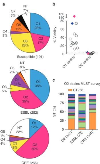
NT 7% O7 5% O5 11% O4 3% O1 28% 150 140 80 60 % Viability 40 20 0 O1 strains O2 (242)ESBL (73)CRE (144) O2 strains O2 17% O3 28% NT 8% NT 22% O5 2% ESBL (252) O4 11% O2 35% O1 38% O1 12% O3 5% O3 9% O5 1% O4 4% O2 50% CRE (266) Susceptible (191) O2 strains MLST survey ST258 100 75 ST (%) 50 25 0a
b
c
Fig. 1Klebsiella pneumoniae serotype O2 prevalence in MDR strains. a LPS serotype-specific antibodies were used to determine the serotype of 709 K. pneumoniae clinical isolates from 38 different countries by western blot. Pie charts represent the distribution of each serotype as a percentage of the total in each antibiotic resistant group. Susceptible, ESBL and CRE strains are categorized based on their MIC profile as described in Methods. More information about the source of the isolates is listed in Supplementary Table1.b Serum sensitivity assays were performed in 45% pooled human sera using a panel of 12 O1 isolates, 12 O2 isolates and two separate isogenic pairs genetically converting an O2 strain to O1 (Kp961842, magenta diamonds) or an O1 strain to O2 (Kp1131115, blue diamonds). Percent viability is the ratio between CFUs present in buffer control media and CFUs present in human sera after 2 h growth.c A subset of serotype O2 isolates was further analyzed by Sanger or next generation sequencing to determine ST types. The CRE ST258 sequence type is represented by the shaded orange portion of each bar to highlight the percent of ST258 isolates observed in each category
Results
O2 LPS serotype prevalence in multidrug-resistant strains. We
surveyed O-antigen serotype prevalence in a geographically
diverse strain collection consisting of 709 clinical isolates
col-lected between 2012 and 2014, from 162 different hospitals in 38
countries, spanning six continents and from a variety of body
infection sites (Fig.
1
a, Supplementary Table
1
). Isolates were
intentionally selected to represent comparable numbers of MDR
(both ESBL and CRE) and fully susceptible isolates to generate
the
first large-scale analysis comparing serotype distribution as a
function of antibiotic resistance. The serotype distribution
observed among antibiotic susceptible strains was largely in line
with serotype frequencies previously determined in the 1990s,
including the predominance of the O1 serotype
20,21. However,
with the development of ESBL and later emergence of CRE
strains in the last two decades, O2 serotype appears to be the
dominant serotype within both of these distinct MDR bacterial
populations in comparison to the susceptible strain population
(Fig.
1
a). This result is unexpected in light of previous
findings
that O1 strains are much more resistant to complement
depen-dent killing than other O serotypes
22–25. Therefore, we evaluated
the relative serum susceptibility of randomly selected O1 and
O2 strains from the above clinical isolate collection to determine
if O2 strains were resistant to human serum. However, we found
O2 strains were much more sensitive to human serum killing
than strains expressing the related O1 serotype (>2000 times
more sensitive on average, Fig.
1
b). Furthermore, isogenic
con-version of an O1 strain (Kp1131115) to O2 by deletion of the
wbbYZ locus resulted in a dramatic 21-fold decrease in serum
resistance, and reciprocal conversion of an O2 strain (Kp961842)
to O1 by the addition of these genes resulted in a 90-fold increase
in serum resistance (Fig.
1
b).
One explanation for the dominance of the O2 serotype within
the MDR population could be an association with clonal
outbreaks, such as the pervasive CRE Klebsiella global
“super
clone” ST258
31–33. While we found the vast majority (85%) of
ST258 isolates to be of the O2 serotype, only 49% of the CRE O2
isolates and just 5% of ESBL O2 isolates typed as ST258 (Fig.
1
c).
Therefore, simple clonal expansion does not wholly explain the
O2 prevalence in MDR strains, particularly within the ESBL
subset. While Klebsiella carbapenem resistance is most often
attributed to the acquisition of plasmid encoded kpc genes, other
genes such as oxa and the emerging ndm
34,35are also of concern.
Interestingly, O2 was also the predominant serotype in isolates
that carry any of these genes (Supplementary Fig.
1
b). These data
suggest therapeutics targeted against O2 LPS could provide better
coverage against MDR isolates than those specifically targeting
only a major sequence type such as ST258.
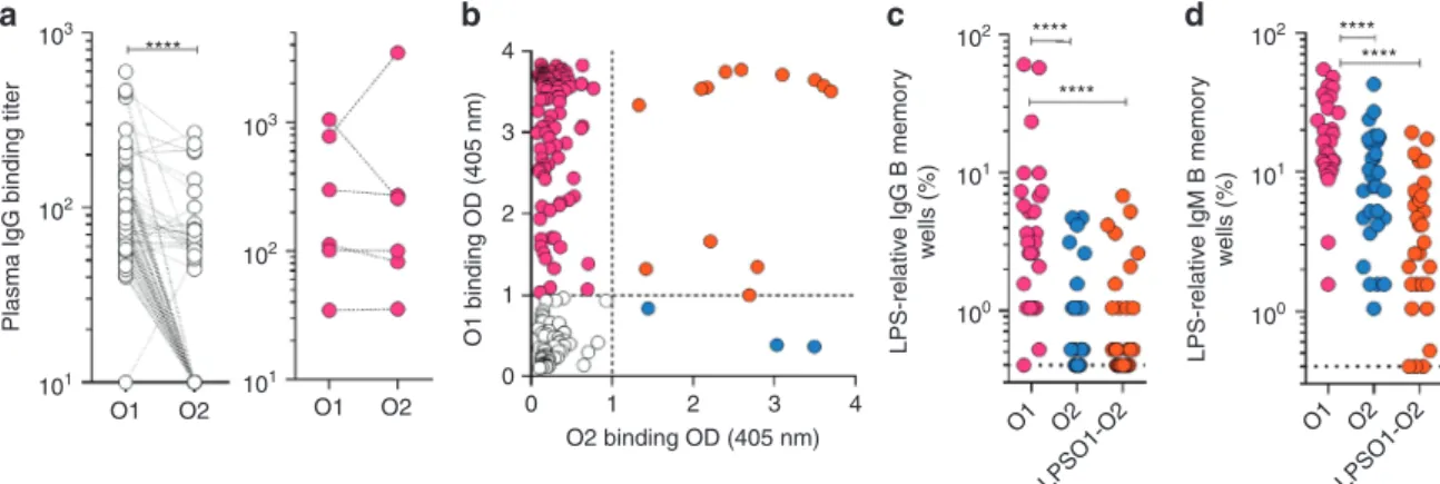
Klebsiella O2 LPS is less immunogenic than O1 LPS. O2 MDR
prevalence suggests an unrecognized selective advantage for these
strains, particularly since it was confirmed that O2 strains are
markedly more serum susceptible and because this increased
prevalence is not entirely attributable to the success of a particular
clone. While identifying antibodies for our serotype screen, we
observed a much lower frequency of O2 specific B cells compared
to O1 or O1/O2 cross reactive B cells, possibly due to a lower
immunogenicity of the O2-antigen. To address this, human
immune responses to O1 or O2 LPS were determined using
plasma samples from 103 healthy donors and 6 O2 KP infected
ICU patients. Titers indicated a dominant IgG response to O1
LPS compared to O2 (Fig.
2
a, c, d). Of note, 5 out of 6 ICU
plasma samples had anti-O2 LPS IgG antibodies, but those titers
were paralleled by high titers of anti-O1 antibodies suggesting a
significant fraction cross-react with O1 LPS (Fig.
2
a). We also
performed a clonal analysis using in vitro polyclonal stimulation,
which selectively activates memory B cells in unfractionated
tonsillar lymphocytes (antigen-specific memory B cell repertoire
analysis, AMBRA
36), (Fig.
2
b–d, Supplementary Fig.
2
). The
frequency of LPS reactive memory B cells was higher in the IgM
repertoire as compared to the IgG repertoire (Fig.
2
c, d),
con-sistent with the concept that the IgM B cell memory repertoire is
dominated by anti-polysaccharide antibodies. Interestingly, the
frequency of O2-specific memory B cells, both IgM and IgG, was
significantly lower as compared to O1 specific. Together, these
results suggest that O2 specific antibody responses may be
103 **** 103 102 **** **** **** **** 101 100 102 101 100 4 3 2 1 0 0 1 2 3 4 O2 binding OD (405 nm) 102 102 101 101 O1 O2 O1 LPSO1-O2 O2 O1 LPSO1-O2 O2 O1 O1 binding OD (405 nm) LPS-relativ e IgG B memor y w ells (%) LPS-relativ e IgM B memor y w ells (%) O2
Plasma IgG binding titer
a
b
c
d
Fig. 2 Analysis of the human antibody response toK. pneumoniae O1 and O2 LPS. a O1 and O2-specific IgG antibodies were measured by ELISA in plasma derived from 103 healthy donors (left panel) and 6 ICU patients (right panel) diagnosed with O2K. pneumoniae infection. Shown are the endpoint titers based on serial dilutions. Lines between data points indicate data acquired from the same sample.b–d Analysis of antigen-specific repertoires (AMBRA, antigen-specific memory B cell repertoire analysis) from tonsillar IgG and IgM memory B cells of 33 donors (30 of the 33 were available for IgM analysis, Supplementary Fig.2). Total tonsillar lymphocytes were plated and stimulated with R848 and IL-2 and the 10-day culture supernatants (192 cultures per donor) were analyzed by ELISA for the presence of IgG or IgM antibodies that bind to O1 LPS, O2 LPS or uncoated control plates. The OD values obtained from the analysis of uncoated plates were subtracted from the analysis of the O1 and O2 specific responses. b Shown are ELISA OD values of individual cultures from the analysis of one representative donor. Cut-off values are indicated by a dotted line. O1, O2 and O1/O2 reactive cultures are represented as magenta, blue and orange circles, respectively.c, d Each data point represents the frequency of the cultures (n = 192) scoring as positive for IgG (c) or IgM (d) reactivity to O1 or O2 LPS for each of the donors analyzed, according to the cut-off values shown in b. Data was analyzed using the Wilcoxon matched-pair signed rank test to assess significance. ****p < 0.0001
disproportionately lower than O1 or O1/O2 cross-reactive
anti-body responses.
To further investigate the immunogenicity of O1 and O2, mice
and rabbits were immunized with purified O1 or O2 LPS. While
O1 LPS-immunized animals had high titers against O1 LPS, O2
LPS-immunized animals had very low titers against O2 LPS
(Fig.
3
a, b). The poor antibody response to O2 LPS in animals is
consistent with the human data. Mice were also immunized with
either member of an isogenic O1/O2 bacteria pair. Mice
immunized with the O1-expressing wild-type (WT) strain
generated high titers of antibodies against O1 LPS, while those
immunized with the isogenic O2-expressing
ΔwbbYZ strain did
not generate a detectable response towards O2 LPS (Fig.
3
c).
Either immunization generated equivalent titers to outer
membrane protein A (OmpA) and to a total carbohydrate extract
(Fig.
3
d). Additionally, both LPS serotypes stimulated a
NF-κB-driven luciferase reporter gene to the same degree, indicating that
O2 LPS is not defective in stimulating TLR4-driven inflammatory
0.8 0.6 0.4
Binding to O1 LPS (OD)
Binding to O1 LPS (OD) Binding to O2 LPS (OD)
0.2 0.0 2.5 2.0 1.5 1.0 0.5 0.0 Binding to O1 LPS (OD) 2.5 2.0 1.5 1.0 0.5 0.0
Binding to KP OmpA (OD)
4 3 2 1 0 2.5 2.0 1.5 1.0 0.5 0.0 Binding to O2 LPS (OD) 2.5 2.0 1.5 1.0 0.5 0.0
Binding to KP lysate (OD) Macrophage activation (%)
2.5 1500 1000 500 0 2.0 1.5 1.0 0.5 0.0 0.8 0.6 0.4 Binding to O2 LPS (OD) 0.2 0.0 CFA/IFA Ctrl O1 LPS O2 LPS CFA/IFA Ctrl O1 LPS O2 LPS CFA/IFA Ctrl WT (O1) ΔwbbYZ (O2) WT (O1) ΔwbbYZ (O2) WT (O1) ΔwbbYZ (O2) WT (O1) O1 LPS O2 LPS ΔwbbYZ (O2) O1 LPS O2 LPS CFA/IFA Ctrl O1 LPS O2 LPS 10–5 10–4 10–3 10–2 10–1
Log [serum dilution]
10–5
10–6 10–4 10–3 10–2 10–1
Log [serum dilution]
10–5 10–6 O1 O2 Core Core D-galactan I D-galactan II D-galactan I LPS O-antigen repeats 180 O1 O2 64 19 6 n n n wbbYZ 10–4 10–3 10–2 10–1
Log [serum dilution]
10–5
10–6 10–4 10–3 10–2 10–1 10–3 10–2 10–1 100 101 102 103
Log [serum dilution] (LPS) (ng/ml)
10–5
10–6 10–4 10–3 10–2 10–1
Log [serum dilution]
10–5 10–4 10–3 10–2 10–1
Log [serum dilution]
10–2 10–1 100 101 102
Log [serum dilution]
10–2 10–1 100 101 102
Log [serum dilution]
a
b
c
d
e
responses (Fig.
3
e). O2 LPS is less complex in structure and has a
lower molecular weight than O1 LPS (Fig.
3
f, g) which may
contribute to its reduced antigenicity, consistent with prevailing
findings that T-cell-independent type 2 antigens require larger
molecular weight, repetitive antigenic epitopes to effectively
cross-link B-cell receptors to stimulate antibody responses
24,37, 38. Together, these data illustrate the weak antigenicity of
O2-antigen and suggest that less antibody-mediated protection
against O2 strains may contribute to their higher prevalence,
particularly among recently emerging MDR strains against which
antibiotic therapy is marginal.
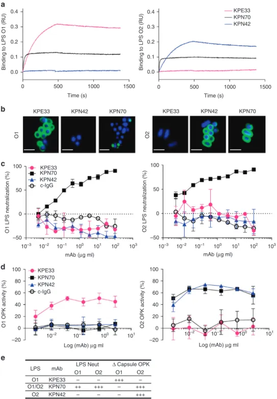
O1 and O2 LPS specific mAbs mediate opsonophagocytic
killing. In an effort to
find functional protective mAbs, human
peripheral blood and tonsil memory B cells were next isolated
based on reactivity against purified O1 and O2 LPS, the most
prevalent serotypes found in clinical isolates (Fig.
1
a). All
anti-LPS mAbs isolated were IgG2, consistent with the concept that
this is the predominant IgG subclass produced in response to
polysaccharides, but were converted and recombinantly expressed
as human IgG1 for functional analysis. Several O1/O2
cross-reactive antibodies were identified and exemplified by mAb
KPN70 which exhibited very high binding affinity to both O1
(~KD
= 0.18 nM) and O2 (~KD = 0.06 nM) LPS, likely
recogniz-ing the shared O1/O2 polysaccharide component
D-galactan I
(Fig.
4
a). Similarly, anti-O1 mAbs with strong binding specificity
were readily identified and exemplified by mAb KPE33 (~KD =
1.8 nM). Consistent with the above data, O2 specific mAbs were
exceedingly rare. Nevertheless, mAb KPN42 was isolated and
determined to be highly specific for O2 LPS (~KD = 4.3 nM).
Further binding analysis on O1 LPS showed no competition
between KPE33 and KPN70, indicating that they recognize
dif-ferent epitopes on O1 LPS (Supplementary Fig.
3
). Similarly,
KPN42 did not block KPN70 binding to O2 LPS, however, the
higher affinity mAb KPN70 partially blocked the binding of
KPN42. This uni-directional blocking might be due to allosteric
or steric hindrance caused by the binding of a high affinity mAb
(in this case KPN70) near the binding region of a lower affinity
mAb (KPN42). Overall, these binding data suggest recognition of
distinct, albeit proximal, epitopes on O2 LPS by mAbs KPN70
and KPN42, and on O1 LPS by mAbs KPN70 and KPE33.
Binding to whole bacteria was also analyzed by confocal
micro-scopy. KPE33 and KPN42 uniformly bound the bacterial surface
of encapsulated O1 and O2 strains, respectively, while the
cross-reactive mAb KPN70 displayed a similar uniform pattern on the
O2 strain, but exhibited a punctuate staining pattern on the
O1 strain (Fig.
4
b). The reduced binding of KPN70 to the surface
of O1 strains compared to purified O1 LPS is likely due to limited
accessibility of this epitope in the context of O1 vs. O2 whole
bacteria. In support of this hypothesis, KPN70 bound to O2 but
not O1 strains as measured by
flow cytometry (Supplementary
Fig.
6
a). In addition, strains that were converted from O1 to
O2 showed enhanced KPN70 binding, and strains that were
converted from O2 to O1 had reduced binding (Supplementary
Fig.
6
b). O-antigen was confirmed to be the target antigen of each
of the mAbs (KPE33, KPN42 and KPN70) as they do not bind to
mutant strains deficient in O-antigen expression (ΔwecA
mutants, Supplementary Fig.
6
c).
Additional assays were performed to assess the functional
activity of the mAbs. LPS neutralization assays demonstrated that
both O1 and O2 LPS were potent stimulators of NF-κB driven
expression of luciferase. Interestingly, only KPN70 exhibited LPS
neutralization activity (Fig.
4
c), indicating that antibody binding
to O-antigen does not necessarily result in blocking LPS
activation of host target cells. Next, the mAbs were assessed for
opsonophagocytic killing activity against luminescent wild-type
and isogenic capsule-deficient K. pneumoniae strains. None of the
mAbs mediated killing of wild-type encapsulated strains
(Supplementary Fig.
4
). When capsule-deficient mutant strains
were used, the O1-specific mAb KPE33 mediated dose-dependent
killing of the O1, but not the O2, target strain (Fig.
4
d).
Conversely, the O2-specific mAb KPN42 effectively mediated
killing of the O2 strain only. Interestingly, the O1/O2
cross-reactive mAb KPN70 mediated opsonophagocytic killing activity
only against the O2, but not the O1, target strain, possibly due to
the lack of uniform binding on the bacterial cell surface as seen by
confocal imaging (Fig.
4
b).
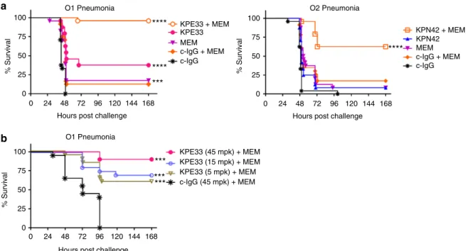
Anti-O-antigen in vivo protection is serotype specific. Given
their opsonophagocytic killing of two prominent LPS serotypes
(summarized in Fig.
4
e), we tested the protective potential of
these mAbs in murine models of K. pneumoniae infection against
wild-type encapsulated strains. Since each mAb targets a unique
LPS antigen (O1 or O2), murine models were developed
inde-pendently as a mimic of various clinical K. pneumoniae
infec-tions. The serotype-specific anti-O1 (KPE33) and anti-O2
(KPN42) mAbs significantly improved survival outcomes
com-pared to the negative control mAb when used to treat infection
with an O1 or O2 strain, respectively, in acute murine pneumonia
(Fig.
5
a) and bacteremia (Fig.
5
b) models. In the bacteremia
model, mortality is more rapid with a lower bacterial challenge
dose than the pneumonia model resulting in a narrower
ther-apeutic window. As such, the mAbs were most effective when
administered prophylactically (Fig.
5
b) vs. in treatment
(Supple-mentary Fig.
5
b), though either mAb delivery was significantly
protective. Despite its high affinity binding to both O1 and O2
LPS and its potent LPS neutralizing activity, the anti-O1/O2
cross-reactive mAb KPN70 failed to improve survival outcome in
the murine pneumonia model with an O1 strain and only
moderately improved survival with an O2 strain (Fig.
5
a).
Interestingly, KPN70 had some activity against both an O1 strain
and O2 strain in bacteremia (Fig.
5
b, Supplementary Fig.
5
b). The
Fig. 3K. pneumoniae O2 LPS is less immunogenic than K. pneumoniae O1 LPS. Purified O1 and O2 LPS were used for immunization of mice (a) or rabbits (b) with 0.5 mg LPS per animal. Immune sera were collected from O1 and O2 immunized animals and used to assess antibody titer against KP O1 or O2 LPS by ELISA. Sera from mock immunized animals with adjuvant only (black open circle) were used as a negative control.c Mice were immunized subcutaneously with either heat killed Kp1131115 (WT, an O1 strain) or the isogenic matched Kp1131115ΔwbbYZ (ΔwbbYZ, O2 serotype). Sera were collected 7 days after thefinal injection and used to assess antibody titer against K. pneumoniae O1 or O2 LPS by ELISA. d Sera collected in c were used to assess antibody binding to a protein target (OmpA) and to proteinase K bacterial lysate by ELISA.e Immune activation of cells in vitro was compared for purified O1 and O2 LPS. RAW264.7 cells stably transfected with a NF-κB driven luciferase promoter were stimulated with various doses of K. pneumoniae O1 or K. pneumoniae O2 LPS for 2 h. Percent activation was calculated as the amount of luminescence in stimulated cells vs. non-stimulated. Error bars indicate s.d. of each data point.f A schematic representation of KP O1 and O2 LPS. The gray region represents the membrane proximal LPS core region, the black region represents theD-galactan I (D-gal I) repeating O antigen sugars, the hatched grayfilled region represents the O1 specificD-galactan II (D-gal II) repeating O antigen sugars encoded by thewbbYZ locus. Deletion of this locus converted the O1 strain (Kp1131115 WT) to an O2 expressing strain (Kp1131115ΔwbbYZ). g Silver stain of a SDS-PAGE gel showing the relative molecular weight ofK. pneumoniae O1 LPS (lane 2) vs. K. pneumoniae O2 LPS (lane 3)0.4 0.3 0.2 Binding to LPS O1 (RU) 0.1 0.0 0.4 0.3 0.2 Binding to LPS O2 (RU) 0.1 0.0 0 500 KPE33 100 50 0 O1 LPS neutralization (%)
O1 OPK activity (%) O2 OPK activity (%)
–50 100 80 60 40 20 0 –20 100 80 60 40 20 0 –20 10–3 10–2 10–2 10–1 10–1 100 100 mAb (μg ml) Log (mAb) μg ml KPE33 KPN70 KPN42 c-IgG KPE33 KPN70 KPN42 c-IgG 101 101 10–2 10–1 100 Log (mAb) μg ml 101 102 103 100 50 0 O2 LPS neutralization (%) –50 10–3 10–2 10–1 100 mAb (μg ml) 101 102 103 O1 O2 KPN42 KPN70 KPE33 KPN42 KPN70 1000 Time (s) 1500 0 500 1000 Time (s) 1500 LPS mAb O1 O1
LPS Neut Δ Capsule OPK
O1/O2 O2 O2 O1 O2 KPE33 KPN70 KPN42 – – – – – – – ++ +++ +++ +++ +++ KPE33 KPN70 KPN42
a
b
c
d
e
Fig. 4 In vitro characterization of anti-O antigen mAbs. a Association/dissociation curves ofK. pneumoniae O1 or K. pneumoniae O2 LPS binding to mAbs KPE33, KPN70, and KPN42 were measured by ForteBio Octet using protein A capture probe pre-loaded with each mAb.b Confocal images of mAb binding to an O1 (Kp43816) or O2 (Kp9148) strain (scale bar, 2μm). c LPS neutralization was measured using RAW264.7 cells transfected with a NK-kB driven luciferase promoter stimulated with 2 ng ml−1of purified O1 or O2 LPS in the presence of KPE33, KPN70, KPN42 or the isotype-matched control mAb (c-IgG). Percent inhibition was calculated as the amount of luminescence (RLU) in treated wells divided by the RLU in wells stimulated with LPS alone multiplied by 100.d Opsonophagocytic killing assays were performed using various concentrations of mAbs in the presence of the phagocytic cell line HL-60. The capsule-deficient luminescent mutants Kp43816ΔcpsB and Kp8570ΔcpsB were used as the O1 and O2 serotype targets, respectively. Percent killing was calculated based on the amount of luminescence after 2.5 h incubation in treated wells vs. wells with bacteria only. An isotype-matched mAb (c-IgG) is used as a negative control.e Summary of the in vitro functional activities against O1 and O2K. pneumoniae for each mAb. All error bars indicate s.d. at each data point
activity of KPN70 against O1 suggests that LPS neutralization in
the absence of opsonophagocytic killing may preferentially
pro-vide limited benefit in the bacteremia model vs. the pneumonia
model. KPE33 also protected against dissemination to distal
organs from necrotic tissue at the burn site in a murine model of
thermal injury (Fig.
5
c), suggesting the utility of anti-O-antigen
mAbs to protect in multiple infection models. Further in vivo
testing using additional strains (including Kp8045, a highly
virulent, mucoid K1 strain
39) in a pneumonia model
(Supple-mentary Fig.
5
a), confirmed the high level of serotype-specific
protection mediated by the KPE33 and KPN42 mAbs as well as
the inferior activity of the cross-reactive, LPS neutralizing mAb
KPN70. Interestingly, KPE33 and KPN42 showed in vivo activity
even though mAb in vitro activity was restricted to
capsule-deficient strains (Fig.
4
d, Supplementary Fig.
4
). The data from
this panel of mAbs, in concert with recently published data
showing that an opsonophagocytic killing deficient mutant
KPE33 loses activity in vivo
40, supports opsonophagocytic killing
activity against specific epitopes as a major mechanism of in vivo
protection, at least in the pneumonia model. Conversely, a
ben-eficial role for LPS neutralization may be more limited and
dependent on the type of infection.
100 % Survival 75 50 25 0 0 24 48 72 96 120 144 168 Hrs post infection 100 % Survival 75 50 25 1011 1010 109 108 108 107 106 105 104 103 102 108 109 107 106 105 104 103 102 101 108 107 106 105 104 103 102 101 CFU/g CFU/g CFU/g CFU/g 0 0 24 48 72 96 120 Hrs post infection 100 % Survival 75 50 25 0 0 24 48 72 96 120 Hrs post infection KPE33 (45 mpk) KPE33 (15 mpk) c-IgG (45 mpk) O1 Pneumonia O1 Bacteremia O2 Bacteremia 100 % Survival 75 50 25 0 0 24 48 72 96 120 144 168 Hrs post infection O2 Pneumonia KPE33 (10 mpk) KPE33 (15 mpk) KPE33 (5 mpk) KPE33 (1 mpk) KPN70 (15 mpk) KPN70 (5 mpk) KPN70 (1 mpk) c-IgG (15 mpk) KPN42 (10 mpk) KPN42 (2 mpk) KPN42 (15 mpk) KPN42 (5 mpk) KPN42 (1 mpk) KPN70 (15 mpk) KPN70 (5 mpk) KPN70 (1 mpk) c-IgG (15 mpk) KPN42 (0.4 mpk) KPN70 (10 mpk) c-IgG (10 mpk) KPE33 (2 mpk) KPE33 (0.4 mpk) KPN70 (10 mpk) c-IgG (10 mpk)
***
****
****
****
***
**
**
*
****
****
***
***
**
Liver Spleen**
Skin Lung**
**
a
b
c
Fig. 5 Anti-O1 and anti-O2 mAbs are protective in vivo. a Therapeutic activity of the mAbs was tested in C57BL/6 mice infected intranasally with aK. pneumoniae O1 (Kp1131115, 6 × 10⁷CFU) or O2 (Kp961842, 2 × 10⁸CFU) strain. KPE33 (anti-O1), KPN42 (anti-O2) and KPN70 (anti-O1/O2) mAbs were administered intravascular 1 h post infection at the doses indicated. An isotype-matched mAb (c-IgG) was used as a negative control.b Prophylactic activity of the mAbs was tested in C57BL/6 mice infected intraperitoneal with the O1 strain Kp1131115 (3 × 10⁶CFU) or the O2 strain Kp961842 (1 × 10⁷ CFU). mAbs were administered IP 24 h prior to infection at the doses indicated; survival was monitored for 5 days post infection.c CF1 mice received a dorsal burn followed byK. pneumoniae O1 (Kp1131115, 5 × 10⁶) bacterial inoculation subcutaneously at the burn site. KPE33 (45 or 15 mpk) or isotype-matched control mAb (c-IgG, 45 mpk) were administered IP 24 h post infection, and organs were harvested 48 h post infection to determine bacterial CFU. All graphs are representative of at least three separate experiments. Mantel-Cox analysis was done to assess significant survival benefit compared to the control mAb. For thermal injury CFU comparison, Kruskal Wallace one way Anova analysis was done to compare differences in CFU between KPE33 treated vs. control IgG treated groups, ****p < 0.0001, ***p < 0.001, **p < 0.01
Anti-O-antigen mAbs protection is synergistic with antibiotics.
Clinical application of anti-KP antibodies would likely be in
combination with current standard of care antibiotic treatments,
including a carbapenem such as meropenem. We therefore
assessed the potential for the anti-O1 and O2 mAbs to
comple-ment antibiotic therapy using sub-therapeutic doses of antibiotics
to simulate insufficient drug exposure against drug-resistant
strains, as described previously
10,11,41. These experiments were
performed using identical conditions to the pneumonia model
described in Fig.
5
a with the additional administration of
mer-openem. A sub-therapeutic dose of mAb KPE33 administered in
combination with a sub-protective meropenem dose provided
significant protection in the murine pneumonia model against a
intermediately meropenem-resistant O1 strain (MIC
= 2 µg ml
−1).
Statistical analysis confirmed synergistic adjunctive activity
compared to either drug administered alone (Fig.
6
a). The
effi-cacy of the mAb was maintained even when administered 24 h
post infection (Fig.
6
b). Similarly, a sub-therapeutic dose of mAb
KPN42 in combination with a dose approximating a
human-equivalent level of meropenem, also provided significant
protec-tion in this model against a highly meropenem-resistant O2 CRE
ST258 strain (MIC
= 32 µg ml
−1, eight times more than the
human breakpoint) (Fig.
6
a). These data clearly illustrate that
mAbs targeting O-antigen could not only provide protection
when dosed sufficiently in monotherapy, but could also provide
complementary synergistic activity in adjunctive therapy with
antibiotics. In conclusion, these results indicate that
anti-O-antigen antibodies can provide additional and complementary
benefits to antibiotic therapy even against highly
carbapenem-resistant KP strains and further substantiate the concept of
antibody help in antibiotic therapy
10, 11, 41. Without this
additional antibody-mediated help, O2 strains may preferentially
persist under antibiotic pressure.
Discussion
K. pneumoniae is a common enteric bacterial species in human
gut
flora, therefore basal levels of antibody to immunodominant
antigens are to be expected and were indeed evident in our survey
of tonsillar and circulating memory B cells. In our efforts to
identify protective mAbs against K. pneumoniae, only a small
fraction of antibodies recognizing whole bacteria possessed potent
opsonophagocytic killing activity, the majority of which
recog-nized LPS O-antigen. It is reasonable to consider that humoral
immunity to O-antigen may offer some level of protection against
the establishment of systemic infections in healthy individuals,
and these antigens are currently under clinical investigation as a
vaccine target against other pathogens such as nontyphoidal
Salmonella, Vibrio cholera and Shigella sonnei
42–46, though in
each case the antibody activity was verified against
unencapsu-lated strains only. Our study determined for the
first time that the
O2 serotype comprises a significantly larger fraction of ESBL
(35%) and CRE (50%) strains in comparison to fully susceptible
(17%) KP strains suggesting a selective advantage for MDR
strains of the O2 serotype despite their susceptibility to serum
killing. Much attention has been focused on the particularly
successful MDR carbapenem-resistant ST258 sequence type of K.
pneumoniae, which we serotyped as almost exclusively O2.
However, as ST258 only accounts for half of CRE strains and a
much smaller fraction of the earlier emerging ESBL strains,
simple clonal outbreaks cannot completely account for the
increased prevalence of the O2 serotype.
100 75 50 % Survival 25 0 0 24 48 72 96 120 144 168 Hours post challenge
100 75 50 % Survival 25 0 0 24 48 72 96 120 144 168 Hours post challenge
O1 Pneumonia
a
b
O1 Pneumonia KPE33 + MEM KPE33 (45 mpk) + MEM KPE33 (15 mpk) + MEM KPE33 (5 mpk) + MEM c-IgG (45 mpk) + MEM****
****
***
***
***
***
100 75 50 % Survival 25 0 0 24 48 72 96 120 144 168 Hours post challengeO2 Pneumonia
****
KPE33 MEM c-IgG + MEM c-IgG KPN42 + MEM KPN42 MEM c-IgG + MEM c-IgGFig. 6 Antibody synergy with antibiotic. a Mice were infected intranasally as in Fig.5with theK. pneumoniae O1 (Kp1131115, 6 × 10⁷CFU) or O2 (Kp961842, 2 × 10⁸CFU) strain then treated 1 h post infection with suboptimal doses of mAb (KPE33, 1 mg kg−1or KPN42, 0.2 mg kg−1given IV) and antibiotic (meropenem, 7.5 mg kg−1for O1 strain; 50 mg kg−1for O2 strain, given subcutaneous) or a combination of both mAb and antibiotic. Graphs represent the combined data from three separate experiments with a total of 24 mice in each group. Mantel-Cox analysis was done to assess significant survival benefit compared to the control mAb (a) or control mAb + MEM (b), ****p < 0.0001, ***p < 0.001, **p < 0.01. The Bliss independence method was used to determine synergistic activity of the combination therapy vs. either monotherapy alone;p = 2.5 × 10⁻⁶for KPE33+MEM combination andp = 7.7 × 10⁻⁵for KPN42+MEM combination.b Mice were infected intransal with a KP O1 strain (Kp8045, 1 × 10⁴CFU). Meropenem (1.5 mpk) was administered subcutaneous 4 h post infection and mAb KPE33 was administered IV 24 h post infection at the indicated doses. All graphs are representative of at least three separate experiments. An isotype-matched mAb (c-IgG) was used as a negative control. Mantel-Cox analysis was done to assess significant survival benefit compared to the control mAb+MEM b, ****p < 0.0001, ***p < 0.001, **p < 0.01
A possible explanation for the unexpected prevalence of the
serum-susceptible O2 serotype is the lower immunogenicity of
the O-antigen and the consequent failure to induce antibodies
that synergize with antibiotics. Thus, the combination of drug
resistance and lack of immunogenicity observed in this study may
contribute to the success of O2 strains (including the
ST258 subset) particularly in the context of broad spectrum
antibiotic pressure. It is notable that strains without a detectable
O-antigen (non-serotypeable) were also more prevalent in CRE
isolates. This observation further supports the concept that strains
with less immunogenic O antigen or lacking O antigen expression
may be selected under antibiotic pressure. However,
immuno-genic O1 strains are also prevalent among ESBL isolates and
slightly increased in prevalence compared to their representation
in susceptible isolates. In this instance, other O1-specific
mechanisms of immune evasion may play a role such as their
resistance to human serum killing (Fig.
1
b) and/or the increased
presence of virulence genes in O1 isolates vs. other serotypes
16.
Human serum sensitivity may play a role in bacterial clearance in
patients, but would not necessarily be expected to translate to
mouse models as species-specific differences in serum sensitivity
have been clearly demonstrated for several pathogens, including
K. pneumoniae
26–30.
A diminishing pipeline for new antibiotics and the now
recognized negative impact of broad-spectrum antibiotics on the
beneficial microbiome highlight the necessity of developing new
antibacterial strategies
47. The diversity of capsular
poly-saccharides of K. pneumoniae has been suggested by many to be
an immunodominant feature that obscures the accessibility of
antibodies to other surface targets. Here, we demonstrate that it is
possible to identify highly protective mAbs to the less diverse
O-antigen. Despite the relative rarity of O2 antigen-specific B cells, it
was possible to identify a mAb (KPN42) with high protective
activity. Potent mAbs targeting the O1 antigen were also
identi-fied. Interestingly, these mAbs had no opsonophagocytic killing
activity against WT strains in vitro. However, in vitro activity
against capsule knock-out strains was found to be predictive of
activity against WT encapsulated strains in vivo, suggesting
capsule may be expressed at lower levels or less consistently
in vivo. Consistent with previous studies with other bacterial
pathogens
10,11, the anti-KP mAbs isolated in this study afforded
synergistic adjunctive protection to drug-resistant strains,
including a representative strain of the problematic ST258 CRE
clone. These data further support an under-appreciated role for
humoral immunity in antibiotic therapy against KP and indeed
the background impact of immunity on bacterial drug resistance
observed with vaccines
9. Furthermore, these studies provide some
encouragement that mAb-based strategies may offer future
solutions for combatting antibiotic refractory bacteria.
Methods
K. pneumoniae strains. All K. pneumoniae isolates were purchased from America Type Culture Collection (ATCC), Eurofin or IHMA (a complete list of strains can be found in Supplementary Table2), and cultures were maintained in 2xYT media at 37 °C supplemented with antibiotics when appropriate. For capsule mutant strain generation, approximately one kilobase of DNAflanking either side of the cpsB/manC gene was PCR amplified and cloned into the pir-dependent plasmid pDMS19748. This plasmid was then electroporated into Klebsiella strains and plasmid integrants selected on LB agar containing 10μg ml−1tetracycline or gen-tamycin. After propagation of recombinants in the absence of selection, clones that resolved the integrated plasmid were selected by growth on LB agar containing 10% sucrose. Deletion of manC was confirmed by PCR on lysed colonies with primers specific to the manC gene (AAACAGTTCCTCCGTCTGTTC, GGAA-TAAAGGTGGACTGGTTCT) using the following cycling conditions: 94° for 2 min, 30 cycles of 94° for 30 s, 58° for 30 s, then 72° for 1 min.
Clinical isolates. For serotyping, clinical isolates were obtained from International Health Management Associates (Schaumburg, IL, USA). The isolates were selected
based on MIC profiles (to ensure relatively equal numbers of susceptible, ESBL and CRE strains), then chosen from 6 continents, 38 different countries and at least 162 different hospitals in an attempt to generate a global snapshot of current K. pneumoniae infections as opposed to a sampling of any single outbreak. All isolates were collected between 2012 and 2014 from various sites of infection (a breakdown of infection site can be found in Supplementary Table1).
Isolation and production of LPS serotyping antibodies. Peripheral blood mononuclear cells (PBMC) and sera were separated from buffy coats of healthy blood donors or convalescent patients after K. pneumoniae infection as previously described49. Alternatively, lymphocytes were obtained from tonsils after tissue
homogenization in the presence of DNase I and collagenase. The donors provided written informed consent for the use of these samples, following approval by the Cantonal Ethical Committee of Canton Ticino, Switzerland (for healthy donors) and the Ethical Committee of the Policlinico San Matteo, IRCCS, in Pavia (Italy) (for convalescent ICU donors). Memory B cells were isolated from cryopreserved PMBC or from lymphocytes isolated from tonsils using PE-Cy7-labeled CD19 microbeads (BD Biosciences, clone SJ25C1), followed by isolation with anti PE-beads (Miltenyi Biotec) and by depletion of cells stained with anti-IgM (Jackson ImmunoResearch), anti-IgD (BD Biosciences), and anti-IgA (Novex) antibodies by cell sorting on a FACSAria (BD Biosciences). Memory B cells were immortalized with EBV as described previously36,50Briefly, sorted memory B cells were infected with EBV and seeded into 384 well plates in the presence of CpG TRL9 agonist and allogenic irradiated PBMCs. Immortalized memory B cells were then screened for antibody binding to purified LPS by ELISA. Total mRNA from positive B cell cultures was reverse transcribed in 50µl nuclease-free water (Ambion) using 15 µM specific pri-mer (IgG:5′-TCTTGTCCACCTTGGTGTTGCT; Igκ: 5′ACACTCTCCCCTGTT-GAAGCTCTT; Igλ: 5′-ACTGTCTTCTCCACGGTGCT), 1.8 µl of 25 mM dNTP mix (GE Healthcare), 5µl of 0.1 M DTT, 0.5% v/v Igepal CA-630 (Sigma), 60 U RNAse OUT (Invitrogen), and 100 U Superscript III reverse transcriptase (Invitro-gen). Reverse transcription (RT) was performed at 42 °C 10 min, 55 °C 60 min, and 94 °C 5 min. IgH, Igκ, and Igλ genes were amplified by PCR using HotStar Taq DNA polymerase (Qiagen) and cloned into human Igγ1, Igκ, and Igλ expression vectors, as described51. Recombinant mAbs were produced by transient transfection of EXPI293
cells (Invitrogen) and purified by Protein A chromatography (GE Healthcare) and desalted against PBS. Alternatively, mouse immune sera were generated by purified LPS or bacterial immunization. The specificity of the resulting mAbs and polysera was confirmed by loading 1 μg of purified LPS from serotype reference strains onto an sodium dodecyl sulfate-polyacrylamide gel electrophoresis (SDS-PAGE), followed by transfer onto a nitrocellulose membrane, and probed using our isolated anti-O-antigen mAbs or polyclonal mouse sera (Supplementary Fig.1a).
Enzyme-linked immunosorbent assay. Spectraplate-384 with high protein binding treatment (custom made from Perkin Elmer) or Nunc 96-well high binding plates were coated overnight at 4 °C with 5µg ml−1LPS in phosphate-buffered saline (PBS), pH 7.2, and plates were subsequently blocked with PBS supplemented with 1% BSA (low endotoxin, Sigma-Aldrich). The coated plates were incubated with serial dilutions of our isolated anti-O-antigen human monoclonal antibodies for 1 h at room temperature. The plates were then washed with PBS containing 0.1% Tween-20 (PBS-T), and alkaline phosphatase-affiniPure F(ab’)2 Frag rabbit anti-human IgG, Fcg Frag Specific (Jackson ImmunoResearch, 10,000× dilution) or alkaline phosphatase-goat anti-human IgG (Southern Biotech) were added and incubated for 1 h at room temperature. Plates were washed three times with PBS-T, and P-NitroPhenyl Phosphate (pNPP, Sigma-Aldrich) substrates were added and the absorbance of 405 nm was measured by a microplate reader (Biotek or Molecular Devices). Data were plotted with GraphPad Prism software. AMBRA of IgG and IgM antibodies from tonsillar B cells. Replicate cultures of total tonsillar lymphocytes were seeded at 20,000 cells per well in 96 U-bottom plates (Costar) and stimulated with 2.5μg ml−1R848 (3 M) and 1000 U ml−1 human recombinant IL-2 for 10 days at 37 °C 5% CO2. The cells culture super-natants were collected for further analysis. Spectraplate-384 with high protein binding treatment (Perkin Elmer) or Nunc 96-well high binding plates were coated overnight at 4 °C with 5µg ml−1O1 or O2 LPS in phosphate-buffered saline (PBS). Plates were blocked with PBS 1% BSA (low endotoxin, Sigma-Aldrich) and then incubated with AMBRA supernatant for 1 h at room temperature. After washings with PBS 0.1% Tween the binding of IgG or IgM Abs was detected with goat Alkaline Phosphatase-anti human IgG (Southern Biotech) or Alkaline
Phosphatase-goat anti human IgM (Southern Biotech), respectively. pNPP (Sigma-Aldrich) substrate was added and absorbance measured at 405 nm on a microplate reader (Biotek).
Serotyping ofK. pneumoniae clinical isolates. The serotypes of the KP clinical isolates were determined by western blot analysis with serotype-specific mono-clonal antibodies or mouse polysera. Briefly, purified LPS or bacterial lysates were subjected to SDS-PAGE. Separated proteins and LPS were transferred from gels to nitrocellulose membranes with an iBlot apparatus (Life Technology). Membranes were then blocked with Casein or Odyssey (Li-cor) blocking buffer before being probed with anti-O1, O1/O2, O3, O4, or O5 LPS monoclonal antibodies or anti-O7
and anti-O12 mouse polysera. After repeated washes with PBS-T, blots were incubated with IRDye680 or 800fluorescent secondary antibodies (Li-cor) and visualized with an Odyssey Image Station (Li-cor). In some circumstances, bacterial lysates were treated with 0.4 mg ml−1Proteinase K (ThermoScientific) to remove protein components before the western blot analysis. CRE isolates were defined as resistant to carbapenams (doripenam, imipenam and/or meropenam MIC> = 4 μg ml−1), ESBL isolates defined as resistant to cephalosporins (ceftazidime MIC > = 16μg ml−1) but sensitive to carpapenams (MIC< 4 μg ml−1) and susceptible iso-lates defined by ceftazidime MIC < 16 μg ml−1and carbapenam MIC< 4 μg ml−1. Octet binding assay with anti-LPS mAbs. Anti-LPS mAbs diluted to 0.2µg ml−1 in PBS were immobilized for 10 min at 37 °C on the surface of a protein A coated sensor-chip of an Octet RED96 system (FortéBio). Coated-Biosensors were incu-bated for 10 min with a solution containing 2µg ml−1O1 or O2 LPS (purified from Kp43816ΔcpsB or Kp8570ΔcpsB, respectively) in Kinetics buffer (ForteBio, dilute 10× to 1× with PBS). A dissociation step was then performed incubating the Biosensor for 20 min in Kinetics buffer. Changes in the number of molecules bound to the biosensor caused a shift in the interference pattern that was recorded in real time and off-rate analyses were performed to determine mAbs KD (M). Blockade of binding assay. Human anti-LPS mAbs were biotinylated using the EZ-Link NHS-PEO solid phase biotinylation kit (Pierce). Labeled mAbs were tested for binding to LPS by ELISA and the optimal concentration of each mAb to achieve 70% maximal binding was determined. Unlabeled mAbs were serially diluted and added to ELISA 96-well plates (Corning) coated overnight at 4 °C with 5µg ml−1 LPS in PBS. After 1 h, biotinylated anti-LPS mAbs were added at the concentration achieving 70% maximal binding and the mixture was incubated at room tem-perature for 1 h. Plates were washed and antibody binding was revealed using Alkaline Phosphatase-streptavidin (Jackson Immunoresearch). After washing, pNPP substrate (Sigma-Aldrich) was added and plates were read at 405 nm. The percentage of inhibition was calculated as a percent decrease of signal vs. the maximum signal achieved with the labeled mAb.
MLST analysis. Multi-locus sequence type (MLST) analysis was performed using the method and primers as described elsewhere52. Briefly, a bacterial colony was
resuspended in 40μl DNase free water and used as template for PCR reactions using primers specific to each of the seven MLST genes with the inclusion of a universal 3′ sequence tag on each primer (rpoB forward GTTTTCCCAGTCAC-GACGTTGTAGGCGAAATGGCWGAGAACCA, reverse TTGTGAGCGGA-TAACAATTTCGAGTCTTCGAAGTTGTAACC; gapA forward
GTTTTCCCAGTCACGACGTTGTATGAAATATGACTCCACTCACGG, reverse
TTGTGAGCGGATAACAATTTCCTTCA-GAAGCGGCTTTGATGGCTT; mdh forward GTTTTCCCAGTCAC-GACGTTGTA CCCAACTCGCTTCAGGTTCAG, reverse
TTGTGAGCGGATAACAATTTCCCGTTTTTCCCCAGCAGCAG; pgi forward GTTTTCCCAGTCACGACGTTGTAGAGAAAAACCTGCCTGTACTGCTGGC, reverse
TTGTGAGCGGATAACAATTTCCGCGCCACGCTTTA-TAGCGGTTAAT; phoE forward GTTTTCCCAGTCACGACGTTGTAACC-TACCGCAACACCGACTTCTTCGG, reverse
TTGTGAGCGGATAACAATTTCTGATCAGAACTGGTAGGTGAT; infB for-ward GTTTTCCCAGTCACGACGTTGTACTCGCTGCTGGACTATATTCG, reverse TTGTGAGCGGATAACAATTTC CGCTTTCAGCTCAAGAACTTC; tonBforward GTTTTCCCAGTCACGACGTTGTACTTTATACCTCGGTA-CATCAGGTT, reverse
TTGTGAGCGGATAA-CAATTTCATTCGCCGGCTGRGCRGAGAG). After exonuclease clean up using Exo-SapIT (Affymetrix), the resulting template was used for a BigDye Terminator (Affymetrix) PCR reaction according to the manufacturer’s protocol using uni-versal primers for all 7 genes (forward GTTTTCCCAGTCACGACGTTGTA, reverse TTGTGAGCGGATAACAATTTC) followed by analysis on the ABI 3730 (Applied Biosystems). SeqScape software (ABI) was used to match sequence data to the public database (http://bigsdb.pasteur.fr/klebsiella/klebsiella.html) and to determine the ST of each strain. In some cases, assembled whole genome sequences were used to determine ST type.
Opsonophagocytic killing assays. Opsonophagocytic killing activity of anti-O antigen mAbs was tested against O1 and O2 strains. Briefly, log phase cultures of luminescent KP strains 8570ΔcpsBlux (O2) and 43816ΔcpsBlux (O1) were diluted to ~2 × 10⁶cells ml−1. Bacteria, 5 × 10⁵dimethylformamide (DMF) differentiated HL-60 cells, 1:10 diluted cleared baby rabbit serum (Cedarlane), and a series dilution of antibodies (2 ng–2500 ng m1) were mixed in 96-well plates and incu-bated at 37 °C for 2 h with shaking (250 rpm). The relative light units (RLUs) were then measured using an Envision Multilabel plate reader (Perkin Elmer). The percent killing was determined by comparing RLU derived from assays with no antibodies to RLU derived from assays with anti-KP or negative control mAbs. Confocal imaging. K. pneumoniae strains were inactivated with formaldehyde, washed and re-suspended in PBS to achieve an OD600of 3. In each experimental condition, 50µl of bacterial suspension were centrifuged and re-suspended in 5% FBS in PBS. The tested mAbs were added to the bacterial suspension at afinal
concentration of 10µg ml−1and incubated for 2.5 h at 4 °C. After two wash steps, fluorescent anti-human IgG secondary antibody (Jackson Immunoresearch) and DAPI were added to the pellet to afinal concentration of 15 µg ml−1and 4µg ml−1, respectively, and incubated for 40 min at 4 °C. Bacteria were washed twice with PBS, resuspended in a Mowiol-based mounting medium and mounted on microscopy slides. Slides were air-dried O/N in the dark and images were taken using the TCS SP5 microscopy system from Leica using a ×100 objective and confocal settings.
LPS neutralization assays. A murine RAW264.7 macrophage cell line was engineered to carry afirefly luciferase reporter gene under the control of an NF-κB-responsive promoter (RAW264.7-lux). Serially diluted antibody stocks were mixed with purified KP LPS (2 ng ml−1final concentration) and incubated at 4 °C for 1 h. Antibody/LPS mixtures were then added to assay plates containing pre-seeded RAW264.7-lux cells (5000 cells per well). Plates were incubated for 2.5 h at 37 °C with 5% CO2then Steady Glo solution (Promega) was added to each well and incubated for another 20 min protected from light. The RLUs were measured using an Envision Multilabel plate reader (Perkin Elmer). The percentage of inhibition was determined by comparing RLU derived from assays with no antibodies to RLU derived from assays with anti-KP or negative control mAbs.
Bacterial infection models. C57BL/6 mice were received from Jackson labora-tories and maintained in a special pathogen free facility. All animal experiments were conducted in accordance with IACUC protocol and guidance. K. pneumoniae strains were grown on agar plates overnight and diluted in saline at proper con-centration just prior to infection. The inoculum titer was determined by plating a serial dilution of bacteria onto agar plates. Mice were inoculated with 5 × 10³to 2 × 10⁸CFU of KP clinical isolates either intranasally (pneumonia model) or intra-peritoneally (bacteremia model). Anti-LPS monoclonal antibody and human IgG1 control antibody were administered IV 1 h post-bacterial challenge (therapeutic dosing) or IP 24 h prior to infection (prophylactic dosing). Mouse survival was monitored daily. For the thermal injury model, CF1 mice (Charles River Labora-tories) received a dorsal burn under anesthetic for 5 s followed by administration of 5 × 10⁶bacteria subcutaneously at the burn site. MAb was administered IP 24 h post infection, organs were harvested at 48 h post infection and bacterial CFU was determined by serial dilution of organ homogenate. For antibody and antibiotic combination studies, both were given 1 h post infection (mAb delivered IV, mer-openem delivered subcutaneously at 7.5 mg kg−1in O1 model or 50 mg kg−1in O2 model). For extended timecourse experiments, meropenem was delivered 4 h post infection and mAb 24 h post infection. Survival data was plotted in Prism and statistical analysis was determined using the Log-rank Mantel-Cox test. To determine synergy in the antibody/antibiotic combination experiments, the Bliss independence method for drug combination synergy test was used as described elsewhere53. Briefly, if the rate of survival at the end of study for drug A alone is ra
and the survival rate for drug B alone is rb, then the expected survival rate for drug A and drug B combination is rBliss= ra+ rb− rarbassuming that the two drugs are bliss independent. The difference between the observed survival rate raband the expected rate is defined as the synergy index.
Ethics statement. The donors provided written informed consent for the use of blood samples, following approval by the Cantonal Ethical Committee of Canton Ticino, (Switzerland), for healthy donors and the Ethical Committee of the Poli-clinico San Matteo, IRCCS, in Pavia (Italy) for convalescent and intensive care donors. All animal studies were performed under the guidance and protocol approval of the MedImmune Institutional Animal Care and Use Committee. Additional oversight was also provided by Office of Research Protections (ORP), US Army Medical Research and Material Command (USAMRMC), Animal Care and Use Review Office (ACURO).
Data availability. The authors declare that all relevant data supporting thefindings of the study are available in this article and its Supplementary Informationfiles, or from the corresponding author upon request.
Received: 5 May 2017 Accepted: 14 November 2017
References
1. McKenna, M. Antibiotic resistance: the last resort. Nature 499, 394–396 (2013). 2. Iredell, J., Brown, J. & Tagg, K. Antibiotic resistance in Enterobacteriaceae:
mechanisms and clinical implications. BMJ 352, h6420 (2016).
3. Taur, Y. & Pamer, E. G. The intestinal microbiota and susceptibility to infection in immunocompromised patients. Curr. Opin. Infect. Dis. 26, 332–337 (2013). 4. Badal, R. E. et al. Etiology, extended-spectrum beta-lactamase rates and
antimicrobial susceptibility of Gram-negative bacilli causing intra-abdominal infections in patients in general pediatric and pediatric intensive care
units-global data from the Study for Monitoring Antimicrobial Resistance Trends 2008 to 2010. Pediatr. Infect. Dis. J. 32, 636–640 (2013).
5. van Duin, D. & Paterson, D. L. Multidrug-resistant bacteria in the community: trends and lessons learned. Infect. Dis. Clin. North Am. 30, 377–390 (2016). 6. Russotto, V. et al. Bloodstream infections in intensive care unit patients:
distribution and antibiotic resistance of bacteria. Infect. Drug Resist. 8, 287–296 (2015).
7. Wyres, K. L. et al. Extensive capsule locus variation and large-scale genomic recombination within the Klebsiella pneumoniae clonal group 258. Genome Biol. Evol. 7, 1267–1279 (2015).
8. Wyres, K. L. & Holt, K. E. Klebsiella pneumoniae population genomics and antimicrobial-resistant clones. Trends Microbiol. 24, 944–956 (2016). 9. Lipsitch, M. & Siber, G. R. How can vaccines contribute to solving the
antimicrobial resistance problem? mBio 7, e00428-16 (2016).
10. DiGiandomenico, A. et al. A multifunctional bispecific antibody protects against Pseudomonas aeruginosa. Sci. Transl. Med. 6, 262ra155 (2014). 11. Hua, L. et al. Assessment of an anti-alpha-toxin monoclonal antibody for
prevention and treatment of Staphylococcus aureus-induced pneumonia. Antimicrob. Agents Chemother. 58, 1108–1117 (2014).
12. Baumgartner, J. D. et al. Prevention of gram-negative shock and death in surgical patients by antibody to endotoxin core glycolipid. Lancet 2, 59–63 (1985).
13. Ziegler, E. J. et al. Treatment of gram-negative bacteremia and septic shock with HA-1A human monoclonal antibody against endotoxin. A randomized, double-blind, placebo-controlled trial. The HA-1A Sepsis Study Group. N. Engl. J. Med. 324, 429–436 (1991).
14. Ziegler, E. J. et al. Treatment of gram-negative bacteremia and shock with human antiserum to a mutant Escherichia coli. N. Engl. J. Med. 307, 1225–1230 (1982).
15. Greenman, R. L. et al. A controlled clinical trial of E5 murine monoclonal IgM antibody to endotoxin in the treatment of gram-negative sepsis. The XOMA Sepsis Study Group. JAMA 266, 1097–1102 (1991).
16. Follador, R. et al. The diversity of Klebsiella pneumoniae surface polysaccharides. Microb. Genom. 2, e000073 (2016).
17. Whitfield, C., Richards, J. C., Perry, M. B., Clarke, B. R. & MacLean, L. L. Expression of two structurally distinct D-galactan O antigens in the lipopolysaccharide of Klebsiella pneumoniae serotype O1. J. Bacteriol. 173, 1420–1431 (1991).
18. Vinogradov, E. & Perry, M. B. Structural analysis of the core region of the lipopolysaccharides from eight serotypes of Klebsiella pneumoniae. Carbohydr. Res. 335, 291–296 (2001).
19. Wang, Q. et al. Target-agnostic identification of functional monoclonal antibodies against Klebsiella pneumoniae multimeric MrkAfimbrial subunit. J. Infect. Dis. 213, 1800–1808 (2016).
20. Hansen, D. S. et al. Klebsiella pneumoniae lipopolysaccharide O typing: revision of prototype strains and O-group distribution among clinical isolates from different sources and countries. J. Clin. Microbiol. 37, 56–62 (1999). 21. Trautmann, M. et al. O-antigen seroepidemiology of Klebsiella clinical isolates
and implications for immunoprophylaxis of Klebsiella infections. Clin. Diagn. Lab. Immunol. 4, 550–555 (1997).
22. Sahly, H. et al. Increased serum resistance in Klebsiella pneumoniae strains producing extended-spectrum beta-lactamases. Antimicrob. Agents Chemother. 48, 3477–3482 (2004).
23. Merino, S., Camprubi, S., Alberti, S., Benedi, V. J. & Tomas, J. M. Mechanisms of Klebsiella pneumoniae resistance to complement-mediated killing. Infect. Immun. 60, 2529–2535 (1992).
24. Hsieh, P. F. et al. D-galactan II is an immunodominant antigen in O1 lipopolysaccharide and affects virulence in Klebsiella pneumoniae: implication in vaccine design. Front. Microbiol. 5, 608 (2014).
25. McCallum, K. L., Schoenhals, G., Laakso, D., Clarke, B. & Whitfield, C. A high-molecular-weight fraction of smooth lipopolysaccharide in Klebsiella serotype O1:K20 contains a unique O-antigen epitope and determines resistance to nonspecific serum killing. Infect. Immun. 57, 3816–3822 (1989).
26. Brown, G. C. The complementary activity of mouse-serum. J. Immunol. 46, 319–323 (1943).
27. Muschel, L. H. & Muto, T. Bactericidal reaction of mouse serum. Science 123, 62–64 (1956).
28. Warren, H. S. et al. Resilience to bacterial infection: difference between species could be due to proteins in serum. J. Infect. Dis. 201, 223–232 (2010). 29. Siggins, M. K. et al. Absent bactericidal activity of mouse serum against invasive
African nontyphoidal Salmonella results from impaired complement function but not a lack of antibody. J. Immunol. 186, 2365–2371 (2011).
30. Szijarto, V. et al. Endotoxin neutralization by an O-antigen specific monoclonal antibody: A potential novel therapeutic approach against Klebsiella pneumoniae ST258. Virulence, http://dx.doi.org/10.1080/21505594.2017.1279778 (2017). 31. Lee, C. R. et al. Global dissemination of carbapenemase-producing klebsiella
pneumoniae: epidemiology, genetic context, treatment options, and detection methods. Front. Microbiol. 7, 895 (2016).
32. Gaiarsa, S. et al. Genomic epidemiology of Klebsiella pneumoniae in Italy and novel insights into the origin and global evolution of its resistance to carbapenem antibiotics. Antimicrob. Agents Chemother. 59, 389–396 (2015). 33. Kitchel, B. et al. Molecular epidemiology of KPC-producing Klebsiella
pneumoniae isolates in the United States: clonal expansion of multilocus sequence type 258. Antimicrob. Agents Chemother. 53, 3365–3370 (2009). 34. Pitout, J. D., Nordmann, P. & Poirel, L. Carbapenemase-producing Klebsiella
pneumoniae, a key pathogen set for global nosocomial dominance. Antimicrob. Agents Chemother. 59, 5873–5884 (2015).
35. Jean, S. S. et al. Carbapenemase-producing Gram-negative bacteria: current epidemics, antimicrobial susceptibility and treatment options. Future Microbiol. 10, 407–425 (2015).
36. Pinna, D., Corti, D., Jarrossay, D., Sallusto, F. & Lanzavecchia, A. Clonal dissection of the human memory B-cell repertoire following infection and vaccination. Eur. J. Immunol. 39, 1260–1270 (2009).
37. Vos, Q., Lees, A., Wu, Z. Q., Snapper, C. M. & Mond, J. J. B-cell activation by T-cell-independent type 2 antigens as an integral part of the humoral immune response to pathogenic microorganisms. Immunol. Rev. 176, 154–170 (2000). 38. Mond, J. J., Lees, A. & Snapper, C. M. T cell-independent antigens type 2.
Annu. Rev. Immunol. 13, 655–692 (1995).
39. Fang, C. T. et al. Klebsiella pneumoniae genotype K1: an emerging pathogen that causes septic ocular or central nervous system complications from pyogenic liver abscess. Clin. Infect. Dis.: Off. Publ. Infect. Dis. Soc. Am. 45, 284–293 (2007).
40. Cohen, T. et al. Anti-LPS antibodies protect against Klebsiella pneumoniae by empowering neutrophil-mediated clearance without neutralizing TLR4. JCI Insight 2, 92774 (2017).
41. Song, Y. et al. PcrV antibody-antibiotic combination improves survival in Pseudomonas aeruginosa-infected mice. Eur. J. Clin. Microbiol. Infect. Dis.: Off. Publ. Eur. Soc. Clin. Microbiol. 31, 1837–1845 (2012).
42. Wu, Y. et al. Development of a live attenuated bivalent oral vaccine against Shigella sonnei shigellosis and typhoid fever. J. Infect. Dis. 215, 259–268 (2016). 43. Lin, J. et al. Monoclonal antibodies to shigella lipopolysaccharide are useful for
vaccine production. Clin. Vaccine Immunol. 23, 681–688 (2016). 44. Leitner, D. R. et al. Lipopolysaccharide modifications of a cholera vaccine
candidate based on outer membrane vesicles reduce endotoxicity and reveal the major protective antigen. Infect. Immun. 81, 2379–2393 (2013).
45. Watson, D. C., Robbins, J. B. & Szu, S. C. Protection of mice against Salmonella typhimurium with an O-specific polysaccharide-protein conjugate vaccine. Infect. Immun. 60, 4679–4686 (1992).
46. Simon, R. et al. Sustained protection in mice immunized with fractional doses of Salmonella Enteritidis core and O polysaccharide-flagellin glycoconjugates. PLoS ONE 8, e64680 (2013).
47. Fraenkel-Wandel, Y., Raveh-Brawer, D., Wiener-Well, Y., Yinnon, A. M. & Assous, M. V. Mortality due to blaKPC Klebsiella pneumoniae bacteraemia. J. Antimicrob. Chemother. 71, 1083–1087 (2016).
48. Edwards, R. A., Keller, L. H. & Schifferli, D. M. Improved allelic exchange vectors and their use to analyze 987Pfimbria gene expression. Gene 207, 149–157 (1998).
49. Beltramello, M. et al. The human immune response to Dengue virus is dominated by highly cross-reactive antibodies endowed with neutralizing and enhancing activity. Cell Host Microbe 8, 271–283 (2010).
50. Corti, D. & Lanzavecchia, A. Efficient methods to isolate human monoclonal antibodies frommemory B cells and plasma cells. Microbiol. Spectr. http://doi: 10.1128/microbiolspec.AID-0018-2014 (2014).
51. Tiller, T. et al. Efficient generation of monoclonal antibodies from single human B cells by single cell RT-PCR and expression vector cloning. J. Immunol. Methods 329, 112–124 (2008).
52. Diancourt, L., Passet, V., Verhoef, J., Grimont, P. A. & Brisse, S. Multilocus sequence typing of Klebsiella pneumoniae nosocomial isolates. J. Clin. Microbiol. 43, 4178–4182 (2005).
53. Zhao, W. et al. A new bliss independence model to analyze drug combination data. J. Biomol. Screen. 19, 817–821 (2014).
Acknowledgements
This work was funded by MedImmune, LLC, a wholly owned subsidiary of AstraZeneca Pharmaceuticals and a grant from the Defense Advanced Projects Research Agency (DARPA). We thank Professor Piero Marone from Policlinico San Matteo in Pavia, Italy for providing the sera samples from convalescent patients.
Author contributions
M.E.P., Q.W., P.W., J.S., D.C., E.C., A.D.M., M.B., A.L., and C.K.S. contributed to the experimental design and writing of this manuscript. M.E.P., Q.W., M.P., A.D.M., J.B., R. C., M.B., X.X., E.C., W.Z., M.M.C., S.B., F.Z., A.D., and E.S. performed experiments or provided critical reagents.
Additional information
Supplementary Informationaccompanies this paper at https://doi.org/10.1038/s41467-017-02223-7.
Competing interests:M.E.P., Q.W., M.P.,J.B, R.C, E.S, X.X, W.Z., M.M.C., A.D., P.W., J. S., C.K.S. are current or former employees of MedImmune/AstraZeneca and may own stock or stock options in the company. Patents describing the activity of the antibodies in this work have beenfiled by MedImmune. A.D.M., M.B, E.C., S.B, F.Z, A.L, D.C. are employees of HumAbs BioMed and may currently hold Humabs stocks or stock options. The remaining authors declare no competingfinancial interests.
Reprints and permissioninformation is available online athttp://npg.nature.com/ reprintsandpermissions/
Publisher's note:Springer Nature remains neutral with regard to jurisdictional claims in published maps and institutional affiliations.
Open Access This article is licensed under a Creative Commons Attribution 4.0 International License, which permits use, sharing, adaptation, distribution and reproduction in any medium or format, as long as you give appropriate credit to the original author(s) and the source, provide a link to the Creative Commons license, and indicate if changes were made. The images or other third party material in this article are included in the article’s Creative Commons license, unless indicated otherwise in a credit line to the material. If material is not included in the article’s Creative Commons license and your intended use is not permitted by statutory regulation or exceeds the permitted use, you will need to obtain permission directly from the copyright holder. To view a copy of this license, visithttp://creativecommons.org/ licenses/by/4.0/.