HAL Id: inserm-00991193
https://www.hal.inserm.fr/inserm-00991193
Submitted on 14 May 2014
HAL is a multi-disciplinary open access
archive for the deposit and dissemination of
sci-entific research documents, whether they are
pub-lished or not. The documents may come from
teaching and research institutions in France or
abroad, or from public or private research centers.
L’archive ouverte pluridisciplinaire HAL, est
destinée au dépôt et à la diffusion de documents
scientifiques de niveau recherche, publiés ou non,
émanant des établissements d’enseignement et de
recherche français ou étrangers, des laboratoires
publics ou privés.
endothelial cells: is smoking reduced or nicotine-free
products really safe?
Pooja Naik, Neel Fofaria, Shikha Prasad, Ravi Sajja, Babette Weksler,
Pierre-Olivier Couraud, Ignacio Romero, Luca Cucullo
To cite this version:
Pooja Naik, Neel Fofaria, Shikha Prasad, Ravi Sajja, Babette Weksler, et al.. Oxidative and
pro-inflammatory impact of regular and denicotinized cigarettes on blood brain barrier endothelial cells:
is smoking reduced or nicotine-free products really safe?. BMC Neuroscience, BioMed Central, 2014,
15 (1), pp.51. �10.1186/1471-2202-15-51�. �inserm-00991193�
R E S E A R C H A R T I C L E
Open Access
Oxidative and pro-inflammatory impact of regular
and denicotinized cigarettes on blood brain
barrier endothelial cells: is smoking reduced or
nicotine-free products really safe?
Pooja Naik
1, Neel Fofaria
3, Shikha Prasad
1, Ravi K Sajja
1, Babette Weksler
4, Pierre-Olivier Couraud
5,6,7,
Ignacio A Romero
8and Luca Cucullo
1,2*Abstract
Background: Both active and passive tobacco smoke (TS) potentially impair the vascular endothelial function in a causative and dose-dependent manner, largely related to the content of reactive oxygen species (ROS), nicotine, and pro-inflammatory activity. Together these factors can compromise the restrictive properties of the blood–brain barrier (BBB) and trigger the pathogenesis/progression of several neurological disorders including silent cerebral infarction, stroke, multiple sclerosis and Alzheimer’s disease. Based on these premises, we analyzed and assessed the toxic impact of smoke extract from a range of tobacco products (with varying levels of nicotine) on brain microvascular endothelial cell line (hCMEC/D3), a well characterized human BBB model.
Results: Initial profiling of TS showed a significant release of reactive oxygen (ROS) and reactive nitrogen species (RNS) in full flavor, nicotine-free (NF, “reduced-exposure” brand) and ultralow nicotine products. This release correlated with increased oxidative cell damage. In parallel, membrane expression of endothelial tight junction proteins ZO-1 and occludin were significantly down-regulated suggesting the impairment of barrier function. Expression of VE-cadherin and claudin-5 were also increased by the ultralow or nicotine free tobacco smoke extract. TS extract from these cigarettes also induced an inflammatory response in BBB ECs as demonstrated by increased IL-6 and MMP-2 levels and up-regulation of vascular adhesion molecules, such as VCAM-1 and PECAM-1.
Conclusions: In summary, our results indicate that NF and ultralow nicotine cigarettes are potentially more harmful to the BBB endothelium than regular tobacco products. In addition, this study demonstrates that the TS-induced toxicity at BBB ECs is strongly correlated to the TAR and NO levels in the cigarettes rather than the nicotine content.
Keywords: Tobacco, In vitro, Smoking, Oxidative stress, Blood–brain barrier, Inflammation, Nicotine, Permeability, Nicotine Free, Ultralow nicotine, alternative
* Correspondence:luca.cucullo@ttuhsc.edu
1Department of Pharmaceutical Sciences, Texas Tech University Health
Sciences Center, School of Pharmacy, 1300 S. Coulter Street, Amarillo TX 79106, USA
2Center for Blood Brain Barrier Research, Texas Tech University Health
Sciences Center, Amarillo, TX 79106, USA
Full list of author information is available at the end of the article
© 2014 Naik et al.; licensee BioMed Central Ltd. This is an Open Access article distributed under the terms of the Creative Commons Attribution License (http://creativecommons.org/licenses/by/2.0), which permits unrestricted use, distribution, and reproduction in any medium, provided the original work is properly credited. The Creative Commons Public Domain Dedication waiver (http://creativecommons.org/publicdomain/zero/1.0/) applies to the data made available in this article, unless otherwise stated.
Background
Tobacco smoke (TS) is a major public health hazard, ac-counting for more than 5.4 million premature deaths worldwide and over 440,000 deaths each year in the United States alone [1]. In addition to the onset of vari-ous forms of cancer [2], smoking has been associated with the pathogenesis and/or progression of a number of major neurological disorders. These include, but are not limited to, silent cerebral infarction (SCI) [3], stroke [4] due to the pro-coagulant and atherogenic effects of smoking [5,6] and cerebral aneurysms [7]. There is also a strong correlation between smoking and an increased risk for multiple sclerosis [8,9], Alzheimer’s disease, small vessel ischemic disease (SVID) and neurodevelop-mental damage during pregnancy [10]. Although it is possible to explain some of the neuropathological ef-fects of TS with nicotine specific pathways [11], the pre-cise harmful mechanisms activated by tobacco smoke remain unclear. Thus the neuropathology of cigarette smoking and underlying pathogenic pathways remain largely unknown, although TS-dependent impairment of blood–brain barrier (BBB) function is certainly a critical prodromal factor.
A burgeoning yet incomplete body of evidence sug-gests that cerebrovascular inflammation and impairment of endothelial physiology are primarily responsible for a large number of neurological disorders associated with BBB dysfunction [12]. This provides a solid link to TS-dependent impairment of BBB function whereas cigarette smoke extracts have been shown to act as a powerful activator of immune/inflammatory response pathways altering the integrity/function of the BBB [13,14].
Mainstream TS contains over 4000 chemical com-pounds including a harmful cloud of free radicals and other reactive oxygen (ROS) and nitrogen species (RNS) contained in both the gaseous phase and the tar [15]. At the vascular level free radicals can lead to oxida-tive damage of endothelial cells [16] involving DNA strand breakage and inflammation [17-19]. Active and passive to-bacco smoking can spawn these highly reactive oxygen species (hydrogen peroxide, epoxides, nitric oxide (NO), nitrogen dioxide, peroxynitrite (ONOO) [20]) beyond the levels which the human body can eliminate effectively. In fact, several studies have shown that: 1) chronic smokers suffer from antioxidant shortage caused by increased anti-oxidative mobilization in response to systemic anti-oxidative stress evoked by ROS-enriched TS [21,22]; 2) antioxi-dant supplementation reduces the oxidation and in-flammation induced by TS in animals and cells [14,23]; 3) TS contributes to a pro-atherosclerotic environment by triggering a complex pro-inflammatory response and mediates the recruitment of leukocytes [24] through cytokine signaling.
The tobacco industry has developed “reduced exposure” and “light” products containing lower levels of nicotine, ni-trosamines or other chemicals deemed to be potentially toxic. However, experimental and clinical data supporting the claim that these products reduce the health hazard of tobacco smoking are lacking. To date, only a handful of studies have investigated the effect of TS on BBB function and integrity, thus limiting our understanding of mecha-nisms involved in TS-related toxicity at BBB and associated risks for neuropathological disorders.
Therefore, in our study we investigated the effects of vari-ous tobacco products (including ultralow nicotine and tobacco-free cigarettes) on BBB endothelium in vitro, using a well characterized human BBB endothelial cell line (hCMEC/D3; [25,26]. Data from this study indicates that smoking-related dysfunction of BBB endothelial physiology (e.g., increased oxidative stress, impaired tight junction ex-pression/distribution, etc.) positively correlate with the total content of tar of various tobacco products and associated oxidative stress (ROS and NO output) rather than nicotine content.
Results
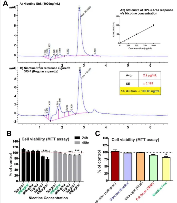
Exposure to nicotine concentrations equivalent to that observed in plasma in chronic human smoker does not affect endothelial cell viability
HPLC studies were performed to determine the dilution factor for freshly prepared 3R4F cigarette-derived CSE stock solution necessary to achieve CSE exposure yielding 100 ng/ml of nicotine (Figure 1A). This nicotine concentra-tion was chosen to model the plasma levels seen in human smokers [27-29]. 3R4F cigarette was used as a reference to calculate the dilution factor for the CSE stock which was then uniformly applied to all the test cigarettes. As shown in Figure 1B, 100 ng/ml of nicotine did not affect the cell viability at 24 and 48 h exposure. Cytotoxic effects of nico-tine exposure were observed at higher concentrations (10 and 100 μg/ml/24 h; 1, 10, 100 μg/ml/48 h). Note also that 24 h exposure to 5% diluted CSE from test cigarettes did not affect endothelial viability with the exception of NF-derived extracts (see Figure 1C). A small yet significant decrease in cell viability was observed in response to NF-derived CSE exposure, as compared to controls (CSE-free PBS or 100 ng/ml nicotine treatment).
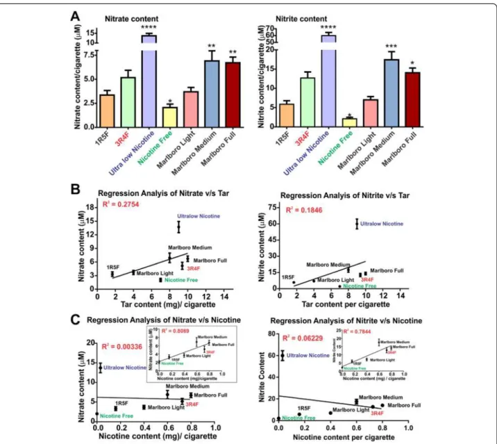
Nitrate and nitrite levels in CSE correlates with corresponding cigarette’s tar and nicotine content
A generation of highly carcinogenic tobacco-specific nitro-samines (TSNA) [30] has been suggested to arise from the reaction of amines with nitrite derived from nitrate in the tobacco [31]. We measured nitrate, NO3−/nitrite andNO2− content of CSE derived from 1R5F (ultralight), 3R4F (full flavor), Ultralow nicotine and NF (a non-tobacco based product) cigarettes (Figure 2A). In addition, commercially
Figure 1HPLC and viability studies to select the CSE concentration for the study. A) HPLC analysis to determine nicotine concentration in CSEshowed that 5% CSE had nicotine concentration comparable to the physiological concentration in a chronic smoker (100 ng/ml) n = 10. (B) Cell viability studies following increasing concentration of nicotine at 24 and 48 h and (C) 5% diluted CSE from ultralow, 1R5F (equivalent to ultralight cigarettes), 3R4F (equivalent to full flavor cigarettes), ultralow nicotine and tobacco free (nicotine free - NF) using MTT assay. Note that 5% CSE from all tested brand but NF did not cause a statistically significant decrease in cell viability. n = 3 individual experiments.
available Marlboro light, medium and full cigarettes were also analyzed for comparison. NO3−/NO2− content in CSE from ultralow nicotine cigarettes was significantly higher than any other brand tested including 3R4F (p < 0.0001, compared to light cigarettes), as observed with Marlboro full or medium cigarettes (p < 0.01, compared to light cigarettes, Figure 2A). In contrast, NO3−/NO2− content of NF cigarette was significantly lower (p < 0.05) when compared to “light” cigarettes (Figure 2A). A posi-tive correlation between NO3−/NO2−content of CSE and tar for the corresponding cigarette brands was also ob-served as demonstrated by the regression analysis shown
in Figure 2B. Importantly, the ultralow nicotine brand stands out in terms of NO3−/NO2− content when com-pared to other brands containing a similar amount of tar. NO3−/NO2− positively correlate with the nicotine content of corresponding cigarette types with an ex-ception of the ultralow nicotine brand (Figure 2C and insets). Results from the regression analyses (tar and nicotine versus NO3−/NO2−) suggests that an alteration of the tobacco product to reduce the nicotine content, such as in ultralow nicotine and NF brands, could be re-sponsible for the higher output of NO3−/NO2− during cigarette combustion.
Figure 2Nitrate/Nitrite content profiling of CSE from tested tobacco products. (A) Nitrate/nitrite content increased proportionally to the amount of tar in the cigarettes where statistically significant higher nitrate was found in ultralow nicotine than ultralight cigarette (P < 0.001, n = 10 biological replicates). Nicotine free cigarettes which were non-tobacco based did not have significant nitrate/nitrite content. Regression analysis of Nitrate/Nitrite correlates with tar (B) but not nicotine content unless ultralow nicotine cigarettes are removed from the analytical pool (C).
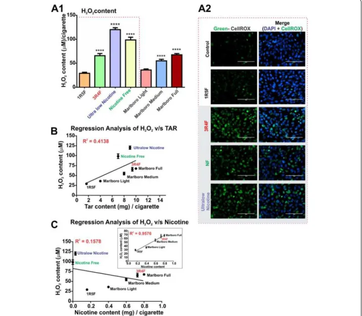
Release of hydrogen peroxide (H2O2) in CSE increases with
the tar content of cigarettes and leads to progressive oxidative damage in BBB ECs
TS is a major exogenous source of free radicals con-tained in both gaseous phase and tar, which can spawn sustained high levels of ROS (e.g., H2O2) that may
dir-ectly affect the BBB integrity. Thus, we determined the amount of H2O2 release in the CSE and assessed its
oxidative effect on BBB endothelial cell cultures. As shown in Figure 3A1, the highest levels of H2O2were
found in CSE from ultralow nicotine and NF
(tobacco-free) cigarettes when compared to light products (p < 0.0001). H2O2 content in CSE from 3R4F (full flavor)
and Marlboro medium/full cigarettes, although statisti-cally higher that light cigarettes, was considerably less compared with ultralow nicotine and NF products. This was in accordance with measurements of cellular oxidative stress (CellROX® Green Reagent, Figure 3A2) and revealed the highest level of cellular oxidation in endothelial cultures chronically exposed (24 h) to Ul-tralow nicotine and NF smoke extracts. Interestingly, 3R4F cigarettes released lower amounts of H2O2 than
Figure 3Hydrogen Peroxide content profiling of CSE from tested tobacco products. (A1) H2O2content increased proportionally to the
amount of tar in the cigarettes where statistically significant higher H2O2was found in full flavor, ultralow nicotine and nicotine free cigarettes
than light cigarette (P < 0.001), (n = 10 CSE preparations). (A2) Immunofluorescence analysis of oxidative stress in ECs (HCMEC/D3 cell line) caused by CSE exposure from 1R5F, 3R4F, ultralow nicotine and NF cigarettes versus controls: Note that most significant oxidative responses were observed in EC cultures exposed to CSE treatment (24 h) derived from 3R4F, ultralow nicotine and NF cigarette (n = 3 biological replicates). Regression analysis of H2O2correlates with tar (B) but not nicotine content unless ultralow nicotine and NF cigarettes are removed from the
Ultralow nicotine and NF cigarettes which also demon-strated a comparable oxidative stress potential in endothe-lial cells. Furthermore, regression analysis of H2O2content
revealed a direct relationship between tar content of the corresponding cigarette brand (Figure 3B) and H2O2. A
positive correlation between H2O2 and nicotine content
was also observed but only with the exclusion of NF and ul-tralow cigarettes from the analytical pool (Figure 3C).
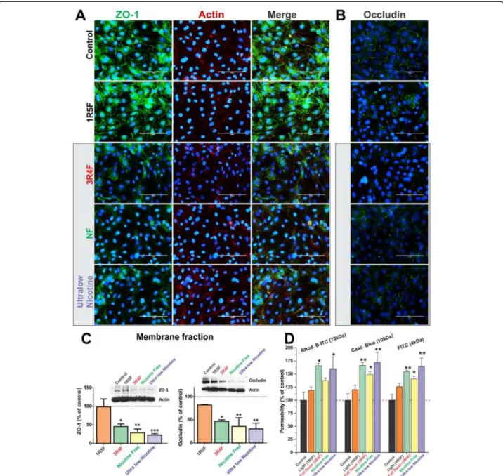
Exposure to CSE from 3R4F, NF and ultralow nicotine cigarettes negatively impacts ZO-1 and occludin expression/distribution as well as BBB integrity
Immunofluorescence analysis of BBB endothelial confluent monolayers revealed a significant loss of ZO-1 at cell-cell junctions following exposure to CSE from 3R4F, Ultralow and NF cigarettes and to a lesser extent in 1R5F treated cultures (compared to controls, Figure 4A). Results were further confirmed by western blot (WB) analysis of the corresponding membrane fractions (Figure 4C, left panel) in which exposure to ultralow nicotine CSE caused the most significant reduction of ZO-1 expres-sion at the membrane level (p < 0.0005, Figure 4C). However, actin distribution and expression were not altered by CSE exposure when compared to controls. Parallel immunofluorescence analyses also revealed a similar down-regulation and altered pattern of distri-bution at cell-cell junction for occludin (Figure 4B). Results were also confirmed by WB analysis of occlu-din expression in the corresponocclu-ding membrane frac-tions (Figure 4C, right panel). Expression levels of occludin were severely impaired in endothelial cultures exposed to CSE from 3R4F cigarette. The effect was even more significant in culture exposed to NF and ultra-low nicotine smoke extracts (see Figures 4B & C). Note also that a modest (not statistically significant) alteration in the distribution and expression level of occludin was observed in cell cultures treated with 1R5F-derived CSE when com-pared to controls. As expected, alteration in TJ expression/ distribution impacted BBB integrity as demonstrated by permeability measurements to dextran molecules (see Figure 4D). Increased permeability to 70 kDa dextran was noted in Transwells exposed to TS extracts from NF, ultralow and 3R4F although results were deemed significant (p < 0.05) only for the last two conditions. On the other hand, permeability to lower molecular weight dextrans (10 and 4 kDa) was significantly in-creased for all the three conditions mentioned above.
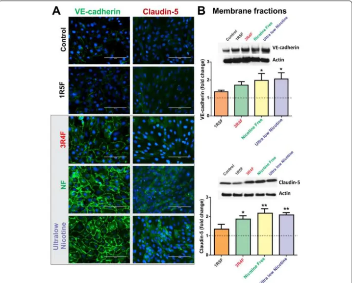
Further, immunofluorescence analysis revealed a marked up-regulation of VE-cadherin at cell-cell contacts in re-sponse to CSE exposure from 3R4F, NF and ultralow nico-tine (Figure 5A). Results were confirmed by parallel WB analysis of corresponding membrane fractions (Figure 5B) demonstrating a statistically significant (p < 0.05) increase in VE-cadherin expression following exposure to NF and
ultralow nicotine derived CSE, compared to controls and cultures exposed to 1R5F. A noticeable (although not sta-tistically significant) increase in VE-cadherin expression was also observed in endothelial cultures treated with 3R4F-derived CSE. Similarly, claudin-5 expression was up-regulated by exposure to CSE derived from 3R4F, ultralow nicotine and NF cigarettes, as demonstrated by immunofluorescence and WB analyses of correspond-ing membrane fractions (Figure 5A & B). Interestcorrespond-ingly, the patterns/level of expression of these junction proteins remarkably reflects the oxidative stress/H2O2 content of
the CSE extracts of the respective brands (as shown in Figure 3A1 & A2).
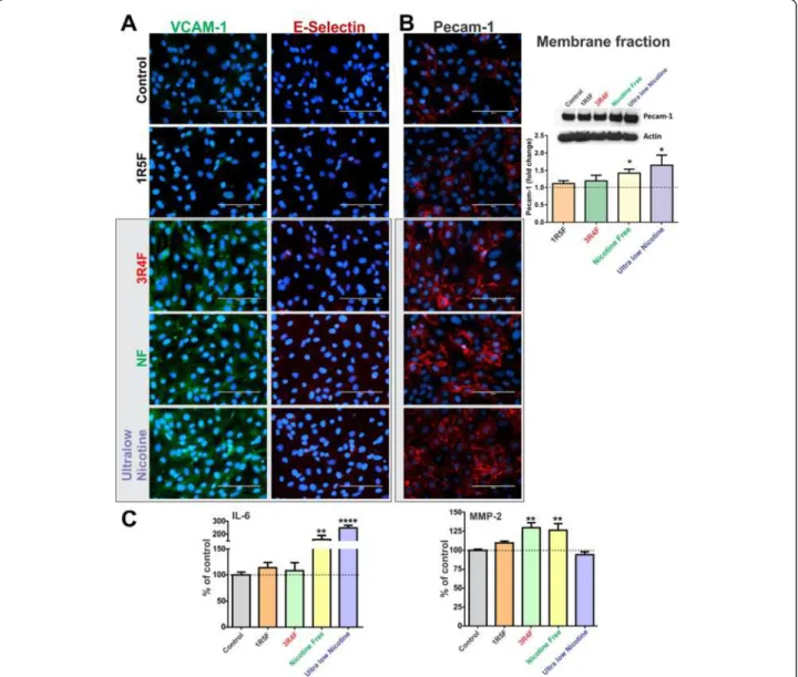
Exposure to CSE from 3R4F, NF and ultralow nicotine cigarettes promotes the pro-inflammatory activation of BBB endothelial cells
As shown in Figure 6A, endothelial cell expression of vas-cular endothelial adhesion molecule-1 (VCAM-1) was up-regulated following exposure to 3R4F, NF and ultralow nicotine CSEs, as compared to controls and 1R5F cigarette treated cultures. However, no expression changes were ob-served with respect to E-selectin. In addition, immunofluor-escence analysis indicated an up-regulation of endothelial Platelet Endothelial Cell Adhesion Molecule-1 (PECAM1) expression following exposure to 3R4F, NF and ultralow nicotine cigarettes (Figure 6B). These results were further supported by WB analysis of the corresponding membrane fractions (Figure 6B).
Importantly, analysis of the culture conditioned media by ELISA revealed a significant increase of interleukin-6 (IL-6) release from the endothelial cells exposed to either NF (p < 0.01) or ultralow nicotine cigarette extracts (p < 0.005), compared to controls (Figure 6C). A modest, yet significant increase in the release of matrix metalloproteinase-2 (MMP-2) was also observed in cultures treated with either 3R4F or NF smoke extracts, but not ultralow nicotine (Figure 6C). However, MMP-9, IL-1β and TNF-α levels in the conditioned media from all treatment conditions were below the reading sensitivity (data not shown).
Discussion
ROS, despite being essential for biological systems [32] have the potential to cause extensive oxidative damage to cells and tissues if their levels become excessive [33,34]. At the vascular level ROS can cause oxidative damage of endothe-lial cells [16] including DNA strand breakage and inflam-mation [17]. In addition to ROS, nicotine can equally elicit oxidative stress and tissue injury [35,36] and has been shown to exacerbate brain edema following focal ischemia [37,38]. Oxidants in the gaseous phase of cigarette smoke, including nicotine and various ROS species, ([15,20] can pass through the lung alveolar wall and raise systemic oxi-dative stress [39]. This can lead to oxioxi-dative damage to cells
and tissues, including the brain vascular system and the BBB, over a period of sustained exposure to TS (e.g., chronic smokers) and facilitate the pathogenesis and pro-gression of neurological disorders [40-42]. Thus, existing evidence strongly suggests a role for TS-dependent oxida-tive and inflammatory stress in the development of CNS pathologies. In fact, the cerebrovascular endothelium is highly vulnerable to oxidative stress resulting in loss of BBB function and integrity via altered expression and dis-tribution of intercellular TJ complexes [43,44].
In this study we assessed and compared the effects of various tobacco products on human BBB endothelial cells in relation to their corresponding oxidative potential. Spe-cifically, several studies have demonstrated that cigarette smoke contains high concentrations of NO which may dir-ectly affect the integrity of the BBB. For this purpose we measured ROS as well as NO3−/NO2−content (Figures 2 & 3) of tobacco smoke from 1R5F (ultralight), 3R4F (full fla-vor), NF (tobacco free) and ultralow nicotine cigarettes. The NO3−/NO2−analysis revealed a direct correlation with
Figure 4Effect of CSE exposure (24 h) on endothelial expression and distribution of ZO-1, Occludin and actin filaments. (A) Down-regulation of ZO-1 expression and disruption and cell-cell junctions were progressively more significant following exposure to 3R4F, NF and ultralow nicotine CSEs. Down-regulation and disruption at cell-cell contacts of occludin were also observed (B). Immunofluorescence analyses were confirmed by WB of corresponding membrane fractions (C). Loss of BBB integrity was further assessed by permeability measurements to dextran molecules ranging from 3 to 70 kDa (D) n = 3 biological replicates, *p < 0.05, **p < 0.01, ***p < 0.001 compared to controls.
the content of tar of the respective cigarettes. However, this did not hold true for NF products, whose tar content (comparable to medium strength cigarettes) produced the least amount of nitrate and nitrite. When we compared the NO3−/NO2−output with corresponding nicotine
con-tent, a significant correlation was not found, unless ultra-low nicotine brand were removed from the pool (Figure 2C - insets). Together these results suggest that NO3−/NO2− is relatively independent of nicotine con-tent while holding a strong correlation with that of tar.
Tar being a byproduct derived from combustion of to-bacco or analogous products, alteration of toto-bacco (e.g., ultralow nicotine products) or replacement with alterna-tive products (NF cigarette) to reduce nicotine content in a bid to decrease addiction potential, may result in an unwanted increase of nitrate/nitrite output, and risk for
health hazard. In fact, tobacco nitrate levels have been previously reported to correlate with the formation of non-specific volatile nitrosamines (e.g., N-nitrosodimethylamine, N-nitroso-diethylamine, N-nitrosoethylmethyl- amine, etc.), and non-volatile Tobacco-Specific Nitrosamines (TSNAs) such as 4-(methylnitrosamino)-1-(3-pyridyl)-1-butanone (NNK, nicotine-derived nitrosamine ketone) which have been associated with carcinogenicity of tobacco smoke [30,31].
Interestingly, H2O2 content measured in ultralow
nico-tine and tobacco free (NF) cigarettes, considered “reduced-exposure” products, was significantly higher than any other brand including medium and full flavor (see Figure 3). Re-gression analysis of H2O2also revealed a strong correlation
with the tar content of the respective cigarettes but not with that of nicotine unless both products were to be re-moved from the pool. These results strongly correlate with
Figure 5Effect of CSE exposure (24 h) on endothelial expression and distribution of VE-cadherin and Claudin-5. (A) Immunofluorescence analysis of BBB endothelial cultures revealed a significant up-regulation of VE-cadherin (at cell-cell junctions) and claudin-5 expression following exposure to 3R4F, NF and ultralow nicotine CSEs. (B) Immunofluorescence analyses were confirmed by WB of corresponding membrane fractions. n = 3 biological replicates, *p < 0.05, **p < 0.01 compared to controls.
the increased oxidative stress generated in BBB endothelial cultures (see Figure 3A2) and revealing that the highest level of oxidation is in endothelial cells that are exposed to ultralow and NF cigarette smoke extracts. Interestingly, the oxidative stress potential of 3R4F cigarettes was comparable to that of ultralow and NF cigarettes despite releasing lower amounts of H2O2. This can be attributed to the higher
con-tent of nicotine in 3R4F cigarettes since nicotine equally contributes to oxidative stress (Das et al. 2012). Taken to-gether, these results suggest that alteration and/or substi-tution of tobacco with alternative products in order to reduce nicotine content was responsible for the increased H2O2output measured in these “denicotinized” cigarette
products.
Previous reports by Hossain and co-workers [14] have shown a dose dependent loss of BBB integrity directly correlating to TS-derived oxidative stress. Furthermore, loss of BBB function and integrity caused by TS expos-ure was prevented or at least reduced by antioxidant vitamins. These findings by others clearly support our results which outlined a strong correlation between the impairment of tight junction protein expression/distri-bution and BBB integrity with the oxidative stress gen-erated by the TS extracts. As clearly shown in the results (see Figure 4) BBB endothelial ZO-1 expression and distribution is completely deregulated upon expos-ure to TS extract from 3R4F, NF and Ultralow Nicotine cigarettes. This is also reflected in the increased BBB
Figure 6Immunofluorescence analysis of BBB endothelial expression of VCAM-1 and E-selectin (A) and PECAM1 (B), following exposure to CSEs from 1R5F, 3R4F, NF and ultralow nicotine cigarettes. Immunofluorescence analysis of PECAM1 was confirmed by WB of corresponding membrane fractions. (C) Release of proinflammatory cytokines IL-6 was up-regulated in endothelial cultures exposed to NF and ultralow nicotine CSE while MMP-2 levels were increased by CSE from 3R4F and NF but not ultralow nicotine. n = 3 biological replicates, *p < 0.05, **p < 0.01, ****p < 0.0001 compared to controls.
permeability to dextran paracellular markers observed under the same conditions.
ZO-1 is a cytoplasmic accessory protein which plays a crucial role in BBB integrity by connecting transmem-brane proteins (such as occludin, claudins and JAM) to cytoskeletal proteins and is actively involved in signal trans-duction and transcriptional modulation [45,46]. Interest-ingly, the effect of CSE on ZO-1 expression/distribution reflects the overall oxidative potential of the corresponding cigarettes (see Figure 3), thus suggesting a correlation be-tween TS-dependent oxidative potential and dysregulation of TJs and BBB integrity. ZO-1 TJ protein closely associates with the actin cytoskeletal network. When we observed the actin structure with respect to ZO-1, it appeared intact. In addition, membrane expression of occludin was signifi-cantly down-regulated as evidenced by the WB analysis of the corresponding membrane fractions. Similar to ZO-1, membrane distribution of occludin was also altered deteri-orating from a homogenous pattern at cell-cell junctions in controls to a patchy distribution in cultures exposed to 3RF4, NF and ultralow nicotine cigarettes. This can be a reflection of the parallel loss of ZO-1 which provides a po-sitioning system and anchoring scaffold for the transmem-brane TJ proteins.
In contrast to ZO-1 and occludin, the expression of VE-cadherin and claudin-5 was proportionally increased with respect to the oxidative potential of the corresponding CSE treatment. In fact, as shown in Figure 5, VE-cadherin mem-brane expression was progressively up-regulated by expos-ure to 3RF4, NF and ultralow nicotine cigarettes, although statistical significance was proven only for the last two cigarette products. In parallel, claudin-5 membrane expres-sion was similarly up-regulated (see Figure 5B). This is in agreement with emerging evidences suggesting that VE-cadherin controls claudin-5 expression by preventing the nuclear accumulation of FoxO1 and beta-catenin which re-press the claudin-5 promoter [47] thus reducing its expres-sion. Although, these results were surprising, they actually seem to be in agreement with the above mentioned obser-vations. In fact, recent in vitro studies have shown a direct positive correlation between VE-cadherin expression and oxidative stress [48] suggesting this being part of a cytopro-tective response mechanism. In fact, VE-cadherin acts as a master regulator of various endothelial functions including modulation of cell-cell adhesion, angiogenesis, and vascular permeability to leukocytes in response to VCAM-1 activa-tion [49], whose expression level was also increased (see Figure 6). Note also that an up-regulation of claudin-5 (in this case mediated by VE-cadherin) does not necessarily translate into an improved BBB integrity. Although this is true from a biological standpoint under normal circum-stances we have to take into consideration that the mere expression of TJ proteins is not sufficient as a standalone determinant for BBB integrity. Other important factors play
a significant role here such as the link between TJ proteins with the cytoskeleton. An important interaction mediated by first order regulatory proteins such as ZO-1 is of critical importance for the positioning and interaction of TJ proteins with their homologues on adjacent endothelial cells. Moreover, although claudin-5 was up-regulated (see Figure 5) the pattern of expression presented as an homogenous distribution throughout the cells and lacked a demarcated membrane localization which does not suggest improvements of cell-cell adhesion. This hypothesis well copes with the evident loss of barrier functions outlined by the increased permeability to dextran markers.
In addition, a similar increase in PECAM-1 expression was observed as well as an increased endothelial release of IL-6 and MMP-2 (see Figure 6). Regarding MMP-2, previous reports by others have shown how ROS regu-late the activity of vascular matrix metalloproteinases in vitro including MMP-2 and MMP-9 [50] which have an implication in atherosclerotic plaque stability. Expres-sion and activation of MMP-2 has been demonstrated as a key event in oxidative stress injury to heart [51] and hyperglycaemia promoted BBB dysfunction [52]. To-gether these results strengthen the link between tobacco smoke, it’s corresponding oxidative and inflammatory stress, and potential risk for BBB dysfunction. Although outside the scope of the present work, more studies will be necessary to dissect the molecular mechanisms in-volved in the generation of cellular oxidative stress at the brain microvascular endothelium by CSE and its impact on BBB function and integrity.
Conclusion
In summary, this study is one of the first attempts to as-sess and compare the potential toxic impact of various cigarette products on BBB endothelial cells using whole smoke extracts. We further correlated the oxidative and inflammatory potential of these cigarette products with respect to their tar, nicotine, H2O2and nitric oxide
con-tent. We also clearly showed that the alteration of to-bacco in an attempt to reduce cigarette nicotine content to attenuate addiction can result in an increased toxicity and endothelial inflammatory response. This can ultim-ately impair the BBB function and increase the risk for the pathogenesis of a number of CNS disorders.
Methods
Materials and reagents
The antibodies used in this study were obtained from the following sources: Rabbit ZO-1 (#8193), rabbit anti-claudin-3 (#341700), rabbit anti-VE-cadherin (#D87F2), rabbit anti-VCAM-1 (#12367), mouse PECAM-1 (#89C2) from Cell Signaling Technology (Danvers, MA, USA); mouse anti-E-selectin (#S 9555), β-actin (#A5441) from Sigma-Aldrich (St. Louis, MO, USA); donkey anti-rabbit
(#NA934) and sheep anti-mouse (#NA931) HRP-linked secondary antibodies from GE Healthcare (Piscataway, NJ, USA); mouse anti-claudin 5 (#35-2500), goat anti-rabbit (#A11008) and anti-mouse (#A21422) conjugated to Alexa Fluor® 488 and 555 from Invitrogen (Camarillo, CA, USA). Sterile cultureware was obtained from Fisher Scientific (Pittsburgh, PA, USA), while other reagents and chemicals were purchased from Sigma-Aldrich (St. Louis, MO, USA) or Bio-Rad laboratories (Hercules, CA, USA).
TS preparation
Concentrated cigarette smoke extracts (CSE) were prepared according to the Federal Trade Commission (FTC) stand-ard smoking protocol (35 ml draw, 2 second puff duration, 1 puff per 60 seconds), using a Single Cigarette Smoking Machine (SCSM, CH Technologies Inc., Westwood, NJ, USA), as shown in Figure 7. This protocol resulted in ap-proximately 8 puffs per cigarette. Mainstream cigarette smoke was bubbled through an impinger into phosphate buffered saline (PBS) to generate a concentrated CSE stock (100%) solution that was further diluted to desired concen-trations and was used immediately for the experiments described below. Four types of cigarettes were used for this study, as shown in Figure 8: a) 1R5F cigarettes equivalent to commercial ultralight brands with 1.67 mg tar and 0.160 mg nicotine per cigarette b) 3R4F cigarettes equiva-lent to full flavor brands with 9.4 mg tar and 0.726 mg nico-tine per cigarette (obtained from University of Kentucky); c) reduced nicotine spectrum cigarettes (obtained from NIH/NIDA) equivalent to ultralow nicotine brands with 0.03 mg nicotine and 9 mg tar per cigarette; and iv) commercially available nicotine-free cigarette from non-tobacco based source with a tar content of 7 mg per cigarette. Commercially available light, medium and full flavor cigarettes from Marlboro were also used for com-parative profiling experiments.
Cell culture
The immortalized hCMEC/D3 cell line was donated by Dr. Couraud (INSERM, Paris). The hCMEC/D3 cells
(passage 28–32) were seeded on collagen coated culture flasks (2.5-3 × 10⁴/cm2) or glass slides (4 × 10⁴/cm2) and maintained at 37°C with 5% CO2 exposure in EBM-2
basal medium (Lonza, Walkersville, MD, USA) supple-mented with 5% FBS (Atlanta Biologicals, Lawrenceville, GA, USA), chemically defined lipid concentrate (Life Technologies, Carlsbad, CA, USA), growth factors, anti-biotic/antimycotic (1:1, Atlanta Biologicals, GA, USA and HEPES (10 mM). Medium was changed every 2 days until the cells reached confluence. Monolayer integrity of hCMEC/D3 cells at confluence was con-firmed by phase contrast microscopy and the expres-sion of endothelial cell-specific phenotypic markers at cell-cell junctions, as previously described [26]. For treat-ment, cell monolayers were exposed to CSE concentration (5-20%) diluted from freshly prepared smoke extracts as described above. Cultures exposed to CSE-free vehicle (PBS) served as controls.
Cell viability assay
The effects of CSE exposure on cell viability were deter-mined by MTT (3 (4,5-dimethylthiazol-2yl)-2,5-diphe-nyltetrazolium bromide) assay. Briefly, HCMEC/D3 cells were passaged in a 96-well plate and allowed to attach for a period of 48 h. Following exposure to CSE, cells were incubated with 10 μM MTT for 5 h at 37°C. MTT was removed and DMSO was added to solubilize the formazan crystals for 20 min. Color development corre-sponding to viable cells was quantitated by measuring absorbance at 520 nm.
Nitric oxide (NO) content analysis
Cigarette smoke was bubbled through an impinger into PBS using the CSM-SCCM smoking machine as described above. NO content of the different types of cigarettes was determined indirectly through the estimation of nitrate/ni-trite content using Griess reagent reaction based NO kit from R&D Systems, according to manufacturer’s guidelines.
Figure 7Smoke Preparation according to ISO/FTC protocol. Concentrated Smoke solution was prepared from each cigarette using CSM-SCSM Cigarette Smoking Machine according to ISO/FTC determination parameters. These require a puff volume of 35 ml with duration of 2 s at interval of 60s.
Hydrogen peroxide (H2O2) content analysis
H2O2 content in smoke extracts of various types of
cigarettes was determined by TBR4100 free radical analyzer with 100 μm HPO sensor. Briefly, aliquots from cigarette preparation were titrated in PBS to ob-tain a sensor reading which was extrapolated against a hydrogen peroxide standard curve to quantitate the amount per cigarette.
HPLC analysis of CSE preparation
For sample preparation, CSE obtained from CSM was sub-jected to liquid/liquid extraction using dichloromethane. Briefly, 500 μl aliquot of CSE was mixed with 5 μl of 1 M NaOH followed by the addition of 2 ml DCM. After centri-fugation of the mixture at 1500 g for 10 min, the upper aqueous layer was discarded. The lower organic layer was evaporated under nitrogen gas, and the precipitate was re-suspended in mobile phase, filtered and then injected on to the column. Nicotine (Sigma Aldrich, St. Louis, MA, USA #36733) dissolved in mobile phase was used to prepare the standard curve. Isocratic separation was performed on Agilent A1220 HPLC System (Agilent Technologies, Santa Clara, CA, USA) coupled to a UV detector, using Zorbax Rx-C18 column (4.6x150mm, 5 μm) with an inline guard column filter. The mobile phase consisted of 50 mM KH2PO4buffer with 10 mM sodium heptane 1-sulfonate,
pH adjusted to 3.0 using orthophosphoric acid and metha-nol (70:30 v/v). The flow rate was set to 1 ml/min with column temperature at 30°C and injection volume was 50 μl. Wavelength corresponding to maximum absorption of nicotine (259 nm) was used.
ELISA
Following exposure to CSE, the cell culture conditioned media was collected and stored at −20°C until analysis. Levels of pro-inflammatory cytokines such as IL-1b, TNF-alpha, IL-6 and matrix metalloproteinases (MMPs) such as MMP-2 and MMP-9 were measured by Quantikine ELISA kits from R&D systems as per manufacturer’s instructions.
Immunofluorescence analysis
Cells were grown in two-well chamber slides specifically for these studies. After treatment, cells were fixed with formaldehyde (15mins at 4°C). Following three PBS washes, cells were blocked using 5% goat serum (Sigma-Aldrich, St. Louis, MO, USA) in PBS at room temperature for 50 min and incubated with primary antibodies prepared in 5% GSA overnight at 4°C. After three rinses with PBS, cells were incubated for 1 h at RT with Alexa Fluor® 488 or 555 conjugated goat anti-rabbit or anti-mouse antibodies, respectively (1:1000). Thereafter, cells were rinsed and counterstained with DAPI in Prolonged Gold Anti-fade mounting media (Invitrogen, OR, USA). Slides were cover slipped and left for overnight drying in the dark before examination with EVOS digital inverted fluorescence microscope. Cells stained with secondary antibodies alone were used as negative controls.
Western blotting
Briefly, cells were lysed in ice-cold Urea-Tris buffer containing Phosphatase and Protease Inhibitors (Roche Diagnostics, Indianapolis, IN, USA), sonicated and centrifuged at 14000 rpm, 4°C for 15 min. Protein con-centration was determined by Bradford assay (Bio-Rad laboratories (Hercules, CA, USA # 5000006). Denatured samples containing equal protein (40 μg) were subjected to SDS-PAGE (10% or 4-15% gradient gel) and electrotrans-ferred to PVDF membranes (2 hr transfer at 100 V). Mem-branes were blocked for 2 h (RT) with 5% non-fat dry milk in Tris buffered saline (TBS) containing 0.1% Tween-20 (TTBS) and subsequently incubated with rabbit (1:1000) or mouse (1:500) primary antibodies. After 4 washes (10 min each) with TTBS, membranes were incubated with anti-rabbit or anti-mouse (1:2000) HRP-conjugated secondary antibodies (2 h, RT) and washed with TTBS. Bands were detected by enhanced chemiluminescence using Amersham ECL™ Prime with ChemiDoc™ XRS system. Membranes were subsequently stripped and probed for β-actin (1:1000) as a loading control. Band densities were analyzed by Quantity One Software.
Tar and Nicotine content of the main
tobacco
products used in the study
Figure 8Tar and Nicotine content of the main tobacco products used in the study. The following table states the tar and nicotine composition of the various cigarettes used in the study. (+1R5F and 3R4F are the names of these cigarettes provided by University of Kentucky for research purposes).
Measurement of BBB integrity: dextran permeability
Differential effects of TS exposure on BBB integrity was assessed by measuring paracellular permeability (luminal to abluminal) to labeled dextrans (4-70 kDa) as previously de-scribed [53]. After 24 h exposure to TS extracts, a mixture of labeled dextrans in PBS (FITC- 4 kDa, 5 mg/ml; Cascade Blue®- 10 kDa, 5 mg/ml; and Rhod. B-ITC - 70 kDa, 5 mg/ ml) was added to the luminal compartment. Abluminal samples (50 μL) were collected over 30 min and replaced with equal volume of fresh media to allow sink conditions. Dextran fluxes were determined by fluorescent measure-ments using the appropriate excitation and emission wavelengths. Permeability measurements were reported as percentage of controls (the permeability coefficients of con-trols were as follow: FITC 0.198 ± 0.009 × 10⁻³ cm/min;
Cascade Blue® 0.0953 ± 0.007 × 10⁻³cm/min and Rhod.
B-ITC 0.007 ± 0.0005 × 10⁻³cm/min).
Statistical analyses
Data from all experiments were expressed as mean ± stand-ard error of mean (S.E.M) and analyzed by one-way ANOVA using GraphPad Prism Software Inc. (La Jolla, CA, USA). Post hoc multiple comparisons were performed with Tukey’s test. P values ≤ than 0.05 were considered statistically significant.
Competing interests
The authors declare that they have no competing interests.
Authors’ contributions
PN conceived the study and elaborated its design. PN also performed most of the experiments and drafted the manuscript. NF carried out major western blot analysis. SP carried out (with PN) immunofluorescence and permeability studies. RS provided direction to this project and helped draft the manuscript. BW, PC and IR provided the necessary cell line to conduct these studies. They also assisted with data analysis and manuscript editing/ revisions. LC supervised the project, the data analysis and provided guidance during manuscript preparation and revisions. All authors have read and approved the final version of the manuscript.
Acknowledgements
These studies were supported by NIH/NIDA R01-DA029121-01A1 and in part by. A.R.D.F to Dr. Luca Cucullo.
Author details
1Department of Pharmaceutical Sciences, Texas Tech University Health
Sciences Center, School of Pharmacy, 1300 S. Coulter Street, Amarillo TX 79106, USA.2Center for Blood Brain Barrier Research, Texas Tech
University Health Sciences Center, Amarillo, TX 79106, USA.3Department of
Biomedical Sciences, Texas Tech University Health Sciences Center, Amarillo, TX 79106, USA.4Weill Cornell Medical College, New York, NY, USA.5Inserm,
U1016, Institut Cochin, Paris, France.6CNRS, UMR8104, Paris, France. 7Université Paris Descartes, Sorbonne Paris Cité, Paris, France.8Department of
Life, Health and Chemical Sciences, Open University, Milton Keynes, UK.
Received: 23 December 2013 Accepted: 7 April 2014 Published: 23 April 2014
References
1. Smoking-attributable mortality, years of potential life lost, and productivity losses–United States, 2000–2004. MMWR Morb Mortal Wkly Rep 2008, 57:1226–1228.
2. Hecht SS: Cigarette smoking: cancer risks, carcinogens, and mechanisms. Langenbecks Arch Surg 2006, 391:603–613.
3. Howard G, Wagenknecht LE, Cai J, Cooper L, Kraut MA, Toole JF: Cigarette smoking and other risk factors for silent cerebral infarction in the general population. Stroke 1998, 29:913–917.
4. Mannami T, Iso H, Baba S, Sasaki S, Okada K, Konishi M, Tsugane S: Cigarette smoking and risk of stroke and its subtypes among middle-aged Japanese men and women: the JPHC study cohort I. Stroke 2004, 35:1248–1253. 5. Miller GJ, Bauer KA, Cooper JA, Rosenberg RD: Activation of the coagulant
pathway in cigarette smokers. Thromb Haemost 1998, 79:549–553. 6. Mast H, Thompson JL, Lin IF, Hofmeister C, Hartmann A, Marx P, Mohr JP,
Sacco RL: Cigarette smoking as a determinant of high-grade carotid artery stenosis in hispanic, black, and white patients with stroke or transient ischemic attack. Stroke 1998, 29:908–912.
7. Chalouhi N, Ali MS, Starke RM, Jabbour PM, Tjoumakaris SI, Gonzalez LF, Rosenwasser RH, Koch WJ, Dumont AS: Cigarette smoke and inflammation: role in cerebral aneurysm formation and rupture. Mediators Inflamm 2012, 2012:271582.
8. Salzer J, Hallmans G, Nystrom M, Stenlund H, Wadell G, Sundstrom P: Smoking as a risk factor for multiple sclerosis. Mult Scler 2013, 19:1022–1027. 9. Hedstrom AK, Hillert J, Olsson T, Alfredsson L: Smoking and multiple
sclerosis susceptibility. Eur J Epidemiol 2013, 28:867–874.
10. Chang RC, Ho YS, Wong S, Gentleman SM, Ng HK: Neuropathology of cigarette smoking. Acta Neuropathol 2013, 127:53–69.
11. Piao WH, Campagnolo D, Dayao C, Lukas RJ, Wu J, Shi FD: Nicotine and inflammatory neurological disorders. Acta Pharmacol Sin 2009, 30:715–722. 12. Rosenberg GA: Neurological diseases in relation to the blood–brain
barrier. J Cereb Blood Flow Metab 2012, 32:1139–1151.
13. Hossain M, Sathe T, Fazio V, Mazzone P, Weksler B, Janigro D, Rapp E, Cucullo L: Tobacco smoke: a critical etiological factor for vascular impairment at the blood–brain barrier. Brain Res 2009, 1287:192–205. 14. Hossain M, Mazzone P, Tierney W, Cucullo L: In vitro assessment of
tobacco smoke toxicity at the BBB: do antioxidant supplements have a protective role? BMC Neurosci 2011, 12:92.
15. Valavanidis A, Vlachogianni T, Fiotakis K: Tobacco smoke: involvement of reactive oxygen species and stable free radicals in mechanisms of oxidative damage, carcinogenesis and synergistic effects with other respirable particles. Int J Environ Res Public Health 2009, 6:445–462. 16. Raij L, Demaster EG, Jaimes EA: Cigarette smoke-induced endothelium
dysfunction: role of superoxide anion. J Hypertens 2001, 19:891–897. 17. Chen HW, Chien ML, Chaung YH, Lii CK, Wang TS: Extracts from cigarette
smoke induce DNA damage and cell adhesion molecule expression through different pathways. Chem Biol Interact 2004, 150:233–241. 18. Valko M, Izakovic M, Mazur M, Rhodes CJ, Telser J: Role of oxygen radicals
in DNA damage and cancer incidence. Mol Cell Biochem 2004, 266:37–56. 19. Pryor WA, Stone K, Zang LY, Bermudez E: Fractionation of aqueous
cigarette tar extracts: fractions that contain the tar radical cause DNA damage. Chem Res Toxicol 1998, 11:441–448.
20. Pryor WA, Stone K: Oxidants in cigarette smoke. Radicals, hydrogen peroxide, peroxynitrate, and peroxynitrite. Ann N Y Acad Sci 1993, 686:12–27. 21. Sobczak A, Golka D, Szoltysek-Boldys I: The effects of tobacco smoke on
plasma alpha- and gamma-tocopherol levels in passive and active cigarette smokers. Toxicol Lett 2004, 151:429–437.
22. Dietrich M, Block G, Norkus EP, Hudes M, Traber MG, Cross CE, Packer L: Smoking and exposure to environmental tobacco smoke decrease some plasma antioxidants and increase gamma-tocopherol in vivo after adjustment for dietary antioxidant intakes. Am J Clin Nutr 2003, 77:160–166.
23. Willcox JK, Ash SL, Catignani GL: Antioxidants and prevention of chronic disease. Crit Rev Food Sci Nutr 2004, 44:275–295.
24. Masubuchi T, Koyama S, Sato E, Takamizawa A, Kubo K, Sekiguchi M, Nagai S, Izumi T: Smoke extract stimulates lung epithelial cells to release neutrophil and monocyte chemotactic activity. Am J Pathol 1998, 153:1903–1912.
25. Weksler B, Romero IA, Couraud PO: The hCMEC/D3 cell line as a model of the human blood brain barrier. Fluids Barriers CNS 2013, 10:16.
26. Weksler BB, Subileau EA, Perriere N, Charneau P, Holloway K, Leveque M, Tricoire-Leignel H, Nicotra A, Bourdoulous S, Turowski P, Male DK, Roux F, Greenwood J, Romero IA, Couraud PO: Blood–brain barrier-specific properties of a human adult brain endothelial cell line. FASEB J 2005, 19:1872–1874.
27. Henningfield JE, Stapleton JM, Benowitz NL, Grayson RF, London ED: Higher levels of nicotine in arterial than in venous blood after cigarette smoking. Drug Alcohol Depend 1993, 33:23–29.
28. Khanna A, Guo M, Mehra M, Royal W III: Inflammation and oxidative stress induced by cigarette smoke in Lewis rat brains. J Neuroimmunol 2013, 254:69–75.
29. Abbruscato TJ, Lopez SP, Roder K, Paulson JR: Regulation of blood– brain barrier Na, K,2Cl-cotransporter through phosphorylation during in vitrostroke conditions and nicotine exposure. J Pharmacol Exp Ther 2004, 310:459–468.
30. Hoffmann D, Brunnemann KD, Prokopczyk B, Djordjevic MV: Tobacco-specific N-nitrosamines and areca-derived N-nitrosamines: chemistry, biochemistry, carcinogenicity, and relevance to humans. J Toxicol Environ Health 1994, 41:1–52.
31. Fischer S, Spiegelhalder B, Preussmann R: Preformed tobacco-specific nitrosamines in tobacco–role of nitrate and influence of tobacco type. Carcinogenesis 1989, 10:1511–1517.
32. Djordjevic VB: Free radicals in cell biology. Int Rev Cytol 2004, 237:57–89. 33. Rao R: Oxidative stress-induced disruption of epithelial and endothelial
tight junctions. Front Biosci 2008, 13:7210–7226.
34. Kong Q, Lin CL: Oxidative damage to RNA: mechanisms, consequences, and diseases. Cell Mol Life Sci 2010, 67:1817–1829.
35. Jain A, Flora SJ: Dose related effects of nicotine on oxidative injury in young, adult and old rats. J Environ Biol 2012, 33:233–238.
36. Zhou X, Sheng Y, Yang R, Kong X: Nicotine promotes cardiomyocyte apoptosis via oxidative stress and altered apoptosis-related gene expression. Cardiology 2010, 115:243–250.
37. Paulson JR, Yang T, Selvaraj PK, Mdzinarishvili A, Van der Schyf CJ, Klein J, Bickel U, Abbruscato TJ: Nicotine exacerbates brain edema during in vitro and in vivo focal ischemic conditions. J Pharmacol Exp Ther 2010, 332:371–379. 38. Catanzaro DF, Zhou Y, Chen R, Yu F, Catanzaro SE, De Lorenzo MS,
Subbaramaiah K, Zhou XK, Pratico D, Dannenberg AJ, Weksler BB: Potentially reduced exposure cigarettes accelerate atherosclerosis: evidence for the role of nicotine. Cardiovasc Toxicol 2007, 7:192–201. 39. Yamaguchi Y, Nasu F, Harada A, Kunitomo M: Oxidants in the gas phase of
cigarette smoke pass through the lung alveolar wall and raise systemic oxidative stress. J Pharmacol Sci 2007, 103:275–282.
40. McQuaid S, Cunnea P, McMahon J, Fitzgerald U: The effects of blood–brain barrier disruption on glial cell function in multiple sclerosis. Biochem Soc Trans 2009, 37:329–331.
41. Weiss N, Miller F, Cazaubon S, Couraud PO: The blood–brain barrier in brain homeostasis and neurological diseases. Biochim Biophys Acta 2009, 1788:842–857.
42. Deane R, Zlokovic BV: Role of the blood–brain barrier in the pathogenesis of alzheimer’s disease. Curr Alzheimer Res 2007, 4:191–197.
43. Freeman LR, Keller JN: Oxidative stress and cerebral endothelial cells: regulation of the blood–brain-barrier and antioxidant based interventions. Biochim Biophys Acta 1822, 2012:822–829.
44. Coisne C, Engelhardt B: Tight junctions in brain barriers during central nervous system inflammation. Antioxid Redox Signal 2011, 15:1285–1303. 45. Abbott NJ, Patabendige AA, Dolman DE, Yusof SR, Begley DJ: Structure and
function of the blood–brain barrier. Neurobiol Dis 2010, 37:13–25. 46. Liu WY, Wang ZB, Zhang LC, Wei X, Li L: Tight junction in blood–brain
barrier: an overview of structure, regulation, and regulator substances. CNS Neurosci Ther 2012, 18:609–615.
47. Gavard J, Gutkind JS: VE-cadherin and claudin-5: it takes two to tango. Nat Cell Biol 2008, 10:883–885.
48. Lei Y, Stamer WD, Wu J, Sun X: Oxidative stress impact on barrier function of porcine angular aqueous plexus cell monolayers. Invest Ophthalmol Vis Sci 2013, 54:4827–4835.
49. Ley K: Leukocytes talking to VE-cadherin. Blood 2013, 122:2300–2301. 50. Rajagopalan S, Meng XP, Ramasamy S, Harrison DG, Galis ZS: Reactive
oxygen species produced by macrophage-derived foam cells regulate the activity of vascular matrix metalloproteinases in vitro. Implications for atherosclerotic plaque stability. J Clin Invest 1996, 98:2572–2579. 51. Ali MA, Schulz R: Activation of MMP-2 as a key event in oxidative stress
injury to the heart. Front Biosci (Landmark Ed) 2009, 14:699–716.
52. Shao B, Bayraktutan U: Hyperglycaemia promotes cerebral barrier dysfunction through activation of protein kinase C-beta. Diabetes Obes Metab 2013, 15:993–999.
53. Santaguida S, Janigro D, Hossain M, Oby E, Rapp E, Cucullo L: Side by side comparison between dynamic versus static models of blood–brain barrier in vitro: a permeability study. Brain Res 2006, 1109:1–13.
doi:10.1186/1471-2202-15-51
Cite this article as: Naik et al.: Oxidative and pro-inflammatory impact of regular and denicotinized cigarettes on blood brain barrier endothelial cells: is smoking reduced or nicotine-free products really safe? BMC Neuroscience 2014 15:51.
Submit your next manuscript to BioMed Central and take full advantage of:
• Convenient online submission
• Thorough peer review
• No space constraints or color figure charges
• Immediate publication on acceptance
• Inclusion in PubMed, CAS, Scopus and Google Scholar
• Research which is freely available for redistribution
Submit your manuscript at www.biomedcentral.com/submit