HAL Id: hal-01865462
https://hal.uca.fr/hal-01865462
Submitted on 26 Nov 2018
HAL is a multi-disciplinary open access archive for the deposit and dissemination of sci-entific research documents, whether they are pub-lished or not. The documents may come from teaching and research institutions in France or abroad, or from public or private research centers.
L’archive ouverte pluridisciplinaire HAL, est destinée au dépôt et à la diffusion de documents scientifiques de niveau recherche, publiés ou non, émanant des établissements d’enseignement et de recherche français ou étrangers, des laboratoires publics ou privés.
selection during deep brain stimulation surgery for
essential tremor
Ashesh Shah, Jerome Coste, Jean Jacques Lemaire, Erik Schkommodau,
Simone Hemm-Ode
To cite this version:
Ashesh Shah, Jerome Coste, Jean Jacques Lemaire, Erik Schkommodau, Simone Hemm-Ode. Use of quantitative tremor evaluation to enhance target selection during deep brain stimulation surgery for essential tremor. 49th annual conference of the German Society for Biomedical Engineering, German Society for Biomedical Engineering, Sep 2015, Lûbeck, Germany. pp.488-492, �10.1515/cdbme-2015-0117�. �hal-01865462�
1
Use of quantitative tremor evaluation to enhance target selection
during deep brain stimulation surgery for essential tremor
Ashesh SHAH1, Jérôme COSTE2,3, Jean-Jacques LEMAIRE2,3,
Erik SCHKOMMODAU1, Simone HEMM-ODE1,*
1. Institute for Medical and Analytical Technologies, University of Applied Sciences and Arts Northwestern Switzerland, Gruendenstrasse 40, 4132 Muttenz, Swtizerland. 2. Image-Guided Clinical Neuroscience and Connectomics (EA7282), Université
Clermont Auvergne, Clermont-Ferrand, France.
3. CHU de Clermont-Ferrand, Service de Neurochirurgie, Clermont-Ferrand, France. *. Corresponding author: E-mail: [email protected]
Deep brain stimulation (DBS), an effective surgical treatment for Essential Tremor (ET), requires test stimulations in the thalamus to find the optimum site for permanent electrode implantation. During these test stimulations, the changes in tremor are only visually evaluated. This, along with other parameters, increases the subjectivity when comparing the efficacy of different thalamic nuclei. We developed a method to quantitatively evaluate tremor during the test stimulations of DBS surgery and applied to 6 ET patients undergoing this treatment. From the quantitative data collected, we identified effective stimulation amplitudes for every test stimulation position and compared it with the ones identified visually during the surgery. We also classified the data based on the thalamic nuclei in which the center of the stimulating contact was present during test stimulations. Results indicate that, to achieve the same reduction in tremor, on average, the stimulation amplitude identified by our method was 0.6 mA lower than those identified by visual evaluation. The comparison of the different thalamic nuclei showed that stimulations in the Ventro-oral and the Intermediolateral nuclei of the thalamus result in higher reduction in tremor for similar stimulation amplitudes as the frequently targeted Ventrointermediate nucleus. We conclude that our quantitative tremor evaluation method is more sensitive than the widely used visual evaluation. Using such quantitative methods will aid in identifying the optimum target structure for patients undergoing DBS.
2
1 Background
Deep Brain Stimulation (DBS) is now a routinely used surgical treatment for movement disorders like Essential Tremor (ET) [1]. Electrical stimulation of certain brain structures is performed by implanting electrodes and connecting them to a subcutaneously implanted neurostimulator. Although the use of DBS is increasing, the optimum target structure is still debated. The limited knowledge of the mechanisms of action of DBS being one of the reasons, we believe the under-utilization of intraoperatively obtained data is another.
A typical DBS surgery is preceded by a planning session during which a trajectory is planned from an entry point in the skull to the target structure on the patient’s anatomical images using stereotactic planning software. With the aid of stereotactic equipment, this trajectory is then explored during the surgery. In most of the surgeries, neuronal activity is recorded and test stimulations are performed at pre-planned positions on this trajectory while the patient is awake. The final implant position is decided after comparing the reduction in tremor, needed stimulation current for inducing those reductions and the side effects observed during the test stimulations. In the routing clinical practice, the symptom evaluation methods are subjective [2]. Tremor is evaluated by a visual rating of the change compared to its level observed before stimulation. We have previously described the method of evaluating tremor during DBS quantitatively by using accelerometers [3]. We used this method to intraoperatively evaluate tremor from 6 ET patients who participated in a clinical study in the University Hospital in Clermont-Ferrand, France. In the current paper, we describe the result of the comparison between visual and quantitative evaluations. Further, we classified the results based on the anatomical location of the electrode and compared them to identify the target structure which is the most effective in reducing tremor.
2 Method
2.1 Surgical protocol
The primary goal of the DBS surgery remains the same for all surgical centres: to determine the optimal implantation position. However, the actual procedure may differ significantly. Lemaire et al have described, in detail, the routine surgical procedure at the University Hospital in Clermont-Ferrand [4]. For treating ET patients using DBS, the primary target structure is the Ventrointermedius nucleus (VIM) of the thalamus. At the University Hospital in Clermont-Ferrand, during the planning session on patient’s images using a commercial stereotactic surgery planning software (iPlan Stereotaxy, Brainlab, Germany), the surgical team identifies and outlines different nuclei in the thalamus using MRI images of the patient [5]. After the definition of target and entry point, test stimulation positions are planned on the resulting trajectory. After performing micro-electrode recording (MER) at these positions, test stimulations are performed. For each test stimulation, the stimulation current is varied from 0 to 3 mA in steps of 0.2 mA and the changes in tremor are observed visually. The highest reduction in
3
tremor (Visually identified Change, VC) and the corresponding stimulation amplitude (Visually identified Amplitude, VA) at which it was observed are noted for every test stimulation location. Along with these, the occurrence of side-effects, their type and their corresponding amplitudes are also noted. On completion of all test stimulations, these data are discussed upon by the surgical team and a position is determined as the “final implant location” for the chronic DBS electrode.
2.2 Acceleration data recording
Tremor was quantified during the surgery using a commercial acceleration sensor evaluation board (STEVALMKI022VI, ST Micro, Switzerland). Time-stamped acceleration values from 3 axes were obtained at a frequency of 400 Hz and a range of 8 g. The evaluation board was fixed in an in-house developed non-conductive printed plastic case which was attached to the patient’s wrist using a Velcro strap. The acceleration data were recorded by in-house developed software installed on a laptop that was interfaced with the sensor via an USB cable. Furthermore, the recording software was also interfaced with the electro-physiology system to synchronize the acceleration data recording with the test stimulation data.
For the simplification of acceleration data recording and analysis, a data recording protocol was defined for all the surgeries. Acceleration data recording is started before the beginning of the test stimulation to record the data representing the baseline tremor for that test stimulation, and is continued till all the stimulation amplitudes are tested. The acceleration data and the related information about the test stimulation position and amplitude were saved in data files for offline analysis [3].
2.3 Post-operative data analysis
Acceleration data were analysed post-operatively using Matlab (Mathwork, USA). In brief, the resultant acceleration was calculated from the three axes and filtered to remove low frequency (2 Hz) movements and high frequency noise (10 Hz). Statistical features were extracted from these filtered acceleration data and the features from different stimulation amplitudes were normalized (%) using the features from the baseline data. These normalized features were used to identify a dataset of 2 values for each test stimulation: 1.) The change in acceleration features (Quantitatively identified Change, QC) at the visually identified best stimulation amplitude VA and 2.) The stimulation amplitude (Quantitatively equivalent Amplitude, QA) at which the quantitative change QC was similar to VC. These data were then statistically compared to the corresponding values from the visual evaluation (VC vs QC and VA vs QA) using the Wilcoxon two-sided signed rank test.
In order to analyse the correlation with the anatomical position of the electrode, the anatomical structure present at the centre of the stimulating electrode was determined. Data were then grouped based on the thalamic nucleus attributed to the test stimulation position. Furthermore, from the acceleration data of every test stimulation, we also calculated the minimum stimulation amplitude required to obtain a 75% reduction in the statistical features compared to baseline (Amp75). These data along with the occurrence of side-effects were used to compare the efficiency of different structures in reducing the patient’s tremor by a fixed amount.
4 2.4 Patients
The above method was applied to 6 ET patients under a clinical study at the University Hospital in Clermont-Ferrand (2011-A00774-37 / AU905). Out of the 167 test stimulations that were performed, 148 positions were found where both evaluation methods identified a reduction in tremor.
3. Results
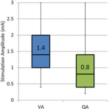
The Wilcoxon signed rank test revealed a statistically significant difference between VA and QA (W=4256, p< 0.001, alpha = 0.001) which can also be observed in the box plot of the two data sets (Figure 1). On the other hand, the difference between VC and QC at the visually identified amplitude VA was not statistically significant (W = 3004, p=0.72, alpha = 0.001).
Figure 1. Box plot comparing visually identified effective stimulation amplitude VA to
quantitatively identified effective stimulation amplitude QA for the same clinical improvement.
From the analysis of the electrode position during test stimulation, it was found that the positions were distributed in 7 thalamic nuclei: Center Median (CM), Intermedio-Lateral (InL), Ventrointermediate (VIM), Ventrocaudal lateral (VCL), Ventrocaudal medial (VCM), Ventro-Oral (VO) and also in Preleminiscal Radiations (PreR). Table 1 shows the average values of the different parameters extracted from the visual and the quantitative evaluations. The number of evaluations in the CM region (3) of the thalamus is small compared to the others and thus the significance of the results is very low. The values of QA are lower than VA for all the structures, the difference between them being the largest for InL, VO and the VIM.
5
Table 1. Average visual and quantitative parameters for different thalamic nuclei
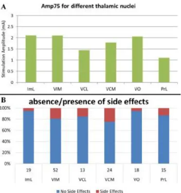
Figure 2A shows the average minimum stimulation amplitude required to see 75% reduction in tremor quantitatively while 2B shows the number of test stimulations with and without side-effects (red and blue respectively). The VCL and the PreR achieve similar reductions in tremor at lower amplitudes. But these structures also have higher occurrence of side effects. The VCM require higher stimulation amplitudes than VCL and PreR, but also has high occurrence of side-effects. The InL and the VO require similar stimulation amplitudes as the VIM but have much lower occurrence of side effects. The VIM requires the highest stimulation amplitude to achieve 75% reduction, but also has high occurrence of side-effects.
Figure 2. Comparison of efficacy of different thalamic nuclei. A) Average stimulation
amplitude required to achieve 75% reduction in tremor. B) Stacked column plot of number of test stimulations with and without side-effects.
4. Discussion
Various methods have been used previously to quantitatively evaluate tremor [6], but only a handful have been reported to be used during DBS surgery. Shamir et al. [7] used goniometers for Parkinson’s patients undergoing DBS, but only during MER to identify borders of sub-thalamic nucleus and not to quantitatively evaluate tremor
6
during test stimulations. Other quantitative evaluation techniques have been used to optimize the stimulation parameters of DBS but only after the implant position of the electrode was already decided [8, 9]. To the best of our knowledge, no other group has previously used quantitative tremor evaluation to compare the efficacy of different thalamic nuclei.
One of the observations of the application of our method to the current set of patients was that the use of quantitative evaluation during DBS surgery did not increase the duration of the surgery or cause any discomfort to the patient. Moreover, our results suggest that this quantitative method is more sensitive than the current visual evaluation methods used during the surgery. The significant difference (0.6 mA) between average values of VA and QA shows that stimulation parameters may be further optimized by using quantitative tremor evaluation.
The anatomical analysis of the data obtained using quantitative tremor evaluation suggests that the conventional target, VIM, may not be the most efficient target for ET patients undergoing DBS surgery. All other structures required stimulation amplitude equal to or lower than the VIM to reduce tremor by 75%. However, after looking at the number of positions with side effects, the VO and InL seem to be more efficient in reducing tremor and limiting side-effects of the therapy. This seems to confirm published data [5] suggesting that parts of the ventro-oral nucleus (VO) could be appropriate targets as well.
The results have also indicated some limitations of the current method. One of the main factors that influences the proper functioning of the method is the recording of baseline data. A minimum baseline recording of 5 seconds is necessary to extract proper statistical features. However, in case a baseline recording is not available for the current position, the baseline data of the previous recording can be used to perform the analysis. Furthermore, the current analysis method is post-operative. The next step for this method will to perform real-time data analysis so that the results are available to the surgical team when they identify the final implant site.
5. Conclusion
The current paper presents the results of quantitative tremor evaluation during DBS surgery for ET patients. The results indicate that our method is more sensitive to changes in tremor than the current visual evaluation methods. Furthermore, the results of the anatomical analysis suggest that the thalamic sub-structures InL and VO are more efficient targets for DBS than the conventional and targeted VIM.
7
Author's Statement: Conflict of interest: Authors state no conflict of interest. Material
and Methods: Informed consent: Informed consent has been obtained from all individuals included in this study. Ethical approval: The research related to human use has been complied with all the relevant national regulations, institutional policies and in accordance the tenets of the Helsinki Declaration, and has been approved by the authors’ institutional review board or equivalent committee.
References
[1] Sarem-Aslani A, Mullett K. Industrial Perspective on Deep Brain Stimulation: History, Current State, and Future Developments. Front. Integr. Neurosci. 2011;5. [2] Hemm S, Wĺrdell K. Stereotactic implantation of deep brain stimulation electrodes: a review of technical systems, methods and emerging tools. Med Biol Eng Comput 2010;48:611–24.
[3] Shah A, Coste J, Schkommodau E, Lemaire JJ, Hemm-Ode S. Using acceleration sensors to quantify symptoms during deep brain stimulation surgery. Biomedical Engineering / Biomedizinische Technik 2013.
[4] Lemaire J, Coste J, Ouchchane L, Hemm S, Derost P, Ulla M, Siadoux S et al. MRI anatomical mapping and direct stereotactic targeting in the subthalamic region: functional and anatomical correspondence in Parkinson’s disease. Int J CARS 2007;2:75–85.
[5] Vassal F, Coste J, Derost P, Mendes V, Gabrillargues J, Nuti C, Durif F et al. Direct stereotactic targeting of the ventrointermediate nucleus of the thalamus based on anatomic 1.5-T MRI mapping with a white matter attenuated inversion recovery (WAIR) sequence. Brain Stimul. 2012;5:625–33
[6] Mansur PHG, Cury LKP, Andrade AO, Pereira AA, Miotto GAA, Soares AB, Naves ELM. A review on techniques for tremor recording and quantification. Crit Rev Biomed Eng 2007;35:343–62.
[7] Shamir RR, Eitan R, Sheffer S, Marmor-Levin O, Valsky D, Moshel S, Zaidel A et al. Intra-operative Identification of the Subthalamic Nucleus Motor Zone Using Goniometers. Information Processing in Computer-Assisted Interventions. Berlin, Heidelberg: Springer Berlin Heidelberg; 2013. p. 21–29.
[8] Papapetropoulos S, Jagid JR, Sengun C, Singer C, Gallo BV. Objective monitoring of tremor and bradykinesia during DBS surgery for Parkinson disease. Neurology 2008;70:1244–9.
[9] Journee HL, Postma AA, Staal MJ. Intraoperative neurophysiological assessment of disabling symptoms in DBS surgery. Neurophysiol Clin 2007;37:467–7