HAL Id: hal-02562545
https://hal.archives-ouvertes.fr/hal-02562545
Submitted on 4 May 2020
HAL is a multi-disciplinary open access archive for the deposit and dissemination of sci-entific research documents, whether they are pub-lished or not. The documents may come from teaching and research institutions in France or abroad, or from public or private research centers.
L’archive ouverte pluridisciplinaire HAL, est destinée au dépôt et à la diffusion de documents scientifiques de niveau recherche, publiés ou non, émanant des établissements d’enseignement et de recherche français ou étrangers, des laboratoires publics ou privés.
Removal of microplastics from the environment. A
review
Mohsen Padervand, Eric Lichtfouse, Didier Robert, Chuanyi Wang
To cite this version:
Mohsen Padervand, Eric Lichtfouse, Didier Robert, Chuanyi Wang. Removal of microplastics from the environment. A review. Environmental Chemistry Letters, Springer Verlag, 2020, 18 (3), pp.807-828. �10.1007/s10311-020-00983-1�. �hal-02562545�
Environmental Chemistry Letters (2020) 18:807–828 https://doi.org/10.1007/s10311-020-00983-1
Removal of microplastics from the environment. A review
Mohsen Padervand1,2 · Eric Lichtfouse3 · Didier Robert4 · Chuanyi Wang1
Abstract
The production of fossil fuel-derived, synthetic plastics is continually increasing, while poor plastic waste management has recently induced severe pollution issues. Microplastics are plastic particles smaller than 5 mm. Microplastics are ubiq-uitous and slowly-degrading contaminants in waters and soils. Microplastics have long residence time, high stability, high potential of being fragmented and can adsorb other contaminants. Many aquatic species contain microplastics, which are in particular easily accumulated by planktonic and invertebrate organisms. Then, microplastics are transferred along food chains, leading to physical damages, decrease in nutritional diet value and exposure of the living organism to pathogens. Raw plastics contain chemical additives such as phthalates, bisphenol A and polybrominated diphenyl ethers that may induce toxic effects after ingestion by living organisms. Furthermore, the adsorption capability of microplastics makes them prone to carry several contaminants. Methods to remove microplastics from water and other media are actually needed. Here, we review microplastics occurrence, transport, raw polymers and additives, toxicity and methods of removal. Removal methods include physical sorption and filtration, biological removal and ingestion, and chemical treatments. Mechanisms, efficiency, advantages, and drawbacks of various removal methods are discussed.
Keywords Microplastic pollution · Environment · Removal method
Introduction
The global production of plastics has highly increased since 1950 to improve human life and reached almost 381 million tons in 2015 (Ritchie and Roser 2018). This increase has, however, induced global plastic pollution, making plastics
pollutants of concern (MacArthur et al. 2016). Microplastics are plastics with size lower than 5 mm, originating from the exfoliation and degradation of many types of plastic-based products released into ecosystems (Zhang et al. 2018). Microplastics has been reported in ocean sediments (Van Cauwenberghe et al. 2013), urban and rural areas (Hirai et al. 2011), freshwaters (Faure et al. 2015) and seawaters (Law et al. 2014). Most reports suggest an accumulation of microplastics in aquatic environments, and, as a conse-quence, a higher exposure of living organisms to microplas-tics and their degradation by-products (Andrady 2011; Sun et al. 2019).
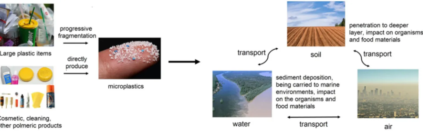
Microplastics are categorized as primary microplastics, which are raw materials used in domestic and personal care products, and secondary microplastics arising from the degradation of raw plastic particles by physical, chemi-cal, and biological processes in the environment (Galgani et al. 2013). Long-term durability due to their polymeric structure and easy transport between different habitats make microplastics of high concern for biologists and environmen-talists (Fig. 1). Major raw polymers include polyethylene terephthalate (PET), polyurethane (PU), polystyrene (PS), polyvinylchloride (PVC), polypropylene (PP), polyesters,
* Mohsen Padervand [email protected]; [email protected] * Chuanyi Wang [email protected] Eric Lichtfouse [email protected] Didier Robert [email protected]
1 School of Environmental Science and Engineering, Shaanxi
University of Science and Technology, Xi’an 710021, China
2 Department of Chemistry, Faculty of Science, University
of Maragheh, Maragheh, Iran
3 Aix-Marseille Univ, CNRS, IRD, INRAE, Coll France,
CEREGE, 13100 Aix-en-Provence, France
4 ICPEES, Université de Lorraine, 12 rue Victor Demange,
polyethylene (PE) and polyamide (PA, nylon). Poor plastic waste management has resulted in ubiquitous microplastics occurrence (Gilani et al. 2019; Thompson 2015). Several reports show that long-term exposure to microplastics causes chronic toxicity, yet there is no evidence on their acute fatal effects (Li et al. 2018a; Sussarellu et al. 2016). Microplas-tic toxicity is controlled by different routes depending upon their chemical structure and additives used as linkage dur-ing polymerization (Meeker et al. 2009; Sussarellu et al. 2016). As an example, polystyrene microplastics are able to be transferred in blood, causing reproductive disruption in marine filter feeders (Law et al. 2014).
To our best knowledge, this is the first review on micro-plastics removal. We discuss microplastic additives, occur-rence, transport and toxicity, then we review removal methods. Removal methods include sorption and filtration, removal based on chemical phenomena, and biological ingestion treatments. Advantages, disadvantages and effi-ciency of different methods are compared at the end.
Microplastic sources, transport, polymers
and additives
Microplastic sources and occurrence
Microplastics can be found worldwide in coastal regions and aquatic ecosystems in various size fractions due to the transport phenomena including wind and ocean cur-rents. Primary sources are household sewage discharge including polymeric particles from cosmetic and cleaning products, feedstocks used to manufacture plastic products, and plastic pellets or powders used for air blasting (Jiang 2018). Progressive fragmentation of larger plastic items under the atmospheric conditions, e.g., by mechanical degradation and UV light exposure, thus contributing to the entrance of considerable amounts of microplastics to
the environment, is the secondary source of microplastics (Eriksen et al. 2014). This increases plastic debris availabil-ity for being ingested by a large variety of organisms and highlights the appearance of further environmental hazards (Thompson et al. 2009).
Wastewater treatment plants are also a major source of microplastics release (Browne et al. 2011; Long et al. 2019). Whereas large plastic particles are efficiently removed dur-ing wastewater treatment, microplastics often bypass the treatment units, thus entering and accumulating in the aquatic environment (Murphy et al. 2016). Noteworthy, a large number of water treatment plants are located near the ocean and seawater, thus inducing a high microplastic release source. For instance, in mainland China, about 1873 wastewater plants (56%), out of 3340, with 78 × 106 m3/day of treatment capacity, are located in coastal regions where their effluents can be directly or indirectly discharged into aquatic ecosystems (Jin et al. 2014). To address this issue, many researchers are investigating the fate, occurrence, detection and removal of microplastics in the water treat-ment plants (Beljanski 2016; Carr et al. 2016; Sun et al. 2019).
Microplastic transport
Sea and ocean are viewed as the major sinks for microplas-tics, whereas freshwaters and terrestrial environments are the main sources. Indeed, early research found that micro-plastic litter reaching oceans by rivers contains particles found in soils (Horton et al. 2017a). This implies that freshwaters and soils are also sinks of microplastics, as evidenced by high concentrations of microplastics in some terrestrial and freshwater areas (Nizzetto et al. 2016). The long-term durability of microplastic fibers found in deeper layers (~ 25 cm) of agricultural soils treated by sewage sludge as fertilizer (Zubris and Richards 2005), suggests a gradual transport in solid media, then further
accumulation at depth; thus making agricultural and forest soils more likely to retain microplastics compared to urban areas (Lwanga et al. 2017).
As rivers carry a huge volume of plastic particles over the large distances, microplastics probably settle out along with sinking sediments, particularly where flow energy drops, for instance in retard-moving riverbeds. Accord-ingly, futher sediment deposition of microplastics in lakes where water flow is the lowest and sedimentation rate is high, should induce high accumulation (Corcoran et al. 2015).
The shape diversity, small size, lightweight and low density of microplastics contribute to their widespread transport and facile dispersal across large distances on land and within aquatic systems by storm sewers, wind and other natural currents (Horton and Dixon 2018). Larger size and higher density result in facile sinking and sedi-ment deposition of the microplastics (Horton et al. 2017b). Furthermore, irregularly shaped microplastics with jagged geometry and sharp ends are more likely retained under-water, rather than returning to the surface, whereas spheri-cal particles show a higher tendency to stay at the surface (Ballent et al. 2012; Lagarde et al. 2016).
Microplastics transport pathways in the air are not fully understood (Horton and Dixon 2018). Noteworthy, in the air, there are few dispersal boundaries, compared to water systems. Nonetheless, microplastics transport within the atmosphere is not totally independent of aquatic and ter-restrial pollutions, and here further investigations are needed to elucidate the mechanisms (Dris et al. 2016).
As another major concern, due to their hydrophobic-ity and high surface area/volume ratio, microplastics are highly susceptible to sorb and carry persistent organic pollutants such as polychlorinated biphenyls (PCB), dichlorodiphenyltrichloroethane (DDT) and polyaromatic polycyclic aromatic hydrocarbons (PAH), which can be subsequently transferred to coastal regions and be des-orbed inside living organisms (Browne et al. 2013). Conse-quently, the concentration of organic pollutants in coastal areas is expected to increase several orders of magnitude as a result of pollutant transport by microplastics. Micro-plastic morphology and transport are thus major charac-teristics controlling the other waterborne pollutants (Cole et al. 2011).
Microplastic raw polymers and additives
Polymeric ingredients of primary microplastics mainly include polyethylene, polypropylene and polystyrene, depending upon the type of the products manufactured by the factory; while secondary microplastics are predominantly made of polyester, acrylic and polyamide, forming fibers in the environment (Jiang 2018). The microplastic number in
the inland freshwaters of Wuhan in China ranged between 1660.0 ± 639.1 and 8925 ± 1591 numbers/m3; here the major types were polyethylene terephthalate and polypropylene (Wang et al. 2017). Low-density polyethylene has been also identified as the dominant type of microplastics.
Microplastics contain a large variety of chemical addi-tives such as bisphenol A, phthalates and polybrominated diphenyl ethers, which are used during raw plastic synthesis to improve plasticity (Besseling et al. 2014, Murphy 2001). These additives are endocrine disruptors, and thus may exhibit toxic effects upon release. The concentration of such plasticizers in plastic debris of remote and urban beaches is up to 35 ng/g in remote beaches and up to 700 ng/g in urban beaches for bisphenol A; between 0.1 and 400 ng/g in remote beaches and up to 9900 ng/g in urban beaches for polybrominated diphenyl ethers; and up to 3940 ng/g for phthalates (Hirai et al. 2011). These plastic additives have been detected in most microplastic polymers (Jiang 2018). Researchers also reported the leaching of bisphenol A and nonylphenol from silicone and polycarbonate microplas-tics (Fasano et al. 2012). Accumulation of such chemicals in human bodies through biological phenomena is also reported (Talsness et al. 2009). The most alarming exposure route to microplastics for human is food, where the adverse effects of the chemical additives and mechanism of entrance to the organs are still unexplored (Wright and Kelly 2017). Accordingly, many efforts must be devoted to finding effi-cient strategies to abate the presence of microplastics in the environment. While there have been published reports on characterizing sources, occurrence, fate, methods for detec-tion, and environmental effects; to date, few research and review papers have discussed removal processes of micro-plastics from contaminated systems.
Toxicity of microplastics
Toxicity from the chemical structure
The potential toxicity of microplastics arises from unre-acted monomers, oligomers and chemical additives leaked from the plastic in the long rub (Thompson et al. 2004). Monomers and oligomers are both able to migrate from food packaging materials (Piringer and Baner 2008). As the concentration of the residuals reaches specific limits, they can be potentially absorbed by human bodies via different pathways. For instance, the presence of polystyrene residuals in food materials is reported to cause serious health issues, while epoxy resins made of bisphenol A are absorbed by living tissues, then interfer with the rate of cell division (Lau and Wong 2000).
Chemical additives are used during polymers manufac-turing for improving the products performance. Additives
include functional additives such as plasticizers, heat stabi-lizers, flame retardants, antioxidants, colorants, e.g. soluble azo-colorants and pigments, fillers such as kaolin and clay, and reinforcements, e.g. carbon and glass fibers. These addi-tives are another source of toxicity. For example, researchers found that the release level of some phthalates from baby bottles was in the range of 50–150 μg/kg of food content after the contact time of 120 min at 70 °C (Simoneau et al. 2012). The release level of bisphenol A from food packag-ing items was estimated to be in the range of 100–800 ng/L, while the values were in the range of μg/L for some phtha-lates under the same conditions (Fasano et al. 2012). Most of these additives are not chemically bound to the bulk plastic structures, implying easier release.
Nobre et al. (2015) studied the toxicity of raw and beach-stranded microplastics on the development of embryos of
Lytechinus variegatus, simulating leaching of the
chemi-cal additives into the water column and interstitial water by assays of elutriate and pellet–water interface, respectively. They found that raw microplastics induced more toxicity, enhancing anomalous embryonic development by 58.1% and 66.5% for the former and latter evaluation method, respectively. Their results also implied that the leaching of chemical compounds strongly depends upon the media compartment in which microplastics accumulate, and upon the exposure pathway. Hahladakis et al. (2018) reviewed migration and release rate, fate, and potential toxicity effects of additives on organisms and environment. The release of volatile compounds, e.g., benzene, toluene, ethylbenzene, styrene and methylene chloride, from plastics can also con-tribute to chronic health effects (Andrady 2017; Huff et al. 2010; Wexler and Gad 1998).
Toxicity from physical properties
Microplastics exert damage through the effect of a relatively large surface area/volume ratio. They absorb hydrophobic pollution from water, then carry this pollution to other habi-tats (Setälä et al. 2014). A study of the effect of phenan-threne-loaded low-density polyethylene glycol microplastics on biomarker responses in juvenile African catfish revealed significant tissue changes in the liver and brain of the organ-ism (Karami et al. 2016).
The ingestion of microplastics by biota is a common way to induce toxicological effects (Hämer et al. 2014). Polysty-rene microplastics enhance the bioavailability of fluoran-thene compounds to marine mussels (Mytilus spp.) after 7 days of exposure under controlled experimental condi-tions (Paul-Pont et al. 2016). These results mean that ther is a higher fluoranthene concentration in mussels exposed to fluoranthene-loaded microplastics than those exposed to pure fluoranthene. Highest levels of antioxidant markers and histopathological damages were also observed for the
former case. They explained the mechanism by interactions between the cell wall components of the marine mussels, e.g. p-glycoprotein, involved in pollutant excretion, and the microplastics surface.
Zhang et al. (2017) investigated the adverse effects of microplastics on the photosynthesis of the marine microal-gae Skeletonema costatum. They found that the maximum growth inhibition ratio reached up to 39.7% after 96 h of exposure to microplastics with average diameter of 1 μm. Their results show a drastic decrease in chlorophyll content (20%) and photosynthetic efficiency (32%) after exposure to high concentration of microplastic (50 mg/L), leading to negative effects on microalgae growth. According to the results of morphological studies and scanning electron microscopy (SEM), they proposed both adsorption and aggregation of microplastics on the outer surface of micro-algae as the most probable mechanism of toxicity.
Size dependency of microplastics toxicity was also con-firmed by Lu et al. (2016) who investigated the exposure effects of polystyrene microplastics to zebra fish. They stated that a 7-day exposure resulted in accumulating the micro-plastics with size of 5 μm in liver, gill and gut, while those with size of 20 μm were just found in fish gill and gut. More-over, lipid accumulation and inflammation of liver, oxidative stress, and adverse alterations in the metabolism profile of the fish liver were the main toxicity outcomes.
The shape and texture of the ingested microplastics also influence their toxicity and absorption capability. According to Au et al. (2015), polypropylene microplastic fibers were more toxic than polyethylene microplastic spherical particles to the freshwater amphipod, Hyalella azteca. They attrib-uted this to the longer residence time of the fibers in gut, which modifies the ability of food processing, thus leading to serious changes in sublethal endpoints.
Toxicity from microorganisms carried by microplastics
The potential of microplastics to carry pathogenic bacteria has been explored by Kirstein et al. (2016). They observed
Vibrio parahaemolyticus bacterial strains on some
polyeth-ylene, polypropylene and polystyrene marine microplas-tic parmicroplas-ticles gathered from North Sea. They highlighted the need for further consideration of health impacts of microbial assemblages in microplastics. A 10-day expo-sure to five types of ~ 70 μm microplastics led to intesti-nal damage including splitting of enterocytes and cracking of villi in zebrafish Danio rerio and nematode C. elegans, as model organisms of freshwater (Lei et al. 2018). They also demonstrated that 2-day exposure of 5.0 mg/m2 of microplastics considerably reduced calcium levels and sur-vival rates, and inhibited body length and reproduction of
C. elegans. They suggested that oxidative stress and
intes-tinal damages are the main toxicity effects of microplastics. Prata (2018) reviewed the potential toxicity of airborne microplastics and adverse effects of their low environmental concentrations on human health. They discussed the diseases aroused from airborne microplastics and pathophysiological mechanisms of toxicity including dust overload, oxidative stress, translocation, and gene mutation. They proposed that exposure to low atmospheric concentrations can contribute to incidence of cardiovascular, respiratory, and interstitial lung diseases.
Overall, the toxicity of microplastics arises from raw chemical additives, adsorption and transport of pollut-ants and microbes, and release to life and the environment. Toxicity depends physically on size and shape of microplas-tics. Biota and humans are affected by toxic effects of micro-plastics via mechanisms including sorption and aggregation in different organs, ingestion, and exertion of physical dam-ages and disturbing the life systems. Research is needed to clarify how microplastics induce tissue changes and patho-logical disorders.
Removal of microplastics using sorption
and filtration methods
Adsorption on green algae
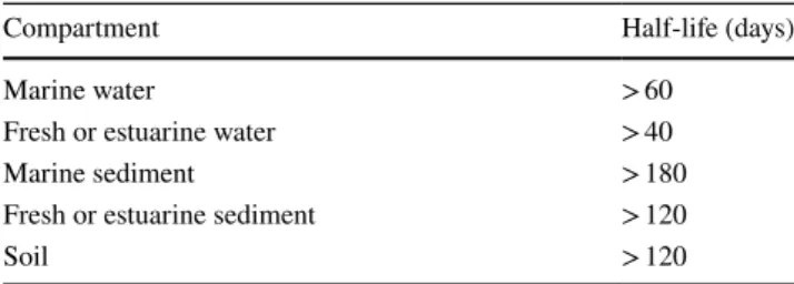
The presence of microplastics in aquatic environments is expected to be more critical than other pollutants due to several harmful effects and death of organisms, e.g. fishes, mammals, marine birds and reptiles, arising from their entanglement and bioaccumulation (Cole et al. 2011; Gra-ham and Thompson 2009; Gregory 2009). Their persistence and low degradability call for removal methods. Microplas-tics are generally categorized as persistent materials but they degrade more of less depending upon their nature and chemical structure. Microplastics with half-life times lower than the values defined in terms of REACH criteria for per-sistency (Table 1) can be considered as degradable, and do not pose a threat to the environment (Verschoor 2015). Microplastics are prone to sorb a large variety of waterborne
contaminants on the surface, carry them and desorb into the new habitats (Rios et al. 2007). Their large surface area/ volume ratio makes adsorption of other contaminants likely. Sundbaek et al. (2018) studied the adherence behavior of fluorescent microplastic particles on the surface of an edible marine microalgae, seaweed, named Fucus
vesicu-losus. The diameter size of the polystyrene microplastics
was ~ 20 μm, while the plant cells of the sorbent contained very narrow microchannels to restrict the translocation of polystyrene microplastics into the tissues. The results revealed a high sorption of microplastics (~ 94.5%), mainly near the cut surfaces of the seaweed, which is explained by the role of released alginate compounds from cell walls in the cut regions. Indeed, because of the gelatinous charac-teristics of this anionic polysaccharide substance, alginate is able to improve the adherence of polystyrene particles on the seaweed’s surface (Martins et al. 2013). This paper and the other researches on microalgae capabilities to sorb tiny plastic particles accentuate the effective role of micro-plastics’ surface charge and microalgae’s surface characters (Bhattacharya et al. 2010; Nolte et al. 2017). Investigating the adsorption of 20–500 nm polystyrene particles onto uni-cellular green algae, Pseudokirchneriella subcapitata, Nolte et al. (2017) concluded that positively charged polystyrene microplastics are more efficiently adsorbed on the algae’s surface than those with negative charge.
Overall, the sorption of microplastics on algae surface strongly depends on particles’ surface charge. Positively charged microplastics have higher tendency to be sorbed more efficiently, which is explained by the presence of an anionic polysaccharide in the algal chemical structure. Removal using membrane technology
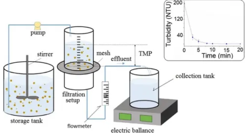
Li et al. (2018b) reported the use of dynamic membranes for the efficient removal of microplastics from a synthetic wastewater (Fig. 2). They investigated the effect of influent flux and particles concentration on the removal efficiency of dynamic membranes formed on a diatomite platform with 90 μm of supporting mesh during filtration of the syn-thetic wastewater. Excellent filtration of microplastices was obtained in 20 min by decreasing the turbidity from 195 NTU for the influent to less than 1 for the effluent (Ersahin et al. 2017; Horton and Dixon 2018). Dynamic membrane formation is facilitated at higher influent fluxes and micro-plastics concentrations.
Ward (2015) designed an efficient microplastic removal tool based on polymer coatings as an elongated mesh screen. He claimed that the tool has good durability and has the advantage of being easily fabricated from com-modious materials. Other tool advantages included the absence of electrical power and mechanical devices.
Table 1 Persistency criteria for contaminants in different media according to the REACH Annex XIII (Verschoor 2015)
Compartment Half-life (days)
Marine water > 60
Fresh or estuarine water > 40
Marine sediment > 180
Fresh or estuarine sediment > 120
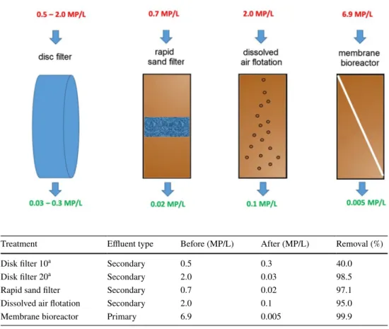
Membrane bioreactors, however, exhibit higher capacitie than simple dynamic membranes for the removal of micro-sized plastics (Lares et al. 2018; Talvitie et al. 2017a). Kno-block et al. (1994) studied the purification capability of a coupled system, taking advantage of porous membranes in combination with biological processes. Successful removal of a large variety of complex industrial wastewaters by mem-brane bioreactors confirms the suitability of this technol-ogy for the treatment for high-strength contaminants such as polymeric debris and microplastics (Gurung et al. 2016). Talvitie et al. (2017a) investigated the removal of various types of microplastics from wastewater treatment plant effluents using advanced final-stage treatment technologies including membrane bioreactor, disk filter, rapid sand fil-tration, and dissolved air floating (Fig. 3). They concluded that the membrane bioreactor eliminated 99.9%, from 6.9 to 0.005 microplactic particle per L (Table 2). They also showed that membrane bioreactor, rapid sand filtration and dissolved air floating removed microplastics of any size, even the smallest size fractions of 20–100 μm. Moreover, the removal efficiency did not depend upon the microplastics shape; particularly, textile fibers which were predominant in both influents and effluents during the treatment, were effi-ciently removed. Analysis of the samples by Fourier trans-form infrared spectroscopy (FTIR) indicated that the mem-brane bioreactor highly decreased the number of polymers in the final effluent, too, which highlights the good sorption capacity of the setup to trap microplastics of various chemi-cal structures.
Membrane technologies were successfully used to remove microplastics from polluted aquatic environments. The removal efficiency over the membranes particularly depends on its durability, influent flux, size, and concentration of the microplastics. The combination of porous membranes with biological processes could enhance the removal efficiency up to 99.9%.
Removal using advanced filtration technologies in wastewater treatment plants
Lares et al. (2018) recently studied the performance of a municipal wastewater treatment plant operating based on a pilot-scale, combined membrane bioreactor–conventional activated sludge methodology for the removal of micro-plastics. Their study took 3 months and sampling cam-paigns were every 2 weeks. The wastewater samples were collected from a municipal water treatment plant located next to the city center of Mikkeli in Finland. Including an aeration tank, for mixing the wastewater with air to activate microorganisms, and a sedimentation tank, where the sludge is separated from the treated wastewater, for the biologi-cal degradation and secondary purification, respectively, a conventional activated sludge system is also expected to improve the activity (Gurung et al. 2016). Results revealed a better removal of microplastics (99.4%) using a membrane bioreactor compared to the conventional activated sludge treatment system (98.3%). Microplastics concentration in the final effluent of the former system was estimated to be 0.4 ± 0.1 MP/L, which was lower in comparison with that evaluated for the later process (1.0 ± 0.4 MP/L). They also stated that the main reason for observing a narrow range of final microplastics concentrations is probably due to opting for different processing steps and wastewater samples than previous works (Leslie et al. 2017; Mintenig et al. 2017; Murphy et al. 2016).
11 wastewater treatment plants in Changzhou, China, were studied for their efficiency to remove microplastics by following the abundance, color, shape and dimensional changes during the removal steps (Ma et al. 2019). All plants that used several treatment steps such as subsequent tanks for floating and sedimentation, and filtration processes, elim-inated more than 90% of microplastics from the influents, with a final removal efficiency reaching 97.15%. The most important reasons that cause variation in removal efficiencies
Fig. 2 Dynamic membrane experimental setup and graph showing the decrease in turbid-ity with time when micro-plastics are removed (Li et al.
2018b). TMP transmembrane pressure, NTU nephelometric turbidity unit
might be daily processing volume, different raw water and type of treatment processes. Large-size microplastics were less abundant in the effluents, in agreement with previous reports (Horton and Dixon 2018). In addition, fiber rayon and polyethylene terephthalate were the main ingredients of these microplastics, and reached the highest removals.
The microplastics removal methodology used by munici-pal sewage treatment plants was studied by researchers in Beijing, China (Yang et al. 2019). The influents were ini-tially treated by a series of processes including aerated grit chamber, primary and secondary sedimentation tanks fol-lowing A2O treatments, e.g., anaerobic, anoxic and aerobic. Finally, advanced treatment processes including denitrifica-tion, ultrafiltradenitrifica-tion, ozonation and ultraviolet are applied to complete the cycle and remove microplastics from waste-water. The results of FTIR analysis revealed the presence of 18 polymers in the effluent, in which polyethylene tereph-thalate and polyester had the highest abundances of 42.26 and 19.1%, respectively. 58.84% of the microplastics popu-lation in influents was removed during the primary step of
aerated grit treatment, while the removal efficiency reached to 71.67% following the advance treatment processes. Although the overall removal efficiency in the present sew-age treatment plant, of 95.16%, was outstandingly less than that of for membrane bioreactors, of 99.9%, it was compa-rable to the efficiency of dissolved air flotation technolo-gies and sand filters (Talvitie et al. 2017b). Noteworthy, the treatment processes currently employed at sewage treatment plants are not necessarily designed for the removal of micro-plastics. Regardless, these processes are able to eliminate a large portion of microplastics from the wastewater. The researchers finally estimated that sewage treatment plants release almost 0.59 × 10⁹ items of microplastic into the aquatic ecosystems.
Overall, sorption and filtration methodologies show a good efficiency for the treatment for microplastics-con-taining wastewater, mainly in combination with other pro-cedures such as biological and sedimentation processes. To achieve higher removal efficiency, membrane bioreactors are simultaneously used with other advanced physical–chemical
Fig. 3 Microplastics removal efficiency, in terms of number of microplastics per liter, MP/L, after treatment by final-stage treatment technologies (Talvitie et al. 2017a)
Table 2 Average microplastic concentrations before and after treatment with different technologies
Data are given in number of microplastics per liter of effluent
a Pore size in μm (Talvitie et al. 2017a)
Treatment Effluent type Before (MP/L) After (MP/L) Removal (%)
Disk filter 10a Secondary 0.5 0.3 40.0
Disk filter 20a Secondary 2.0 0.03 98.5
Rapid sand filter Secondary 0.7 0.02 97.1
Dissolved air flotation Secondary 2.0 0.1 95.0
treatments in most wastewater treatment plants all over the world, but it has still been confirmed that these plants act as microplastics sources to aquatic environment.
Chemical methods to treat microplastics
Many wastewater treatment plants worldwide use coagula-tion and agglomeracoagula-tion processes to form enlarged contami-nant particles that are easier to separate (Hu et al. 2012; Lee et al. 2012; Shirasaki et al. 2016). These processes involve Fe- and Al-based salts and other coagulants to bind tiny particles via uptake-complexation mechanisms initiated by a ligand exchange, thus forming strong bonds between waste particles (Chorghe et al. 2017).
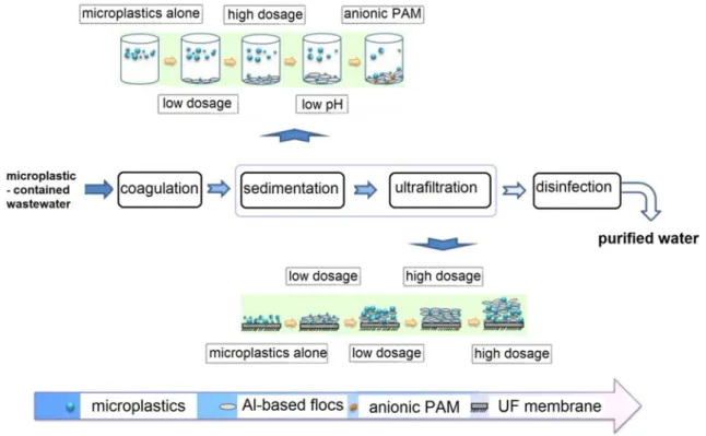
Ariza-Tarazona et al. (2019) recently studied the removal of polyethylene microplastics using iron and aluminum salt coagulants and ultrafiltration (Fig. 4). The experi-ments were carried out under different concentrations of Al3+ and Fe3+ ions, and the results indicated that Al3+
has better performance than Fe3+. Also, the microplas-tic removal efficiency was scarcely modified by the pH of the solution at low concentration of Al coagulant source, 0.5 mM, whereas removal efficiency decreased by increas-ing the pH, particularly for small-sized microplastics, of diameter lower than 0.5 mm. They found that polyacryla-mide (PAM), an enhancing coagulation agent, increased the removal efficiency for small microplastics much better than for large particles under high Al dosage of 5 mM. This highlights the greater growth rate of small microplastics in the presence of cationic polyacrylamide. The growth rate was highly enhanced when anionic polyacrylamide was used for the removal efficiency for the smaller microplas-tics (d < 0.5 mm): here the removal was raised from 25.83% without polyacrylamide to 61.19% with 15 mg/L polyacryla-mide, while it just increased from 4.27% to 18.34% for larger microplastics, of 2–5 mm diameter.
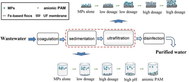
Ariza-Tarazona et al. (2019) also explored the removal of the microplastics with the same method but using FeCl3·6H2O coagulation agent (Fig. 5). They found that the
Fig. 4 Microplastic removal by coagulation, sedimentation and ultrafiltration (UF) (Ma et al. 2019). The effect of anionic polyacrylamide (PAM), pH and the formation of Al-based flocs on the removal efficiency is well represented
removal at neutral pH enhanced as the coagulant concentra-tion increased and, this trend being clearer for small micro-plastics, of d lower than 0.5 mm. Similarly, the removal effi-ciency was accelerated at high pH and high concentration, 2 mM, of the coagulant and for lower size of the micro-plastics. However, in this case and under high dosage of the coagulation agent, 2 mM, anionic polyacrylamide acted much better than the cationic one to improve the removal rate of polyethylene microplastics. This can be mechanisti-cally explained based on facile formation of Fe-based flocs during the coagulation process, during which anionic poly-acrylamide makes the products dense enough to be easily trapped and separated.
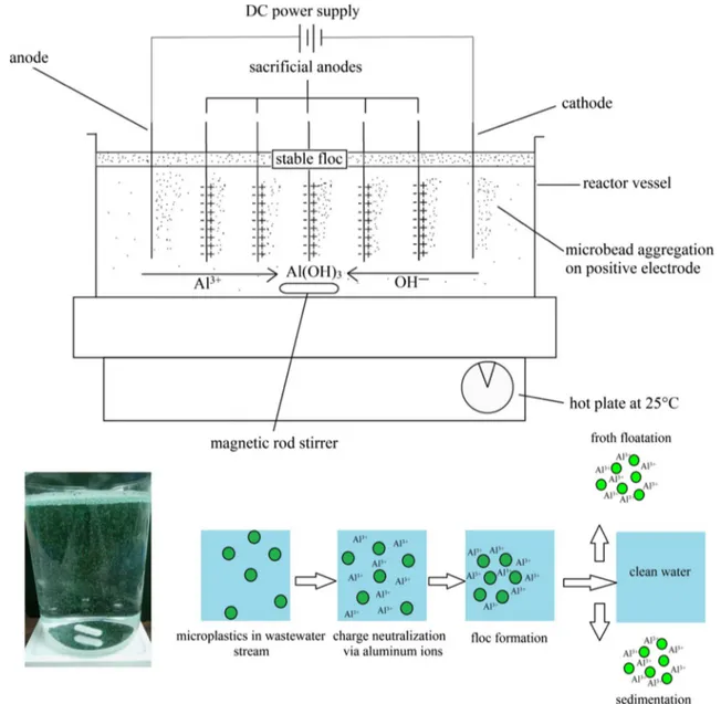
Researchers have also used the robust and environmen-tally compatible electrocoagulation technique (Perren et al. 2018), which allows sludge minimization, energy efficiency, cost-effectiveness, and flexibility to automation, to remove the polyethylene microplastics in a stirred-tank batch reactor (Fig. 6). In situ formation of the metal hydroxide coagulants is initiated by the reaction of metal ions such as Fe2+ and Al3+ released from sacrificial electrodes in a water stream with hydroxyl anions of the media; Eqs. 1–4 display the anodic and cathodic reactions, respectively:
(1) M(s)→ Mn+(aq)+ ne− (2) 2H2O(l)→ 4H + (aq)+O2(g)+4e − (3) Mn+(aq)+ ne− → M(s) (4) 2H2O(l)+2e − → H2(g)+2OH−
The produced coagulants break up the colloids and sta-bilize the suspended microparticles surface charges, which allows the particles to get close to each other sufficiently for making interactions via van der Waals forces (Akbal and Camcı 2011). The coagulants simultaneously form a sludge blanket to trap the suspended microplastics in the wastewater sample. The results show a removal efficiency higher than 90% for all experiments using proposing electrocoagula-tion. The highest removal efficiency, 99.24%, was obtained with pH of 7.5 and NaCl concentration of 0–2 g/L. Further-more, the lowest tested current density of 11 A/m2, which is the best in view of energy consumption, was the most efficient for achieving the highest removal rate.
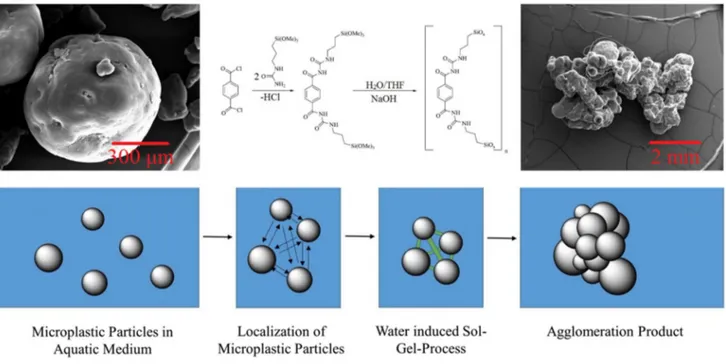
Herbort et al. (2018) suggested agglomeration based on alkoxy-silyl bond formation via sol–gel reactions as a new sustainable removal approach for treatment for the micro-plastics originating from inert products of the textile and cosmetic industries. Functionalized molecular precursors were initially synthesized in an inert atmosphere, then used for bio-inspired alkoxy-silyl formation. Meanwhile, micro-plastics adhered together to form large three-dimensional agglomerates which can, afterward, be removed using cost-efficient filtration methods (Fig. 7). The sol–gel formed in this way is similar to hybrid organic–inorganic silica gels with a large variety of benefits and usages in sensors and optical materials, medicine, and corrosion protection (Nicole et al. 2004).
The mechanisms of degradation of microplastics are not fully known. Brandon et al. (2016) studied the degra-dative changes of the chemical structure of two types of microplastics including polypropylene and polyethylene (5) Mn+(aq)+ nOH−→ MOHn(s)
Fig. 5 Removal of polyethylene microplastics from the wastewater using Fe3+ coagulation, sedimentation and ultrafiltration processes
(Ma et al. 2019). The effect of polyacrylamide (PAM), pH and the
formation of Fe-based flocs on the removal efficiency is well repre-sented. MPs microplastics, UF ultrafiltration
Fig. 6 Bench-scale reactor setup for the removal of microplastics using electrocoagulation method in which Al3+ acts as coagulation agent
(Per-ren et al. 2018)
for 3 years under simulated realistic weather conditions. According to FTIR analysis, they found some slight nonlin-ear changes with time in moieties like carbonyl, hydroxyl and carbon–oxygen bonds, implying microplastics slow degradation.
The exposure of macroplastics to elements (Colom et al. 2003; Gulmine et al. 2003), microorganisms (Pathak 2017), catalysts (Hazrat et al. 2015), and photo-active materials (Li et al. 2010) has been widely reported but there are no large number of publications on microplastics degradation. Liu et al. (2019) studied the long-term aging behavior of poly-styrene and polyethylene microplastics treated by a com-bined heat-activated persulfate and Fenton method in the aquatic environment. According to their results, O/C ratio
and the average size of the microplastics play a role in the adsorption capacity and surface properties, which affect the microplastic oxidation rate significantly. Degradation of polyethylene microplastics in artificial seawater under dark and UV radiation to evaluate the structural and morphologi-cal alterations has been recently reported (Da Costa et al. 2018; Pathak 2017). FTIR analysis of the initial materials and products (Fig. 8) evidenced the stronger degradative role of the artificial seawater compared to UV illumination. This was confirmed by an increase of the organic content of the medium. As a control experiment, the researchers exposed microplastics to UV light alone for the same period of time and did not observe significant changes in the chemi-cal structure, thus showing implying that salt is needed to
form oxidized sites. SEM images also revealed the effect of salt on microplastic surface morphology, in which observ-able crack lines were clearly detected. This strengthens the fact that media salinity facilitates microplastic degradation (Vasile and Pascu 2005).
Photocatalytic degradation of the organic contaminants is a destructive process triggered by absorbed photons on the surface of a semiconducting material for generating hyperac-tive radicals. Depending upon the semiconductor band gap, the energy of the incident photons and, subsequently, the suitable light source must be chosen. Photooxidation degra-dation, that is oxidation in the presence of light source and
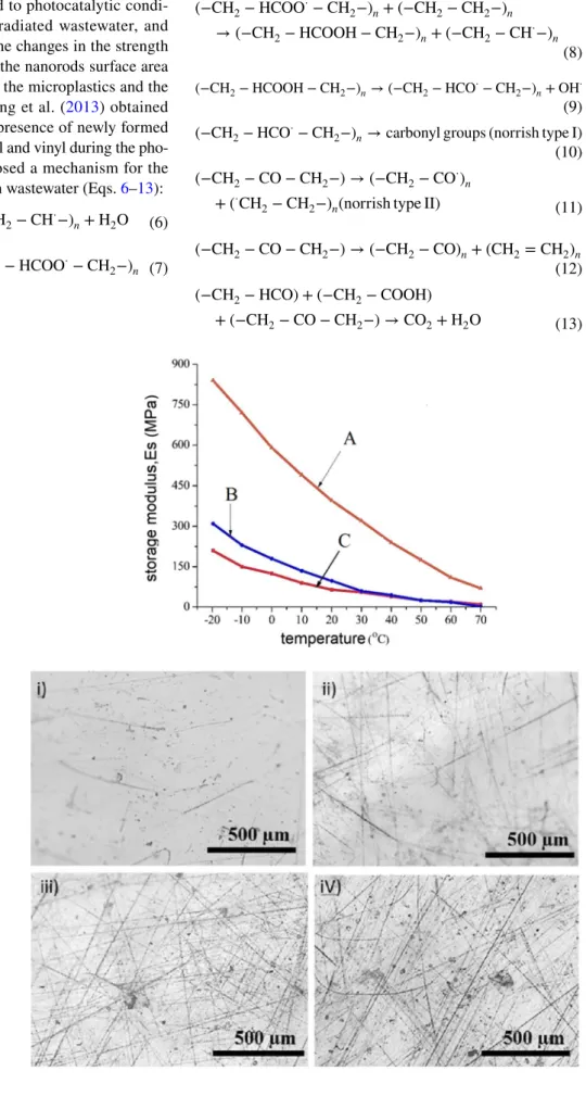
air, is the mostly common process for the breaking of poly-meric chains (Yousif and Haddad 2013). Different mecha-nisms were suggested for the photochemical processes in the aqueous phase, in which hydroxyl radicals strongly promote the degradation reactions (Li et al. 2010). Heterogeneous photocatalytic degradation of low-density polyethylene microplastics has been recently investigated in the aquatic media over the rod-like ZnO nanoparticles (Tofa et al. 2019). From the optical images, morphological changes including the appearance of wrinkles, brittleness, cracks and spots on photo-exposed surfaces of the microplastics were observed (Fig. 9). Also, the results revealed variations in elasticity
Fig. 7 Removal of polyethylene microplastics using alkoxy-silyl-induced agglomeration. The microscopy images of the microplastic before agglomeration (above left) and after that (above right). The
synthesis reaction in the middle exhibits the formation process of the coagulation agent (Nicole et al. 2004)
Fig. 8 Fourier transform infra-red (FTIR) spectra of polyeth-ylene microplastics before and after an 8-week treatment with artificial seawater (Da Costa et al. 2018). The appearance of new bands assigned to the oxi-dized groups in the spectrum of the treated sample confirms the microplastic transformation
properties of the sample exposed to photocatalytic condi-tions in comparison with nonirradiated wastewater, and this directly is in correlation to the changes in the strength of chemical bonds. Accordingly, the nanorods surface area influenced the stiffness degree of the microplastics and the photocatalytic performance. Liang et al. (2013) obtained FTIR data which confirmed the presence of newly formed functional groups such as carbonyl and vinyl during the pho-tocatalytic treatment, they proposed a mechanism for the mineralization of microplastics in wastewater (Eqs. 6–13):
(6) (−CH2−CH2−)n+OH⋅→ (−CH2−CH⋅−)n+H2O
(7) (−CH2−CH⋅−)n+O2→ (−CH2−HCOO⋅−CH2−)n
Fig. 9 Decrease of the elasticity of microplastics after photocata-lytic treatment with: A: ZnO (10 mM, 5 h), B: ZnO (3 mM, 5 h) and C: control. Optical images of the treated microplas-tics, i photo-irradiated, only, for 175 h, ii photo-irradiated in the presence of the ZnO nanorods, 3 mM for 5 h, iii photo-irradiated in the presence of the ZnO nanorods, 10 mM for 5 h, iv photo-irradiated in the presence of the ZnO nanorods, 20 mM for 5 h, representing the development of holes, cracks, and spots (Tofa et al. 2019)
(8) (−CH2− HCOO⋅− CH2−) n+ (−CH2− CH2−)n → (−CH2− HCOOH − CH2−)n+ (−CH2− CH⋅−)n (9) (−CH2−HCOOH − CH2−)n→ (−CH2−HCO⋅−CH2−)n+OH⋅ (10) (−CH2−HCO⋅−CH
2−)n→ carbonyl groups (norrish type I)
(11) (−CH2−CO − CH2−) → (−CH2−CO⋅)
n + (⋅CH
2−CH2−)n(norrish type II)
(12) (−CH2−CO − CH2−) → (−CH2−CO)n+ (CH2=CH2)n
(13)
(−CH2− HCO) + (−CH2− COOH)
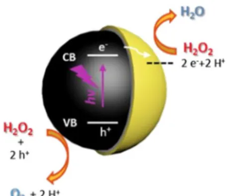
The design and degradative capability of the TiO2-based micro- and nanodevices for the photocatalytic treatment for microplastics have been studied (Sekino et al. 2012). Holding high potential for water purification, remediation of environmental pollutions and medicinal usages, photo-active micromotors have attracted huge attention (Eskandarloo et al. 2017; Zhang et al. 2018). Wang et al. (2019) reported the use of a Au-decorated TiO2-based micromotor for the efficient photocatalytic removal of polystyrene microplas-tics from wastewater under UV illumination (Fig. 10). The micromotor propulsion is supplied by photochemical reactions in water and hydrogen peroxide initiated by elec-tron–hole generation processes. Although the lack of selec-tivity was the most important disadvantage of this system. The need for low concentrations of H2O2 for promoting the phoretic interactions, which enable the micromotor to move, makes this system actually non applicable in real wastewater.
Recently, Ariza-Tarazona et al. (2019) reported the ability of a protein-based N-TiO2 photocatalyst to degrade the poly-ethylene microplastics in solid and aqueous phases. Accord-ing to their results, photocatalytic treatment for the pollution resulted in 1.1% of the mass loss in the solid phase with the rate constant of 12.2 × 10⁻⁴/h after 20 h of treatment time. These values were reported to be 6.4% and 38.2 × 10⁻⁴/h in the aqueous phase. They demonstrated that surface area of the semiconductor and interactions between the photo-catalyst surface and microplastics significantly influenced
the removal efficiency. The SEM images and FTIR spectra have also provided good evidences to confirm the structural changes during the mineralization process.
To conclude, coagulation and agglomeration methods have been investigated to remove microplastics in the labo-ratory. The efficiency depends on the type of coagulant, pH, the chemical composition of the media and the concentra-tions. Although salinity of the media and the presence of illumination source are reported to affect modify the degra-dation of microplastic structures in the aquatic environment, degradative processes have been rarely studied. Of special interest are photocatalytic nanomaterials which make use of both irradiation and catalytic degradation.
Biological removal and ingestion
Microplastic removal by marine organisms
Harrison et al. (2011) published “Interactions Between Microorganisms and Marine Microplastics: A Call for Research” to highlight the outstanding potential of micro-organisms, including picoeukaryotes, bacteria and archaea, to facilitate the biological degradation of microplastics in coastal sediments. They discuss the interactions between synthetic microplastics and marine microorganisms. Many reports present the biological degradation of natural and
Fig. 10 Photocatalytic removal of polystyrene microplastics in water by a Au–Ni–TiO2
micro-motor (Wang et al. 2019). The microscopy images represent the efficient interactions leading to photodegradation reaction of the microplastics on the surface of the micromotor
synthetic macroplastics (Ahmed et al. 2018; Bettas Ardis-son et al. 2014).
Polyethylene microplastics fragmentation and size alter-ation ingested by Antarctic Krill (Euphausiasuperba), a planktonic crustacean, were studied by a group of environ-mentalists in Australia (Dawson et al. 2018). The mecha-nism of fragmentation and type of interactions between microplastics and zooplankton, in which biota-facilitated degradation occurs, still remains unclear but the experi-ments confirmed that smaller microplastics are much eas-ily fragmented under environmental conditions (Ter Halle et al. 2016). Electron microscopic evidences confirmed the shrinking of polyethylene microplastics through fragmen-tation, where the physical size decreased from ~ 31 μm for
the microplastics to less than 1 μm for the fragmentation products (Fig. 11) (Dawson et al. 2018). The findings thus provide evidence for the biologically-mediated transforma-tion of microplastics to nanoplastics.
The preliminary results of a four-month study on removal of high-density polyethylene secondary microplastics in sea-water using two types of indigenous marine communities including Agios consortium and Souda consortium were published by Cocca et al. (2017). According to the recorded weight reduction results, the Souda consortium was more efficient. Interestingly, their results on monitoring the cell content and populations suggested that microplastics acted as a rich carbon source to feed the organisms. While the protein content of both communities decreased during the
Fig. 11 Fate of microplastics ingested by Antarctic krill. Microplas-tics on a filter paper isolated from digested krill with auto-fluorescent mandible (a), digestive gland tissue (b), midgut and digestive gland tissue (c), mandible with the fragments embedded into the surface (d), and fecal pellet with microplastics under bright-field and
fluores-cence microscopy (f). This figure typically represents the pathway of microplastics transformation to nanoplastics inside the organism. WB whole bead, FB fragmented bead, M mandible, DG digestive gland,
experiment, the increase in cell’s carbohydrate content implies enhancement in microbial tendency to adhere to the microplastics surfaces, which is the first step in microbial polymer degradation (Sivan 2011).
An important concern about the microplastics is their small size and negligible weight which enables them to eas-ily spread over the long distances by wind-driven free ocean surface layer movements (Ivleva et al. 2017). Studies have also indicated that microplastics in sediments are normally in the range of 0–3146 particles/kg of dry weight sediment, which highlights the urgent need of designing comprehen-sive experimental routes for the removal from aquatic eco-systems (Maes et al. 2017). The growth of microbial assem-blages on the surface of microplastics should form a new platform prone to settle other organisms like microscopic fungi and microalgae. Although these microorganism com-munities can probably catalyze the metabolic reactions of microplastics resulting in their further biodegradation, the transport of nonnative organisms, which does not naturally occur in such environments, can induce negative effects on marine ecosystems variety (Urbanek et al. 2018).
Paço et al. (2017) explored the capability of fungus
Zale-rion maritimum, a naturally occurring fungus in marine
eco-systems, for the polyethylene microplastics biodegradation based on mass and size variations of the microplastics in a batch reactor (Fig. 12). They measured the concentration of the contaminant at various time intervals to recognize the reaction rate order and found that biodegradation kinet-ics of the microplastkinet-ics obeys to fractional order which can be described in terms of the complex mechanism expected for such reactions (Boudart and Djéga-Mariadassou 2014).
The evidences from biological content measurements, such as protein reduction and carbohydrate increase with expo-sure time, revealed that the Z. maritimum community pos-sibly uses the microplastics as a nutrient source, an iden-tical finding described earlier (Sivan 2011). Noteworthily, the results of the electron and optical microscopy clearly revealed that the biological compounds were present on the surface of the microplastic pellets, typically, highlighting the aforementioned potential of such organisms for trig-gering the effective biological degradation of microplastics from their surfaces. From FTIR analysis, the increases in the new bands’ intensity appearing at regions 3700–3000, 1700–1500 cm−1, and 1200–950 cm−1, are, respectively, attributed to the hydroperoxide and hydroxyl groups, car-bonyl groups, and double bonds, implying the oxidative degradation of polyethylene microplastics. Such variations were already observed in another work in which low-density polyethylene microplastics were photocatalytically removed from wastewater under visible light (Tofa et al. 2019). Microplastic removal by bacteria
Auta et al. (2017) investigated the removal of different microplastics composed of polyethylene, polystyrene, polyethylene terephthalate and polypropylene by Bacillus
cereus and Bacillus gottheilii, two types of Bacillus bacterial
strains isolated from mangrove sediments (Fig. 13). In addi-tion to scanning the morphological and structural changes using electron microscopy and FTIR analyses, the rate of biodegradation was assessed via measuring the micro-plastics weight loss. The fastest mass reduction (0.0019/ day) and shortest degradation half-life (363.16 days) was found using B. cereus isolate and polystyrene microplas-tics, whereas B. gottheilii on polyethylene gave 0.0016/ day and 431.25 days. Microplastics biodegradation can also be monitored performed by scanning electron micros-copy (SEM), where several cracks, holes and erosions were observed (Sowmya et al. 2014). By comparing the degrada-tion results, bond cleavages and chemical alteradegrada-tions scanned by FTIR analysis, B. gottheilii appeared as a better potential microplastic degrader.
Microplastic ingestion
The number of papers discussing microplastics inges-tion by organisms as a removal strategy is limited (Arossa et al. 2019; Cole et al. 2013; Hall et al. 2015). Based on the results of coherent anti-Stokes Raman scattering and fluorescence microscopy, Cole et al. (2013) suggested that zooplankton’s function and health can be negatively affected by micro-sized polystyrene plastic debris. They also stated
Fig. 12 Structural and morphological changes of the treated micro-plastics through biodegradation over fungus Z. maritimum in a batch reactor model (Paço et al. 2017). a NMR spectra, b FTIR patterns, c optical images and d electron microscopy images of the treated microplastics after 28 days. NMR: nuclear magnetic resonance. FTIR: Fourier transform infrared
that microplastics adherence to the external carapace of zoo-plankton considerably influenced the organism feeding, a crucial hindrance factor against normal growth conditions in marine wildlife (Van Franeker et al. 2011). Although this work was not aimed to introduce a way for microplastics removal, the results reveal the high capacity of zooplankton
to remove 1.7–30.6 μm polystyrene microplastics through uptake and ingestion inside the organism body, in which their intestinal tracts can hold the microplastics up to 7 days after entrance.
Hall et al. (2015) also reported capture and ingestion of polypropylene microplastics by scleractinian corals with a
Fig. 14 Polyethylene microplastics removal from wastewater over Red Sea clams (Arossa et al. 2019). Experiments involved four con-centrations of microplastics over two different sizes of T. Maxima
clams. Removal was done by retention, trapping inside the clam, and adhesion on the surface
Fig. 13 Removal of different types of microplastics over two types of Bacillus bacte-rial strains, B. cereus and B.
gottheilii (Auta et al. 2017). a Initial growth of bacterial population implies the use of microplastics as nutrient. The further decrease exhibits the progress in biodegradation. b Appearance of new bands in the Fourier transform infrared (FTIR) spectra of the treated samples confirms microplastic biodegradation. c Scanning electron microscopy (SEM) images show numerous erosions and cracks on the surface of the microplastics after the biologi-cal treatment
consumption rate of 50 μg plastic cm−2/h, and evidenced the presence of microplastics in mesenterial tissue within gut cavity of the coral for at least 24 h. However, the mecha-nism of the affection which potentially interferes with coral growth, physiology, and survival still remains unclear. This research was important since inshore coral reefs ecosystem, the coastal areas, is easily available as touristic regions for long periods and many of water treatment plants also daily release their effluents containing microplastics debris, as mentioned above, to such environments.
The evaluation of the capability of the Red Sea giant clam, Tridacna maxima, for the removal of 53–500 μm polyethylene microplastics from wastewater (Fig. 14) has been reported by Arossa et al. (2019). Their results revealed the key role of clam’s shells to remove the microplastics by sorption on the surface, as they resulted in 66.03% of removal from the wastewater. The authors suggested that this marine biota can be used for rapid removal of the anthro-pogenic microplastics debris from aquatic surfaces in the regions with low concentration of microplastics, a fact partly demonstrated by previous researchers (Martí et al. 2017). The effect of microplastics initial concentration and clam’s size on the removal rates through active, microplas-tics retention inside the body, and/or passive, trapping by the shells, pathways was investigated. The results indicated that big clams tend to retain bigger microplastics inside their bodies, while small clams did not show any sharp preference for them. Moreover, at any concentration, the microplastics easily penetrated into the digestive system of all clams used in this experiment. According to their measurements, the passive route of removal accounts averagely for 35.95% of the microplastics, leading to a significant increase in the den-sity of microplastics attached to clam’s shells and normally, the larger clams adsorb higher concentrations of the micro-plastics. As a whole, this research opened a new window for environmental pollution management making use of the marine biota possessing complex surface and structure. But, finding the mechanism of interactions between the micro-plastics and organisms needs to be explored. Accordingly, we will be able to opt the type of sorbent biota and opera-tional conditions for achieving the highest microplastics removal efficiency.
In brief, microplastics can provide a platform to grow a variety of microbial assemblages. Biological degrada-tion using microorganisms, e.g., zooplankton, marine fungi
and bacterial strains, has been confirmed to be suitable to remove microplastics at low concentrations. The mechanism of interactions and fragmentation is not well understood. Microplastics, in the most of case studies, acted as a nutri-ent source for the organisms. This could not convince us to consider “ingestion” as a removal strategy to treat the microplastic pollutions.
A comparison of treatment methods for removal of the microplastics from the environment is represented in Table 3. The table briefly summarizes the advantages, dis-advantages, and the efficiency of the projects utilized for the last few years. It is obvious that membrane-included technologies are still the most reliable techniques to remove microplastics in the practical applications. However, it seems that finding the suitable alternatives to couple with these methodologies is the main goal of the ongoing projects.
Conclusion
After short surveying on sources, occurrence, and transport pathways of microplastics, recent studies on three major approaches including chemical, biological, and physical methods to remove them from the environments have been summarized. Sorption and filtration processes coupled with membrane bioreactors lead to removing a high percentage of microplastics in the influents entering into the water treatment plants but these systems acted themselves as daily microplas-tic sources since the effluents are directly released to aquamicroplas-tic environments. Conventional activated sludge strategy is also used to treat wastewaters in water treatment plants but shows lower efficiency than membrane bioreactor technologies which makes it a less popular methodology. Electrocoagula-tion and agglomeraElectrocoagula-tion have also become reliable techniques for easy separation of microplastics but are needed to be cou-pled with eminent extra filtration stages. Photocatalytic degra-dation using TiO2 and ZnO semiconductors is experimentally revealed to be a suitable method among the aforementioned strategies. FTIR and electron microscopy analyses are widely served to elucidate any structural alterations during the degra-dation process. Biological degradegra-dation has been investigated over the bacterial strains and marine organisms. Appearance of hydroperoxide and hydroxyl groups, carbonyl groups, and double-bond characteristic bands in the FTIR patterns of the treated microplastics suggests the oxidative mechanism for
Table 3 Com par ison of differ ent me thods f or t he r emo val of micr oplas tics fr om t he en vir onment Me thod Efficiency% Adv ant ag es Dr awbac ks Ref er ences Adsor ption on g reen micr oalg ae 94.5 High affinity of t he cut sur faces t o sorb tin y micr oplas tic par ticles, selectivity based on micr oplas tics sur face c har ge Nonr ecy clable me thod, c hemical adher -ence of t he micr oplas tics t o t he sur face and poisoning it Lag ar de e t al. ( 2016 ) and Zhang e t al. ( 2017 ) Dynamic membr anes > 90 Lo w filtr ation r esis tance, lo w tr ans-membr ane pr essur e, easy oper ation, nonc hemical tr eatment muc h cleaning t o a void e xcessiv e mem-br ane f ouling, ener gy demand, sludg e accumulation due t o its flat s tructur e Li e t al. ( 2018b ) Membr ane bior eact ors > 99
Using combined adv
anced tr eatment me thods wit h por ous membr anes, Shape dependency of t he r emo val per -cent ag e, membr ane f ouling Lar es e t al. ( 2018 ) Con ventional activ ated sludg e 98 Robus t, cos t-effectiv e, fle xible, tr eating a wide r ang e of influent concentr ations, applicable f or lar ge-scale tr eatments long r etention times in t he t ank , t he lar ge sediment ation sur face, t he high cos t of ener gy and t he pr
ocessing and disposal
of sludg e Gur ung e t al. ( 2016 ) W as tew ater tr eatment plants > 95
Efficient mix of sor
ption-biological tr eat-ment pr ocesses, lo w maintenance cos ts, sim ple oper ation A ct as secondar y micr oplas tic sour ces, disability t o tr
eat small-sized micr
o-plas tics, sludg e agg reg ation, lar ge mec hanical de vices Leslie e t al. ( 2017 ), Long e t al. ( 2019 ) and Mintenig e t al. ( 2017 )
Classic coagulation and agg
lomer ation me thods 61 Suit able t o r emo ve small micr opar ticles, contr ollable oper ational conditions, sim ple mec hanical de vices Addition of c hemicals t o media, nonus-able f or lar ge micr oplas tics, Herbor t e t al. ( 2018 ) and Ma e t al. ( 2019 ) Electr ocoagulation > 90 No c hance of secondar y pollution, suit able f or t he r emo val of smalles t par ticles, sludg e minimization, ener gy efficiency , cos t-effectiv eness, fle xibility to aut omation Repeated need of r eplacing t he sacr ificial anode, cat hode passiv ation, nonusable in ar eas wit hout electr icity Per ren e t al. ( 2018 ) Pho tocat alytic deg radation No need t o add o ther c hemicals, e xtend-able applications t o solar light as a rene
wable and pollution-fr
ee ener
gy
sour
ce, efficient miner
alization, en vi-ronment all y fr iendl y r emediation Lac k of selectivity , lo
wer efficiency
com-par ed t o t he o ther me thods, difficult t o scale up, g ener ation of secondar y-type or ganic pollut ants, hug e pho tor eact ors se tup, ener gy consuming, pho tocat aly st reco ver y Sekino e t al. ( 2012 ), T of a e t al. ( 2019 ), W ang e t al. ( 2019 ) and Ar iza-Tar azona et al. ( 2019 ) Biological deg radation
Depends on type of micr
obial community , mos tly > 20 Sim
plicity and saf
ety f or lar ge-scale use, lo w oper ating cos ts, pr acticall y applica-ble in differ ent en vir onments, fle xibility to handle a wide r ang e of w as tew ater char acter
istics and flo
ws Agg reg ation of t he micr obial assem-blag es on t he sur face, en vir onment al conditions canno t be easil y contr olled, difficulties t o anal ysis of t he pr oducts in lar ge scale, lac k of r epr oducibility , find -ing t he suit able micr obial community Ar ossa e t al. ( 2019 ), A ut a e t al. ( 2017 ), Cole e t al. ( 2013 ), Da wson e t al. ( 2018 ), Hall e t al. ( 2015 ) and P aço e t al. ( 2017 )
biological removal. Ingestion by organisms has still not con-sidered as a removal strategy but Red Sea giant clam exhibits good potential for microplastics sorption on the shells besides its degradation in digestive system.
Acknowledgements This work is financially supported by SAFEA of China (High-End Foreign Expert Project # G20190241013) and the scientific research startup fund of Shaanxi University of Science and Technology.
References
Ahmed T, Shahid M, Azeem F, Rasul I, Shah AA, Noman M, Hameed A, Manzoor N, Manzoor I, Muhammad S (2018) Biodegradation of plastics: current scenario and future prospects for environmen-tal safety. Environ Sci Pollut Res 25(8):7287–7298. https ://doi. org/10.1007/s1135 6-018-1234-9
Akbal F, Camcı S (2011) Copper, chromium and nickel removal from metal plating wastewater by electrocoagulation. Desalination 269(1–3):214–222. https ://doi.org/10.1016/j.desal .2010.11.001
Andrady AL (2011) Microplastics in the marine environment. Mar Pollut Bull 62(8):1596–1605. https ://doi.org/10.1016/j.marpo lbul.2011.05.030
Andrady AL (2017) The plastic in microplastics: a review. Mar Pollut Bull 119(1):12–22. https ://doi.org/10.1016/j.marpo lbul.2017.01.082
Ariza-Tarazona MC, Villarreal-Chiu JF, Barbieri V, Siligardi C, Cedillo-González EI (2019) New strategy for microplastic deg-radation: green photocatalysis using a protein-based porous N-TiO2 semiconductor. Ceram Int 45(7):9618–9624. https ://doi.
org/10.1016/j.ceram int.2018.10.208
Arossa S, Martin C, Rossbach S, Duarte CM (2019) Microplastic removal by red sea giant clam (Tridacna maxima). Environ Pol-lut 252:1257–1266. https ://doi.org/10.1016/j.envpo l.2019.05.149
Au SY, Bruce TF, Bridges WC, Klaine SJ (2015) Responses of
Hya-lella azteca to acute and chronic microplastic exposures.
Envi-ron Toxicol Chem 34(11):2564–2572. https ://doi.org/10.1002/ etc.3093
Auta H, Emenike C, Fauziah S (2017) Screening of Bacillus strains isolated from mangrove ecosystems in Peninsular Malaysia for microplastic degradation. Environ Pollut 231:1552–1559. https ://doi.org/10.1016/j.envpo l.2017.09.043
Ballent A, Purser A, de Jesus Mendes P, Pando S, Thomsen L (2012) Physical transport properties of marine microplastic pollution. Biogeosci Discuss. https ://doi.org/10.5194/bgd-9-18755 -2012
Beljanski A (2016) Efficiency and effectiveness of a low-cost, self-cleaning microplastic filtering system for wastewater treatment plants. 2016 NCUR
Besseling E, Wang B, Lürling M, Koelmans AA (2014) Nano-plastic affects growth of S. obliquus and reproduction of D.
magna. Environ Sci Technol 48(20):12336–12343. https ://doi. org/10.1021/es503 001d
Bettas Ardisson G, Tosin M, Barbale M, Degli-Innocenti F (2014) Bio-degradation of plastics in soil and effects on nitrification activity. A laboratory approach. Front Microbiol 5:710–723. https ://doi. org/10.3389/fmicb .2014.00710
Bhattacharya P, Lin S, Turner JP, Ke PC (2010) Physical adsorption of charged plastic nanoparticles affects algal photosynthesis. J Phys Chem C 114(39):16556–16561. https ://doi.org/10.1021/ jp105 4759
Boudart M, Djéga-Mariadassou G (2014) Kinetics of heterogeneous catalytic reactions. Princeton University Press, Princeton. https ://doi.org/10.1007/978-3-662-05981 -4_13
Brandon J, Goldstein M, Ohman MD (2016) Long-term aging and deg-radation of microplastic particles: comparing in situ oceanic and experimental weathering patterns. Mar Pollut Bull 110(1):299– 308. https ://doi.org/10.1016/j.marpo lbul.2016.06.048
Browne MA, Crump P, Niven SJ, Teuten E, Tonkin A, Galloway T, Thompson R (2011) Accumulation of microplastic on shorelines worldwide: sources and sinks. Environ Sci Technol 45(21):9175– 9179. https ://doi.org/10.1021/es201 811s
Browne MA, Niven SJ, Galloway TS, Rowland SJ, Thompson RC (2013) Microplastic moves pollutants and additives to worms, reducing functions linked to health and biodiversity. Curr Biol 23(23):2388–2392. https ://doi.org/10.1016/j.cub.2013.10.012
Carr SA, Liu J, Tesoro AG (2016) Transport and fate of microplastic particles in wastewater treatment plants. Water Res 91:174–182.
https ://doi.org/10.1016/j.watre s.2016.01.002
Chorghe D, Sari MA, Chellam S (2017) Boron removal from hydrau-lic fracturing wastewater by aluminum and iron coagulation: mechanisms and limitations. Water Res 126:481–487. https :// doi.org/10.1016/j.watre s.2017.09.057
Cocca M, Di Pace E, Errico ME, Gentile G, Montarsolo A, Mossotti R (2017) Proceedings of the international conference on microplas-tic pollution in the mediterranean sea. Springer, Berlin Cole M, Lindeque P, Halsband C, Galloway TS (2011)
Microplas-tics as contaminants in the marine environment: a review. Mar Pollut Bull 62(12):2588–2597. https ://doi.org/10.1016/j.marpo lbul.2011.09.025
Cole M, Lindeque P, Fileman E, Halsband C, Goodhead R, Moger J, Galloway TS (2013) Microplastic ingestion by zooplankton. Environ Sci Technol 47(12):6646–6655. https ://doi.org/10.1021/ es400 663f
Colom X, Canavate J, Sunol J, Pages P, Saurina J, Carrasco F (2003) Natural and artificial aging of polypropylene–polyethylene copolymers. J Appl Polym Sci 87(10):1685–1692. https ://doi. org/10.1002/app.11613
Corcoran PL, Norris T, Ceccanese T, Walzak MJ, Helm PA, Marvin CH (2015) Hidden plastics of Lake Ontario, Canada and their potential preservation in the sediment record. Environ Pollut 204:17–25. https ://doi.org/10.1016/j.envpo l.2015.04.009
Da Costa JP, Nunes AR, Santos PS, Girão AV, Duarte AC, Rocha-Santos T (2018) Degradation of polyethylene microplastics in seawater: insights into the environmental degradation of poly-mers. J Environ Sci Health Part A 53(9):866–875. https ://doi. org/10.1080/10934 529.2018.14553 81
Dawson AL, Kawaguchi S, King CK, Townsend KA, King R, Huston WM, Nash SMB (2018) Turning microplastics into nanoplastics through digestive fragmentation by Antarctic krill. Nat Commun 9(1):1001. https ://doi.org/10.1038/s4146 7-018-03465 -9
Dris R, Gasperi J, Saad M, Mirande C, Tassin B (2016) Synthetic fibers in atmospheric fallout: a source of microplastics in the environment? Mar Pollut Bull 104(1–2):290–293. https ://doi. org/10.1016/j.marpo lbul.2016.01.006
Eriksen M, Lebreton LC, Carson HS, Thiel M, Moore CJ, Borerro JC, Galgani F, Ryan PG, Reisser J (2014) Plastic pollution in the world’s oceans: more than 5 trillion plastic pieces weighing over 250,000 tons afloat at sea. PLoS ONE 9(12):111913–111927.
https ://doi.org/10.1371/journ al.pone.01119 13
Ersahin ME, Tao Y, Ozgun H, Gimenez JB, Spanjers H, van Lier JB (2017) Impact of anaerobic dynamic membrane bioreactor con-figuration on treatment and filterability performance. J Membr Sci 526:387–394. https ://doi.org/10.1016/j.memsc i.2016.12.057
Eskandarloo H, Kierulf A, Abbaspourrad A (2017) Light-harvest-ing synthetic nano-and micromotors: a review. Nanoscale 9(34):12218–12230. https ://doi.org/10.1039/C7NR0 5166B