HAL Id: hal-03093259
https://hal.archives-ouvertes.fr/hal-03093259v2
Submitted on 4 Jan 2021HAL is a multi-disciplinary open access archive for the deposit and dissemination of sci-entific research documents, whether they are pub-lished or not. The documents may come from teaching and research institutions in France or abroad, or from public or private research centers.
L’archive ouverte pluridisciplinaire HAL, est destinée au dépôt et à la diffusion de documents scientifiques de niveau recherche, publiés ou non, émanant des établissements d’enseignement et de recherche français ou étrangers, des laboratoires publics ou privés.
12-HETE is a regulator of PGE2 production via COX-2
expression induced by a snake venom group IIA
phospholipase A2 in isolated peritoneal macrophages
Vanessa Moreira, José María Gutiérrez, Bruno Lomonte, Marco Aurélio
Ramirez Vinolo, Rui Curi, Gerard Lambeau, Catarina Teixeira
To cite this version:
Vanessa Moreira, José María Gutiérrez, Bruno Lomonte, Marco Aurélio Ramirez Vinolo, Rui Curi, et al.. 12-HETE is a regulator of PGE2 production via COX-2 expression induced by a snake venom group IIA phospholipase A2 in isolated peritoneal macrophages. Chemico-Biological Interactions, Elsevier, 2020, 317, pp.108903. �10.1016/j.cbi.2019.108903�. �hal-03093259v2�
12-HETE is a regulator of PGE2 production via COX-2 expression induced by a snake venom group
IIA phospholipase A2 in isolated peritoneal macrophages
Vanessa Moreiraa,*, José María Gutiérrez b, Bruno Lomonteb, Marco Aurélio Ramirez Vinoloc, Rui Curid, Gérard Lambeaue and Catarina Teixeiraf
a
Departamento de Farmacologia, Universidade Federal de Sao Paulo, Sao Paulo/SP, Brazil
b
Instituto Clodomiro Picado,Facultad de Microbiología, Universidad de Costa Rica, San José, Costa Rica;
c
Departamento de Genética, Evolução e Bioagentes, Instituto de Biologia, Universidade de Campinas, Campinas/SP, Brazil;
d
Departamento de Fisiologia, Instituto de Ciências Biomédicas, Universidade de Sao Paulo, Sao Paulo, Brazil
e Université Côte d’Azur, CNRS, IPMC, Valbonne Sophia Antipolis, France; f
Laboratório de Farmacologia, Instituto Butantan, Sao Paulo/SP, Brazil;
*Correspondence: Dr V Moreira, Ph.D. Mailing address: Departamento de Farmacologia, Universidade Federal de Sao Paulo (UNIFESP), Rua 3 de maio 100, Ed. INFAR, 3rd floor, CEP 04044-020, Sao Paulo, Brazil. Tel: +55-11-55764848. Fax: +55-11-55764447.
Abstract
The snake venom miotoxin (MT)-III is a group IIA secreted phospholipase A2 (sPLA2) with
pro-inflammatory activities. Previous studies have demonstrated that MT-III has the ability to stimulate
macrophages to release inflammatory lipid mediators derived from arachidonic acid metabolism.
Among them, we highlight prostaglandin (PG)E2 produced by the cyclooxygenase (COX)-2
pathway, through activation of nuclear factor (NF)-B. However, the mechanisms coordinating this process are not fully understood. This study investigates the regulatory mechanisms exerted by
other groups of bioactive eicosanoids derived from lipoxygenase (LO), in particular
12-hydroxyeicosatetraenoic (12-HETE), on group IIA sPLA2-induced (i) PGE2 release, (ii) COX-2
expression, and (iii) activation of signaling pathways p38 mitogen-activated protein kinases
(p38MAPK), protein C kinase (PKC), extracellular signal-regulated kinase 1/2 (ERK1/2), and
NF-B. Stimulation of macrophages with group IIA sPLA2 resulted in release of 12-HETE without
modification of 12-LO protein levels. Pre-treatment of these cells with baicalein, a 12-LO inhibitor,
decreased the sPLA2-induced PGE2 production, significantly reduced COX-2 expression, and
inhibited sPLA2-induced ERK; however, it did not affect p38MAPK or PKC phosphorylation. In
turn, sPLA2-induced PGE2 release and COX-2 expression, but not NF-B activation, was attenuated
by pre-treating macrophages with PD98059 an inhibitor of ERK1/2. These results suggest that, in
macrophages, group IIA sPLA2-induced PGE2 release and COX-2 protein expression are distinctly
mediated through 12-HETE followed by ERK1/2 pathway activation, independently of NF-B activation. These findings highlight an as yet undescribed mechanism by which 12-HETE regulates
one of the distinct signaling pathways for snake venom group IIA sPLA2-induced PGE2 release and
Keywords: snake venom group IIA phospholipase A2, prostaglandin E2, cyclooxygenase-2,
12-hydroxyeicosatetraenoic acid, 12-lipoxygenase, macrophages
1. Introduction
Secreted phospholipases A2 (sPLA2) are a group of lipolytic enzymes with a range of biological
functions, including metabolism and production of lipid compounds, cell membrane degradation or
repair and production of second messengers [1,2], both in physiological or physiopathological
conditions. These enzymes are abundantly expressed in nature and have been classified into 11
distinct groups (IB, IIA, IIC, IID, IIE, IIF, III, V, X, XIIA and XIIB), and further subdivided into
secreted sPLA2, cytosolic PLA2 and Ca2+ independent PLA2, on the basis of their localization,
source, amino acid sequences, and biochemical characteristics [2,3]. Among sPLA2s, we highlight
the group IIA sPLA2s because it has been considered clinically relevant due to its involvement in
several inflammation related diseases, such as cardiovascular diseases, immune disorders,
Alzheimer’s disease, and cancer [4-8]. On the other hand, some sPLA2 from the same group (IIA)
are present in snake venoms and are one of the major components implicated in acute inflammatory
effects caused by viperid snake venoms [9-11].
The PLA2s hydrolyze the membrane phospholipids at the sn-2 position, generating free fatty
acids such as arachidonic acid (AA). This fatty acid is a substrate for various enzymatic pathways,
mainly cyclooxygenases (COX), including the isoforms COX-1 and COX-2 and lipoxygenases
(LO), as 5- and 12-LO types, generating diverse lipid mediators, named eicosanoids, such as
prostaglandins (PG), leukotrienes (LT) and hydroxyeicosatetraenoic acids (HETE) [12-14]. Among
prostaglandins, PGE2 is the major lipid mediator involved in pro-inflammatory events, represented
by vasodilation, nociception, and increased vascular permeability processes [6,15,16]. LTB4, in turn,
neutrophils and potent chemotaxis effect on inflammatory response [17,18]. 12-HETE, the main
product generated from AA metabolism by 12-LO and of interest in this study, has been associated
with amplification of inflammatory processes [4,19,20], and is known to regulate COX-2 expression
and generation of its major product, PGE2 [21-24]. Few studies have reported that this regulatory
effects exerted by 12-HETE involve activation of protein kinases, such as p38MAPK, ERK1/2, and
PI3K [25-28]. Three isoforms of 12-LO have been described: platelet-type 12-LO; macrophage or
leukocyte-type LO; and epidermal-type LO [29]. HETE generated by leukocyte-type
12-LO has been demonstrated as inducing inflammatory events such as vasodilation and increased
adhesion molecule expression, as well as monocyte migration and the release of inflammatory
mediators, such as MCP-1/CCL2, IL-6, and TNF- [28,30-33].
PGE2 produced by COX-2 pathway on AA was intensely demonstrated to be involved in the
local inflammatory response induced by the snake venom group IIA sPLA2, named myotoxin-III
(MT-III), isolated from Bothrops asper venom [34-36]. This sPLA2 promotes an acute
inflammatory response including edema, pain, and leukocyte accumulation at the site of injection
by enhancing the production of inflammatory mediators, highlighting PGE2, LTB4 and
pro-inflammatory cytokines [35-38]. Our studies have demonstrated that stimulation of isolated
macrophages in culture by sPLA2 MT-IIIresult in COX-2-dependent release of PGE2 and activation
of distinct protein kinases, including those of the MAPK family (p38MAPK), PI3K and PKC, as
well as nuclear factor kappa B (NF-B) [35,36,39]. These effects corroborate the demonstration that membrane cleavage products generated by group IIA sPLA2s are important bioactive mediators
involved in the release of inflammatory PGE2, through their activation of intracellular signaling
mechanisms in various cells [40-42]. Despite this knowledge, the signal transduction pathways
leading to sPLA2 MT-III-promoted protein expression of COX-2 and PGE2 production in
macrophages still need to be complemented, and the role of lipids mediators from other AA
effect played by 12-LO or 12-HETE in a group IIA sPLA2-induced COX-2 expression and PGE2
release by macrophage has never been investigated. Taking into account, this study investigated the
involvement of 12-LO and 12-HETE in group IIA sPLA2-induced COX-2 protein expression and
PGE2 release in macrophages, as well as its role on signaling pathways mediated by p38MAPK,
PKC or ERK1/2 in a group IIA sPLA2-induced inflammatory effects.
2. Materials and Methods
2.1. Reagents
12-HETE and PGE2 enzyme immunoassay (EIA) kits, rabbit polyclonal anti-murine 12-LO and
COX-2, and baicalein were purchased from Cayman Chemicals (Ann Arbor, MI, USA); mouse
monoclonal anti--actin was from Sigma Aldrich Co. (St. Louis, MO, USA); peroxidase-conjugated sheep anti-mouse or donkey anti-rabbit antibodies were from GE Healthcare (Buckinghamshire,
UK). PD98059 was purchased from Calbiochem-Novabiochem (La Jolla, CA, USA). Antibodies to
phospho-ERK (p-p44/42 MAPK) and ERK (p44/42 MAPK) were from Cell Signaling Technology
(Danvers, MA, USA). RPMI 1640 was purchased from Sigma-Aldrich. Ethanol absolute for
analysis was obtained from Merck (Darmstadt, Germany). The salts used were purchased from
Merck, GE Healthcare (Buckinghamshire, UK) and Bio-Rad (Hercules, CA, USA).
2.2. Animals
Male Swiss mice (18-20 g) were used. The animals were housed in temperature-controlled rooms,
with a relative humidity of 65.3 ± 0.9% and a 12 h dark-light period, and received water and food
ad libitum. The animal- and research protocols used in this study followed the Instituto Butantan,
SP, Brazil’s Ethical Committee guidelines for the use of animals in experiments (CEUAIB, protocol
number 1323/14), and international policies on experimental animal care. All efforts were made to
2.3. Phospholipase A2
The group IIA sPLA2 named MT-III was isolated by ion-exchange chromatography on
CM-Sephadex C-25 from Bothrops asper venom [43]. A final purification step using high-performance liquid chromatography (HPLC) semi-preparative C8 column was performed as previously described [44]. Homogeneity was demonstrated by analytical reverse phase HPLC. The absence of endotoxin
contamination in the sPLA2 preparation was demonstrated by the quantitative Limulus amebocyte
lysate (LAL) test [45], which revealed undetectable levels of endotoxin (<0.125 EU/mL).
2.4. Collection and culture of resident peritoneal macrophages and pharmacological treatments
Resident peritoneal macrophages were harvested by washing the peritoneal cavities of the mice
with 2 mL of apyrogenic saline solution. Aliquots of the washes were used to count total cell
numbers in a Neubauer´s chamber after dilution (1:20, v/v) in Turk solution. Aliquots of either
1x10⁶ or 5x10⁶ cells/mL were pre-incubated in 24- and 6-well polystyrene culture plates, respectively, for 3 h, with RPMI 1640 medium supplemented with 1% of L-glutamine and 100 μg/mL of gentamicin, at 37C and 5% CO2 atmosphere. Non-adherent cells were removed by three
washes with RPMI medium. After cell adhesion, peritoneal cells, which were initially composed of
40-50% F4/80 positive cells and more than 30% CD19 positive cells, became predominant in F4/80
positive cells (more than 90% of the cells). This was demonstrated by performing flow cytometry
using F4/80-fluorescein isothiocyanate (Clone A3–1, Bio-rad, Hercules, CA, USA) and
CD19-phycoerythrin antibodies (Clone 1D3, BD Bioscience, San Jose, CA, USA), and a FACSCalibur
flow cytometer (BD Bioscience). When appropriate, distinct set of culture cells were incubated with
40 M of baicalein [46,47], an inhibitor of 12-LO or 50 µM of PD98059 [48], a selective inhibitor of ERK1/2, or appropriate vehicle (DMSO 0,01%) for 30 min before sPLA2 MT-III at 0.4 µM
[38,39] or saline solution (control).After this time, the plates were centrifuged at 500 g for 6 min at
22C and the supernatants were stored at -80ºC for quantification of 12-HETE or PGE2 by EIA kit.
The cells were collected to determine the protein expression of COX-2, 12-LO, p-ERK, and
ERK1/2 by western blotting or to analysis the nuclear translocation of NF-B by electrophoretic mobility shift assay (EMSA).
2.5. Quantification of PGE2 and 12-HETE concentrations
Concentrations of PGE2 and 12-HETE were determined by EIA, using commercial kits from
Cayman Chemicals. In brief, 50 μL aliquots of cell supernatant were incubated with the eicosanoids
conjugated with acetylcholinesterase and the specific rabbit antiserum, in 96-well plates coated with
anti-rabbit IgG mouse monoclonal antibody. After addition of the substrate, the absorbance of the
samples was recorded at 405 nm in a microplate reader (Labsystem Multiskan, Midland, ON,
Canada), and concentrations of PGE2 or 12-HETE estimated from standard curves.
2.6. Western blotting
COX-2 and 12-LO protein expression was detected in macrophages by western blotting. Aliquots of
1x10⁶ cells were lysed with 100 μL of sample buffer (0.5 M Tris-HCl, pH 6.8, 20% SDS, 1% glycerol, 1 M β-mercaptoethanol, 0.1% bromophenol blue) and boiled for 10 min. The samples
were subjected to SDS-polyacrylamide gel electrophoresis (SDS-PAGE) on 10% bis-acrylamide
gels overlaid with a 5% stacking gel. Proteins were then transferred to a nitrocellulose membrane
(GE Healthcare) using a Mini Trans-Blot® from Bio-Rad. The membrane was blocked for 1 h with 5% w/v nonfat dry milk in Tris-buffered saline-Tween 20 (TTBS) (20 mM Tris, 100 mM NaCl and
0.5% Tween 20), and incubated with primary antibodies against COX-2 (1:1500) or 12-LO
(1:5000) and -actin (1:2000). To analyze the activation of ERK1/2, p38MPAK or PKC induced by sPLA2 MT-III, the membrane was blocked for 1 h in TTBS/5% w/v BSA, and incubated with
antibodies against p-p44/42 MAPK or p44/42 MAPK or p-p38MAPK or p38MAPK or p-PKC or
PKC, respectively, at 4°C, with gentle shaking, overnight. The membrane was then washed and
incubated with an appropriate secondary antibody conjugated to horseradish peroxidase. Detection
was performed using the enhanced chemiluminescence method (GE Healthcare). Densities of the
band were determined by an LAS Imager 3000 (GE Healthcare) using the image analysis software
from Quantity One® (GE Healthcare).
2.7. Electrophoretic mobility shift assay (EMSA)
NF-B binding capacity was assessed by EMSA. Nuclear extracts from peritoneal adherent macrophages (3x10⁶ cells/well) were obtained as previously described [49], and protein
concentration determined by the Bradford method [50]. NF-B binding capacity was evaluated as previously described [51]. Briefly, end-labeled [γ-32P] ATP oligonucleotides containing an NF-κB consensus-binding site (5’-AGTTGAGGGGACTTTCCCAGGC–3’) were incubated for 20 min at room temperature with 5 μg of nuclear extract protein. DNA-protein complexes were then separated
on a 5.5% non-denaturing polyacrylamide gel, using a running buffer of 45 mM Tris, 45 mM
borate, and 1 mM EDTA buffer. The gels were vacuum dried (at 80°C) and subjected to
autoradiography. The blots were analyzed by scanner densitometry (STORM 840, Dynamic
Molecular, Sunnyvale, CA, USA), and the results obtained through densitometric analysis. The
results was expressed relative to the control condition (unstimulated control).
2.8. Statistical analysis
Results were expressed as mean SEM. Differences between the groups were analyzed by one-way analysis of variance (ANOVA) followed by Tukey´s range test. Values of probability lower than
3. Results
3.1. Group IIA sPLA2 induces 12-HETE production but not the protein expression of 12-LO
Incubation of isolated macrophages with group IIA sPLA2 significantly increased (p<0.05)
12-HETE concentration from 30 min (78093 10333 pg/mL) up to 6 h (21461 11012 pg/mL) in comparison to respective controls (7170 4176 pg/mL at 30 min; 2200 541.5 at 6 h) (Figure 1A). Maximal release occurred at 30 min, with a 90% increase.
Since 12-LO is a key enzyme in the 12-HETE biosynthetic pathway, the effect of sPLA2 on
the expression of this protein was assessed in isolated macrophages by western blotting analysis.
The expression of 12-LO did not differ (p>0.05) between macrophages incubated with sPLA2 and
those incubated with saline only (Figures 1B and 1C).
3.2. 12-HETE is involved in COX-2 expression and PGE2 production induced by group IIA sPLA2
in macrophages
The Fig 2A and B show that pretreatment of macrophages with baicalein at 40 M, but not at 20 M, significantly reduced (p<0,05) sPLA2-induced COX-2 protein expression in comparison
to cells incubated with sPLA2 (positive control). From this result, the concentration of 40 µm was
elected for the proposed studies. Pre-treatment of macrophages with baicalein resulted in a
significant reduction (p<0,05) of release of PGE2 induced by group IIA sPLA2 (Figure 2C).
Moreover, this pretreatment significantly inhibited sPLA2-induced release of 12-HETE (p<0,05) in
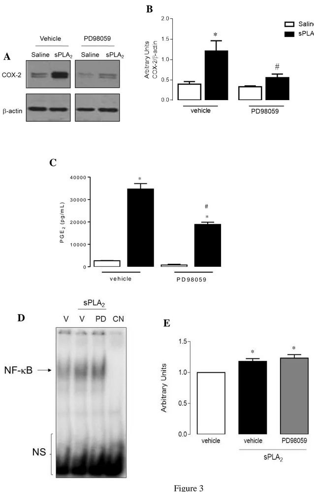
3.3 ERK1/2 is an important component in the signaling of COX-2 expression and PGE2 production
induced by group IIA sPLA2 in macrophages
sPLA2-induced COX-2 protein expression and PGE2 levels, respectively, in macrophages pretreated
with PD98059 or with vehicle only, were examined (Figures 3A-C). Unstimulated macrophages
showed a weak basal COX-2 protein expression and PGE2 release when pre-incubated with or
without inhibitors of kinases (Figures 3A -C). In contrast, pre-incubation of cells with PD98059, an
ERK1/2 activation inhibitor significantly reduced COX-2 protein expression and PGE2 release (by
82% and 48%, respectively) induced by this venom group IIA sPLA2, in comparison to stimulated
cells.These results implicate ERK1/2 signaling protein in the COX-2 pathway induced by group
IIA sPLA2. Next, to determine whether activation of NF-B is mediated by an ERK1/2 signaling
pathway, we evaluated the translocation of NF-B to the macrophage nucleus under sPLA2
stimulation. Pre-treatment of resident peritoneal macrophages with PD98059 did not alter group IIA
sPLA2.induced activation of NF-B (Figure 3D). These results suggest evidence ERK1/2 is not
involved in by sPLA2-induced activation of NF-B. The downstream factors activated by ERK1/2
remain to be investigated.
3.4 12-HETE is involved in ERK1/2 phosphorylation induced by group IIA sPLA2
In order to evaluate the role of the derived 12-LO mediator in the effects of group IIA
sPLA2, the effects of baicalein on sPLA2-stimulated p38MAPK, PKC, and ERK1/2 protein kinases
phosphorylation were tested. As assessed by western blotting results showed that the activation of
PKC or p38MAPK kinases phosphorylation by group IIA sPLA2 were not modified by
pre-treatment with baicalein compound (Figures 4A-B). On the other hand, sPLA2-induced ERK1/2
protein phosphorylation was significantly inhibited by baicalein, when compared with macrophages
were not affected by baicalein (Figure 4E). To better demonstrate this last effect, the Fig. 5A and B
showed that the treatment of macrophages in culture with 0.4 µM of sPLA2 induced significantly
ERK1/2 phosphorylation from 5 up to 30 min (p 0.05), indicating that group IIA sPLA2 triggers
signaling pathway mediated by ERK1/2.
4. Discussion
In our present study, we showed for the first time a group IIA sPLA2 isolated from snake
venom induced early and sustained release of 12- (S)-hydroxy-eicosatetraenoic (12-HETE) by
macrophages. Bee venom (Apis mellifera) and snake venom (Bungarus multicinctus) sPLA2s also
stimulate production of 12- (S)-hydroxy(peroxy)eicosatetraenoic (12-H(P)ETE) by platelets [53],
which is an active precursor of 12-HETE [29, 54], indicating that venom sPLA2s can upregulate
12-LO pathway. In addition an association between mammalian PLA2s from groups IIA and IV and
platelet-type 12-LO during the inflammatory response has also been demonstrated by some studies
[39, 55]. In the present study, our data evidenced similar active association between PLA2 and
leukocyte-type 12-LO which is in turn the isoform expressed by macrophages [55,56].
We have previously reported that group IIA sPLA2 from Bothrops asper snake venom
MT-III triggers inflammatory activity at the site of the injection [35,57], inducing a prominent release of
PGE2 from macrophages by up-regulating COX-2 expression, which is dependent on activation of
distinct protein kinases [39]. The involvement of 12-LO as the underlying mechanism of sPLA2
-induced up-regulation of COX-2 was confirmed by inhibiting expression of COX-2 protein with
baicalein [58-60]. Thus, this group IIA sPLA2 activates downstream pathways required for
up-regulation of COX-2 expression and PGE2 production through the catalytic activity of 12-LO and
release of 12-HETE, accompanied by high production of AA induced by sPLA2 MT-III [35]. These
data are in agreement with findings that 12-HETE directly or 12-LO activity are involved in COX-2
expression and PGE2 production in pancreatic, mesangial cells and human colonic myofibroblasts
acid mobilization through up-regulation of cytosolic PLA2 in human macrophages by group V
sPLA2, in order to amplify inflammatory mechanisms [62]. To our best knowledge, this is the first
evidence for the role of 12-HETE in the regulation of snake venom group IIA sPLA2-induced
COX-2 protein expression and PGE2 biosynthesis.
12-HETE elicits inflammatory, immunomodulatory and leukocyte chemoattraction [63,64].
In turn, it stimulates signaling pathways involving protein kinases that play regulatory roles on the
route of cyclooxygenase activity and production of lipid mediators [21,23, 65-67]. The 12-HETE
mediator is known to activate p38 mitogen-activated protein kinases (p38MAPK), protein kinase C
(PKC), and extracellular signal-regulated kinases1/2 (ERK1/2) to up-regulate inflammatory effects
in various cell types [25-28,33,61,68]. Our findings showed that inhibition of 12-LO by baicalein
reduced ERK1/2 activation in cells stimulated by group IIA sPLA2, indicating participation of
ERK1/2 in the pathway triggered by 12-HETE in our experimental condition. Interestingly,
baicalein did not affect sPLA2-induced activation of p38MAPK and PKC evidencing that
phosphorylation of these kinases is largely independent of 12-LO up-regulation in isolated
macrophages. This finding highlights that the up-regulation of 12-LO by group IIA sPLA2 regulates
ERK1/2 phosphorylation in isolated macrophages. The lipid mediator 12-HETE is then an upstream
activator of ERK1/2 upon stimulation by group IIA sPLA2. These results add relevant knowledge
about the signal pathways triggered by group IIA sPLA2s leading to the production of PGE2. This
sequential activation of 12-HETE and ERK1/2 has also been demonstrated to mediate mucus
secretion in chronic pulmonary inflammatory diseases [66] and pancreatic cancer and epidermoid
carcinoma cells proliferation [25,36, 66].
In the present study, group IIA sPLA2 was found to induce the phosphorylation of ERK1/2
and this effect is consistent with other studies showing activation of ERK as key link of
inflammation, cell proliferation and lipid body formation induced by both mammalian group IIA
group IIA sPLA2-induced PGE2 production and COX-2 expression, is largely dependent on ERK1/2
phosphorylation, in agreement with previous reports that have evidenced the involvement of
ERK1/2 pathway in AA metabolism by sPLA2s [52, 72, 74-76]. Surprisingly, our data showed that
this downstream of activation is independent of nuclear translocation of NF-B. Our previous study has demonstrated that production of PGE2 and expression of COX-2 induced by sPLA2
MT-III result from both NF-B-linked p38MAPK and PKC and NF-B-independent PI3K kinase pathways [39]. Considering this information we hypothesize that a transcription factor other than
NF-B is involved in the present described pathway.
Thus, the present study report that 12-LO and its product 12-HETE play an important role in
the upstream cascade involved in sPLA2-induced ERK1/2 phosphorylation carry on in COX-2
protein up-regulation and PGE2 production, as illustrated in Figure 6. Taken together, by this study
we revealed a new aspect of the effect of group IIA sPLA2 in macrophage inflammatory functions
by demonstrating that distinct arachidonic acid metabolism products may positively regulate
crosswise of group IIA sPLA2-induced-COX-2 and -PGE2 up-regulation. Therefore, these findings
bring new insights into the mechanisms of amplification of the inflammatory response induced by a
snake venom group IIA sPLA2.
Acknowledgements
The authors thank Renata Hage do Amaral for technical assistance. This investigation was
supported by a research grant from Fundação de Amparo à Pesquisa do Estado de São Paulo
(FAPESP) Brazil (07/03336-9). C.T. is recipient of CNPq - PQ (grant 306099/2008-0), V.M. was
recipient of Pos doctoral fellowship from FAPESP-Brazil (grant 07/03337-5) and CNPq (grant
202077/2008-0), M.A.R.V. was recipient of FAPESP-Brazil (grant 12/10653-9) and R.C. is
References:
[1] M. Murakami, Y. Taketomi, H. Sato, K. Yamamoto Secreted phospholipase A2 revisited J Biochem., 150 (2011), pp. 233-255.
[2] M. Murakami, H. Sato, Y. Miki, K. Yamamoto, Y. Taketomi A new era of secreted
phospholipase A₂ J. Lipid Res., 56 (2015), pp. 1248-1261.
[3] M. Murakami, Y. Taketomi, C. Girard, K.Yamamoto, G. Lambeau Emerging roles of secreted
phospholipase A2 enzymes: Lessons from transgenic and knockout mice Biochimie, 92 (2010),
pp. 561-582.
[4] N.D. Quach , R.D. Arnold, B.S. Cumming Secretory phospholipase A2 enzymes as
pharmacological targets for treatment of disease Biochem. Pharmacol., 90 (2014), pp. 338-348.
[5] M. Murakami, Y. Taketomi Secreted phospholipase A2 and mast cells Allergol. Int., 64 (2015), pp. 4-10.
[6] J.S. Rana, M. Cote, J.P. Després, M.S. Sandhu, P.J. Talmud, E. Ninio, N.J. Wareham,
J.J. Kastelein, A.H. Zwinderman, K.T. Khaw, S.M. Boekholdt Inflammatory biomarkers and the
prediction of coronary events among people at intermediate risk: the EPIC-Norfolk prospective population study Heart, 95 (2009), pp. 1682-1687.
[7] P. Gałecki, E. Gałecka, M. Maes, M. Chamielec, A. Orzechowska, K. Bobińska, A. Lewiński, J.J. Szemraj The expression of genes encoding for COX-2, MPO, iNOS,
and sSPLA2-IIA in patients with recurrent depressive disorder Affect. Disord., 138 (2012), pp.
360-366.
[8] M. Menschikowski, A. Hagelgans, U. Schuler, S. Froeschke, A. Rosner, G. Siegert, Plasma
levels of phospholipase A2-IIA in patients with different types of malignancies: prognosis and association with inflammatory and coagulation biomarkers Pathol. Oncol. Res., 19 (2013), pp.
839-846.
[9] M. Chacur, E.D. Milligan, E.M. Sloan, J. Wieseler-Frank, R.M. Barrientos, D. Martin, S. Poole, B. Lomonte, J.M. Gutiérrez, S.F. Maier, Y. Cury, L.R. Watkins, Snake venom phospholipase A2s
(Asp49 and Lys49) induce mechanical allodynia upon peri-sciatic administration: involvement of spinal cord glia, proinflammatory cytokines and nitric oxide Pain, 108 (2004), pp. 180-191.
[10] C.F. Teixeira, E.C. Landucci, E. Antunes, M. Chacur, Y. Cury Inflammatory effects of snake
venom myotoxic phospholipases A2 Toxicon, 42 (2003), pp. 947-962.
[11] F. Chaves, G. León, V.H. Alvarado J.M. Gutiérrez Pharmacological modulation of edema
induced by Lys-49 and Asp-49 myotoxic phospholipases A2 isolated from the venom of the
[12] W.S. Powell, J. Rokach Biosynthesis, biological effects, and receptors of
hydroxyeicosatetraenoic acids (HETEs) and oxoeicosatetraenoic acids (oxo-ETEs) derived from arachidonic acid Biochim. Biophys. Acta, 1851 (2015), pp. 340-355.
[13] D. Sacerdoti, P. Pesce, M. Di Pascoli, S. Brocco, L. Cecchetto, M. Bolognesi, Arachidonic
acid metabolites and endothelial dysfunction of portal hypertension Prostaglandins Other Lipid
Mediat., 120 (2015), pp. 80-90.
[14] K. Kawahara, H. Hohjoh, T. Inazumi, S. Tsuchiya, Y. Sugimoto Prostaglandin
E2-induced inflammation: Relevance of prostaglandin E receptors Biochim. Biophys. Acta, 1851
(2015), pp. 414-421.
[15] A.N. Hata, R.M. Breyer Pharmacology and signaling of prostaglandin receptors: Multiple
roles in inflammation and immune modulation Pharmacol. Ther., 103 (2004), pp. 147-166.
[16] K. Omori, T. Kida, M. Hori, H. Ozaki, T. Murata Multiple roles of the PGE2 -EP receptor
signal in vascular permeability Br. J. Pharmacol., 171(2014), pp. 4879-4889.
[17] B. Colom, J.V. Bodkin, M. Beyrau, A. Woodfin, C. Ody, C. Rourke, T.Chavakis,
K. Brohi, B.A. Imhof, S. Nourshargh Leukotriene B4-neutrophil elastase axis drives neutrophil
reverse transendothelial cell migration in vivo Immunity, 42 (2015), pp. 1075-1086.
[18] M. Le Bel, A. Brunet, J. Gosselin, Leukotriene B4, an endogenous stimulator of the innate
immune response against pathogens J. Innate Immun., 6 (2014), pp.159-68.
[19] A.C. Duchez, L.H. Boudreau, G.S. Naika, J. Bollinger, C. Belleannée, N. Cloutier, B. Laffont, R.E. Mendoza-Villarroel, T. Lévesque, E. Rollet-Labelle, M. Rousseau, I. Allaeys, J.J. Tremblay, P.E. Poubelle, G. Lambeau, M. Pouliot, P. Provost, D. Soulet, M.H Gelb, E. Boilard, Platelet
microparticles are internalized in neutrophils via the concerted activity of 12-lipoxygenase and secreted phospholipase A2-IIA Proc. Natl. Acad. Sci. USA, 112 (2015), E3564-E3573.
[20] D.A. Taylor-Fishwick, J. Weaver, L. Glenn, N. Kuhn, G. Rai, A. Jadhav, A. Simeonov, A. Dudda, D. Schmoll, T.R. Holman, D.J. Maloney, J.L. Nadler Selective inhibition of
12-lipoxygenase protects islets and beta cells from inflammatory cytokine-mediated beta cell dysfunction Diabetologia, 58 (2015), pp. 549-557.
[21] X. Han, S. Chen, Y. Sun, J.L. Nadler, D. Bleich Induction of cyclooxygenase-2 gene in
pancreatic beta-cells by 12-lipoxygenase pathway product 12-hydroxyeicosatetraenoic acid,
Mol. Endocrinol. 16 (2002) 2145-2154.
[22] K.J. Woo, J.H. Lim, S.I. Suh, Y.K. Kwon, S.W. Shin, S.C. Kim, Y.H. Choi, J.W. Park, T.K. Kwon Differential inhibitory effects of baicalein and baicalin on LPS-induced
cyclooxygenase-2 expression through inhibition of C/EBPbeta DNA-binding activity,
[23] Z. G. Xu, S.L. Li, L. Lanting, Y.S. Kim, N. Shanmugam, M.A. Reddy, R. Natarajan
Relationship between 12/15-lipoxygenase and COX-2 in mesangial cells: potential role in diabetic nephropathy, Kidney Int. 69 (2006) 512-519.
[24] M. Niknami, S. Vignarajan, M. Yao, S. Hua, P.K. Witting, Y. Kita, T. Shimizu, P. Sved, M.I. Patel, Q. Dong Decrease in expression or activity of cytosolic phospholipase A2alpha
increases cyclooxygenase-1 action: A cross-talk between key enzymes in
arachidonic acid pathway in prostate cancer cells, Biochim. Biophys. Acta 1801(2010) 731-737.
[25] S. Agarwal, C. Achari, D. Praveen, K.R. Roy, G.V. Reddy, P. Reddanna Inhibition of
12-LOX and COX-2 reduces the proliferation of human epidermoid carcinoma cells (A431) by modulating the ERK and PI3K-Akt signaling pathways, Exp. Dermatol. 18 (2009) 939-946.
[26] X.Z. Ding, W.G. Tong, T.E. Adrian 12-lipoxygenase metabolite 12(S)-HETE stimulates
human pancreatic cancer cell proliferation via protein tyrosine phosphorylation and ERK activation, Int. J. Cancer. 94 (2001) 630-636.
[27] R.A. Stockton, B.S. Jacobson Modulation of cell-substrate adhesion by arachidonic acid:
lipoxygenase regulates cell spreading and ERK-inducible cyclooxygenase regulates cell migration in NIH-3T3 fibroblasts, Mol. Biol. Cell 12 (2001) 1937-1956.
[28] Y. Wen Y, J. Gu, G.E. Vandenhoff, X. Liu, J.L. Nadler Role of 12/15-lipoxygenase in the
expression of MCP-1 in mouse macrophages, Am. J. Physiol. Heart Circ. Physiol, 294 (2008)
H1933-H1938.
[29] T. Yoshimoto, Y. Takahashi Arachidonate 12-lipoxygenases, prostaglandins Other Lipid Mediat. 68-69 (2002) 245–262.
[30] M.K. Patricia, J.A. Kim, C.M. Harper, P.T. Shih, J.A. Berliner, R. Natarajan, J.L. Nadler, C.C. Hedrick Lipoxygenase products increase monocyte adhesion to human aortic endothelial
cells, Arterioscler Thromb. Vasc. Biol. 19 (1999) 2615–2622.
[31] K.B. Reilly, S. Srinivasan, M.E. Hatley, M.K. Patricia, J. Lannigan, D.T. Bolick, G.
Vandenhoff, H. Pei, R. Natarajan, J.L. Nadler,C.C.Hedrick 12/15-Lipoxygenase activity mediates
inflammatory monocyte/endothelial interactions and atherosclerosis in vivo, J. Biol. Chem. 279
(2004) 9440–9450.
[32] J. Faulkner, C. Pye, M. Al-Shabrawey, A.A. Elmarakby Inhibition of 12/15-Lipoxygenase
Reduces Renal Inflammation and Injury in Streptozotocin-Induced Diabetic Mice, J. Diabetes
Metab. 6 (2015) pii: 555. doi:10.4172/2155-6156.1000555.
[33] Y. Wen, J. Gu, S.K. Chakrabarti, K. Aylor, J. Marshall, Y. Takahashi, T. Yoshimoto, J.L. Nadler The role of 12/15-lipoxygenase in the expression of interleukin-6 and tumor
[34] J.M. Gutiérrez, B. Lomonte Phospholipase A2 myotoxins from Bothrops snake venoms,
Toxicon 33(1995) 1405-1424.
[35] V. Moreira, J.M. Gutiérrez, A.M. Soares, S.R. Zamunér, E. Purgatto, C.F. Teixeira Secretory
phospholipases A(2) isolated from Bothrops asper and from Crotalus durissus terrificus snake venoms induce distinct mechanisms for biosynthesis of prostaglandins E2 and D2 and
expression of cyclooxygenases, Toxicon 52 (2008) 428-439.
[36] V. Moreira, J.M. Gutiérrez, R.B. Amaral, B. Lomonte, E. Purgatto, C. Teixeira, A
phospholipase A₂ from Bothrops asper snake venom activates neutrophils in culture: expression of cyclooxygenase-2 and PGE₂ biosynthesis, Toxicon 57 (2011) 288-296.
[37] M. Chacur, I. Longo, G. Picolo, J.M. Gutiérrez, B. Lomonte, J.L. Guerra, C.F. Teixeira, Y. Cury Hyperalgesia induced by Asp49 and Lys49 phospholipases A2 from Bothrops asper
snake venom: pharmacological mediation and molecular determinants, Toxicon 41 (2003)
667-678.
[38] J.P. Zuliani, C.M. Fernandes, S.R. Zamuner, J.M. Gutiérrez, C.F. Teixeira Inflammatory
events induced by Lys-49 and Asp-49 phospholipases A2 isolated from Bothrops asper snake
venom: role of catalytic activity, Toxicon 45 (2005) 335-346.
[39] V. Moreira, B. Lomonte, M.A. Vinolo, R. Curi, J.M. Gutiérrez, C. Teixeira An Asp49
phospholipase A2 from snake venom induces cyclooxygenase-2 expression and prostaglandin
E2 production via activation of NF-κB, p38MAPK, and PKC in macrophages, Mediators
Inflamm. 2014 (2014) 105879.
[40] V. Ruipérez, J. Casas, M.A. Balboa, J. Balsinde Group V phospholipase A2-derived
lysophosphatidylcholine mediates cyclooxygenase-2 induction in lipopolysaccharide-stimulated macrophages, J. Immunol. 179 (2007) 631-638.
[41] D.Y. Kim, H.J. Song, J.H. Jeong, J.S. Suh, U.D. Sohn Regulation of lysophosphatidic
acid-induced COX-2 expression by ERK1/2 activation in cultured feline esophageal epithelial cells,
Arch. Pharm. Res. 31 (2008) 1331-1338.
[42] M. Hughes-Fulford , Y. Chen, R.R. Tjandrawinata Fatty acid regulates gene expression and
growth of human prostate cancer PC-3 cells, Carcinogenesis 22 (2001) 701-707.
[43] B. Lomonte, J.M. Gutiérrez A new muscle damaging toxin, myotoxin II, from the venom of
the snake Bothrops asper (terciopelo), Toxicon 27 (1989) 725-733.
[44] D. Mora-Obando, C. Díaz, Y. Angulo, J.M. Gutiérrez, B. Lomonte Role of enzymatic activity
in muscle damage and cytotoxicity induced by Bothrops asper Asp49 phospholipase A2 myotoxins: are there additional effector mechanisms involved?, Peer Journal, 2:e569 (2014).
[45] K. Takayama, D. H. Mitchell, Z. Z. Din, P. Mukerjee, C. Li, D. L. Coleman Monomeric Re
lipopolysaccharide from Escherichia coli is more active than the aggregated form in the Limulus amebocyte lysate assay and in inducing Egr-1 mRNA in murine peritoneal macrophages, J. Biol. Chem. 269 (1994) 2241-2244.
[46] C.Y. Peng, S.L. Pan, Y.M. Huang, J.H Guh, Y.L. Chang, C.M. Teng Baicalein attenuates
intimal hyperplasia after rat carotid balloon injury through arresting cell-cycle progression and inhibiting ERK, Akt, and NF-kappaB activity in vascular smooth-muscle cells, Naunyn.
Schmiedebergs Arch. Pharmacol. 378 (2008) 579-588.
[47] Z. Qi, F. Yin, L. Lu, L. Shen, S. Qi, L. Lan, L. Luo, Z. Yin Baicalein reduces
lipopolysaccharide-induced inflammation via suppressing JAK/STATs activation and ROS production, Inflamm. Res. 62 (2013) 845-855.
[48] S. Nishimoto, E. Nishida MAPK signalling: ERK5 versus ERK, EMBO Rep. 7 (2006) 782-786.
[49] Y. Rong, M. Baudry Seizure activity results in a rapid induction of nuclear factor-kappa B
in adult but not juvenile rat limbic structure, J. Neurochem. (1996) 67:662-668.
[50] M.M. Bradford A rapid and sensitive method for the quantitation of microgram quantities
of protein utilizing the principle of protein-dye binding, Anal. Biochem. 72 (1976) 248-254.
[51] M.A. Vinolo, H.G. Rodrigues, E. Hatanaka, F.T. Sato, S.C. Sampaio, R. Curi Suppressive
effect of short-chain fatty acids on production of proinflammatory mediators by neutrophils,
J. Nutr. Biochem. 22 (2011) 849-855.
[52] V. Moreira, P.C.C. Souto, M.A.R Vinolo, B. Lomonte, J. M. Gutiérrez, R. Curi, C. Teixeira A
catalytically-inactive snake venom Lys49 phospholipase A2 homolog induces expression of
cyclooxygenase-2 and production of prostaglandins through selected signaling pathways in macrophages, Eur. J. Pharmacol. 708 (2013) 68-79.
[53] M.J. Coffey, B. Coles, M. Locke, A. Bermudez-Fajardo, P.C. Williams, G.E. Jarvis, V.B. O'Donnell Interactions of 12-lipoxygenase with phospholipase A2 isoforms following
platelet activation through the glycoprotein VI collagen receptor, FEBS Lett. 576 (2004)
165-168.
[54] C.R. Pace-Asciak Pathophysiology of the hepoxilins. Biochim Biophys Acta. 1851(2015): 383-396.
[55] T. Yoshimoto, Y. Takahashi Arachidonate 12-lipoxygenases. Prostaglandins Other Lipid Mediat. 68-69(2002): 245–262.
[56] Yoshimoto T., S. Yamamoto Arachidonate 12-lipoxygenase. J Lipid Mediat Cell Signal 12(1995):195-212.
[57] J.P. Zuliani, J.M. Gutiérrez, L.L. Casais e Silva, S. C. Sampaio, B. Lomonte, C.F.P. Teixeira
Activation of cellular functions in macrophages by venom secretory Asp-49 and Lys 49 phospholipases A(2), Toxicon 46 (2005) 523-532.
[58] W.G. Tong, X.Z. Ding, R.C. Witt, T.E. Adrian Lipoxygenase inhibitors attenuate growth of
human pancreatic cancer xenografts and induce apoptosis through the mitochondrial pathway, Mol. Cancer Ther. 1 (2002) 929–935.
[59] R. Yoshimura, K. Inoue, Y. Kawahito, M. Mitsuhashi, K. Tsuchida, M. Matsuyama, H. Sano, T. Nakatani Expression of 12-lipoxygenase in human renal cell carcinoma and growth
prevention by its inhibitor. Int. J. Mol. Med. 13 (2004) 44-46.
[60] X.M. Xu, G.J. Yuan, J.J. Deng, H.T. Guo, M. Xiang, F. Yang, W. Ge, S.Y. Chen Inhibition of
12-lipoxygenase reduces proliferation and induces apoptosis of hepatocellular carcinoma cells in vitro and in vivo, Hepatobiliary Pancreat. Dis. Int. 11 (2012) 193-202.
[61] J.F. Di Mari, J.I. Saada, R.C. Mifflin, J.D. Valentich, D.W. Powell HETEs enhance
IL-1-mediated COX-2 expression via augmentation of message stability in human colonic myofibroblasts, Am. J. Physiol. Gastrointest. Liver Physiol. 293 (2007) G719-G728.
[62] M.A. Balboa, R. Pérez, J. Balsinde. Amplification mechanisms of inflammation: paracrine
stimulation of arachidonic acid mobilization by secreted phospholipase A2 is regulated by cytosolic phospholipase A2-derived hydroperoxyeicosatetraenoic acid. J Immunol. 171(2003):
989-394.
[63] A. Nicolaou, M. Masoodi, K. Gledhill, A.K. Haylett, A.J. Thody, D.J. Tobin, L.E. Rhodes The
eicosanoid response to high dose UVR exposure of individuals prone and resistant to sunburn,
Photochem. Photobiol. Sci. 11 (2012): 371-380.
[64] L. E. Rhodes, K. Gledhill, M. Masoodi, A.K. Haylett, M. Brownrigg, A.J. Thody, D.J. Tobin, A. Nicolaou The sunburn response in human skin is characterized by sequential
eicosanoid profiles that may mediate its early and late phases FASEB J. 23(2009): 3947–3956.
[65] S.K. Chakrabarti, B.K. Cole, Y. Wen, S.R. Keller, J.L. Nadler 12/15-lipoxygenase products
induce inflammation and impair insulin signaling in 3T3-L1 adipocytes, Obesity, 17 (2009)
1657-1663.
[66] C.K. Szekeres, K. Tang, M. Trikha, K.V Honn Eicosanoid activation of extracellular
signal-regulated kinase1/2 in human epidermoid carcinoma cells, J. Biol. Chem. 275 (2000),
38831-38841.
[67] F.L. Chen, X.Z. Wang, J.Y. Li, J.P. Yu, C.Y. Huang, Z.X. Chen 12-lipoxygenase induces
apoptosis of human gastric cancer AGS cells via the ERK signal pathway, Dig. Dis. Sci. 53
[68] I. Garcia-Verdugo, F. BenMohamed, S. Tattermusch, D. Leduc, G. Charpigny,
M. Chignard, M. Ollero, L. Touqui A role for 12R-lipoxygenase in MUC5AC expression by
respiratory epithelial cells, Eur. Respir. J. 40 (2012) 714-723.
[69] R. Martín, C. Cordova, M.L. Nieto Secreted phospholipase A2-IIA-induced a phenotype of
activated microglia in BV-2 cells requires epidermal growth factor receptor transactivation and proHB-EGF shedding, J. Neuroinflammation 9 (2012) 154.
[70] J.A. Yu, S. Kalatardi, J. Dohse, M.R. Sadaria, X. Meng, D.A. Fullerton, M.J. Weyant Group
IIa sSPLA2 inhibition attenuates NF-κB activity and promotes apoptosis of lung cancer cells,
Anticancer Res. 32 (2012) 3601-3607.
[71] M. Hernández, S.L. Burillo, M.S. Crespo and M.L. Nieto Secretory phospholipase
A2 activates the cascade of mitogen-activated protein kinases and cytosolic phospholipase A2
in the human astrocytoma cell line 1321N1, J. Biol. Chem. 273 (1998) 606-612.
[72] K.C. Giannotti, E. Leiguez, V. Moreira, N.G. Nascimento, B. Lomonte, J.M. Gutiérrez, R.L Melo, C. Teixeira A Lys49 phospholipase A2, isolated from Bothrops asper snake venom,
induces lipid droplet formation in macrophages which depends on distinct signaling pathways and the C-terminal region, Biomed Res Int. 2013 (2013) 807982.
[73] E. Leiguez, J.P. Zuliani, A.M. Cianciarullo, C.M. Fernandes, J.M. Gutiérrez, C. Teixeira A
group IIA-secreted phospholipase A2 from snake venom induces lipid body formation in
macrophages: the roles of intracellular phospholipases A2 and distinct signaling pathways, J.
Leukoc. Biol. 90 (2011) 155-166.
[74] E. Kikawada, J.V. Bonventre, J.P. Arm Group V secretory SPLA2 regulates
TLR2-dependent eicosanoid generation in mouse mast cells through amplification of ERK and cSPLA2alpha activation, Blood. 110 (2007) 561-567.
[75] W.K. Han, A. Sapirstein, C.C. Hung, A. Alessandrini, J.V. Bonventre Cross-talk between
cytosolic phospholipase A2 alpha (cSPLA2 alpha) and secretory phospholipase A2 (sSPLA2) in
hydrogen peroxide-induced arachidonic acid release in murine mesangial cells: sSPLA2 regulates cSPLA2 alpha activity that is responsible for arachidonic acid release, J. Biol.
Chem. 278 (2003) 24153-24163.
[76] V. Ruipérez, A.M. Astudillo, M.A. Balboa. J. Balsinde Coordinate Regulation of
TLR-Mediated Arachidonic Acid Mobilization in Macrophages by Group IVA and Group V Phospholipase A2s. J. Immunol. 182 (2009) 3877-3883.
Figures Figure 1
A
B
C
Figure 2
A
C
B
D
Figure 3
A
B
C
0 1 0 0 0 0 2 0 0 0 0 3 0 0 0 0 4 0 0 0 0 * * # v e h ic le P D 9 8 0 5 9 P G E2 ( p g /m L )D
E
Figure 4
B
C
D
E
A
Figure 5
A
B
0 .0 0 .2 0 .4 0 .6 0 .8 * * 0 1 5 1 5 3 0 T im e ( h ) A rb it r a r y U n it s p -E R K / -a c ti n * 0 .0 0 .5 1 .0 1 .5 2 .0 0 1 5 1 5 3 0 T im e ( h ) s P L A2 A rb it ra ry U n it s E R K / -a c ti nC
Legends
Fig. 1. Effect of group IIA sPLA2 on the release of 12-HETE and protein expression of 12-LO in
macrophages. Cells (1x10⁶) were incubated with MT-III (0.4 µM) or saline (control) for 15 min or 0.5, 1, 3 or 6 h. 12-HETE was quantified in culture supernatants by specific EIA (A); western
blotting analysis of 12-LO and β-actin (loading control) (B); bar graph shows densitometric analysis
of immunoreactive 12-LO in AU, normalized with -actin (C). Results are expressed as mean ± SEM from 3 to 4 experiments. *p< 0.05 as compared to the corresponding macrophages incubated
with saline solution. #p< 0.05 as compared to macrophages incubated with sPLA2 at 15 min or 1, 3,
or 6 h.
Fig. 2. Effect of baicalein on COX-2 expression, PGE2, and 12-HETE production induced by group
IIA sPLA2 in isolated macrophages. Resident peritoneal macrophages (1x10⁶ cells) were pretreated
for 30 min with baicalein (40 μM) and then incubated for 3 h with MT-III (0.4 µM). Western
blotting analysis of COX-2 and β-actin (loading control) of cells treated with MT-III (A); bar graph
exhibits densitometric analysis of immunoreactive COX-2 in AU, normalized with -actin (B); PGE2 and 12-HETE were quantified in culture supernatants by specific EIA (C, D). Results are
expressed as mean ± SEM from 3 to 4 experiments. *p< 0.05 as compared with macrophages
incubated with saline solution. #p< 0.05 as compared with macrophages incubated with sPLA2 and
vehicle.
Fig. 3. Effect of PD98059 on COX-2 protein expression, PGE2 release, and NF-B activation
induced by group IIA sPLA2 in isolated macrophages. MT-III activates NF-B in cultured
macrophages. (A,C) Nuclear extracts were prepared and assayed for B probe activity with 32 P-labeled double-stranded oligonucleotide B using EMSA. (B, D) Densitometric analysis of NF-B
band intensities. Results are expressed as mean ± SEM from 3 experiments. *p< 0.05 as compared
with control value. NS=Non-specific band; V=Vehicle; NC=Negative control.
Fig. 4. Effect of baicalein on p-PKC, p-p38 MAPK, and p-ERK protein expression induced by
group IIA sPLA2 in macrophages. Resident peritoneal macrophages (1x10⁶ cells) were pretreated
for 30 min with baicalein (40 μM) or vehicle (control). They were then incubated for 30 min with
MT-III (0.4 µM) and western blotting of PKC and PKC (A); p38MAPK and p38MAPK (B);
p-ERK and p-ERK (C), and β-actin (loading control) of cells treated with MT-III. Bar graph shows
densitometric analysis of immunoreactive bands of p-ERK and ERK in AU, both normalized with
-actin (D and E). Results are expressed as mean ± SEM from 3 experiments. *p< 0.05 as compared with macrophages incubated with saline. #p< 0.05 as compared with macrophages incubated with sPLA2 and vehicle.
Fig. 5. ERK phosphorylation induced by group IIA sPLA2 in isolated macrophages. Resident
peritoneal macrophages (1x10⁶ cells) were incubated for 1, 5, 15, or 30 min with MT-III (0.4 µM). Western blot analysis of p-ERK and ERK and β-actin (loading control) (A); densitometric analysis
of immunoreactive bands of p-ERK and ERK in AU, both normalized with -actin (D and E). Results are expressed as mean ± SEM from 3 experiments. *p< 0.05 as compared with
macrophages incubated for zero time.
Fig. 6. Schematic model for mechanisms involved in group IIA sPLA2-induced COX-2 expression
and PGE2 release in macrophages. MT-III induces AA release from macrophage membrane. In the
12-LO-shuttling pathway, the released AA is used for production and release of 12-HETE. The
possible association of 12-HETE with specific receptors also activates the ERK1/2 cascade, thus
activation. On the other hand, 12-HETE is not associated with alternative cascades sPLA2
-stimulated p38MAPK and PKC, both involved in NF-B activation and COX-2 protein expression and PGE2 production.