CONFORMATIONAL STUDIES OF THE BETA AMYLOID PROTEIN AND IN VITRO MODELS FOR THE EFFECT OF APOLIPOPROTEIN
E
ON FIBRIL FORMATION IN ALZHEIMER'S DISEASE
by
Krista Carole Evans B.S. Chemistry, Ithaca College
(1991)
Submitted to the Department of Chemistry in Partial Fulfillment of the
Requirements for the Degree of DOCTOR OF PHILOSOPHY
at the
Massachusetts Institute of Technology June 1996
© 1996 Massachusetts Institute of Technology All rights reserved
Signature of Author
Department of Chemistry May 8, 1996
Certified
by-Peter T. Lansbury, Jr. Associate Professor of Chemistry Thesis Supervisor Accepted
by-Dietmar Seyferth Chairman, Department Committee on Graduate Students
Science
OF TECH!NOLOGY
JUN
12
1996
This doctoral thesis has been examined by a Committee of the Department of Chemistry as follows:
Professor JoAnne Stubbe
Chairoerson Professor Peter T. Lansburv, Tr.
Thesis Supervisor Professor Jonathan King.
(
CONFORMATIONAL STUDIES OF THE BETA AMYLOID PROTEIN
AND IN VITRO MODELS FOR THE EFFECT OF APOLIPOPROTEIN
E
ON FIBRIL FORMATION IN ALZHEIMER'S DISEASE
by
Krista Carole Evans
Submitted to the Department of Chemistry on May 8, 1996 in Partial Fulfillment of the Requirements for the Degree of Doctor of Philosophy
ABSTRACT
The presence of amyloid plaques is one of the hallmarks of Alzheimer's disease (AD) brains. The plaque is an ordered, fibrous protein aggregate of which the major component is the p protein, a 39-43 amino acid peptide. The P protein is a normal product of the constitutive proteolytic processing of the P amyloid precursor protein, and has been detected in the blood and CSF of healthy individuals. Exposure to extrinsic factors such as metals or exogenous proteins has been proposed to be a risk factor for AD.
Rodents encode for a
P
protein that differs from the human variant by only three residues (R5G, Y10F, H13R) but do not form amyloid plaques. 11-28, which has a conformation similar to 11-40 as measured by circular dichroism (CD), and 11-28 rodent analogs were examined by CD and the effect of metals on their conformations was examined. Unlike the human analog which undergoes large conformational changes in the presence of zinc, the rodent analog shows little change. The H13R mutation in the rodent sequence appears to be responsible for the decreased interaction of the peptide with zinc.The
3
protein also forms fibrillar aggregates in vitro according to a nucleation-dependent mechanism. The apolipoprotein E genotype has recently been recognized as a susceptibility factor for AD. The APOE4 allele correlates with an earlier onset of AD and with increased amyloid deposition in the brain. Apolipoprotein E (apoE) proteins may effect the rate of in vivo amyloidogenesis. ApoE3 inhibits amyloid nucleation at an apoE3:p protein molar ratio of 1:1000. ApoE4, which cannot form a disulfide dimer, is an equipotent or less potent inhibitor. The apoE3 dimer is a significantly more potent inhibitor than apoE4. The 22 kD receptor-binding region of recombinant apoE isoforms had an inhibitory activity comparable to the native protein. The inhibitory activity of apoE was further narrowed to a 19 kD variant. Finally, a Cys57 22 kD apoE3 mutant was a significantly less efficient inhibitor of amyloid fibril formation. Thesis Supervisor: Dr. Peter T. Lansbury, Jr.Table of Contents
L ist of Figu res... 7
Acknowledgements... 9
A b b rev iation s ... 11
Chapter 1 The Role of the
3
Amyloid Protein in Alzheimer's Disease... 12C linical R esearch... ... 14
A D pathology ... 15
A m yloid Fibril ... ... 17
The causative role of P amyloid in AD... ... 19
M etabolism of A PP ... 21
G enetic research ... ... 24
Therapeutic options ... ... 29
References for chapter 1... 32
Chapter 2 Metal-Binding Studies of the 0 Protein... ... 35
C ircular dichroism ... 36
Solution structure of the
1
protein ... ... 39A mutation in the
3
protein causes HCHWA-D ... . 40Mammalian sequence variants ... ... ... 41
Effect of Metals on the conformation of the
1
protein ... 42Peptide A nalogs... 44
Analysis of the 0 protein and peptide analogs by circular dichroism... 47
Circular dichroism studies of 13-40 ... ... 49
Circular dichroism studies of 01-17... ... 55
Circular dichroism studies of 13-28 analogs ... 57
-28 R esu lts ... 57
1
-28 Rat Results...601
-28 Y 10F Results ... ... 64 11-28 R5G Results ... 66 11-28 H13R Results... 67 01-28 R5G,Y10F Results ... 69 1 1-28 E 22Q R esults... 72 D iscu ssion ... 74 E xp erim ental... 78 M aterials ... ... 78Peptide synthesis (Fmoc) ... ... 78
Peptide synthesis (Boc)... 79
Synthesis of P protein analogs ... ... 80
Purification and characterization of peptides... 82
Circular Dichroism Spectroscopy ... ... 82
References for chapter 2... 84
Chapter 3 Apolipoprotein E and its Relationship with Alzheimer's Disease.... 86
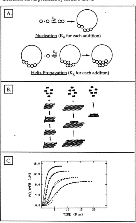
Mechanisms for protein polymerization ... ... 88
Nucleation-dependent polymerization of the P protein ... 91
Apolipoprotein E ... ... 94
Structure of apoE... ... 96
Function of apoE... 99
The connection of apolipoprotein E with AD ... 101
Effect of apoE on brain morphology, memory, and cognition in A D ... ... ... ... 102
Proposed role of apoE in AD ... 104
ApoE-deficient mice as a model for AD... 105
Results from In vitro studies of apoE and the
1
protein... 106ApoE is an inhibitor of amyloid formation ... 106
ApoE does not inhibit seeding of amyloid fibrils ... 110
ApoE does not effect the structure or final solubility of 11-40... 111
Inhibition of amyloid formation by the apoE3 dimer ... 112
Binding studies of the
1
protein with apoE ... 117D iscu ssion ... ... 117
E xp erim ental.. ... ... ... ... ... 121
M aterials ... 121
Aggregation assay (unstirred) ... ... 121
Aggregation assay (stirred) ... 122
Seeding experiments ... 122
Radioassay ... 122
Solubility Determ ination ... 123
Fourier-Transform Infrared Microscopy ... 123
Electron M icroscopy ... 123
SDS-polyacrylamide gel electrophoresis ... 124
Binding studies of ý1-40 with apoE... ... 124
References for chapter 3... .... ... 125
Chapter 4 Models of the N-Terminal Domain of Apolipoprotein E... 128
Proteolysis of apoE yields two intact domains... 128
Conformational change of apoE resulting from lipid-binding... 129
Proposed inhibitory mechanism of apoE 22 kD domain ... 132
Truncated apoE variant ... ... ... ... 133
Expression of apoE 22 kD variants ... 134
Cell transformation and protein induction ... 136
Purification of 22 kD apoE isoforms ... ... 139
Inhibition of P protein amyloid formation by the 22 kD domain... 139
The 22 kD domain is an effective amyloid inhibitor... 139
Inhibition of P protein aggregation by 2% 22 kD apoE isoforms... 142
Inhibition of amyloid formation is sensitive to apoE concentrations ... 143
Inhibitory activities of apoE3 and apoE4 are not distinguishable ... 144
Creation of a 19 kD truncated apoE protein ... ... 145
The Cys57 22 kD apoE3 appears to have diminished inhibitory effects... 150
Fibril morphology may be altered in the presence of the 22 kD apoE3... 153
Circular dichroism of 22 kD apoE proteins ... 155
The 22 kD apoE isoforms have a large c-helical content ... 156
Stability studies of the apoE proteins... 156
Discussion... ... 165
Future D irections ... ... ... 167
Experim ental ... 169
M ateria ls ... .... ... .. .. . ... ... 169
Aggregation assay (stirred) ... ... 169
Preparation of com petent cells... ... 169
Purification of plasm id DNA ... 170
Transform ation of E. Coli ... 171
Protein Induction... ... . 172
PCR protocol ... 173
Synthesis of truncated apoE DNA inserts ... 174
Purification of PCR products... 175
Restriction digest of DNA ... 175
Ligation of PCR insert and vector ... ... 176
Purification of apoE... ... 176
Circular dichroism of protein samples ... 178
List of Figures
1.1 Structure of Congo Red ... 18
1.2 C ro ss- fib ril... 19
1.3 Biogenesis of the
P
protein from APP... ... 221.4 APP mutations associated with early-onset FAD ... . 25
2.1 Sequence variability in the primate and rat P proteins ... 41
2.2 Full-length and truncated
1
protein peptides... ... 452.3 Peptide analogs of the
P
protein ... ... 462.4 CD spectra of 31-40 vs. HFIP ... 50
2.5 CD spectra of 31-40 vs. concentration... 51
2.6 CD spectra of 11-40 (repetitive scanning)... ... 52
2.7 CD spectra of 11-40 vs. salt ... ... 53
2.8 CD spectra of 31-40 vs. zinc ... 54
2.9 CD spectra of 11-40 vs. copper ... ... 55
2.10 CD spectra of 11-17 vs. TFE ... ... 56
2.11 CD spectra of the concentration dependence of 31-17... 57
2.12 C D spectra of 31-28 ... ... 58
2.13 CD spectra of 01-28 vs. zinc ... 59
2.14 CD spectra of 31-28 vs. copper ... ... 60
2.15 CD spectra of the concentration dependence of 11-28 Rat ... 61
2.16 CD spectra of 11-28 Rat vs. zinc ... ... 62
2.17 CD spectra of 31-28 Rat vs. copper... .... ... 63
2.18 CD spectra of 31-28 Y10OF vs. zinc ... ... 64
2.19 CD spectra of 31-28 Y10F vs. copper ... 65
2.20 CD spectra of 11-28 R5G vs. zinc ... ... 67
2.21 CD spectra of 31-28 H13R vs. zinc... ... 68
2.22 CD spectra of 31-28 H13R vs. copper ... ... 69
2.23 CD spectra of 31-28 R5G,Y10F vs. zinc ... ... 70
2.24 CD spectra of 11-28 R5G,Y10F vs. copper ... 71
2.25 CD spectra of 13-28 E22Q vs. zinc... ... 72
2.26 CD spectra of 13-28 E22Q vs. copper ... ... 73
3.1 Nucleation-dependent mechanism for microtubule formation ... 90
3.2 Nucleation-dependent mechanism for hemoglobin aggregation ... 92
3.3 Mechanism for nucleation-dependent aggregation of amyloid... 93
3.4 Amino acid sequence of human apoE2 ... 96
3.5 Predicted secondary structure of apoE... ... 97
3.6 Crystal structure of the 22 kD domain of apoE... . 99
3.7 Correlation of apoE genotype with AD risk and age of onset ... 102
3.9 Aggregation of f1-42 in the presence of apoE ... ... 108
3.10 Inhibition of the aggregation of f1-40 by 1% apoE... 110
3.11 Seeding of amyloid formation... 111
3.12 FTIR of amyloid fibrils in the presence of apoE... 113
3.13 EM of am yloid fibrils... ... ... ... 113
3.14 SDS-PAGE of full-length apoE proteins... 115
3.15 Inhibition of the aggregation of p1-40 by the apoE3 dimer ... 116
3.16 SDS-PAGE of j1-40 incubated with apoE... ... 118
3.17 Mechanism for amyloid formation and the effect of apoE... 119
4.1 Proteolytic fragments of apoE produced by thrombin ... 130
4.2 Proposed opening of the four-helix bundle of 22 kD apoE ... 131
4.3 Circular m ap of pT22K... 135
4.4 DNA gel electrophoresis of the apoE plasmids ... 137
4.5 SDS-PAGE of IPTG inductions of the apoE proteins... 138
4.6 SDS-PAGE of the apoE proteins after size-exclusion ... 140
4.7 SDS-PAGE of the apoE proteins after ion-exchange HPLC ... 141
4.8 Aggregation of f1-40 with the 22 kD and 10 kD apoE domains ... 142
4.9 Aggregation of f1-40 with 2% 22 kD apoE isoforms ... 143
4.10 The effect of apoE concentrations on the aggregation of ý1-40 ... 144
4.11 Aggregation of j1-40 in the presence of apoE3 and apoE4 ... 145
4.12 Translation of cDNA of the 22 kD apoE3... ... 146
4.13 DNA gel electrophoresis of the 19 kD apoE3 plasmids... 148
4.14 SDS-PAGE of the IPTG induction of the 19 kD apoE protein... 149
4.15 Aggregation of 31-40 in the presence of the 19 kD apoE protein ... 150
4.16 Aggregation of f1-40 in the presence of the Cys57 apoE3 mutant... 151
4.17 SDS-PAGE of the apoE3 Cys57 mutant... 152
4.18 Proposed effect of apoE on nucleation and plaque formation ... 155
4.19 CD spectra of the apoE proteins ... 157
4.20 CD spectra of the apoE3 Cys57 mutant ... 158
4.21 CD spectra of apoE2... 160
4.22 CD spectra of apoE3... ... ... 161
4.23 CD spectra of apoE4... 162
4.24 CD spectra of the 19 kD apoE3 protein... ... 163
Acknowledgements
I have found that this section, which is undoubtedly the most critical part of any thesis, is also the most difficult one to write. Where do I begin? Five years ago I came to MIT not knowing what I wanted to do with my life, or even if I really wanted to be here. One of the best decisions that I made during that time was joining Peter Lansbury's group.
I think that in addition to being an extraordinary scientist, Peter is also an incredible mentor. Peter's contagious enthusiasm has always made science exciting. Peter's greatest attribute, however, is his sincere concern for the well-being of his students. Because of this, Peter has earned the loyalty and respect from all of his students, past and present.
I also owe many thanks to every other member of the Lansbury lab. I would never have been successful without their help and support. Paul Weinreb has been my baymate since the beginning, or at least since we were given our own bench. Paul is not only a great scientist, but also a great teacher, mainly because he is so patient. Unfortunately, this trait often made him the target of constant prodding and practical jokes (i.e. gluing his pad of paper to his desk). He always managed to ignore us anyway.
Paul and I are also the last class to know all of the original Lansbury members (Julia Hendrix Miwa, Kurt Halverson, Shimi Cohen-Anisfeld and Annemarie Coffman-Lellouch), and take trips to Martha's Vineyard. I'll never forget sitting around the campfire drinking Southern Comfort through Twizllers listening to Irving Sucholeiki tell us about "Freaks" as only Irv could do, or the time that the skunks attacked the campsite. I'll also never forget the bikeride from Hell. Could it have rained any harder?
I would also like to thank both Joe Jarrett and Jon Come for their helpful suggestions and conversations, and Jon for his ability to make me laugh. Jon always made the lab an enjoyable place to work, even though he made fun of my cat calendar and all of my friends named Heather. Beth Berger was also a great mentor and helped me on many occasions. I look forward to hearing about her exciting travels through Papau New Guinea. I hope that 'Ted Ashburn is successful in his medical career. I wouldn't be surprised if someday he actually made it onto the space shuttle. To Dave Kocisko, I hope the fishing is great wherever the Army takes you.
Jim Harper, Anna Poon, and Raul Zambrano were great additions to the Lansbury lab. Jim always has great suggestions for interesting experiments, and has the ability to fix just about anything. Most importantly, he initiated poker night. I wish him all the best with his new baby. Anna was the first to introduce molecular biology to our lab. Anna added so much to
our group on both a scientific and personal level. I will never be able to listen to "Magic 106" without thinking of Raul. I am sure that Raul will be an incredible doctor. Cheon-Gyu Cho is a great chemist and has become an integral part of the lab. Santosh Nandan was unique in every way, and I hope everything is going well for him in India with his new wife. Kelly Conway, Magdalena Anguiano, Chris May, and Weiguo Zhen are the newest additions to our group and are responsible for carrying on the group traditions after the move to Harvard Medical School. I know that they will continue to do exciting work.
I would never have made it to this point without my chemistry buddy, Heather Nielsen Sugrue. We have shared so much over the years that I don't know where to begin. We managed to make chemistry fun. Remember doing shots before the chemistry banquet, the killer hangover in P-chem lab, taking candy breaks, tormenting Prof. Anderson, running through the chemistry building after jumping in the fountain during senior week, and so much more? I can't believe how much time has passed since sophomore year at Ithaca. Heather will always be a truly special friend, no matter how many miles are between us.
My deepest thanks go to my family who have always had endless faith in me. My parents, Dale and Svend Rasmussen, are the most loving and generous people that I have ever known. No sacrifice was ever too large for them when it came to either myself or my brother, Bob. My family always gave me the freedom to make my own decisions, while letting me know that they were there for me if I needed them. I know that I would never have
achieved so much without them.
My husband, Jeff, is undoubtedly the best thing that ever happened to me. I can't begin to imagine what my life would be like without him. From the first time that I met Jeff at Ithaca almost seven years ago, I knew that he was very special. Jeff is a wonderful and caring person who only sees the best in people. Because of his undying optimism and support, I have accomplished so much more than I ever imagined. I look forward to spending the rest of my life with you, Jeff, which I know will be filled with a lot of excitement, success, and love.
Abbreviations AD AFM ApoE APP Boc BOP CD CSF DIEA DMF DMS DMSO EDT EM FAD Fmoc FTIR HCHWA-D HFIP HPLC IPTG LDL LDMS MS NMR PAM PDMS PCR PMSF PMC PyBOP RPHPLC SDS-PAGE TFE Alzheimer's disease Atomic Force Microscopy Apolipoprotein E
p Amyloid Precursor Protein
t-butoxycarbonyl benzotriazol-1-yl-oxy-tris-(dimethylamino)phosphonium hexafluorophosphate Circular Dichroism Cerebrospinal fluid diisopropylethylamine dimethylformamide dimethylsulfide dimethylsulfoxide ethane dithiol Electron Microscopy
Familial Alzheimer's disease fluorenylmethoxycarbonyl
Fourier-Transform Infrared Spectroscopy
Hereditary Cerebral Hemorrhage with Amyloidosis-Dutch Type
hexafluoroisopropanol
High Performance Liquid Chromatography isopropyl-f-D-thiogalactopyranoside
Low Density Lipoprotein
Laser Desorption Mass Spectrometry Mass Spectrometry
Nuclear Magnetic Resonance phenylacetamidomethyl
Plasma Desorption Mass Spectrometry Polymerase Chain Reaction
phenylmethylsulfonyl fluoride pentamethylchroman-6-sulfonyl bromo-tris-pyrrolidino-phosphonium hexafluorophosphate
Reverse Phase High Performance Liquid Chromatography sodium dodecyl sulfate polyacrylamide gel electrophoresis trifluoroethanol
Chapter 1
The Role of the
3
Amyloid Protein in Alzheimer's
Disease
Alzheimer's disease (AD) is a devastating neurodegenerative disorder that was first described by the German physician Alois Alzheimer in 1907.1 The pathological characteristics are extracellular amyloid deposits and intracellular neurofibrillary tangles (NFTs) that are found among dying neurons in the brains of AD victims.2 AD robs its victims of their most
human qualities: reasoning, memory, abstraction, and judgment. Paranoid or delusional symptoms can often be a problem as well.3 A quote from a
translation of Alzheimer's 1907 case report illustrates the destructiveness of the disease: "The first noticeable symptom of illness...was suspiciousness of her husband. ...believing that people were out to murder her, [she] started to
scream loudly. ...At times she is totally delirious,...and seems to have
auditory hallucinations."4
Clinical symptoms typically begin with subtle short-term memory problems but continue to deteriorate as the disease progresses, until the victim can no longer function independently.5 Memory, language, and
occurs from secondary causes such as pneumonia. The time span between the first symptoms of memory loss to death is ten years on average. However, patients may live anywhere from two to twenty years in an increasingly dependent state.6 This disease exacts a huge emotional, physical, and economic toll on the families of the victims and on society.
Although the disease was initially recognized by Alzheimer in a 55 year old woman, AD is now acknowledged to be the predominant cause of dementia in people over the age of 65. This contrasts with the traditional view of AD which only considered progressive dementia in people younger than 65 to be AD. Dementia in people over 65 was labeled simply as "senile
dementia" and was considered to be a normal part of aging. It was not proposed until 1976 that AD should be redefined to include both presenile (less than 65 years of age) and senile (over 65 years of age) dementia.6 It was first suggested at this time that AD could pose a major public-health threat in the future.
Currently, an estimated 4 million people in the U.S. suffer from AD.7
This large number is due both to increases in the population and to changes in how AD is defined. AD affects predominantly older people, with more than 95% of the patients over 65 years of age. The risk of developing AD rises with increasing age: 1% of the population between the ages 65-74, 7% of ages 75-84, and 25% of ages 85 and older have severe dementia.6
The cost of caring for an AD patient at home approaches $50,000 a year per patient.6 Families often are not able to provide care for relatives in the middle or late stages of AD because they require around the clock care. Overall, the burden that AD currently places on society is extremely high. In 1994, the annual cost for nursing home care for AD alone was estimated to be
$20 billion and the costs for home care was around $80 billion, bringing the total annual cost of AD to approximately $100 billion dollars.6
Unfortunately, all projections predict substantial growth in the population at greatest risk of needing 24-hour institutional care, such as AD patients.8 The extension of life-spans due to medical technology will also cause an increase in the prevalence of chronic conditions, such as AD, and is seen by some as the failure of success. The number of people with AD is expected to climb to 15 million in less than fifty years, placing a huge burden on the health-care industry and added pressure on scientists to elucidate the pathogenic mechanism of AD.
Clinical Research
The lack of a well defined disease mechanism has made it extremely difficult to not only find a therapeutic drug, but to even diagnose the disease in the first place. The first clinical symptoms of memory loss occur well after the initial biological insult which is responsible for initiating the cascade of cellular processes that signal the onset of the disease. Because doctors are unable to diagnose the disease at its starting point, finding a way to prevent the onset of AD will remain a challenge for some time.
There are several other diseases of dementia where the clinical symptoms are very similar to AD such as Pick's disease, Lewy body disease, Creutzfeldt-Jakob disease, and sometimes Parkinson's disease.4 These AD
look-alike diseases complicate matters and make a clinical diagnosis very subjective. A definitive diagnosis of AD can only be made at autopsy. Additionally, accurate diagnosis is difficult in the elderly. Other causes of dementia such as multi-infarct dementia (caused from atherosclerosis), overmedication, and depression become common and confound the
diagnosis of AD.5 There is also debate over whether AD is a single disease or a group of disorders manifesting similar symptoms and pathological changes.9
There are presently several approaches used in the clinical diagnosis of AD, and direct comparison of research findings across countries is complicated by the lack of uniform diagnostic criteria. For example, doctors in the U.S. are much more likely to diagnose dementia as AD than their European counterparts, possibly due to the recent overwhelming national
awareness of AD in the U.S.4
In addition, much more emphasis has been given in the U.S. to the establishment of valid criteria for the clinical diagnosis of AD. Guidelines for the diagnosis of possible, probable, and definite AD have been published as the NINCDS/ADRDA criteria and have been validated both clinically and pathologically.10 However, a uniform international criteria for the diagnosis of AD needs to be implemented in order to compare and contrast studies across borders and address the question of whether or not AD is a heterogeneous disease.
AD Pathology
Extensive neuronal death is observed in AD. A 15% loss of total brain weight is often a result of AD due to the considerable atrophy of the cerebral cortex.11 Two of the hallmarks of AD are the presence of extracellular neuritic
plaques and intracellular neurofibrillary tangles (NFTs). Neuritic plaques are required, by definition, for the diagnosis of definite AD.12 The plaques are
heterogeneous deposits of proteins that were initially termed "amyloid" by the German pathologist Rudolf Virchow in 1853 who erroneously believed them to be composed of a starch-like substance.13 Five years later Friedrich
and Kekule demonstrated that these deposits were made of protein.9 The extreme insolubility of the plaques prevented further determination of their chemical nature until 1971 when Glenner and Wong first extracted the P
protein from vascular plaques using 6N guanidine HC1 and a partial N-terminal sequence was produced.
Senile plaques are a complex, slowly evolving structure, and the time required to generate fully formed, mature plaques may be years or even decades. They are observed in the brain tissue of AD victims after silver staining. The plaques exist as dark, spherical masses with a diameter of 50-100 gtm. The major component of the amyloid plaques was determined in 1984 to be the P amyloid protein, a 39-43 amino acid peptide.14 It was later confirmed
that the sequence of the protein found in the senile plaques was identical to the protein found in the vascular plaques.15
The P protein is derived by proteolysis from the much larger
3
amyloid precursor protein (APP). While the biological function of this protein is still unknown, it was discovered in 1992 that the P protein is a normal product of the constitutive proteolytic processing of APP in healthy individuals.16 Inaddition to the central core of amyloid, which accounts for more than 60% of the total protein, the plaques also contain dystrophic neurites, altered glial cells and a number of other proteins such as NAC (the Non-f-Amyloid Component of AD), ubiquitin, apolipoprotein E (apoE), apolipoprotein
J
(or "Clusterin"), interleukin-1, and al-antichymotrypsin.1 7Along with the senile plaques, AD brain tissue is characterized by variable numbers of NFTs which are dense, highly insoluble bundles of abnormal fibers in the cytoplasm of neurons. Ultrastructurally, NFTs appear as paired helical filaments (PHFs). Each filament is unbranched and 10 nm in width. Two filaments are then twisted about each other to produce the PHF.1 8
The major component of the PHFs is not the
3
amyloid protein, but rather a hyperphosphorylated form of the microtubule-associated protein tau.17 Tau normally binds and stabilizes microtubules and promotes their assembly by polymerizing tubulin. Microtubules are necessary for neurite extension and maintenance, and for transport along axons and dendrites. NFTs occur in several chronic diseases of the human brain.For the most part, the distinction between normal brain aging and AD is quantitative rather than qualitative. Most people who live into their late seventies or eighties will have a few senile plaques and NFTs in their hippocampus at the time of death. However, the brains of patients with progressive dementia of the AD type usually show a marked increase in both plaques and tangles than their age-matched controls.17
Amyloid Fibril
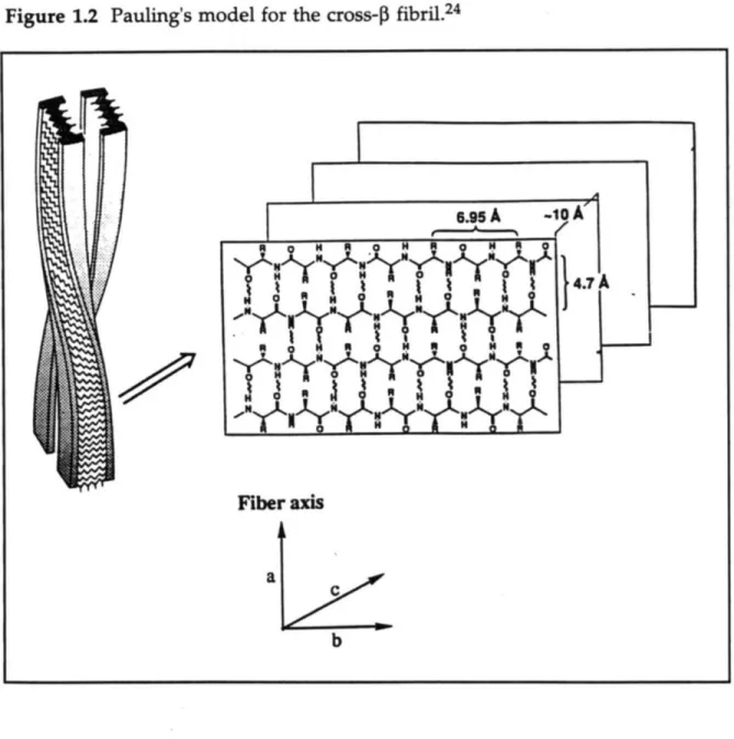
Three properties used to classify a substance as amyloid are birefringent staining with Congo Red (Figure 1.1), fibrillar morphology as seen by electron microscopy (EM), and an X-ray diffraction pattern consistent with a cross-3 fibril. Glenner proposed that the X-ray diffraction pattern of fibrils isolated from the plaques of AD victims were very similar to the cross-0 fibril structure observed by Pauling19in his studies of silk and polyalanine.14
Figure 1.1 Structure of Congo Red.
In the cross-0 fibril model, the protein chains are extended and anti-parallel to the adjacent chains, forming an anti-anti-parallel P-sheet (Figure 1.2). The 3-sheets then stack on top of each other in the fibril. The fibril growth is in the direction of the hydrogen bonding, which is perpendicular to the sheet stacking. Studies by Kurt Halverson and Peter Lansbury showed that the model peptide comprising residues 34-42 of the P protein (334-42) formed fibrils that had a similar structure to the full-length protein, as seen by X-ray diffraction, yet differed slightly in morphology, as seen by EM.20
Since the amyloid fibrils are non-crystalline, structural studies in the past have been limited to low resolution techniques such as EM and X-ray diffraction. Recently, two methods, Fourier-transform infrared spectroscopy (FTIR) and solid-state nuclear magnetic resonance spectroscopy (SSNMR), have been modified and utilized by Peter Lansbury and coworkers in order to create a new method for amyloid structure determination.2 1-23 The method is
based on the spectral deconvolution by selective incorporation of 13C into the
peptide backbone, and has been used to study the structure of 334-42 by measuring distances of up to 6A between twenty pairs of 13C-labeled carbon
Figure 1.2 Pauling's model for the cross-1 fibril.24
The Causative Role of
f
Amyloid in ADSince the original isolation of the B protein from amyloid plaques in 1984, there has been a heated debate over whether the NFTs, the amyloid plaques, or neither is the cause of AD. Some scientists do not believe that amyloid deposition precedes the pathology of AD, but instead argue that senile plaques are produced by the neurites in the plaque periphery as they
Fiber axis
:rýý
degenerate. However, while there is no concrete evidence of the causative role of amyloid in AD, a great deal of research seems to indicate that the amyloid plaques are important in the pathogenesis of AD. Based on the large body of neuropathological, biochemical, molecular biological, and genetic data, the "amyloid-cascade hypothesis" has been made.25 This hypothesis assumes that AD is a disorder of APP processing which may have a number of different causes and that results in the increased production of the P protein. The aggregation of soluble
3
protein into amyloid fibrils then sets off a chain of events that results in the formation of senile plaques which are toxic to the surrounding neurons and induce the formation of NFTs, ultimately leading to cell death.Major support for this hypothesis came from the realization in 1988 that the earliest detectable AD-type structural change in the brains of Down's syndrome (DS) patients, who have three copies of the APP gene and invariably develop AD, is the appearance of diffuse protein deposits.2 5 Diffuse plaques consist of an amorphous, largely non-fibrillar form of the P protein that is hypothesized by some to be an immature form of senile plaques. Additionally, studies of DS patients indicate that NFT's develop after senile plaques have formed.17
The considerable number of genetic causes for AD, which are discussed further below, are associated with a common neuropathological and clinical phenotype. The phenotype is characterized by abundant amyloid plaques in the brain. Most experts agree that amyloid is the cause of at least some forms of AD and that the formation of NFT's follow the formation of the plaques.3
In vitro studies also support the amyloid hypothesis. Aggregation of the soluble 0 protein into amyloid fibrils is required in order for the
3
protein to be toxic to a neuronal cell culture.26 How the fibrillar P protein is toxic tocells in not well understood. However, the accumulation of aggregated 0 protein has been associated with a local inflammatory response resulting from the release of interleukin-10 (IL-10) and other cytokines which then stimulate the production of al-antichymotrypsin.25 This inflammation could
possibly be responsible for augmenting the cytotoxicity of the P protein and the acceleration of APP processing into the 0 protein.
One observation that is often sited to disprove the amyloid-cascade hypothesis is that the total number of amyloid deposits correlates weakly with the presence and degree of dementia. However, a modest correlation does exist, but a better correlate to the degree of dementia in AD is the extent of neuritic degeneration and the degree of loss of synapses.25
Metabolism of APP
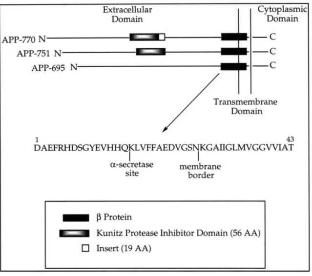
During 1987 and 1988, a number of groups cloned and identified three major forms of APP (695, 751, and 770).27-32 The larger 751 and 770 forms, which are generated by alternate splicing, contain a 56 amino acid domain in the extracellular region that is homologous to the Kunitz family of serine protease inhibitors (KPI) (Figure 1.3). Additionally, the 770 form also contains a 19 amino acid insert. APP is produced in a wide variety of cell types and the relative levels of the APP isoforms vary according to cell type. APP-695 is particularly abundant in the brain and there is evidence that APP-751 is produced in large amounts in brain regions involved in senile plaque formation.3 3
Figure 1.3 Biogenesis of the
P
protein as derived from proteolysis of APP. Cytoplasmic Domain-C
-C
-C
After its synthesis on ribosomes, APP is translocated into the lumen of the endoplasmic reticulum and transported through the Golgi apparatus; some molecules go to the cell-surface. During this process, it undergoes N'-and O'-linked glycosylation, sulfation, N'-and phosphorylation.34 APP is
normally processed in constitutive secretory and putative endosomal-lysosomal pathways. Direct trafficking of [APP from the trans-Golgi network to the endosomal-lysosomal system has been suggested, but has not been proven. Extracellular Domain APP-770 N APP-751 N APP-695 N
ransmembrane
Domain 1 43 DAEFRHDSGYEVHHQKLVFFAEDVGSNKGAIIGLMVGGVVIATI
I
ac-secretase membrane site border [p ProteinI I" Kunitz Protease Inhibitor Domain (56 AA)
O Insert (19 AA)
EN
m
During its intracellular trafficking, APP may undergo one of two alternate cleavages prior to secretion. The first cleavage of APP is within the
0 protein sequence (between Lysl6 and Leul7) by a protease that has been
termed "a-secretase" to produce a large secreted derivative and a small membrane-associated fragment, neither of which contain the entire 0 protein sequence nor is amyloidogenic.3 5 The second "P-secretase" cleavage is at the amino terminus of the 0 protein. Subsequent cleavage by "y-secretase" results in the production of the full-length, amyloidogenic protein. It is believed that most APP molecules are cleaved by a-secretase and that only a small subset of APP is processed by P- and y-secretases to release the 0 protein.25 These
secretases have not yet been identified.
The constitutive secretion of the 0 protein raises the question of how the putative transmembrane domain of APP is cleaved by y-secretase. Additionally, as is evident from the isolation of the
P
protein from plaques present in the brains of AD victims, there is heterogeneity in the y-secretase site resulting in C-terminally truncatedP
proteins ranging from 39 to 43 amino acids in length. There are mutations in APP just outside theP
protein sequence that result in both an altered total amount of P protein as well as altered ratios of the C-terminally truncated P proteins (discussed below).17Cleavage by a-secretase was originally demonstrated in cell-surface APP molecules, but subsequent studies have shown this cleavage, in addition to P-secretase, to also occur during intracellular exocytosis.3 6 P- and y- secretase
cleavages have also been demonstrated during endocytosis, with the resulting release of the 0 protein occurring.37 These studies suggest that constitutive
P
protein production occurs in a weakly acidic vesicle such as the endosome or a late Golgi compartment and is followed by the rapid secretion of the 0 protein.
The biological function of APP has not been determined. The secreted forms that contain the KPI domain might be involved in the regulation of protease activity at the cell surface or in the extracellular environment. APP may also have a role in tissue repair in the brain as it is secreted by blood platelets and has an N-terminal amino acid sequence similar to platelet coagulation Factor Xla-inhibitor which is involved in blood clotting.38 There is evidence that APP promotes cell-cell and cell-extracellular matrix adhesion. APP also promotes survival and growth of neurons in vitro.17
Genetic Research
After the identification of the 3 amyloid protein, research on AD became much more prevalent and scientists began to study with more intensity the contribution of genetics to the development of AD. Presently, data indicates that AD is a heterogeneous disease that exists in two forms. The first is the early-onset familial Alzheimer's disease (FAD), which is hereditary, and the second is the more common late-onset AD. Late-onset AD can be either genetic, as is the case with the inheritance of the APOE4 allele (discussed below), or "sporadic". The term sporadic is used to indicate AD subjects with no family history of AD and does not necessarily imply a nongenetic etiology.
Genetic dissection of late-onset AD is more problematic than early-onset AD primarily because the disease is very common in the elderly, especially in the 75-84 year age group. Therefore, the existence of multiple AD cases in a given family does not necessarily indicate the inheritance of a common genetic factor, and could be the result of chance or could be the result of a combination of both genetic and sporadic forms of AD.
While the vast majority of AD cases are not associated with mutations, a large number of the early-onset AD cases appear to be directly caused by genetic mutations in the APP gene (Figure 1.4). These cases represent some of the strongest evidence supporting a causative role of the
P
amyloid protein in the pathogenesis of at least some forms of AD. There is an extraordinarily high statistical correlation between inheriting one of these rare mutations and developing the disease. These mutations are absent in large numbers of unaffected individuals and have been considered by many to be sufficient to cause the disease. Additionally, the phenotype associated with these mutations is typical of AD both clinically and neuropathologically. It is due to these two facts that the assumption was made that all cases of AD might be the result of amyloid deposition.Figure 1.4 APP mutations associated with early-onset AD.
In some families AD is inherited as an autosomal dominant trait.3 9 The first pathogenic APP mutation was found in a kindred with autosomal dominant hereditary hemorrhage with amyloidosis of the Dutch type (HCHWA-D). In this disease, a mutation in APP results in a Glu to Gln substitution at position 22 of the
3
protein.40 Victims experience strokes afterthe age of 25 with death occurring by the age of 60. In this disease there is ...EVKMDAEFRHDSGYEVHHQKLVFFAEDVGSNKGAIIGLMVGGVVIATvIvIT...
I
II
/I\I
cerebral amyloid angiopathy, but few AD-type amyloid plaques or NFTs are found on autopsy. The identification of the HCHWA-D mutation was the first demonstration that a APP mutation could result in amyloid deposition and neurologic disease.5
Another pathogenic APP mutation was identified in a Dutch family with both cerebral hemorrhage disease and dementia.4 1 The disease in this family is a variant of both HCHWA-D disease and AD. There is a mutation in this disease at codon 692 in APP that results in an Ala to Gly mutation at position 21 of the P protein. An exhaustive screening of early-onset FAD victims has proven these mutations to be very rare because they have never been seen in controls and they are clearly pathogenic mutations.5
Autosomal dominant APP mutations found in a Swedish familial Alzheimer's disease (FAD) pedigree, in which Val is replaced by either Ile, Phe, or Gly at position 717 in APP770, cause a marked increase in the production of a P protein-bearing, carboxyl terminal, APP derivative. The mutations, which were studied in human neuroblastoma cells (M17) transfected with constructs expressing the wild-type or mutant APP, resulted in five times more P protein in the mutant cells than in the cells expressing wild-type APP. The mutant cell line also released six times more AP into the medium. 39 A fourth double mutation (K670N and M671L in APP770) produced a normal total amount of the P protein, but a greater proportion of the variant I1-42.42 This longer variant has previously been shown to form amyloid more rapidly than the shorter P protein variant, P1-40, in vitro.4 3
The clinical, neuropathologic, and genetic characteristics of AD observed in subjects with APP mutations have several features in common.5 The first is that the age of onset is early, anywhere from 41-64 years of age, depending on the specific mutation. The second is that inheritance is
dominant with complete penetration by the early sixties. No APP mutation carrier living past the age of 67 has been reported.5 This indicates that in every type of FAD caused by mutations in the APP gene, the genetic defects are sufficient, but not necessary, for causing AD. Lastly, the clinical and neuropathological features of AD caused by genetic mutations are indistinguishable from those of non-APP mutation AD subjects.
A large step forward in AD research was made with the creation of the first real animal model in 1995, the Athena mouse.44 This transgenic animal
has marked overexpression of an FAD-linked mutant APP gene and forms AD-like plaques.10 However, while the mouse possesses pathology
characteristic of AD (without NFTs), clinical symptoms have yet to be observed.
As further evidence indicating the importance of amyloid in AD, Down's syndrome (DS) patients are trisomic for chromosome 21 and therefore have three copies of the APP gene. DS victims show signs of amyloid formation in childhood and always develop early-onset AD.45 The
increase in the amount of the
P3
protein produced by DS patients appears to be responsible for the onset of the disease.A mutation on chromosome 14, which encodes a recently discovered membrane-associated protein, has been linked to a very early and severe form of FAD. Victims start showing signs of the illness in their early 40's.46 S182 (or presenilin), the protein encoded by the isolated gene on chromosome 14, was found to have five different missense mutations that altered the predicted amino-acid sequence and occurred only in AD patients. The primary structure of the longest open reading frame currently identified predicts a 467 amino-acid protein with seven hydrophobic, putative membrane-spanning domains. The function of S182 may be as a receptor,
channel protein, or structural membrane protein. Mutations in S182 that alter its hypothetical role in the Golgi or membrane-trafficking might enhance
P
protein production.47 It appears that carriers of the chromosome14 mutation do actually have an increased P protein secretion.48 Another mutation on a gene located on chromosome 1 has also been identified.49,50 The product of this gene is a protein with homology to the protein mapped to
chromosome 14.
Finally, it was shown in 1993 that the APOE gene on chromosome 19 is important in AD. Despite the difficulties in determining genetic causes for late-onset AD, the APOE gene has been shown to be a late-onset AD risk factor.51 Scientists determined that people carrying the APOE4 allele, which encodes one of the apoE isoforms, had a 15-fold increase in the risk for developing late-onset AD. Three APOE alleles exist that encode proteins differing from each other at positions 112 and 158: apoE3 (Cys-112, Arg-158), apoE4 (Arg-112, Cys-112), and apoE2 (Cys-112, Cys-158).
A large number of genetic studies have confirmed that the inheritance of one or two APOE4 alleles significantly increases the likelihood of developing AD with the risk increasing with an increasing number of APOE4 alleles.5 1 Additionally, the age of onset of AD is decreased and the number of
amyloid plaques is increased with increasing number of APOE4 alleles. The exact mechanism of action of apoE has not yet been determined.
In one study of familial, late-onset AD, approximately 90% of APOE4 homozygotes were found to have developed the disease by 76 years of age.5 1
While this leads to the conclusion that APOE4 homozygotes will almost inevitably develop AD if they live long enough, it should be noted that the possession of one or two copies of the APOE4 allele is neither necessary nor sufficient for the development of AD.52 Unlike carriers of APP mutations
who have a 100% chance of developing AD, APOE4 heterozygotes and homozygotes may live long into their eighties, nineties, or possibly to over
one hundred years of age without developing AD.
All of the evidence leads to the difficult question: How can all of the biochemical and cellular abnormalities be reconciled into a mechanism that will help to explain the pathogenesis of AD? In addition, while most of the FAD types are linked to genetic mutations, the majority of all AD victims are sporadic late-onset cases with no known genetic defects.
Therapeutic Options
Currently there is no proven therapy for victims of AD and research into possible therapeutic options has been thwarted by the complex nature of the disease. The pathology, which shows on a gross level the destructive nature of AD, is only the end-point of a lengthy disease process and is far removed from the initiating events of the disease. In addition, although the pathology of most AD victims is similar, the causes of AD appear to be heterogeneous. As previously mentioned, there are mutations on chromosomes 21 and 14 that are responsible for some forms of FAD. For late-onset AD, the APOE4 allele is a risk factor, but its presence does not guarantee the onset of AD. The majority of all AD patients (50-60%) do not meet any of the above criteria indicating that other loci contributing to AD remain to be detected and mapped, or that environmental factors, such as the presence of metals, are important.5 Even after all of the genes are located, substantial effort will be required to understand how each susceptibility factor is involved in AD pathogenesis.
The most ambitious efforts aim to identify probable causes and correct them in an early stage. There are approximately 20 companies developing
AD drugs; the majority have focused on finding a way to prevent amyloid
buildup, thereby preventing the extracellular plaques from forming. Scientists hope to use the Athena mouse to test competence of new drug candidates. Characterization of the a-, P-, and y-secretases involved in APP processing is also an important component of research aimed at the rational design of anti-AD drugs.
While several companies are screening small-molecule drugs that either facilitate clearance of p amyloid or prevent it from aggregating in the first place, others are looking for a drugs that delay disease onset by mimicking the proteins that protect neurons or that minimize the secondary damage by scavenging toxic free radicals.7
Once again, these drug candidates address biological mechanisms that are not well understood. Additionally, they are directed at patients in the early stages of disease which is problematic because without any reliable diagnostic test, no one is really sure who is at risk until it is too late. Other companies are looking for drugs that treat the symptoms of AD and are studying genetically engineered cell implants that will deliver nerve-growth factors to areas of the brain that have been damaged by the disease. Drugs that attempt to boost memory and cognition are also being developed.7
AD is a complex and seemingly heterogeneous disorder that can strike its victims in the prime of their lives. Victims of this destructive disease may deteriorate into an increasingly dependent state for as long as 20 years until death. AD is devastating not only to the victims, but also to the families who are often the prime care-givers. The social and economic implications of an
ever-growing elderly population, which is expected to represent one in four people in the U.S. within the next 40 years, are frightening.
Because of the increasing importance of AD, scientists have worked intensely in the hope of developing new therapeutics for the disease. However, this process has been thwarted by the inability of these scientists to not only explain the complicated mechanisms involved in the pathogenesis
of the disease, but to also diagnose AD in its initial stages.
This thesis describes in vitro experiments that were designed to elucidate critical factors in the pathogenesis of AD. Initially, the conformation of the P protein and the effect of mutations on its metal-binding properties was studied (chapter 2). The effect of apolipoprotein E (apoE), a risk factor for late-onset AD, on the aggregation of the 0 protein was also examined (chapter 3). Finally, through the use of molecular biology, several isoforms of a domain of apoE that is critical to its interaction with P protein aggregation were expressed and studied (chapter 4).
References
(1) Alzheimer, A. Allg Z Psychiatrie 1907, 64, 146. (2) Ezzell, C. J. of NIH Res. 1995, 7, 107.
(3) Hardy, J. Nature Genetics 1992, 1, 233.
(4) Rubin, E.H. Advances in Neurology: Alzheimer's Disease; 1990, Raven
Press, LTD, New York, NY.
(5) Schellenberg, G.D. Proc. Natl. Acad. Sci. USA 1995, 92, 8552-8559.
(6) Gregg, D. A Special Report: Alzheimer's Disease; 1994, Harvard Medical School Health Publications Group, Boston, MA.
(7) Gross, N.; Flynn, J. Business Week 1995, 108.
(8) Caring for the Elderly: Reshaping Health Policy, Eisdorfer, C.; Kessler, D.A.; Spector, A.N., Eds.; The John Hopkins University Press:
Baltimore, MD, 1989.
(9) Selkoe, D. Scientific American 1991, 68-78, (10) Martin, E.M. Neurology 1987, 37, 1201. (11) Katzman, R. NEJM 1986, 314, 146.
(12) Khachaturian, Z.S. Arch. Neurol. 1985, 42, 1097-1105. (13) Stone, M.J. J. Am. Soc. Hemat. 1990, 75, 531-545.
(14) Glenner, G.G.; Wong, C.W. Biochem. Biophys. Res. Commun. 1984, 120, 885.
(15) Masters, C.; Simms, G.; Weinman, N.; Multhaup, G.; McDonald, B.; Beyreuther, K. Proc. Natl. Acad. Sci. USA 1985, 82, 4245.
(16) Seubert, P.; Vigo-Pelfrey, C.; Esch, F.; Lee, M.; Dovey, H.; Dovey, H.; Davis, D.; Sinha, S.; Schlossmacher, M.; Whaley, J.; Swidlehurst, C.; McCormick, R.; Wolfert, R.; Selkoe, D.; Lieberburg, I.; Schenk, D. Nature 1992, 359, 325.
(17) Ashall, F.; Goate, A.M. TIBS 1994, 19, 42-46.
(18) Goedert, M.; Sisodia, S.S.; Price, D.L. Curr. Opin. Neurobiol. 1991, 1, 441-447.
(19) Marsh, R.E.; Corey, R.B.; Pauling, L. Acta. Cryst. 1955, 8, 710-715. (20) Halverson, K.; Fraser, P.E.; Kirschner, D.A.; Lansbury, P.T., Jr.
Biochemistry 1990, 29, 2639-2644.
(21) Halverson, K.H.; Sucholeiki, I.; Ashburn, T.T.; Lansbury, P.T., Jr. J. Am. Chem. Soc. 1991, 113, 6701-6703.
(22) Ashburn, T.T.; Auger, M.; Lansbury, P.T., Jr. J. Am. Chem. Soc. 1992, 114, 790.
(23) Spencer, R.G.S.; Halverson, K.J.; Auger, M.; McDermott, A.E.; Griffin, R.G.; Lansbury, P.T., Jr. Biochemistry 1991, 30, 10382-10387.
(24) Halverson, K.J., The Molecular Determinants of Amyloid Deposition in Alzheimer's Disease, Ph.D. Thesis, MIT, 1992.
(25) Selkoe, D.J. J. NIH Res. 1995, 7, 57-64.
(27) Kang, J.; Lemaire, H.G.; Unterbeck, A.; Salbaum, J.M.; Masters, C.L.; Grzeschik, K.H.; Multhaup, G.; Beyreuther, K.; Muller-Hill, B. Nature
1987, 325, 733-736.
(28) Goldgaber, D.; Lerman, M.I.; McBride, O.W.; Saffiotti, U.; Gajdusek, D.C. Science 1987, 235, 877-880.
(29) Tanzi, R.E.; McClatchey, A.I.; Lamperti, E.D.; Villa-Komaroff, L.; Gusella, J.F.; Neve, R.L. Nature 1988, 331, 528-530.
(30) Robakis, N.K.; Ramakrishna, N.; Wolfe, G.; Wisniewski, H.M. Proc. Natl. Acad. Sci. USA 1987, 84, 4190-4194.
(31) Ponte, P.; Gonzalez-DeWhitt, P.; Schilling, J.; Miller, J.; Hsu, D.;
Greenberg, B.; Davis, K.; Wallace, W.; Lieberburg, I.; Fuller, F. Nature 1988, 331, 525-527.
(32) Tanzi, R.E.; Gusella, J.F.; Watkins, P.C.; Bruns, G.A.; St. George-Hyslop, P.; Van Keuren, M.L.; Patterson, D.; Pagan, S.; Kurnit, D.M.; Neve, R.L. Science 1987, 235, 880-884.
(33) Anderson, J.P. EMBO J. 1989, 8, 3627-3632.
(34) Weidemann, A.; Konig, G.; Bunke, D.; Fischer, P.; Salbaum, J.M.; Masters, C.L. et al. Cell 1989, 57, 115.
(35) Esch, F.S.; Keim, P.S.; Beattie, E.C.; Blacher, R.W.; Culwell, T. et al. Science 1990, 248, 1122.
(36) Sambamurti, K.; Shioi, J.; Anderson, J.P.; Pappolla, M.A.; Bobakis, N.K. J. Neurosci. Res. 1992, 33, 319.
(37) Koo, E.H.; Squazzo, S. J. Mol. Biol. 1994, 269, 17386.
(38) Smith, R.P.; Higuchi, D.A.; Broze, G.J. Science 1990, 248, 1126-1128. (39) Cai, X.D.; Golde, T.E.; Younkin, S.G. Science 1993, 259, 514.
(40) Van Broeckhoven, C.; Haan, J.; Bakker, E.; Hardy, J.A.; Van Hul, W.; Wehnert, A.; Vegter-Van Der Vlis, M.; Roos, R.A.C. Science 1990, 248, 1120-1122.
(41) Hendriks, L.; Van Duijn, C.M.; Cras, P.; Cruts, M.; Van Hul, W.; Van Harskamp, F.; Warren, A.; McInnis, M.G.; Antonarakis, S.E.; Martin,
J.J.; Hofman, A.; Van Broeckhoven, C. Nature Genetics 1992, 1, 218-221.
(42) Suzuki, N. et al. Science 1993, 264, 1336-40.
(43) Jarrett, J.T.; Berger, E.P.; Lansbury, P.T., Jr. Biochemistry 1993, 32, 4693-4697.
(44) Games, D.; Adams, D.; Alessandrini, R.; Barbour, R.; Berthelette, P.; Blackwell, C.; Carr, T.; Clemens, J.; Donaldson, T. et al. Nature 1995, 373, 523.
(45) Potter, H. Am. J. Hum. Genet. 1991, 48, 1192.
(46) Sherrington, R.; Rogaev, E.I.; Liang, Y.; Rogaeva, E.A.; Levesque, G.; Ikeda, M.; Chi, H.; Lin, C.; Li, G.; Holman, K.; Tsuda, T.; Mar, L. et al. Nature 1995, 375, 754.
(47) Selkoe, D.J. Nature 1995, 375, 734-735.
(48) Querfurth, H.W.; Wijsman, E.M.; St. George-Hyslop, P.H.; Selkoe, D.J. Mol. Brain Res. 1995, 28, 319-337.
(49) Levy-Lahad, E.; Wijsman, E.M.; Nemens, E.; Anderson, L.; Goddard, K.A.B.; Weber, J.L.; Bird, T.D.; Schellenberg, G.D. Science 1995, 269, 970-973.
(50) Rogaev, E.I.; Sherrington, R.; Rogaeva, E.A.; Levesque, G.; Ikeda, M. et al. Nature 1995, 376, 775-778.
(51) Corder, E.H.; Saunders, A.M.; Strittmatter, W.J.; Schemchel, D.E.; Gaskell, P.C.; Small, G.W.; Roses, A.D.; Haines, J.L.; Pericak-Vance,
M.A. Science 1993, 261, 921.
Chapter 2
Metal-Binding Studies of the
P
Protein
The presence of insoluble protein deposits in the form of senile plaques surrounded by damaged tissue is an invariant feature of AD. In addition to genetic studies that implicate the 0 amyloid precursor protein (APP) in the pathogenesis of AD,1,2 other studies have shown that in its insoluble fibrillar
form the 0 protein is toxic to cultured neurons.3 ,4 Families where AD cosegregates with mutations in the gene on chromosome 21 encoding APP have been identified. The presence of amyloid is also a central pathological event in other diseases such as hereditary cerebral hemorrhage with amyloidosis-Dutch type (HCHWA-D) where a mutation in the
P
protein sequence is responsible for the fatal deposition of amyloid.5 Furthermore,other higher mammals that encode for the same
3
protein develop amyloid deposits while rodents that encode for a P protein containing three amino acid substitutions do not form amyloid plaques.In the absence of any significant quantity of brain derived material, work with synthetically derived material has proven to be necessary and important in studying the effect that a genetic mutation might have on the processing or solubility of the precursor and the resulting fragments. A
mutation may result in a change in the proteolysis of the precursor which could either liberate an insoluble fragment of an otherwise soluble precursor, or cause an increase in the production of an amyloidogenic protein. A mutation might also induce a conformational change that results in the aggregation of that protein.
This chapter describes studies of several model peptides that examine the effect of point mutations and the presence of metals on the secondary structure of the
3
protein. Metal-binding studies were performed on several peptides derived from the P protein and3
protein analogs using circular dichroism (CD) in order to provide information that will help to determine the mechanism of amyloid formation.Circular Dichroism
Circular Dichroism (CD) spectroscopy is a technique commonly used to study the secondary structure of polypeptides and proteins in solution. CD has become a standard tool of many protein chemists. CD spectroscopy uses chirally polarized light comprised of left- and right-circularly polarized components. The CD signal arises from the differential absorption of these components in optically active molecules, and is reported in terms of ellipticity (0) with a unit of degrees. The CD signal of an optically active molecule most often occurs in the same region of the spectrum that it would
normally absorb in standard UV-VIS spectroscopy.
In proteins and polypeptides, amide bonds, aromatic side chains, and disulfide bonds are all optically active. However, the signals from side chains and disulfide bonds are much smaller than that from the backbone amide bond.6 The CD spectrum of proteins is primarily a result of the amide
chromophore, the most informative functionality to study because it is very sensitive to changes in the secondary structure of the polypeptide backbone. The environment in which a protein is studied (i.e. pH, solvent, temperature) can have a large effect on the secondary structure of the protein.
The four major categories of secondary structure, a-helix, sheet, P-turn, and random coil, each produce a characteristic CD spectrum. In an a-helix, the carbonyls and amide groups align such that a macroscopic dipole occurs which results in a strong positive signal at 192 nm and two negative signals at 208 nm and 222 nm.7 The spectrum for a P-pleated sheet shows a maximum near 198 nm and a minimum near 217 nm. However, these transitions may shift depending on the type of 1-sheet present and its degree of twist.7
CD data can be analyzed by a large number of techniques. The method of Greenfield and Fasman is based on the spectra of polylysine and polyglutamate under conditions where "pure" a-helical conformations were obtained. The absorbance at 208 nm is used to calculate the amount of a-helix present.7 In the method of Morrisett, which is based on the work of Greenfield and Fasman, the amount of a-helicity is calculated by using the 222 nm absorption.8 It has been suggested that using the absorption at 208 nm
results in a much more accurate calculation of a-helix in the presence of P-sheet content because there is little contribution from other structures at this wavelength.9 The percent of a-helical content can be calculated by the
following equations according to the methods of Greenfield and Fasman (Equation 2.1) and Morrisett (Equation 2.2):
Equation 2.1 a-helix = ([01208 - 4,000)
(33,000 -4,000)
Equation 2.2 a-helix = ([01222 + 3,000)
(36,000 + 3,000)
Many computer programs have been written that will calculate all of the secondary structural components in a given protein based on its CD spectrum. One of these programs is PROSEC which calculates the component contributions based on the CD spectra derived from crystalline proteins of known secondary structures.10 Other programs are based on the CD spectra of
unnatural polypeptides.
Data obtained from CD spectra can be extremely useful in examining the secondary structure of proteins. However, there are several potential sources of inaccuracy that result from improper interpretation of the data. Fitting programs can lead to false component calculations. Programs based on the structures of proteins may be inadequate for small polypeptides. Conversely, programs based on "pure" peptide structures can be inadequate for studying protein structure because the long peptide homopolymers may not be representative of those structures found in large proteins. The signal produced by an a-helix is proportional to the chain length and there are few examples of homopolymers that depict pure 13-turn structures.1 1
Additionally, it has been shown that unless the CD spectral data extends down to 184 nm, only the a-helical component is accurate.12