HAL Id: tel-00945688
https://tel.archives-ouvertes.fr/tel-00945688
Submitted on 12 Feb 2014
HAL is a multi-disciplinary open access archive for the deposit and dissemination of sci-entific research documents, whether they are pub-lished or not. The documents may come from teaching and research institutions in France or abroad, or from public or private research centers.
L’archive ouverte pluridisciplinaire HAL, est destinée au dépôt et à la diffusion de documents scientifiques de niveau recherche, publiés ou non, émanant des établissements d’enseignement et de recherche français ou étrangers, des laboratoires publics ou privés.
La bioénergétique systémique moléculaire des cellules
cardiaques : la relation structure-fonction dans la
régulation du métabolisme énergétique
compartmentalisé
Marcela Alejandra Gonzalez Granillo
To cite this version:
Marcela Alejandra Gonzalez Granillo. La bioénergétique systémique moléculaire des cellules car-diaques : la relation structure-fonction dans la régulation du métabolisme énergétique compartmen-talisé. Sciences agricoles. Université de Grenoble, 2012. Français. �NNT : 2012GRENV078�. �tel-00945688�
THÈSE
Pour obtenir le grade de
DOCTEUR DE L’UNIVERSITÉ DE GRENOBLE
Spécialité : Physiologie Physiopathologies et Pharmacologie
Arrêté ministériel : 7 août 2006
Présentée par
« Marcela Alejandra GONZÁLEZ GRANILLO »
Thèse dirigée par « Professeur Valdur SAKS » et codirigée par « Docteur Yves USSON »
préparée au sein du Laboratoire de Bioénergétique Fondamentale et Appliquée - INSERM U1055, UJF dans l'École Doctorale Chimie et Science du Vivant
La bioénergétique systémique
moléculaire des cellules
cardiaques : la relation
structure-fonction dans la régulation du
métabolisme énergétique
compartmentalisé.
Thèse soutenue publiquement le « 28 septembre 2012 », devant le jury composé de :
M. Pierre DOS SANTOS
Professeur, Directeur du GIS CHU - UBxS, Rapporteur.
M. Yves TOURNEUR
CR1, Directeur d’INSERM U1060/UNI.LYON1/INRA135, Rapporteur.
M. François BOUCHER
Professeur, Directeur du TIMC-IMAG UM 5525, Président.
Mme. Anne CANTEREAU
Docteur, Ingénieur de recherche CNRS, Plateforme ImageUP, Membre.
M. Andrey KUZNETZOV
Docteur, D. Swaroski research laboratory IMU, Membre.
M. Uwe SCHLATTNER
DOCTORAL THESIS
To obtain the grade ofDOCTOR OF PHILOSOPHY AT THE UNIVESITY OF
GRENOBLE
Specialty: Physiology, Physiopathology and Pharmacology
Arrêté ministériel : 7 août 2006
Presented by
« Marcela Alejandra GONZÁLEZ GRANILLO »
Supervised by « Professor Valdur SAKS » and Co-supervised by « Doctor Yves USSON »
prepared at the Laboratory of Fundamental and Applied Bioenergetics (LBFA) - INSERM U1055, UJF
at the doctoral school of chemistry and life sciences.
Molecular system bioenergetics of
cardiac muscle cells:
structure-function relationship in regulation
of compartmentalized energy
metabolism.
Thesis defended in public on « September 28th, 2012 », in front of the thesis jury. Composition of the jury:
Professor Pierre DOS SANTOS
Director of the GIS CHU - UBxS, Reviewer.
Professor Yves TOURNEUR
Director of the INSERM U1060/UNI.LYON1/ INRA135, Reviewer.
Professor François BOUCHER
Director du TIMC-IMAG UM 5525, President.
Anne CANTEREAU, PhD
Engineer of research at the CNRS, Platform ImageUP, Examiner.
Andrey KUZNETZOV, PhD
D. Swaroski research laboratory IMU, Examiner.
Professor Uwe SCHLATTNER
TABLE OF CONTENTS I. INTRODUCTION……….………1 ‐ 47 1. Philosophical background……….….1 ‐ 4 1.1. Systems Biology: general historical overview………..……1 2. Principles of bioenergetics……….4 ‐ 14 2.1. Molecular System Bioenergetics………..….4 2.2. Energy metabolism in muscle cells………7 2.3. Electron transport chain (ETC)………....9 2.4. ATP synthase………..………..12 2.5. Adenine nucleotide transporter………..………14 3. Muscle cell and functioning………..……….15 ‐ 19 3.1. Contractile module……….………15 3.2. Excitation‐contraction coupling………..17 4. Integrated energy metabolism……….20 ‐ 21 4.1. Intracellular diffusion and compartmentation phenomenon……….20 5. Organization of mitochondria in cardiac cells………21 ‐ 31 5.1. Mitochondrial functioning in cardiac myocytes………..21 5.2. Mitochondria and cytoskeletal interactions………..27 5.3. Tubulins………28 6. Regulation of respiration in heart mitochondria……….31 ‐ 38 6.1. Theories of regulation of mitochondrial respiration……….….31 6.2. The Frank‐Starling law………...32 6.3. The intracellular phosphotransfer network………..……….33 7. Mitochondrial dynamics………...38 ‐ 49 7.1. Fusion and fission……….…….38 7.2. Mitochondria dynamics in different cardiac muscle cell phenotypes.…….43 7.3. Do fusion‐fission events occur in all cells?...47 II. STUDY LIMITATIONS……….……...50 ‐ 51 III. AIM OF THE STUDY……….…………...…….52 IV. METHODS……….……….53 ‐ 77 1. Animals………..…..53 1.1. Cardiomyocytes isolation………....…54 1.2. Dilaceration of adult left‐ventricle fibers………..57 2. Oxygraphy………55 ‐ 56 2.1. Measurements of oxygen consumption……….…..55 2.1.a. Integrity of the outer membrane………..…56 2.1.b. PK‐PEP trapping system………56 2.1.c. Regulation of mitochondrial respiration by creatine in the presence of activated MtCK………...56 2.1.d. Regulation of respiration by exogenous ADP………56 3. Microscopy techniques……….……….57 ‐ 59 3.1. Immunofluorescence………..……57 3.2. Transfection………..…58 3.3. Confocal imaging………...59
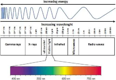
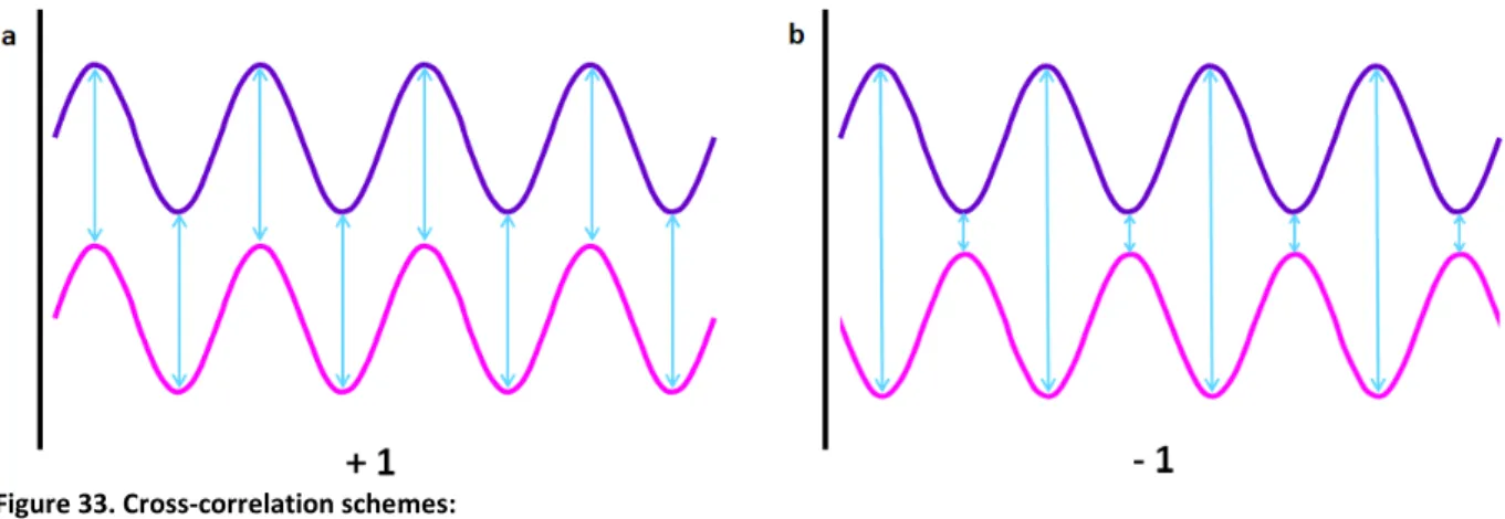
4. Principles of fluorescence imaging………...60 ‐ 63 4.a. Electromagnetic spectrum of radiation (ESR)……….………60 4.b. Fluorescence………..61 4.c. NADH and flavoproteins autofluorescence………..63 5. Principle of confocal microscopy……….65 ‐ 69 5.1. Confocal laser‐scanning microscope (CLSM)……….……..65 5.1.a. Differential interference contrast (DIC)……….…..68 5.1.b. Deconvolution……….68 5.1.c. Two photon microscopy………...68 5.2. Dynascope prototype………..……..69 6. Principles for the dynamics analysis………....69 ‐ 76 6.1. Fourier transform………..……71 6.2. Cross‐correlation………..….72 6.3. Image processing………..….73 6.3.a. Gradient clustering algorithm………..……73 6.3.b. Quantification of mitochondrial fluctuations……….……..74 V. RESULTS………78 ‐ 109 1. Discovery of specific β‐tubulin isotypes in adult rat cardiac cells………..….…78
2. Possible role of structural proteins (tubulin beta II) in the regulation of respiration………..…..86 3. Functional roles of structural proteins in the energy fluxes………..91 4. Fusion‐fission processes in differentiated adult rat cardiac cells………..….96 VI. DISCUSSION………..……….111 ‐ 121 VII. CONCLUSIONS……….……...….122 VIII. FUTURE PERSPECTIVES………..………..….123 IX. REFERENCES……….…………....124 ‐ 132 X. ANNEXES……….……….133 ‐ 144 XI. ACKNOWLEDGMENTS……….…..……145‐146 XII. LIST OF PUBLICATIONS……….………147
FIGURE INDEX Figure 1. Reductionist causal chain 2 Figure 2. Downward causation 2 Figure 3. Hegelian logic of reflection and an approach of the biological sciences 4 Figure 4. General scheme of cellular metabolism 6 Figure 5. Mitochondrion 8 Figure 6. Oxidative phosphorylation in cardiac cells 10 Figure 7. Mitochondrial ATP synthase 12 Figure 8. Mechanism of ATP synthase 13 Figure 9. Electron microscopy of the myofibril organization 16 Figure 10. Major components of the thin and thick filaments in the myofibril 16 Figure 11. Mechanism of the cross‐bridge cycle 17‐18 Figure 12. Intracellular energy unit 22 Figure 13. Mitochondrial creatine kinase functioning for high‐energy metabolite channeling 25 Figure 14. Possible regulation of VDAC by tubulin 26 Figure 15. Tubulin dimer with C‐terminal tails 29 Figure 16. Tubulin portioning between dimmers and polymers 29 Figure 17. Proposed functional arrangement of the intracellular phosphotransfer network 36 Figure 18. Proteins involved in fusion and fission 41 Figure 19. Representation of the three‐dimensional organization of the sarcoplasmic reticulum (SR) as it is seen in a heart muscle fiber 44 Figure 20. Distribution of mitochondria in adult and neonatal rat cardiomyocytes 45 Figure 21. Mitochondrial distribution in NB HL‐1 cells 46 Figure 22. pEGFP‐N1 alpha actinin map vector 59 Figure 23. Electromagnetic spectrum of radiation 60 Figure 24. Newton´s color circle 61 Figure 25. Jablonski energy diagrams 62 Figure 26. Excitation and emission spectral profiles 63 Figure 27. Confocal point sensor principle from Minsky’s patent 65 Figure 28. Mechanics of confocal microscopy 67 Figure 29. Plot of a) fluorescence intensity versus time and b) fluorescence compensation plot 70 Figure 30. Alignment of global motion 72 Figure 31. Plot of one dimensional Fourier transform a) of time signal and b) one spectrum of a square signal 73 Figure 32. Plot of the Fourier expansion of sine waves 73 Figure 33. Cross‐correlation schemes 74 Figure 34. 3‐D diagram of clustering technique in two mitochondria 75 Figure 35. Principle of analysis of velocities if mitochondrial movements in cells in situ 76 Figure 36. Example plots of the mean square distance vs time 77 Figure 37. Fluorescent microscopy imaging of mitochondria fluoroprobes 79 Figure 38. Immunofluorescent confocal microscopy imaging of β‐tubulin isotypes in fixed cardiomyocytes 80 Figure 39. Immunofluorescent confocal microscopy imaging of β‐tubulin isotypes in fixed HL‐1 cells 81 Figure 40. Immunofluorescent confocal microscopy imaging of α‐actinin and βII‐tubulin proteins, and fluorescent staining and autofluorescence of mitochondria fixed cardiac cells 82 Figure 41. Co‐immunofluorescent of α‐actinin and βIII‐tubulin in fixed cardiomyocytes 83
Figure 42. Co‐immunostaining of α‐actinin and βIII‐tubulin in isolated rat heart mitochondria 84 Figure 43. Co‐immunofluorescent labeling of βII‐tubulin and VDAC in fixed HL‐1 cells 84 Figure 44. Western blots 85 Figure 45. Creatine effect on the respiration of permeabilized cardiomyocytes and NB HL‐ 1 cells 86 Figure 46. Specificity test for immunofluorescence Cy5‐conjugated labeling 86 Figure 47. Immunofluorescence labeling of βII‐tubulin with IgG‐FITC 87 Figure 48. Immunofluorescence labeling of βII‐tubulin with Cy5‐conjugated AffiniPure 88 Figure 49. Immunofluorescence kabeling of βII‐tubulin with Cy5‐conjugated AffiniPure after a short proteolysis treatment (trypsin 0.5 μM) of permeabilized cardiomyocytes 88 Figure 50. Comparison of the intracellular distribution of βII‐tubulin, α‐actinin, and mitochondria 90 Figure 51. Effect of trypsin on apparent Km for ADP of mitochondrial respiration in isolated permeabilized cardiomyocytes 91 Figure 52. Respiration activated by 2mM exogenous ADP, Cyt c test to show intactness of MOM, and ATR test to show the total control of ANT on respiration 92 ‐ 93 Figure 53. Respiration of permeabilized cardiomyocytes activated by MgATP (2mM) and creatine 94 Figure 54. Respiration of permeabilized cardiomyocytes activated by MgATP (2mM) and creatine after a short proteolysis 94 Figure 55. Respiration of about 50% of rod‐like intact cells activated by MGATP (2mM) and creatine 95 Figure 56. Regulation of mitochondrial respiration by creatine in the presence of activated MtCK in permeabilized cardiomyocytes, with and without short proteolysis 95 ‐ 96 Figure 57. Example of six different fixed cardiomyocytes 98 Figure 58. Analysis of intensity profiles in fixed adult rat cardiac cells: Plots, Fourier transform and Covariance analyzes of intensity profiles 100 ‐ 102 Figure 59. Image of three dimensional structure of a cardiac fiber 103 Figure 60. Analysis of intensity profiles, Fourier transforms of intensity profiles and Covariance analyzes in fixed adult rat cardiac fiber 106 ‐ 107 Figure 61. Example of a transfected cardiomyocytes with pEGFP‐N1 α‐actinin and MitoTracker® red FM 108 Figure 62. Example of the clustering technique applied in isolated cardiomyocytes transfected with pEGFP‐N1 α‐actinin and labeled with MitoTracker® red FM 108 Figure 63. Plots of intensity profiles, Fourier transforms of intensity profiles and Covariance analyzes of intensity profiles in cultured transfected adult rat cardiac cells 110
TABLE INDEX Table 1 Sequence of rodent tubulin isotypes 29 1. Functional test, oxygraphy 133 ‐ 134 a. Integrity of the outer membrane 133 b.1. PK+PEP trapping system 133 , 134 c. ADP kinetics 134 d. ADP kinetics + creatine 134 2. Immunofluorescence, confocal imaging 135 ‐ 136 a. 1st antibodies 135 , 136 b. 2nd antibodies 135 c. Mitochondrial membrane potential dependent dyers 135 3. Buffers, solutions and media 135 ‐ 144 a. Isolation buffer 135 , 136 b. Digestion solution 136 c. Sedimentation buffers 136 ‐137 c.1. Sedimentation buffer for oxygraphy 136 , 137 c.2. Ca2+ gradient buffers for culturing 137 c.3. Ca2+ stock solution 137 d. Culturing media 137 d.1. Recovery medium 137 d.2. Transfection medium 137 , 138 d.3. Mitomed without Ca2+ solution 138 d.3.1. K‐Lactobionate solution 138 d.3.2. BSA 20% 139 d.3.3. Saponin 139 d.4. Solution A for fibers 139 d.4.1. CaK2EGTA 139 , 140 d.4.2. K2EGTA 140 e. Stock substrate solutions for oxygraphy 140 ‐ 141 e.1. Creatine 140 e.2. Glutamate 140 e.3. Malate 140 ‐ 141 e.4. Cytochrome C 141 e.5. Atractyloside 141 e.6. Sodium pyruvate 141 e.7. Phosphoenolpyruvate (PEP) 141 e.8. NADH 141 e.9. Pyruvate kinase (PK) 141 e.10. Lactate dehydrogenase (LDH) 142 e.11. ATP‐Na2 142 e.12. ADP 142 e.13. 2% BSA solution 142 f. Fixation solutions 142 ‐ 143 f.1. PFA 4% solution 142 f.2. Glutaraldehyde 25% 143 f.3. PFA 4% + Glutaraldehyde 0.1% solution 143 f.4. 1% Triton 143 Table 2 Key points for high quality cardiomyocyte isolation 144
ABBREVIATIONS AK Adenylate kinase α‐KG α‐ketoglutarate KDH α‐ketoglutarate dehydrogenase AMP Adenosine monophosphate ANC Adenine nucleotide carrier ANT Adenine nucleotide translocase β‐FAO β‐fatty acids oxidation BCK Brain creatine kinase Ca2+ Calcium CTT Carboxyl‐terminal tail CoQ Coenzyme‐Q Cr Creatine CK Creatine kinase Cyt c Cytochrome c Drp1 Dynamin‐related protein 1 Δψ Electrochemical gradient ΔμH+ Electrochemical potential of protons ETF Electron‐transferring flavoprotein ETC Electron transport chain G Energy S Entropy H Enthalpy DNA Deoxyribonucleic acid FAD Flavin adenine dinucleotide FMN Flavinmononucleotide FFA Free fatty acids G6P Glucose‐6‐phosphate GLUT‐4 Glucose transporter‐4 GTP Guanosine triphosphate GAPDH Glycealdehyde‐3‐phosphate dehydrogenase HK Hexokinase IMAC Inner‐membrane anion channel Pι Inorganic phosphate IMS Intermembrane space ICEU Intracellular energetic units KHE K+/H+ exchanger MDH Malate dehydrogenase
mitoKATP Mitochondrial ATP‐sensitiveK+ channel
mtDNA Mitochondrial deoxyribonucleic acid
mitoKca Mitochondrial Ca2+‐activated K+ channel
MCU Mitochondrial Ca2+ uniporter
MtCK Mitochondrial creatine kinase Δψm Mitochondrial electrical potential energy MIM Mitochondrial inner membrane MI Mitochondrial interactosome MOM Mitochondrial outer membrane MT Microtubules MAP Microtubule associated protein Mfn Mitofusin MB‐CK Muscle‐brain creatine kinase
MCK Muscle creatine kinase M Myosin NHE Na+H+exchanger NAD+ Niacin adenine dinucleotide NADH Nicotinamide adenine dinucleotide NB HL‐1 Non‐beating HL‐1 OPA1 Optic atrophy PTP Permeability transition pore PiC Phosphate Carrier PFK Phosphofructokinase PCr Phosphocreatine PGK 3‐phosphoglycerate kinase PTM Post‐translational modifications Δp Proton motive force PYR Pyruvate PDH Pyruvate dehydrogenase PK Pyruvate kinase ROS Reactive oxidative species RC Respiratory chain RNA Ribonucleic acid rRNA Ribosomal ribonucleic acid SR Sarcoplasmic reticulum STI Soy trypsin inhibitor SDH Succinate dehydrogenase T Temperature tRNA Transfer ribonucleic acid TCA Tricarboxylic acids Tn Troponin QH2 Ubiquinol VDAC Voltage dependent anion channel
I. INTRODUCTION
1. PHILOSOPHICAL BACKGROUND
1.1. Systems Biology: general historical overview.
Systems Biology describes complex biological systems quantitatively at different levels. It involves the application of experimental, theoretical, and computational techniques to the study of biological organisms at the molecular, cellular, organ, organism, and population level. Its aim is to understand biological processes as integrated systems instead of isolated parts.
Systems biology’s main principle is to consider the interactions between components of the system, resulting in new system-level properties, which do not exist when components are studied separately. An integrated system and specific biological function resulting from interactions between individual components and depends on these level properties. The main aim of each level is to maintain the whole organism’s life. An empowering tool of Systems Biology is mathematical modeling based on firm experimental data, without it mathematical modeling is sometimes just like a computer game. The use of well-established mathematical models can help to merge concepts of dynamics, regulation and control of biological functions. Systems biology interprets biological phenomena as dynamic processes which inherent time resolution depends on the behavior studied [1].
On the other hand, the previous approach of reductionism studies each element of a living organism on its isolated state. It is useful in order to determine intrinsic characteristic properties of objects, which cannot be explored in their native fundamental state. Reductionism is based in the central dogma of molecular biology (Watson and Crick [2]) where information flows from DNA to RNA, from RNA to proteins, to then form protein networks, and so on up through the biological levels to that of the whole organism. This line of thought is well schematized by Denis Noble in his book “The Music of Life” that describes how the, reductionist casual chain begins with the central dogma that
information flows from DNA to proteins, strictly from bottom-up, and extends the same concept through all the higher levels [3].
Figure 1. Reductionist causal chain: one-way system, from the genes to the organism. If it is known all about the
lowest-level elements, genes and proteins, then everything about the organism would be clear. Taken from [3].
In addition, Denis Noble completed the previous diagram by adding the downward forms of causation, such as higher level triggering cell signaling and gene expression; these regulatory loops provide a fine control and a high degree of cellular organization.
Figure 2. Downward causation: completed reductionist causal chain by the addition of downwards forms of causation.
Higher levels trigger cell signaling and gene expression. Protein machinery reads and interprets gene coding. Loops of interacting downward and upward causation can be built between all levels of biological organization. Taken from [3].
By the addition of these loops, it can be seen that genes and proteins are involved and respond to an interaction of an organism with its environment, providing a physiological function and preserving life. Besides in “The Music of Life”, Noble presents the ten principles of system biology [3].
Claude Bernard (1813-1878), a French physiologist and founder of experimental medicine and systems biology was according to Denis Noble. Through “Introduction à l’étude de la Médicine
Expérimentale” in 1865, Claude Bernard established the principles of system biology. In addition, he
stated that ‘the living organism does not really exist in the milieu extérieur but in the liquid milieu
intérieur… a complex organism should be looked upon as an assemblage of simple organism… that
live in the liquid milieu intérieur’; giving the first example of multilevel functionality through his concept of the constancy of the internal environment [4], called homeostasis nowadays.
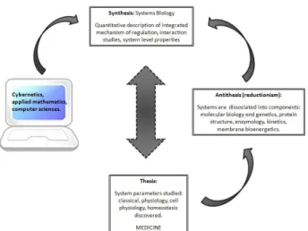
From a philosophical point of view, systems biology follows the dialectic principles of the general historical developments described by a German philosopher, Georg Wilhelm Friedrich Hegel (1770-1831). Hegel considered the real knowledge as an understanding of not only part but the Whole, the Absolute Idea This Absolute Idea of a detailed knowledge of the integrated whole systems is what systems biology wants to understand (i.e. to know all about life in its complexity, an Absolute Idea of living systems) [5].
The dialectic consists of the thesis, antithesis and synthesis; this triadic movement is essential to understand the result. A thesis is first giving rise to its reaction, an antithesis contradicts or refutes the thesis, and a synthesis resolves the tension between a thesis and antithesis.
In addition, systems biology has been also favored by the works of Norbert Wiener (1894-1964), an American mathematician and founder of cybernetics. Cybernetics studies the structure and function of regulatory systems. Due to this, it plays a major role in systems biology, seeking to integrate different levels of information to understand how biological systems function. Wiener through cybernetics developed the theory of feedback regulation and its application to explain the mechanism of homeostasis [5].
Gathering the contributions of Bernard, Hegel and Wiener, it can be established that the development of biological science is a Hegelian dialect movement. In systems biology, these components are again studied in their interaction within the intact systems of interest. Finally, among these principles, transmission of information by feedback mechanisms is most important.
Figure 3. Hegelian logic of reflection and an approach of the biological sciences.
2. PRINCIPLES OF BIOENERGETICS
2.1.Molecular System Bioenergetics
Once the principles of system biology are applied to cellular bioenergetics, the field of Molecular System Bioenergetics arises, which includes the energy metabolism as integrated networks in spatial (organization) and temporal (dynamic) aspects [6].
Bioenergetics is the science that studies the energetic transformations within living cells, explaining how cells obtain energy to live and perform work : mechanically (via motility and contraction), osmotically (via ion transport), or chemically (via biosynthesis), and how a cell maintains its specific structure and metabolism [6].
The molecular system bioenergetics is the study of energy conversion in cells, which allows to explain many classical observations in the cellular physiology of respiration and to understand how a cell senses its energy status. By sensing its energy status the cell is able to adjust its functions under different living conditions. Moreover, it takes into account the spatial and temporal dynamics of intracellular interactions.
Therefore, living organisms are open systems that operate far from the thermodynamic equilibrium; they exchange both matter and energy with their surroundings. This is well described with the first law of thermodynamics that describes the principle of the conservation of energy “in any physical or
chemical change, the total amount of energy in the universe remains constant, although the form of the energy may exchange”.
Moreover, the second law of thermodynamics establishes the tendency in nature toward ever-greater disorder in the universe “the total entropy of the universe is continually increasing”. A process occurs as a result of increasing entropy in the universe. The theory of energy changes during chemical reaction at constant temperature (T), developed by J. Willard Gibbs, shows that the free energy (G) depends on entropy (S) and enthalpy (H):
∆ ∆ ∆
A spontaneously reacting system has always a negative ΔG. Therefore, cells carry out thermodynamically unfavorable energy-requiring (endergonic) reactions, cells couple these reactions to others in order to liberate free energy (exergonic reactions); then the overall process is the sum of the free energy changes, which is negative and never reaches equilibrium [7].
Living systems, take up nutrients - release waste products - and generate work and heat, and are characterized by being in a steady state; the total entropy change can be split into two contributing parts:
∆ ∆ ext ∆ int
where ΔSext is the entropy of catabolism and ΔSint is the entropy of anabolism. In open systems the
energy flows from chemical energy sources (carbohydrates, proteins and lipids) to cells where these substances are metabolized to maintain the living system.
Figure 4. General scheme of cellular metabolism:exchange of matter and energy with the surrounding medium in the cell is called metabolism. The metabolism involves two general processes: catabolism (increases entropy within the surrounding medium) and anabolism (decreases the internal entropy). Both processes are coupled by reactions of cellular energetics, which are the center of cellular life.
Catabolism and anabolism coupling (metabolism) is the way through which free energy is extracted from the medium. Evolution has selected adenine nucleotides to fulfill this important task of coupling catabolism and anabolism (Fig. 4). Cellular energetics is based on synthesis and recycling reactions of ATP, ADP + Pι ↔ ATP + H2O. Therefore, the mass action ratio of the reaction of ATP synthesis is
usually expressed as:
Γ
ι
A high mass action ratio for the reaction of ATP synthesis, supplies free energy for anabolic reactions in cellular work. The phosphorylation potential is the amount of free energy available in ATP system in cells, described as:
Δ ATP Δ °
! ι"
The energy obtained by this potential is used for several cellular processes, such as movement (contraction), ion transport and biosynthesis. We make use of molecular system bioenergetics to investigate the regulation of cardiac cells respiration and energy fluxes in vivo.
2.2.Energy metabolism in muscle cells.
The energy metabolism in living organisms is supported by the oxidation of substrates: carbohydrates and lipids; as well as amino acids which after deamination enter into carbohydrate oxidation pathway. The rate and efficiency of oxidative phosphorylation depend on the substrate source, carbohydrates and lipids, although both contribute to NAD+ and FAD reduction; being the
oxidative phosphorylation activity one element in the determination of the ratio of NADH to FADH2
oxidation. In the heart fatty acids are the preferred fuel for mitochondrial respiration and account for 60-90% of energy production, with glucose oxidation making up 10-40% [8].
In 1948, Eugene Kennedy and Albert Lehninger discovered that mitochondria are the site of oxidative phosphorylation in eukaryotic cells. Moreover, mitochondria have an important role in thermogenesis, apoptosis, signaling, and regulation of ion homeostasis [7]. The word “Mitochondrion” comes from the Greek roots “mitos” - thread, and “chondrion” - grain or granule. It is involved in cellular ATP generation, apoptosis and necrosis processes [9].
Mitochondria have their circular own genome composed of 16-kilobase, its genome contains 37 genes essential for mitochondrial respiration function. Mammalian mitochondrial DNA (mtDNA) encodes 22 mitochondrial rRNAs, two mitochondrial tRNAs, and 13 proteins that make up parts of the oxidative phosphorylation complexes I, III, IV, and V [10].
Mitochondria are composed of compartments that carry out specialized functions:
• Mitochondrial outer membrane (MOM): encloses the entire organelle but it is freely permeable to molecules up to 5 KDa and ions due to the presence of the Voltage-Dependent Anion Channel (VDAC), being VDAC the most abundant protein and it has three isoforms in eukaryotic cells (VDAC1, VDAC2 and VDAC3)
• Intermembrane space (IMS)
• Mitochondrial inner membrane (MIM): forms internal compartments called cristae and inside of it we find the matrix. It is impermeable to most small molecules and ions, including
protons (H+) due to the presence of cardiolipin, this establishes a proton-motive force across
the MIM. The proton-motive force is generated by the complexes of the respiratory chain, also known as the electron transport chain (ETC). Moreover, it bears specific transporters which carry pyruvate, fatty acids, and amino acids or their α-keto derivatives into the matrix to follow the Krebs cycle. ADP and Pι are specifically transported into the matrix as newly synthesized ATP is transported out. It contains the respiratory electron carriers (complexes I-IV), ADP-ATP synthase (F0F1) or complex V, and other membrane transporters.
• Mitochondrial matrix: contains pyruvate dehydrogenase complex; citric acid cycle enzymes; fatty acid β-oxidation enzymes; amino acid oxidation enzymes; mitochondrial DNA (mtDNA), ribosomes; many other enzymes; ATP, ADP, Pι, Mg
2+
, Ca2+, K+; and many soluble metabolic intermediates.
Figure 5. Mitochondrion: outer membrane, inter membrane space, inner membrane. The cristae are the internal
compartments formed by the MIM. These regions expand the MIM, enhancing the ATP synthesis. The mitochondrial matrix is an aqueous solution of enzymes and intermediates of energy metabolism, mtDNA and ribosomes.
The content of mitochondria in skeletal muscles can vary from approximately 1% of cellular volume in the glycolytic muscles to up to 50% in the oxidative muscles. In adult cardiac cells, mitochondria occupy approximately 30-40% of the volume [11], whereas in neonatal cells mitochondrial volume occupies about 20% of the volume [12].
2.3. Electron transport chain (ETC)
In higher eukaryotes, the mitochondrial ETC has four major complexes. Each complex is a collection of proteins working together to produce the catalytic reducing power. The energy of electrons, liberated in a number of redox reactions, is translated into proton pumping by three large complexes (I, III and IV) [13]:
• Complex I: NADH-ubiquinone oxidoreductase, accepts electrons from NADH.
• Complex II: succinate oxidase-ubiquinone reductase, which oxidizes and accepts electrons from succinate, and reduces the ubiquinone pool.
- Coenzyme-Q (CoQ): moves the electrons from complex I and II to complex III.
• Complex III: ubiquinol-cytochrome c oxidoreductase, which transfers electrons from ubiquinol to cytochrome c.
- Cytochrome c (cyt c): moves the electrons from complex III to complex IV.
• Complex IV: cytochrome c oxydase, oxidizes cyt c and reduces molecular oxygen into water. Thus, electron transport between complexes I to IV is coupled to extrusion of protons from the matrix though complexes I, III and IV into the IMS, creating an electrochemical gradient (Δψ) across MIM. This movement of electrons generates an alkaline matrix and an acidic IMS. The electrochemical proton gradient, known as proton motive force (Δp) helps protons to back flow through complex V (ATP synthase complex / F1F0-ATPase synthase), which utilizes the energy to drive
Figure 6. Oxidative phosphorylation in cardiac cells: the sequential oxidation of fuel (e.g. fatty acids and glucose) leads to
the common substrate for the Krebs cycle, aceyl-CoA, which drives the production of the reducing equivalents NADH and FADH2. Electrons are passed to the ETC, where coupled redox reactions mediate proton translocation across MIM to
establish an electrical potential and pH gradient (Δp) that drives ATP synthesis by the mitochondrial ATP synthase. Ion-selective or nonIon-selective mitochondrial channels dissipate energy and alter the ionc balance and volume of the mitochondrial matrix, which is patly compensated by antiporters coupled to H+ movement.
Adenine nucleotide translocase (ANT), glucose-6-phosphate (G-6-P), inner-membrane anion channel (IMAC), mitochondrial Ca²+ uniporter (MCU), mitochondrial Ca²+-activated K+ channel (mitoK
Ca), mitochondrial ATP-sensitiveK+
channel (mitoKATP), phosphate carrier (PIC), permeability transition pore (PTP), pyruvate (PYR), K+/H+ exchanger (KHE),
Na+H+ exchanger (NHE), isocitrate dehydrogenase (IDH), α-ketoglutarate dehydrogenase (KDH), malate dehydrogenase
(MDH), pyruvate dehydrogenase (PDH), succinate dehydrogenase (SDH). Taken from [14] with permission.
Complex I is a large enzyme, which contains a prosthetic flavinmononucleotide (FMN)-containing flavoprotein and six sulfur centers. It has an L-shape, one arm of the L being in the membrane and the other extending into the matrix. Complex I catalyzes two simultaneous and obligated coupled processes: the exergonic transfer to ubiquinone of a hydride ion from NADH and a proton from the matrix:
NADH + H+ + Q → NAD+ + QH2
Complex II is a membrane-bound enzyme in the Krebs cycle, it contains flavin adenine dinucleotide (FAD) prosthetic groups and Fe-S centers. Complex II catalyzes the transfer of electrons from succinate via FAD and Fe-S centers to ubiquinone. The β-oxidation of fatty acids involves transfer of electrons from the substrate to the FAD of the dehydrogenase, and then to electron-transferring flavoprotein (ETF), which in turn passes its electrons to ETF:ubiquinone oxidoreductase. The effect of each of these electron-transferring enzymes is to contribute to the pool of reduced ubiquinone. Ubiquinol (QH2) diffuses in the MIM, to be reoxidized by Complex III.
Complex III is a homodimer. Each dimer has with 11 different subunits and their functional core consist of: cytochrome b with two heme, the Rieske iron-sulfur protein with 2Fe-2S centers, and cytochrome c1 with its heme. Complex III couples the transfer of electrons from QH2 to cytochrome c
with the transport of protons from the matrix to the IMS. The net equation of the Q cycle is QH2 + 2cyt c1 (oxidized) + 2H
+
N → Q + 2 cyt c1 (reduced) + 4H +
P
Complex IV has 13 subunits and carries electrons from cytochrome c to molecular oxygen, reducing it to H2O.
4 Cytc (reduced) + 8H+N + O2 → 4 cyt c (oxidized) + 4H +
N + 2H2O
The four-electron reduction of O2 involves redox centers and occurs without the release of
incompletely reduced intermediates (hydrogen peroxide or hydroxyl fee radical) that would damage cellular components.
Along the ETC/RC the redox potential increases gradually. The energy stored in the electrochemical potential of protons (ΔµH+) has two components: the chemical potential (ΔpH) energy and the mitochondrial electrical potential energy (Δψm) [14]:
∆#H+ 2.3 ' ∆( F ∆ψ
The electrochemical energy inherent to the proton-motive force drives the synthesis of ATP as protons flow passively back into the matrix through ATP synthase. This is the chemiosmotic model proposed by Peter Mitchell:
∆( ∆#H+ F
2.3
F ' ∆( ∆ψ
The efficiency of oxidative phosphorylation is defined by the amount of inorganic phosphate incorporated into ATP per amount of molecule of oxygen consumed.
Pyruvate and acyl-CoA obtained by glycolysis and fatty acid oxidation, respectively, are transported across the mitochondrial membrane and oxidized in the mitochondrial matrix. Pyruvate transport is affected by the difference in pH through MIM, with mitochondrial NADH:NAD+ ratio playing a key
role in the control of β-oxidation.
2.4. ATP synthase
The ATP synthase (F1Fo) is a large complex of the MIM that catalyzes the formation of ATP from ADP
and Pι, accompanied by the flow of protons from the IMS to the matrix. The mitochondrial F1 has
nine subunits of five different types, with the composition α3β3γδε. Each of the three β subunits has
one catalytic site for ATP synthesis. The conformational difference among β subunits extend to differences in their ATP/ADP-binding sites (β-ATP, β-ADP and β-empty). The Fo complex that makes
up the proton pore is composed of three subunits ab2c10-12.
Figure 7. Mitochondrial ATP synthase:F1 is peripherial domain, consisting of three α subunits and three β subunits, one δ,
and a central shaft (the γ subunit). Fo is the integral portion has multiple copies of c, one a and two b subunits. Fo provides a
transmembrane channel through which about four protons are pumped (pink arrows) for each ATP hydrolyzed on the β subunits of F1. Its mechanism involves the rotation of Fo relative to F1 (black arrow). Reprinted from [15] with permission.
The movement of protons (H+) causes the rotation of the central shaft (γ subunit), which comes into
contact with each αβ subunit pair in succession. This produces a cooperative conformational change in which the β-ATP site is converted to the β-empty conformation, and ATP dissociates; the β-ADP site is converted to the β-ATP conformation, which promotes condensation of bound ADP + Pι to form ATP; and the β-empty site becomes a β-ADP site , which loosely binds ADP + Pι, entering from the solvent.
Figure 8. Mechanism of ATP synthase:the F1 complex has three nonequivalent adenine nucleotide-binding sites, one for
each pair of α and β subunits. One of these sites is in the ATP conformation (which binds ATP tightly), a second is in the β-ADP (loose-binding) conformation, and a third is in the β-empty (very-loose-binding). The proton-motive force causes rotation of the central shaft (the γ subunit -green arrow-) which comes into contact with each αβ subunit pair in succession. This produces a cooperative conformational change in which the β-ATP site is converted to the β-empty conformation, and ATP dissociates; the β-ADP site is converted to the β-ATP conformation, which promotes condensation of bound ADP + Pι to form ATP; and the β-empty site becomes a β-ADP site, which loosely binds ADP + Pι entering from the solvent. ATP cannot be released from one site unless and until ADP and Pι are bound at the other. Redrawn from [7].
Proposed by Paul Boyer, the rotational catalysis mechanism in which the three active sites of F1 take
turns catalyzing ATP synthesis. The conformational changes central to this mechanism are driven by the passage of protons through the Fo portion of ATP synthase.
2.5. Adenine nucleotide transporter.
The adenine nucleotide transporter (ANT) is an antiporter that moves ATP4- out of the mitochondria
for every ADP3- moved in. Its activity is favored by the transmembrane electrochemical gradient,
which gives the matrix a net negative charge; the proton movement drives ATP-ADP exchange[7]. Thus, the ATP synthesized inside of the mitochondrion through the oxidative phosphorylation is pumped out of the matrix into the cytosol, while ADP and Pι are moved in the opposite direction (i.e. from the cytosol to the matrix) [16].
Therefore, the electrochemical proton gradient across the MIM is used to drive the formation of ATP and the transport of metabolites across the membrane. The ATP pool is therefore used to drive cellular processes (i.e. contraction). The ANT adjusts ATP supply and the phosphorylation potential of ATP in accordance with cytosolic requirements.
Previous studies [17] have shown a direct cooperation of ATP synthase and ANT, such that entering ADP is handed over directly to the ATP synthase and released as ATP into the endogenous pool. Thereby, it has been proposed [18-21] that a functional micro-compartmentation must be viewed on the background of the physical organization of the IMS and the inner membrane into two sections: the inner cristae membrane surrounding and inner boundary membrane facing the outer membrane, provided by small tubular structures.
Oxidative phosphorylation rate is regulated by the ADP availability to ANT, and the affinity of ANT for ADP is very high with an apparent Km(ADP) between 5 and 20 µM even in presence of ATP at physiological concentration (3-5 mM) [22].
3. MUSCLE CELL AND FUNCTIONING 3.1. Contractile module
The contractile machinery of striated muscles -skeletal and cardiac muscle (the myocytes)- is composed of sarcomeric units, which are joined into highly ordered myofibril structures [23]. Sarcomeres are comprised of thin (i.e. actin, tropomyosin -Tm- and troponin -Tn-) and thick (i.e. myosin) filaments and titin filaments highly ordered the I-band and A-band.
Contraction occurs when myosin heads of the thick filament attach to and exert force on actin molecules in the thin filament. This force causes the thin filament to slide over the thick filament, shortening the sarcomere and contracting the muscle fiber. Ca2+ binding to troponin (Tn) on the thin
filament initiates the force-generating interaction of myosin and actin, and ATP hydrolysis provides the energy for the molecular changes that drive force generation and muscle shortening. Ca2+
regulation is mediated through changes in the thin filament, though modulation can occur through myosin [24].
The arrangement of thick and thin filaments originates the A and I bands. The I band contains the thin filaments and the A band stretches the length of the thick filaments and includes the region where parallel thick and thin filaments overlap. Furthermore, the actin filaments in the I band are limited by the Z lines in a very regular pattern, and the M line is found in the A band [7].
The titin proteins extend from the Z line to the M line, regulating the length of the sarcomere and preventing overextension of the muscle. It provides a third level of filaments organization and is responsible for the resting elasticity muscle. Titin contains multiple α-actinin binding sites on differentially spliced Z repeats, which tightly correlates to the number of α-actinin molecules and hence to Z line thickness. Slow oxidative muscles (e.g. soleus) have more Z lines than fast glycolytic muscles (e.g. extensor digitorum longus) [25]. Moreover, skeletal and cardiac muscles contain different titin isoforms that depend on the physiological demands of fibers type [26].
Figure 9. Electron microscopy of the myofibril organization: the sarcomere consists of thick filaments interleaved at either end with thin filaments. This electron micrograph shows the relaxed state of the muscle, I band, A band, Z line and M line. Taken from [7].
Figure 10. Major components of the thin and thick filaments in the myofibril: thick filament is composed of myosin and titin. Thin filaments consist of actin and tropomyosin. The Z line contains α-actinin. It also shows the M line and H zone.
Skeletal muscles consist of parallel bundles of muscle fibers which fuse together and often span the length of the muscle. Each fiber is made up of a single, very large, multinucleated cell with a diameter of 20-100 µm. In contrast, cardiac myofibers are composed of a series of cardiomyocytes that are joined together end-to-end by intercalated disks to form a functional syncitium. Cardiomyocytes’ contractile apparatus consists of thin actin filaments and thick myosin filaments [8], when a contraction occurs actin and myosin are combined in the presence of ATP to produce the complex actomyosin [27].
Cardiac Z lines are considerably thicker due to more α-actinin cross-links, and consequently the lateral mechanical strength of cardiac myofibrils is 2-10 times higher than skeletal myofibrils in both the relaxed and the rigor state [28]. Meanwhile, titin protein is responsible for the increased tension of cardiac muscle related to an increase in the ventricular volume through modulation of the lattice
spacing between the filaments. In addition, it sets the slack length of cardiac myocytes on the ascending limb of the length-tension curve; this allows the heart to adapt to increased filling with a stronger contraction, a phenomenon called the Frank-Starling law [26].
3.2. Excitation-contraction coupling.
In striated muscle cells, myosin and actin are specialized to transform the chemical energy of ATP into motion. The binding and subsequent hydrolysis of ATP provides the energy that forces cyclic changes in the conformation of the myosin head. Actin-myosin interaction and ATP-hydrolysis are regulated by chemomechanical cross-bridge cycles, in skeletal and cardiac muscle. The contractile properties differ across types of muscle. Moreover, Ca2+ activation is needed for muscle contraction.
Biochemical and structural studies suggest the existence of three states of the thin filament which are in dynamic equilibrium [29]:
• Open conformation: myosin can strongly bind to actin.
• Closed state: where cross-bridges can weakly bind to actin.
• Blocked state: in the absence of Ca2+, Tm “blocks” cross-bridge access to thin filament strong binding sites.
The force exerted by the muscle in the isometric state depends on the number of strongly attached cross-bridges and the force developed by each cross-bridge. This depends on the number of actin binding sites open for strong myosin binding.
Figure 11. Mechanism of the cross-bridge cycle:formation of actin-myosin cross-bridges (AM) in terms of various reagents and products (A) and the corresponding structural changes (B).
Figure 11 A: actin; M: myosin; Pι: inorganic phosphate; f: cross-bridge exerting force; “•” is a strong connection; “~” is weak connection. Reprinted from [24] with permission.
ATP binding to myosin (step 1) is rapid and irreversible. The subsequent detachment of actin from the A˜M∙ATP complex (step 2) is similarly rapid and is caused by an opening between myosin’s upper
and lower regions like the “opening of jaws”. Bending of myosin neck region accompanies step 3, the hydrolytic cleavage of ATP. Following ATP cleavage, myosin again bind weakly to actin at a high rate, but in the absence of Ca2+ Tm sterically blocks access of the myosin head to strong binding sites on
actin. In step 5, Ca2+ is bound to TnC, Tnl detaches from actin, allowing the Tm/Tn complex to slide
over the thin filament surface. This exposes weak binding sites on actin and transiently exposes strong binding sites on actin for binding to the complementary regions in myosin’s domain. The greater the [Ca2+], the greater the fraction of time the Tm/Tn complex allows myosin access to strong
binding sites on actin. Consequently, the rate of strong cross-bridge attachment, the flux through step 5, is dependent on [Ca2+] and Tm position. Strong binding of myosin to actin (Fig. B) is associated
with movement of “closing the jaws”. This movement may allow the neck region of myosin to extend, opening a pathway for inorganic phosphate release from the ATP binding pocket in myosin. Alternatively, closing the jaws might promote Pι release from the binding pocket, which then allows the extension of myosin’s neck region. Step 6 involves myosin neck extension, which is the power stroke that in isometric muscle, stenches an elastic element (represented as S2) by some 10nm and produces a force of ˜2pN/cross-bridge. In nonisometric conditions, shortening of the neck extension
causes the thick and thin filaments to slide past each other. Step 7 is an irreversible isomerization and is strain sensitive. Finally, ADP is released from A∙Mf∙ADP in the reversible step 8 to form the
rigor state A∙Mf. cross-bridges attach and exert force constantly during steps 7, 8 and 1 during
isometric contraction, and force drops to zero when the cross-bridges detach in step 2. During shortening contractions the filaments slide past each other, the strain on the cross-bridge is reduced,
and step 7 occurs more rapidly. This chemomechanical mechanism implies that during an isometric contraction, a cross-bridge remains strongly attached for a relatively long time (>100 ms/cycle). Strongly bound cross-bridges prevent Tm/Tn from returning to its blocked or closed position, maintaining the thin filament in a “switched on” position (Fig. B). In the absence of Ca2+, cross-bridge
detachment at the end of the cycle allows Tm/Tn to cover the strong myosin binding sites on actin and deactivate the thin filament (Fig. A).
Differentiation and maturation of adult mammalian muscle cells lead to specialization, allowing different muscle types to have highly variable contractile abilities. In adult muscle cells, subcellular functions are specifically localized within structural and functional compartments: energy consuming processes take place in the SR and myofibrillar compartments, whereas depending on muscle type, energy production occurs mainly within mitochondria or glycolytic complexes [30].
Experimental studies in muscle cell energetics by Nabuurs [31] showed that ATP and ADP in muscles are bound to macromolecules and that these results in a non-equilibrium state of the creatine kinase (CK) reaction. These data contribute to the explanation of the cellular mechanisms of ATP compartmentation [32]. The energy metabolism of muscle cells, including heart, the compartmentation of adenine nucleotides in the cells is closely related to the CK system role [33]. Diffusion restrictions result in compartmentation of adenine nucleotides, which may explain the need for energy transfer and metabolic signaling networks [22].
In addition, MgADP is an efficient competitive inhibitor of ATPases. Thus, MgADP accumulation in the vicinity of any ATPases slows down the contraction cycle and impairs the Frank-Starling mechanism. Consequently, intracellular energy and metabolic signaling phosphotransfer networks within a cellular structural organization have the potential to protect cells from the excess of cytosolic free Ca2+ and ADP, and regulate respiratory ATP production in close correspondence to ATP consumption.
4. INTEGRATED ENERGY METABOLISM
4.1. Intracellular diffusion and compartmentation phenomenon.
Dense cytoskeleton, sarcoplasmic reticulum (SR), myofibrillar networks and macromolecular traffic result in the presence of physical barriers. Therefore, the cell metabolism is organized in: micro- and macrocompartments, metabolic channeling and functional coupling [34]. Compartmentation refers to the heterogeneity of intracellular diffusion, inasmuch as diffusion of metabolites in an organized intracellular media showing a clear restriction. Local diffusion restrictions are microcompartmentation of metabolites and their channeling within organized multi-enzyme complexes [32].
For many decades, several models have been developed to explain these diffusion restrictions in cells originated by the specific organization of intracellular structures that causes heterogeneity of diffusion, metabolite compartmentation and metabolic channeling [35].
Because of cytoskeletal structures and macromolecular crowding (high concentration of macromolecules) in cells, there is a decrease in the available volume for free diffusion substrates. The mitochondrial matrix is much more viscous than the cytoplasm. In mitochondria more than 60% of the matrix volume is constituted by the high density of enzymes and other proteins [32].
The term of apparent diffusion coefficient used for the analysis of ADP and ATP diffusion is expressed as: Dapp = DFxD0; where D0 is the diffusion coefficient in the bulk water phase, and DFx is a diffusion
factor accounting for all intracellular mechanism locally restricting the particles movement [36-38]. The phenomenon of diffusion restriction studied by Ridgway [39] showed that the apparent coefficient might be decreased by an order of magnitude depending upon the size of the diffusing particles and occupied volume fraction.
Mathematical modeling of permeabilized cardiomyocytes, with a decreased affinity of mitochondria for exogenous ADP in situ, showed that ADP or ATP diffusion in cells is heterogeneous. Also, the apparent diffusion coefficient for ADP and ATP may be locally decreased by several orders of
magnitude [38]. Experimental evidence has shown the existence of distinct ATP pools in mitochondria, and how ATP utilization sites are connected by phosphotransfer networks, notably via Phosphocreatine-Creatine kinase pathway [35, 37].
Phosphotransfer networks function as a bypass and overcome the local restrictions of ATP or ADP diffusion, and hence perform the important task of energy supply and metabolic feedback regulation of respiration [32].
In the heart, the intracellular energy transfer networks are structurally organized. Macromolecules and organelles, surrounding a regular mitochondrial lattice, are involved in multiple structural and functional interactions.
5. ORGANIZATION OF MITOCHONDRIA IN CARDIAC CELLS
5.1. Mitochondrial functioning in cardiac myocytes.
Cells are open systems that operate far from thermodynamic equilibrium while exchanging energy and matter with the external environment. These structurally and functionally organized metabolic systems may be described as cellular metabolic dissipative structures. They have functional enzymatic associations that form a catalytic entity as a whole and carry out their activities relatively independent [40].
The structural and functional unit of striated cardiac muscle cells consisting of distinct mitochondria localized at the level of sarcomeres between z-lines and interacting with surrounding myofibrils, SR, cytoskeleton, and cytoplasmic enzymes, is referred to as intracellular energetic units (ICEUs) [40]. These complexes have specialized metabolic regulatory pathways of energy transfer and feedback, mediated by creatine kinase (CK) and adenylate kinase (AK). The ICEUs are metabolic dissipative structures that help to extract Gibbs free energy and negentropy from the environment [41].
Figure 12. Intracellular Energy Unit (ICEU):structural organization of the energy transfer networks of coupled CK and AK reactions within an ICEU. By the interaction with cytoskeletal elements, mitochondrial and sarcoplasmic reticulum are precisely fixed in respect to the structure of the sarcomere of myofibrils between two Z lines and correspondingly between two t-tubules. Redrawn with permission [40].
The free fatty acids (FFA) are introduced in the cell by a family of plasma membrane proteins. Once in the ICEUs FFA are esterified to acyl-CoA. Acyl-CoAs enter in the β-fatty acids oxidation (β-FAO) pathway, producing acetyl-CoA. The electron-transferring flavoprotein (ETF)-ubiquinon oxidoreductase delivers the electrons produced by the β-FAO directly to the complex III of the respiratory chain (RC). The NADH produced by β-FAO is oxidized in the complex I of the RC, passing two electrons and two protons along. This contributes to the polarization of MIM.
In addition, the glucose is taken up by the glucose transporter-4 (GLUT-4) and oxidized. This oxidation produces pyruvate. Pyruvate dehydrogenase (PDH) complex catalyzes pyruvate into acetyl-CoA. The NADH redox potential from glycolysis enters into the mitochondrial matrix via the
malate-aspartate shuttle. Malate generated in the cytosol enters the matrix in exchange for α-ketoglutarate (αKG) and can be used to produce matrix NADH.
Matrix oxaloacetate returns to the cytosol by conversion to aspartate and glutamate exchange. Acetyl-CoA is oxidized to CO2 in the tricarboxylic acids (TCA) cycle, generating NADH and FADH2.
Both, NADH and FADH2 are further oxidized in the RC (complex I, II) with the final ATP synthesis. The
glycolysis rate is decreased by G6P inhibition of HK.
The mitochondrial interactosome (MI) supercomplex that transfers energy from mitochondria to cytoplasm is composed of ATP synthase, ANT, phosphate carriers (PiC), MtCK and VDAC with bound cytoskeletal proteins. MtCK is responsible of creatine phosphorylation, producing phosphocreatine (PCr). PCr is used to regenerate local ATP by CK and ATPases (actomyosin ATPase, sarcoplasmic reticulum SERCA and ion pumps ATPases). The re-phosphorylation of ADP in MMCK reaction increases the Cr/PCr ratio, which is transferred towards MtCK via CK/PCr shuttle. Calcium released from local intracellular stores during excitation-contraction coupling through calcium-induced calcium release mechanism, 1) activates the contraction cycle by binding to troponin C in the troponin-tropomyosin complex of thin filaments and 2) enters the mitochondria mainly via the mitochondrial Ca2+ uniporter to activate Krebs cycle dehydrogenases: PDH, αKG, IDH.
Previous studies with electron microscopy and confirmed by confocal microscopy have shown that cardiac cells have a very regular arrangement of mitochondria, crystal-like, at the level of A-band of sarcomeres in myofibrils surrounded by the sarcoplasmic reticulum[42].
One component of the ICEUs is the MI, which is used to designate the complex make-up of:
• ATP synthasome: including ATP synthase, adenine nucleotide carrier (ANC) and inorganic phosphate carrier (PiC)
• Mitochondrial creatine kinase (MtCK),
• Regulatory cytoskeletal proteins (such as tubulins and/or linker proteins (LP)).
The role of the MI is to regulate the mitochondrial adenine nucleotide flux and the cytoplasmatic Cr/PCr cycles in the IMS of the heart, skeletal muscle and brain cells. In addition, the MI strongly increases the efficiency of functional coupling between MtCK and the ATP synthasome [40]. The central mechanisms of energy transfer compartmentalization, is the functional coupling between the mitochondrial ATP/ADP translocase (ANT) and the mitochondrial creatine kinase (MtCK).
In addition, the MI functions in the non-equilibrium state caused by the dissipative enzymatic sub-networks (glycolysis, Krebs cycle, fatty acid oxidation, electrons transport chain, shuttles of creatine kinase/ phosphocreatine, malate/aspartate, etc.), structured and connected together by flows and regulating signals.
Therefore, the functional and structural role of MI is to increase the efficiency of the oxidative ATP synthesis and peripheral ATP hydrolysis, avoiding the waste of energy and fulfilling energy requirements. These can be achieved on one side by the ATP/ADP intramitochondrial circuit between ATP synthase and MtCK, through their functional coupling via ANT, and also by the Cr/PCr circuit through the different CK isotypes.
Creatine kinase (CK) has five different isotypes, three of which are cytosolic (dimeric) and two of which are mitochondrial (octameric): BCK, MCK, MB-CK, ubiquitous MtCK and sarcomeric MtCK, respectively [43]. The compartmentalized CK isotypes network is the main ICEUs phosphor-transfer circuit. MtCK was discovered in 1964 by Klingenberg’s lab. It is bound to the outer surface of the MIM through electrostatic interactions involving positive charges of lysine residues and negative charges from cardiolipin which is also associated with the ANT. The ADP formed at the active site of MtCK is transferred into IMS. Then ADP may either return to the matrix via ANT or leave the mitochondria via VDAC.
Figure 13. Mitochondrial creatine kinase functioning for high-energy metabolite channeling: ATP synthesis and ATP export through the MIM via ANT are tightly coupled to transphosphorylation of ATP to PCr by MtCK. PCr is exported into the cytosol by the outer membrane porin. Octameric MtCK binds to cardiolipin at the MIM. Redrawn from [44].
VDAC is the most abundant protein in MOM and is known to be primarily responsible for metabolites flux across the outer membrane [45]. VDAC is a monomeric β-barrel protein of 32 KDa and 2.5-3 nm-diameter. Three isoforms have been characterized in mammals: VDAC1, VDAC2 and VDAC3. VDAC has the ability to adopt fully open state and multiple states with smaller conductance, i.e. the “close states” that are impermeable to ATP but still permeable to small ions, including Ca2+ [46]. These
conformational states are also influenced by the transmembrane voltage. Due to its limited permeability, VDAC and the entire MOM create a dynamic micro-compartmentation of metabolites in the IMS that contributes to MtCK-linked channeling and separate mitochondrial ATP and ADP pools [22].
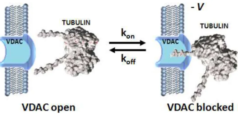
Rostovtseva et al. [46] showed the role of tubulin as a possible regulator of VDAC. They demonstrated the functional interaction between the dimeric tubulin and VDAC. Cytosolic heterodimeric tubulins (αβ-tubulin) are in a dynamic equilibrium with the microtubular network. Furthermore, they proposed a model for tubulin-VDAC interaction, where the C-terminal tail (CTT) penetrates into the channel lumen potentially reaching though the channel. Because of the length of the CTT, tubulin interacts with the positively charged domain of VDAC. Besides regulating VDAC permeability, the apparent affinity of oxidative phosphorylation for cytoplasmic ADP can be modified by tubulin [47].
Figure 14. Possible regulation of VDAC by tubulin: One tubulin CTT partially blocks channel conductance by entering VDAC pore. Redrawn from [46].
The selective permeability of VDAC to ATP/ADP and Cr/PCr has an important influence over the control of respiration rate and energy fluxes displayed by the different components of the MI.
Experimental data showed that tubulin binding to VDAC restricts significantly the availability of ADP to oxidative phosphorylation. In isolated heart mitochondria, the low apparent KmADP increased from 10-20µM up to 169±52µM by the addition of 1µM of dimeric αβ-tubulin [46]. Hence, the VDAC-tubulin interaction participates in the regulation of mitochondrial functioning via the modulation of mitochondrial compartmentation of adenine nucleotides [44].
Accordingly, the structural organization of ICEUs results in local confinement of adenine nucleotides and Cr-PCr coupling in discrete dynamic energetic circuits between actomyosin ATPases and mitochondrial ATPsynthases. Mitochondrial respiration and ADP kinetics are effectively modulated by all these mitochondrial interactions with cytoskeletal elements [36].
ICEU functioning is based on the regulation of mitochondrial ATP synthesis by intracellular energy consuming processes. It adjusts energy extraction from nutrients, to energy used to perform cellular work.
5.2. Mitochondria and cytoskeletal interactions
The cardiomyocyte cytoskeleton is composed of rigid and elastic elements. It maintains the shape of the cell as an elongated cylinder with an elliptical cross-section, even during contraction-relaxation cycles. Proteins of the cardiomyocyte have been categorized into five different families:
• Contractile proteins: functional structural proteins, i.e. myosin, actin, tropomyosin and the troponins.
• Proteins which contribute to cell shape, mechanical resistance, signal transduction and morphological integrity: subdivided into their structural and functional properties
Sarcomeric skeleton: titin, myosin, binding protein C, α-actinin, myomesin, and M-protein.
True ‘cytoskeletal’ proteins: tubulin, desmin and actin.
Membrane-associated proteins: dystrophin, spectrin, talin, vinculin, ankyrin.
Proteins of the intercalated disc: desmosomes consisting of desmoplakin, desmocollin, desmoglein and desmin; adherens junctions with N-cadherin, the catenins and vinculin, and gap junctions with connexin [48].
The major constituent of microfilaments is actin. The microtubules are essentially composed of tubulin, and their assembly and function are regulated by microtubule associated proteins, such as the microtubule associated motors (kinesin, dynein, etc.) and the structurally associated proteins (tau, Microtuble Associated Protein MAPs). Both tubulin and actin exist in equilibrium between polymerized and depolymerised states [49]. Intermediate filaments, which are rather stable, are constituted by desmin and plectin.
The regular arrangement of mitochondria in cardiac cells is mediated by their association with three major cytoskeletal structures: microfilaments, microtubules (MT) and intermediate filaments. The microtubular network, intermediate filaments and microfilaments form specific structures which are vital during the contraction cycle and for the regulation of energy supply [40].
VDAC is not only the most abundant protein in MOM, but also one important element regulating substrates exchange between cytosol and mitochondria. It is known to be primarily responsible for ATP/ADP fluxes across the outer membrane. Previous studies have demonstrated that VDAC’s closed states, impermeable to ATP, are still permeable to small ions including Ca2+.
Several studies have shown that the cell’s permeability depends on its preparation, meaning that a low permeability of the MOM (and hence VDAC) for ADP may be the result of the interaction of mitochondria with some cytoskeletal proteins. Therefore, high values of apparent Km for ADP in permeabilized cells are related to decreased permeability of MOM, by the interaction with cytosolic proteins, such as tubulin [50].
5.3. Tubulins
Tubulin, the subunit protein MT, is an acidic heterodimer, composed of similar 50 kDa globular α and β subunits. Each subunit possess a strongly anionic, extended CTT of ~15 amino acids that represents (α=10, β=20) ~3% of the subunit mass and ~40% of the subunit charge. It binds to guanine nucleotides and polymerize into MT in a GTP-dependent manner. Tubulin is an abundant and stable protein, present in many cells.
The native structures of α- and β-tubulin consist of three domains: 2 globular domains comprising >90% of the protein, and an unstructured CTT. The globular domains consist of an N-terminal domain that binds GTP or GDP and represents about ½ of the protein, and a slightly smaller C-terminal globular domain to which the unstructured tail is grafted. These proteins have binding regions that facilitate longitudinal assembly of the protein into filaments consisting of heat-to-tail, single file alignments of the subunit protein. Moreover, additional binding sites allow the filaments to join together laterally to form sheets of filaments or closed cylinders of filaments called protofilaments [51].