HAL Id: hal-02111165
https://hal-amu.archives-ouvertes.fr/hal-02111165
Submitted on 25 Apr 2019
HAL is a multi-disciplinary open access
archive for the deposit and dissemination of
sci-entific research documents, whether they are
pub-lished or not. The documents may come from
teaching and research institutions in France or
abroad, or from public or private research centers.
L’archive ouverte pluridisciplinaire HAL, est
destinée au dépôt et à la diffusion de documents
scientifiques de niveau recherche, publiés ou non,
émanant des établissements d’enseignement et de
recherche français ou étrangers, des laboratoires
publics ou privés.
A Proposal for a Classification for Recurrent
Endometrial Cancer: Analysis of a French Multicenter
Database From the FRANCOGYN Study Group
Alexandre Bricou, Sofiane Bendifallah, Mathilde Daix-Moreux, Lobna
Ouldamer, Vincent Lavoué, Amélie Benbara, Cyrille Huchon, Geoffroy
Canlorbe, Emilie Raimond, Charles Coutant, et al.
To cite this version:
Alexandre Bricou, Sofiane Bendifallah, Mathilde Daix-Moreux, Lobna Ouldamer, Vincent Lavoué, et
al.. A Proposal for a Classification for Recurrent Endometrial Cancer: Analysis of a French Multicenter
Database From the FRANCOGYN Study Group. International Journal of Gynecological Cancer,
Lippincott, Williams & Wilkins, 2018, 28 (7), pp.1278-1284. �10.1097/IGC.0000000000001296�.
�hal-02111165�
Downloaded from http://journals.lww.com/ijgc by BhDMf5ePHKbH4TTImqenVCscuGFl+NVZJNN5BTUGIp9Dpbm+KtzP1304LzFbyiZLAFGkJCIxha0= on 10/22/2018 Downloadedfrom http://journals.lww.com/ijgcby BhDMf5ePHKbH4TTImqenVCscuGFl+NVZJNN5BTUGIp9Dpbm+KtzP1304LzFbyiZLAFGkJCIxha0=on 10/22/2018
A Proposal for a Classification for Recurrent
Endometrial Cancer
Analysis of a French Multicenter Database From the
FRANCOGYN Study Group
Alexandre Bricou, MD,* Sofiane Bendifallah, MD, PhD,Þ Mathilde Daix-Moreux, MD,*
Lobna Ouldamer, MD, PhD,þ Vincent Lavoue, MD, PhD,§ Ame´lie Benbara, MD,*
Cyrille Huchon, MD, PhD,|| Geoffroy Canlorbe, MD, PhD,Þ Emilie Raimond, MD,¶
Charles Coutant, MD, PhD,# Olivier Graesslin, MD, PhD,¶ Pierre Collinet, MD, PhD,**
Xavier Carcopino, MD, PhD,ÞÞ Cyril Touboul, MD, PhD,þþ§§ Emile Dara
B
, MD, PhD,Þ
Lionel Carbillon, MD, PhD,* and Marcos Ballester, MD, PhD,Þ
for the Groupe de Recherche FRANCOGYN
Objective: Endometrial cancer (EC) recurrences are relatively common with no standardized way of describing them. We propose a new classification for them called locoregional, nodal, metastasis, carcinomatosis recurrences (rLMNC).
Patients and Methods: The data of 1230 women with EC who were initially treated by primary surgery were included in this French multicenter retrospective study. Recurrences were classified based on dissemination pathways: (1) locoregional recurrence (rL); (2) nodal recurrence (rN) for lymphatic pathway; (3) distant organ recurrence (rM) for hematogenous pathway; and (4) carcinomatosis recurrence (rC) for peritoneal pathway. These pathways were further divided into subgroups. We compared recurrence free survival and overall survival (OS) between the 4 groups (rL/rN/rM/rC).
Results: The median follow-up was 35.6 months (range, 1.70Y167.60). One hundred ninety-eight women (18.2%) experienced a recurrence: 150 (75.8%) experienced a single-pathway recurrence and 48 (24.2%) a multiple-single-pathway recurrence. The 5-year OS was 34.1% (95% confidence interval [CI], 27.02%Y43.1%), and the median time to first recurrence was 18.9 months (range, 0Y152 months). The median survival after recurrence was 14.8 months (95% CI, 11.7Y18.8). Among women with single pathway of recurrence, a difference in 5-year
*Department of Obstetrics and Gynecology, Jean-Verdier University Hospital, Assistance Publique des Hoˆpitaux de Paris, University Paris 13; †Department of Gynaecology and Obstetrics, Tenon University Hospital, Assistance Publique des Hoˆpitaux de Paris, University Pierre and Marie Curie; ‡Department of Obstetrics and Gynaecology, Centre Hospitalier Re´gional Universitaire de Tours, Hoˆpital Bretonneau, Tours; §CRLCC Euge`ne-Marquis, Service de Gyne´cologie, CHU de Rennes, Universite´ de Rennes 1; ||Department of Gynaecology and Obstetrics, Centre Hospitalier Intercommunal, Poissy; ¶Department of Obstetrics and Gynaecology, Institute Alix de Champagne University Hospital, Reims; #Centre de Lutte Contre le Cancer Georges Franc¸ois Leclerc, Dijon; **Department of Obstetrics and Gynaecology, Centre
Hospitalier Re´gional Universitaire, Lille; ††Department of Obstetrics and Gynecology, Hoˆpital Nord, Marseille; ‡‡Department of Obstetrics and Gynecology, Centre Hospitalier Intercommunal, Cre´teil; and §§Faculte´ de Me´decine de Cre´teil, UPECVParis XII, Paris, France. Address correspondence and reprint requests to Bricou Alexandre,
MD, Department of Obstetrics and Gynecology, Jean-Verdier University Hospital, Assistance Publique des Hoˆpitaux de Paris, University Paris 13, Avenue du 14 Juillet, 93143 Bondy Cedex, France. E-mail: alexandre.bricou@aphp.fr.
The authors declare no conflict of interest.
Clinical trial identification number: CEROG 2014-GYN-020. URL of the registration site: http://www.cngof.fr/accueil-cngof/
le-cerog.
Supplemental digital content is available for this article. Direct URL citation appears in the printed text and is provided in the HTML and PDF versions of this article on the journal’s Web site (www.ijgc.net).
Copyright* 2018 by IGCS and ESGO ISSN: 1048-891X
OS (PG 0.001) and survival after recurrence (P G 0.01) was found between the 4 rLNMC groups. The carcinomatosis group had the worst prognosis compared with other single recurrence pathways. Women with multiple recurrences had poorer 5-year OS (PG 0.001) and survival after recurrence (PG 0.01) than those with single metastasis recurrence, other than women with peritoneal carcinomatosis.
Conclusions: This easy-to-use and intuitive classification may be helpful to define EC recurrence risk groups and develop guidelines for the management of recurrence. Its prognosis value could also be a tool to select homogenous populations for further trials. Key Words: Endometrial cancer, Recurrences, Relapses, Classification
Received January 30, 2018, and in revised form April 21, 2018. Accepted for publication April 23, 2018.
(Int J Gynecol Cancer 2018;28: 1278Y1284)
E
ndometrial cancer (EC) is the most common gynecologic cancer worldwide with 320,000 new cases each year.1Around 75% of ECs are diagnosed at an early stage with a 5-year overall survival (OS) reaching 80%.2However, EC is a heterogeneous disease with some subsets of women displaying an increased risk of recurrence. Indeed, it causes almost 76,000 deaths per year.1The most usual identified risk factors for
re-currence are the International Federation of Gynecology and Obstetrics (FIGO) stage, depth of myometrial invasion, histolog-ical type and grade, lymphovascular space invasion, and tumor size.3In this specific setting, the European Society for Medical
Oncology (ESMO)/European Society of Gynaecological Oncology/European Society for Radiotherapy and Oncology group recently classified EC women into 4 subgroups according to the risk of recurrence (low risk, intermediate risk, high-intermediate risk, and high risk) to better adapt surgical staging and indications for adjuvant therapies.4
Although overall prognosis of women with surgically treated EC is good, Bendifallah et al5recently reported that at
least 20% of them will experience a recurrence, impacting OS. Moreover, it is now well recognized that more than 70% of recurrences occur within the first 2 to 3 years after initial treatment.6Y8Hence, recurrence events and their treatment are a
crucial aspect in the management of women with EC. Most studies to date focus on predictive factors of recurrence, recurrence pat-terns, and specific treatments.9Y11In this setting, most authors
concur that locoregional recurrences are to be differentiated from distant recurrences.
However, there is currently no consensual standardized description of EC recurrence and definitions vary widely from one study to another.12Y16 This lack of homogeneity limits
comparison of study results by rendering their interpretation somewhat blurred.
Hence, the purpose of this study was to describe the anatomical locations of EC recurrence with a view to devel-oping an easy-to-use classification based on the 4 dissemination pathways (locoregional, lymphatic, blood, and peritoneal) to better assess prognosis.
PATIENTS AND METHODS
Study Population
From January 2001 to December 2013, data of women with histologically proven EC who received primary surgical treatment were abstracted from EC databases of 9 institutions in France (Tours University Hospital, Tenon University Hospital, Dijon Cancer Centre, Rennes University Hospital, Lille University Hospital, Reims University Hospital, Creteil University Hospital, Poissy University Hospital, and Jean Verdier University Hospital) and from the Senti-Endo trial.17
The research protocol was approved by the Institutional Review Board of the Colle`ge National des Gyne´cologues et Obste´triciens Franc¸ais (CEROG 2014-GYN-020).
All enrolled women underwent preoperative imaging examinations (abdominopelvic magnetic resonance imaging and/or computed tomography scan). The following clinical, surgical, pathological, and adjuvant therapy data were collected: women’s age, body mass index (BMI; calculated as weight in kilograms divided by the square of height in meters), surgical procedure, nodal staging, final pathological analysis (histologi-cal type and grade, depth of myometrial invasion, and lymphovascular space invasion status), and type of adjuvant therapy. Histological type 1 included endometrioid tumors (grades 1 to 3), and histological type 2 included serous or clear-cell carcinomas and carcinosarcomas. All women were clas-sified according to the FIGO 2009 classification18after final
pathological analysis and were classified into recurrence risk groups as defined by the ESMO/European Society of Gynaecological Oncology/European Society for Radiother-apy and Oncology guidelines.4
Treatment and Follow-up
All the women underwent primary surgical treatment including at least total hysterectomy with bilateral salpingo-oo-phorectomy, with or without nodal staging (sentinel lymph nodes T pelvic T para-aortic lymphadenectomy), as well as omentectomy. Adjuvant therapy and clinical follow-up were decided by a
International Journal of Gynecological Cancer
&
Volume 28, Number 7, September 2018 Classification for Recurrent ECmultidisciplinary committee and based on the French guidelines. Adjuvant therapy could include vaginal brachytherapy and/or ex-ternal beam radiotherapy (EBRT) and/or chemotherapy (CT). Clinical follow-up consisted of physical examinations and the use of imaging techniques according to the findings. Follow-up visits were conducted every 3 months for the first 2 years, every 6 months for the following 3 years, and once a year thereafter.
Recurrence Events and Classification
Recurrent disease was assessed by physical examination and imaging techniques (computed tomography, magnetic res-onance imaging, ultrasonography, bone scintigraphy, positron emission tomography with the fluorodesoxyglucose (18F) as well as histological findings when feasible. We applied a new classification, called the rLNMC classification, based on the pathway of dissemination: locoregional recurrence (rL), nodal recurrence (rN), distant organ recurrence (rM), and peritoneal recurrence with carcinomatosis (rC) (Table 1). Locoregional pathway recurrence was divided into 2 groups as follows: re-currence in the vaginal vault only (rL1) and rectal and/or bladder involvement (rL2). We defined a locoregional recur-rence that could not be assessed (rLx) and no evidence of
locoregional recurrence as rL0. Nodal recurrences were sepa-rated into 2 groups: for lymph node recurrence in the pelvic, inguinal, and/or infradiaphragmatic para-aortic area (rN1) and for mediastinal or other distance nodal recurrences (rN2). We defined a node recurrence that could not be assessed as rNx and no evidence of the recurrent node site as rN0. Hematogenous recurrences were also divided into 2 groups: single metastasis (rM1) and multiple metastasis (rM2). We defined a metastatic recurrence that could not be assessed as (rMx) and no evidence of the metastatic as (rM0). The peritoneal recurrence rep-resented a single group for pelvic and/or abdominal carci-nomatosis (rC1). We defined a peritoneal recurrence that could not be assessed as rCx and no evidence of carcino-matosis as (rC0). Single recurrence was defined as recur-rence with only one pathway of dissemination. Multiple recurrences were defined as recurrences with more than one pathway of dissemination, Supplemental Digital Content 1, http://links.lww.com/IGC/A754;Supplemental Digital Content 2, http://links.lww.com/IGC/A755.
Statistical Analysis
Recurrence free survival was defined as the length of time from the date of primary surgery to any EC recurrence and was censored at the date of the last follow-up or date of death without recurrence. Overall survival was defined as time from primary surgery to death as a result of any cause and survival after recurrence was defined as time from recurrence diagnostic to death. Kaplan-Meier estimates were used to estimate the event-time distributions, and log-rank test was used to compare the differences in terms of OS between the 4 groups (rL/rN/rM/rC). Values of P G 0.05 were considered to denote significant differences. Data were managed with an Excel database (Microsoft, Redmond, Wash) and analyzed using R 2.15 soft-ware, available online with caTools, rms, presence/absence, and verification libraries (https://www.r-project.org/).
RESULTS
Characteristics of the Population
During the study period, 1230 women with EC who were initially treated by primary surgery were included in this multicenter retrospective study. Two hundred twenty-four women (18.2%) experienced a recurrence. Among them, 26 were ex-cluded owing to incomplete data leaving 198 women for final analysis. The median age of the women with recurrence was 68.5 years (range, 32Y88 years), and their BMI was 29.09 kg/m2 (range, 14Y50.4 kg/m2). The median follow-up was 35.6 months (range, 1.70Y167.60 months) after primary diagnosis and 10.2 months (range, 1Y109 months) after recurrence. The median recurrence free survival was 18.9 months (range, 0Y152 months). The 5-year OS was 34.1% (95% CI, 27.02%Y43.1%). The median survival after recurrence was 14.8 months (95% CI, 11.7Y18.8). One hundred nineteen women (60%) died, and 65 (55%) of these deaths were due to EC recurrence. For the remaining 54 women, the cause of the death was not recorded. The characteristics of the women with recurrence are reported in Table 2.
TABLE 1. rLMNC classification for EC recurrence rLocoregional
rLx A recurrence tumor that cannot be assessed rL0 No evidence of the recurrent tumor site rL1 Recurrence tumor in the vaginal vault only rL2 Centropelvic recurrence tumor with or
without rectal and/or bladder involvement.
rNode
rNx The lymph node recurrence cannot be assessed
rN0 No lymph node recurrence
rN1 Lymphnode recurrence in pelvic, inguinal, and/or infradiaphragmatic para-aortic area rN2 Mediastinal or other distance lymph
node recurrence rMetastasis
rMx Distant organ recurrence cannot be assessed rM0 No distant organ recurrence
rM1 Distant recurrence in one organ
rM2 Distant recurrence in two or more organs. rCarcinomatosis
rCx Carcinomatosis recurrence cannot be assessed rC0 No carcinomatosis recurrence
Patterns of Recurrence According to the
rLNMC Classification
Among the 198 women who experienced recurrence: 52 (21.1%) experienced a locoregional recurrence (vaginal vault [rL1] in 22 cases [8.9%], centropelvic recurrence [rL2] in 30 cases [12.2%]); 50 (20.3%) experienced a nodal recurrence (pelvic, inguinal, and/or para-aortic node recurrence [rN1] in 41 cases [16.7%] and mediastinal or distant nodes [rN2] in 9 cases [3.8%]); 95 (38.6%) experienced a metastatic pathway recur-rence (rM1) in 49 cases (19.9%) and rM2 in 46 cases (18.7%); and 49 (19.9%) experienced a peritoneal carcinomatosis re-currence (rC1).
Among the women who experienced a single recur-rence (n = 150, 75.8%): 30 (20%) experienced a tumor site recurrence (vaginal recurrence [rL1N0M0C0] in 14 cases [46.7%], centropelvic recurrence [rL2N0M0C0] in 16 cases [53.3%]); 26 (17.3%) experienced a nodal recurrence (pelvic, para-aortic, or inguinal areas [rL0N1M0C0] in 18 cases [69.2%] and mediastinal or distance nodes [rL0N2M0C0] in
8 cases [30.8%]); and 65 (43.3%) experienced a metastatic recurrence (only 1 localization [rL0N0M1C0] in 32 cases [49.2%] [15 lung, 8 bone, 6 brain, 2 liver, 1 unspecified], more than 1 [rL0N0M2C0] in 33 cases [50.8%], and peritoneal carcinomatosis recurrence (rL0N0M0C1) in 29 cases [19.4%]). Among the women who experienced a multiple recur-rence (n = 48 [24.2%]): 22 (45.8%) experienced a locoregional recurrence (vaginal vault [rL1] in 8 cases [36.4%], centropelvic [rL2] in 14 cases [63.6%]); 24 (50%) experienced a nodal re-currence (involving pelvic, para-aortic, or inguinal area nodes [rN1] in 23 cases [95.8%] and mediastinal area or distance nodes [rN2] in 1 case [4.2%]); and 30 (62.5%) experienced a metastatic recurrence (1 metastasis [8 lung, 7 liver, 1 unspecified, 1 bone] [rM1] in 17 cases [56.7%], more than 1 metastatic re-currence [rM2] in 13 cases [43.3%] [Table 3], and peritoneal carcinomatosis [rC1] in 20 cases [41.7%]).
Survival Outcomes
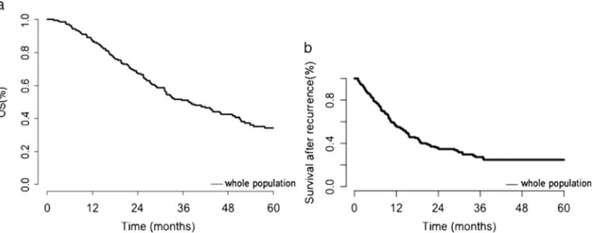
The 5-year OS was 34.1% (95% CI, 27,02Y43,1%) (Fig. 1A). The median survival after recurrence was 14.8 months (95% CI, 11.7Y18.8) (Fig. 1B). Among women with single pathway of recurrence, a difference in 5-year OS (Fig. 2A) and survival after recurrence (Fig. 2B) was found between the 4 rLNMC groups. The carcinomatosis group (rL0N0M0C1) had the worst prognosis compared with other single recurrence pathways. When considering women with multiple recurrences, a difference in 5-year OS (Fig. 3A) and survival after recurrence (Fig. 3B) was found between the rLNMC groups.
DISCUSSION
Our study confirms that the patterns of recurrence in EC are multiple. Moreover, it shows that recurrence sites can be isolated or associated with each other: we identified 246 different recurrences among the 198 women who experienced recurrence with 75.8% experiencing single recurrence and (24.2%) multiple recurrences. These results are in agreement with those of Sohaib et al12who report 48% of multiple-site
recurrences. This is why the rLNMC classification we propose here includes the different pathways of spread: locoregional TABLE 2. Characteristics of women who experienced
a recurrence
Characteristics Population, n = 198 Age, median (range), y 68.49 (32Y88) BMI, median (range), kg/m2 29.09 (14Y50.4) Follow-up, median (range), mo 35.6 (1.70Y167.60) Histological type n (%) Type 1 115 (58) Type 2 83 (42) ESMO classification n (%) Low risk 18 (9.1) Intermediate risk 9 (4.5) Intermediate-high risk 18 (9.1) High risk 151 (76.3) NA 2 (1)
FIGO stage at definitive histology n (%)
I 86 (43.4)
II 26 (13.1)
III 73 (36.9)
IV 13 (6.6) (13)
Adjuvant therapies before recurrence n (%) No adjuvant therapy 30 (15.2) Vaginal brachytherapy alone 13 (6.6) EBRT + brachytherapy 64 (32.3) CT + EBRT 21 (10.6) CT alone 28 (14.1) EBRT + brachytherapy + CT 27 (13.6) EBRT alone 14 (7.1) Brachytherapy + CT 1 (0.5)
NA, not available.
TABLE 3. Women with single or multipathways of recurrence with rLNMC classification
Single recurrence on locoregional site (rL) 30 (15.2%)
rL1N0M0C0 14 rL2N0M0C0 16 Single-nodes recurrence (rN) 26 (13.2%) rL0N1M0C0 18 rL0N2M0C0 8 Single-metastasis recurrence (rM) 65 (32.8%) rL0N0M1C0 32 rL0N0M2C0 33 Single-carcinomatosis recurrence (Rc) 29 (14.6%) rL0N0M0C1 29
Multiple-pathways recurrence (rMpath) 48 (24.2%)
International Journal of Gynecological Cancer
&
Volume 28, Number 7, September 2018 Classification for Recurrent ECextension (contiguous extension), lymphatic dissemination (nodal recurrences), hematogenous dissemination (metastatic recurrences), and peritoneal seeding (carcinomatosis). It is based on clinical and/or imaging data found during the follow-up period with or without histological confirmation and describes localization, type, and focality of recurrences. Moreover, local-ization of recurrence is not the only prognostic factor; prognosis may also depend on the type of recurrence treatment, which is conditioned by (1) initial treatment (radiotherapy), (2) the interval between initial diagnosis and recurrence, and (3) the initial histology and tumor grade.13Hence, our classification
could be completed by the type of early adjuvant treatment used. Consequently, the prefixes Ra for radiotherapy, Ch for CT, and Ht for hormonotherapy could also be recorded in the classification.
In the classification we present here, locoregional re-currence (rL) concerns rere-currences localized in the locoregional area and related to contiguous dissemination (from the initial EC tumor site). In this specific setting, we decided to exclude parenchimatous recurrences from this group and include them in the hematogenous pathway (metastasis) group, even if such recurrences can involve abdominal or pelvic organs. Our division into 2 different subgroups (rL1, rL2) is supported by its impact on OS: a trend for poorer prognosis was found for centropelvic
recurrences (rL2, N0, M0, C0) compared with vaginal vault recurrences (rL1, N0, M0, C0). Therapeutic options are an additional parameter supporting this distribution. Indeed, sur-gical treatment can be more difficult and sometimes not pos-sible for centropelvic recurrences as opposed to isolated vaginal vault recurrence, which can often be salvaged with surgical excision and/or radiotherapy. In this setting, Ackerman et al19
reported a local control rate of up to 79% for isolated vaginal vault recurrences. Moreover, several studies report that survival is better in women with recurrence confined within the vaginal vault rather than in the true pelvis13,20Y22or other sites.23
How-ever, these results must be interpreted with caution because the prognosis seems to be the poorest for women who initially re-ceived external beam radiotherapy.13Concerning centropelvic
recurrences (rL2), the extent of recurrence appears to be an important prognostic factor for local control and survival.24In a
series of 209 women with recurrence, Sartori et al23reported that
the 5-year OS for vaginal, pelvic, and distant recurrence was 68%, 29%, and 8%, respectively.
Concerning the lymphatic pathway, nodal recurrences represented 20.3% of the total recurrence sites in our study and were isolated in 13.1% of cases. These findings are in contradiction with those of Sohaib et al12 who found that
nodal recurrences were the most frequent recurrence site (47%). FIGURE 1. A, Five-year cumulative survival rates for the whole population. B, Cumulative survival rates after recurrence for the whole population.
FIGURE 2. A, Five-year cumulative survival rates for the different single recurrences. B, Cumulative survival rates after recurrence for different single recurrences.
However, their study included 86 patients among whom only 40 (46.5%) had undergone nodal dissection during primary sur-gery.12We distinguish between 2 types nodal recurrence in
our classification: rN1 for all pelvic, inguinal, and/or infradiaphragmatic para-aortic area node recurrence and rN2 for mediastinal or other distant lymph node recurrence. This distinction is based on the possibility of surgical removal for the rN1 group compared with the rN2 group. However, we were not able to distinguish pelvic node involvement from infradiaphragmatic para-aortic or inguinal nodal involvement in the database. Moreover, we did not find any difference in survival between the rN1 and the rN2 groups, when isolated. This can be explained by the fact that isolated rN2 recurrences are rare and that nodal recurrences are often associated with other recurrence sites. However, a few studies in the literature have attempted to distinguish between type of nodal involve-ment and what we consider to be the rN2 group is often clas-sified as distant recurrence sites.5,21,22,25
In our study, metastases represented the first pathway of recurrence and was found in 95 women (38.6%) with a similar distribution between isolated and multiple metastases. This rate is in complete agreement with that reported by Sohaid et al12who found that metastases occurred in 36% of recurrences
and represented a major predictor factor for poor survival. In the current classification, we decided to classify metastatic recurrences as single (rM1) or multiple (rM2). We believe that single metastases could be, in selected cases, eligible for local control (surgery or interventional imaging) with a view to in-creasing the symptom-free interval, which is less feasible for multiple-site metastases. In contrast, we decided not to distin-guish between the different anatomical sites because this would detract from the clarity and ease-of-use of the classification.
In our study, the presence of peritoneal carcinomatosis was linked to a significant decrease in 5-years OS and survival after recurrence. That is why we decided to create a single group because it may represent a specific pathway of dissemination. In the current literature, the real impact of peritoneal carcinoma-tosis on survival remains unclear because it is often associ-ated with other recurrence sites such as distant metastases, extravaginal recurrences, or abdominal recurrences.22,26Y28 Indeed, Ouldamer et al29 recently reported a nomogram to
predict poor prognosis recurrences (ie, peritoneal carcinomatosis
and distant metastases) with a 3-year OS of 33.1% for the peritoneal carcinomatosis group. Finally, the treatment options may differ depending on whether the peritoneal carcinomatosis is isolated or associated to another pathway of dissemination. In this setting, in case of isolated carcinomatosis, the place of cytoreductive surgery with hyperthermic intraperitonal CT is a matter of major interest.
The strengths of our study lie in its multicenter nature and the large number of women with EC recurrence included. However, we cannot exclude an inherent bias linked to its retrospective nature. Indeed, among the 224 women who ex-perienced a recurrence, 26 (11.6%) were excluded owing to incomplete data. Moreover, our database did not allow to dif-ferentiate pelvic nodes involvement, infradiaphragmatic para-aortic area nodes involvement, and inguinal nodes involvement. Finally, during the data collection period, modifications oc-curred in surgical techniques such as lymph node staging and indications for adjuvant therapies with potential impact on prognosis. However, all women were managed in regional re-ferral centers with a systematic multidisciplinary committee approval in accordance with French/Europeans guidelines.30
In conclusion, EC recurrences represent a heterogeneous group of women with various histological characteristics, initial treatments, and anatomical sites of recurrence. Such hetero-geneity has been widely described, but, to our knowledge, this is the first study to propose a classification including standardized definitions of recurrence based on the different pathways of dissemination. A consensual classification would not only make it easier to better define risk groups and develop guide-lines for the surgical or medical management of EC recurrence but also help select homogenous populations for further trials. This first classification for EC recurrence should, naturally, be further developed and updated.
REFERENCES
1. Galaal K, Al Moundhri M, Bryant A, et al. Adjuvant chemotherapy for advanced endometrial cancer. Cochrane Database Syst Rev. 2014;5:CD010681.
2. Amant F, Moerman P, Neven P, et al. Endometrial cancer. Lancet Lond Engl. 2005;366:491Y505.
3. Morrow CP, Bundy BN, Kurman RJ, et al. Relationship between surgical-pathological risk factors and outcome in clinical stage I
FIGURE 3. A, Five-year cumulative survival rates for single and multiple recurrences. B, Cumulative survival rates after recurrences for single and multiple recurrences.
International Journal of Gynecological Cancer
&
Volume 28, Number 7, September 2018 Classification for Recurrent ECand II carcinoma of the endometrium: a Gynecologic Oncology Group study. Gynecol Oncol. 1991;40:55Y65.
4. Colombo N, Creutzberg C, Amant F, et al. ESMO-ESGO-ESTRO consensus conference on endometrial cancer: diagnosis, treatment and follow-up. Radiother Oncol. 2015;117:559Y581.
5. Bendifallah S, Ouldamer L, Lavoue V, et al. Patterns of recurrence and outcomes in surgically treated women with endometrial cancer according to ESMO-ESGO-ESTRO Consensus Conference risk groups: results from the FRANCOGYN study Group. Gynecol Oncol. 2017;144: 107Y112.
6. Fung-Kee-Fung M, Dodge J, Elit L, et al. Follow-up after primary therapy for endometrial cancer: a systematic review. Gynecol Oncol. 2006;101:520Y529.
7. Ben Arie A, Lavie O, Gdalevich M, et al. Temporal pattern of recurrence of stage I endometrial cancer in relation to histological risk factors. Eur J Surg Oncol. 2012;38:166Y169. 8. Kilgore JE, Jackson AL, Ko EM, et al. Recurrence-free and
5-year survival following robotic-assisted surgical staging for endometrial carcinoma. Gynecol Oncol. 2013;129:49Y53. 9. Rauh-Hain JA, Del Carmen MG. Treatment for advanced and
recurrent endometrial carcinoma: combined modalities. Oncologist. 2010;15:852Y861.
10. Del Carmen MG, Boruta DM, Schorge JO. Recurrent endometrial cancer. Clin Obstet Gynecol. 2011;54:266Y277. 11. Morice P, Leary A, Creutzberg C, et al. Endometrial cancer.
Lancet. 2016;387:1094Y1108.
12. Sohaib SA, Houghton SL, Meroni R, et al. Recurrent endometrial cancer: patterns of recurrent disease and assessment of prognosis. Clin Radiol. 2007;62:28Y34; discussion 35Y36. 13. Creutzberg CL, van Putten WL, Koper PC, et al. Survival after
relapse in patients with endometrial cancer: results from a randomized trial. Gynecol Oncol. 2003;89:201Y209.
14. Barlin JN, Wysham WZ, Ferda AM, et al. Location of disease in patients who die from endometrial cancer: a study of 414 patients from a single institution. Int J Gynecol Cancer. 2012;22:1527Y1531.
15. Simpkins F, Papadia A, Kunos C, et al. Patterns of recurrence in stage I endometrioid endometrial adenocarcinoma with lymphovascular space invasion. Int J Gynecol Cancer. 2013;23:98Y104.
16. Wang J, Jia N, Li Q, et al. Analysis of recurrence and survival rates in grade 3 endometrioid endometrial carcinoma. Oncol Lett. 2016;12:2860Y2867.
17. Ballester M, Dubernard G, Le´curu F, et al. Detection rate and diagnostic accuracy of sentinel-node biopsy in early stage endometrial cancer: a prospective multicentre study (SENTI-ENDO). Lancet Oncol. 2011;12:469Y476.
18. Pecorelli S. Revised FIGO staging for carcinoma of the vulva, cervix, and endometrium. Int J Gynaecol Obstet. 2009;105: 103Y104.
19. Ackerman I, Malone S, Thomas G, et al. Endometrial carcinomaVrelative effectiveness of adjuvant irradiation vs therapy reserved for relapse. Gynecol Oncol. 1996;60:177Y183. 20. Jeppesen MM, Jensen PT, Gilsa˚ Hansen D, et al. The nature of early-stage endometrial cancer recurrenceVa national cohort study. Eur J Cancer. 2016;69:51Y60.
21. Sartori E, Pasinetti B, Carrara L, et al. Pattern of failure and value of follow-up procedures in endometrial and cervical cancer patients. Gynecol Oncol. 2007:107(1 Suppl 1):S241YS247. 22. Esselen KM, Boruta DM, del Carmen M, et al. Defining
prognostic variables in recurrent endometrioid endometrial cancer: a 15-year single-institution review. Int J Gynecol Cancer. 2011;21:1078Y1083.
23. Sartori E, Laface B, Gadducci A, et al. Factors influencing survival in endometrial cancer relapsing patients: a Cooperation Task Force (CTF) study. Int J Gynecol Cancer. 2003;13: 458Y465.
24. Curran WJ Jr, Whittington R, Peters AJ, et al. Vaginal recurrences of endometrial carcinoma: the prognostic value of staging by a primary vaginal carcinoma system. Int J Radiat Oncol Biol Phys. 1988;15:803Y808.
25. Tejerizo-Garcı´a A, A´ lvarez-Conejo C, Mun˜oz-Hernando L, et al. Tumor recurrence and tumor-related mortality in endometrial cancer: analysis in 276 patients. Indian J Cancer. 2015;52:682Y684.
26. Moschiano EJ, Barbuto DA, Walsh C, et al. Risk factors for recurrence and prognosis of low-grade endometrial adenocarcinoma; vaginal versus other sites. Int J Gynecol Pathol. 201433:268Y273.
27. Dunn EF, Geye H, Platta CS, et al. Predictive factors of recurrence following adjuvant vaginal cuff brachytherapy alone for stage I endometrial cancer. Gynecol Oncol. 2014;133: 494Y498.
28. Odagiri T, Watari H, Hosaka M, et al. Multivariate survival analysis of the patients with recurrent endometrial cancer. J Gynecol Oncol. 2011;22:3Y8.
29. Ouldamer L, Bendifallah S, Body G, et al. Predicting poor prognosis recurrence in women with endometrial cancer: a nomogram developed by the FRANCOGYN study group. Br J Cancer. 2016;115:1296Y1303.
30. Querleu D, Planchamp F, Narducci F, et al. Clinical practice guidelines for the management of patients with endometrial cancer in France: recommendations of the Institut National du Cancer and the Socie´te´ Franc¸aise d’Oncologie Gyne´cologique. Int J Gynecol Cancer. 2011;21:945Y950.