HAL Id: hal-01948155
https://hal.archives-ouvertes.fr/hal-01948155
Submitted on 1 Oct 2020HAL is a multi-disciplinary open access archive for the deposit and dissemination of sci-entific research documents, whether they are pub-lished or not. The documents may come from teaching and research institutions in France or abroad, or from public or private research centers.
L’archive ouverte pluridisciplinaire HAL, est destinée au dépôt et à la diffusion de documents scientifiques de niveau recherche, publiés ou non, émanant des établissements d’enseignement et de recherche français ou étrangers, des laboratoires publics ou privés.
From nano-structured polycrystalline spheres with
Zn1-xCoxO composition to core-shell Zn1-xCoxO@SiO2
as green pigments
Issam Mjejri, Stéphane Mornet, Manuel Gaudon
To cite this version:
Issam Mjejri, Stéphane Mornet, Manuel Gaudon. From nano-structured polycrystalline spheres with Zn1-xCoxO composition to core-shell Zn1-xCoxO@SiO2 as green pigments. Journal of Alloys and Compounds, Elsevier, 2019, 777, pp.1204-1210. �10.1016/j.jallcom.2018.10.333�. �hal-01948155�
1
From nano-structured polycrystalline spheres with Zn
1-xCo
xO composition
to core-shell Zn
1-xCo
xO@SiO
2as green pigments
Issam Mjejri, Stéphane Mornet, Manuel Gaudon*
Université de Bordeaux, CNRS, ICMCB, 87 Avenue du Dr. Albert Schweitzer, 33608 F-Pessac Cedex, France.
Corresponding Author: *[email protected]
Abstract:
ZnO doped with Co2+, a well-known green pigment which is an alternative to the chromium
based inorganic pigments, has been prepared by a polyol process and investigated in terms of
crystallographic structure and UV−visible properties. Thanks to the obtaining of nanometric crystallite size from our process, the incorporation of a very high concentration of Co2+ in the
ZnO matrix is achieved. Thus, different grades of more or less deep green pigments can be
produced. Furthermore, the obtaining of spherical aggregates allows the easy preparation of
ZnO:Co@SiO2 core-shells, minimizing hence the problems linked to the zinc oxide high
solubility into slightly acidic conditions acidic conditions and the metal cation’s toxicity.
Keywords: Polyol synthesis, Zinc-Cobalt oxide, Green Pigment, Core-shell
I. Introduction
Zinc oxide doped with cobalt, ZnO:Co is a well-known green pigment which is largely
developed, especially as an alternative to the chromium based inorganic pigments [1-6]. Indeed,
the chromium has adverse effects on environment and the chroma a* value is often porrer than
the cobalt green materials [2]. Furthermore, chromium exist in oxides with various oxidation
states [7], and the change of the Cr-O valence bond with the effect of oxygen partial pressure
and/or temperature makes it difficult to uniform the hue of such Cr-based pigments; it is not
2 of the color stability, lower toxicity for, as illustration, building materials. Nevertheless, to get
different grades of green hues, the solubility of cobalt inside the ZnO matrix must be varied
with great amplitude [2]. New synthesis process allowing the introduction of high content of
cobalt inside zinc oxides must be found [8-12]. Also, the toxicity and the chemical stability of
Zn1-xCoxO pigments (the oxide is quite soluble in slightly acidic aqueous solution) has to be
increased. That is why, during recent past years, there are many methods for preparation of
these pigments such as solid state reaction [13], hydrothermal technique [14] co-precipitation
method [15], combustion synthesis [16], High Energy Ball milling processes... But at the
exception of solid state reaction, those methods are easy to introduce impurities, and
experimental process can be expansive and complex. The solid state route, due to the obtaining
of large particle size, gets the disadvantage to lead to the appearing of cobalt secondary phases
for moderate cobalt concentration (depending on the authors); so, the preparation of deep green
hues is compromised. To prevent metal oxide dissolution and their subsequent toxicity effects,
numerous coating strategies involving a thin silica layer have been developed to create
colloidally stable surface-protected nanoparticles [17]. For instance, ZnO nanoparticles have
shown a significant reducing of dissolution in acidic digestive media such as gastric media after
coating with a 28 nm thick silica shell [18]. This increased chemical stability was directly
related to the decrease in cytotoxicity when ZnO @ SiO2 nanoparticles were incubated with
colorectal cell lines.
In this study, we propose a simple and low cost process: polyol co-precipitation route in order
to obtain pure nanometric Zn1-xCoxO with x that can be varied in a wide range. The easy
obtaining of ZnO:Co@SiO2 core-shell powers is also reported, these latter materials exhibiting
a decreased solubility into acidic solution and avoids the possibility of environment pollution
3
II. Experimental
II.1. Powder from polyol synthesis
All of the chemical reagents were purchased from Aldrich and used without further
purification steps. Zinc acetate dihydrate: Zn(CH3OO)2.2H2O and cobalt nitrate
:Co(NO3)2,6H2O were used as zinc and cobalt sources and diethylene glycol (H10C4O3) as
template. In a typical synthesis, zinc and cobalt precursors are introduced in stoichiometric
proportion in order to get 1 g. of Zn1-xCoxO powder (2.924 g) and added to 250 mL H10C4O3.
The resulting mixture was heated under continuous stirring to obtain a green sol refluxed at 180
°C for 1 h. At the end of the reaction, the precipitate was separated by centrifugation, washed
several times with ethanol to remove the organic product and dried in an oven at 80 °C.
In a second step Zn1-xCoxO@SiO2 core-shells were obtained by controlled
hydrolysis-condensation of tetraethoxysilane (TEOS, Si(OC2H5)4) in alcohol medium (EtOH) from a
derived Stöber process. Typically, 100 mg of powder were dispersed in 150 mL of EtOH, and
6.4 mL of NH4OH as hydrolysis-condensation catalyst. TEOS with various quantities
calculated to obtain different SiO2 shell thicknesses was then added drop by drop to the
as-prepared suspension. The suspension was then maintained under agitation at room temperature,
2hrs. Additionally, few drops of HCL aqueous solution (0.2M) can be added at the end of the
maturation step in order to dissolve ZnO cores in case the silica shell is porous.
II.2. Characterization techniques
The powder structure was characterized by X-ray diffraction analysis (Philips PW 1820,
PANalytical X’Pert instrument, 2θ range from 8 to 60° and λ-Cu (Kα1- Kα2). The unit cell parameters were refined by structural pattern matching using the Fullprof® program package.
4 isotropic peak profile function for which the u, v, w and shape parameters are refined; in a more
appropriate model, the powder patterns were also refined using anisotropic peak profile
(function N°7) that allows the crystallite size determination for each (hkl) crystallographic
directions.
ICP measurements allow the determination of the Co/(Zn+Co) molar ratio of the Co-doped
ZnO powder samples. Measurements were carried out on aqueous solutions obtained after
complete dissolution of the powder into HCl with high purity using the Varian ICP-OES 720ES
apparatus.
Scanning electron microscope used for morphological investigations of the Zn1-xCoxO
as-prepared particles is a Jeol-6700F SEM apparatus (using for chemical characterization, 15 kV
as probe potential - 15 mm distance). For the Zn1-xCoxO@SiO2 morphological characterization,
transmission electron microscopy (TEM) images were recorded with JEOL JSM-6700F
(operating at 5 kV) microscope.
The diffuse reflectance of our pigment particles were measured using a Varian Cary 5000
UV-Vis-NIR spectrophotometer in 250 nm - 2500 nm range. From the diffuse reflectance
spectra, the determination of colorimetric parameters in the CIE L*a*b* colorimetric space for
each compound was made using x();y();z() color matching functions which are the numerical description of the chromatic response of the observer (herein, CIE 1931 Standard
Observer function).
III. Results and Discussion
III.1. Physico-chemical characterizations of as-prepared Co2+-doped ZnO powder
All of the chemical reagents were purchased from Aldrich and used without further
purification steps. Zn1-xCoxO compounds with targets for x concentrations equal to 0.5 mol%,
5 These five compounds named here after ZC0, ZC05, ZC1, ZC2, and ZC3 were analyzed on a
structural point of view by X-ray diffraction and on a chemical point of view by ICP
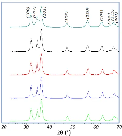
measurements. In Figure 1 are reported the X-ray diffraction patterns of the 5 as-prepared
compounds. The compounds exhibit a pure würtzite structure, i.e. a single ZnO-type solid
solution is obtained whatever the target composition. The efficient doping concentration, which
was previously shown to differ from the target concentration in such a process used for the
obtaining of Zn1-xAlxO or Zn1-xGaxO phases, [19, 20] was titrated using ICP measurements.
Figure 1. X-ray diffraction patterns for various Co2+-doped ZnO oxides obtained by
hydrolysis-condensation in DEG. a) ZC0, b) ZC05, c) ZC1, d) ZC2 and e) ZC3.
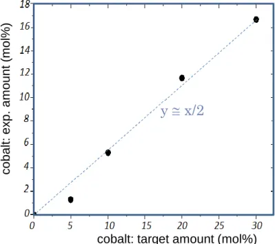
The evolution of the efficient cobalt concentration versus the target one is plotted in Figure
2. The dependency of the efficient concentration versus the target concentration is clearly linear
and the curve’ slope indicates that half the cobalt ions in the raw polyol medium are efficiently
20 30 40 50 60 70
6 introduced in the Zn1-xCoxO compounds: there is a constant factor of 2 in between the efficient
and target cobalt concentrations.
Figure 2. Co2+ concentration efficiently introduced in Co-doped ZnO particles versus the Co2+
target concentration (corresponding to the ratio between Zn2+ and Co2+ ions introduced in the
DEG synthesis medium).
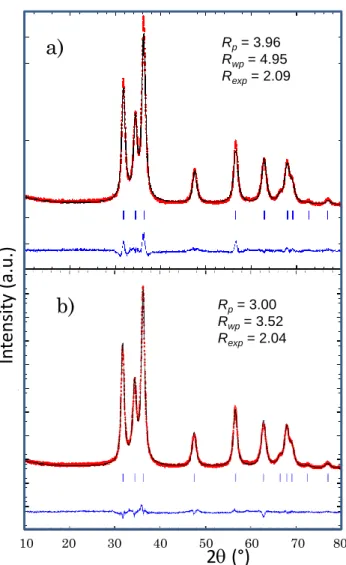
Full pattern matching was performed on the X- ray pattern of the various Zn1-xCoxO
compounds. Two models were used for the full-pattern matching process: (i) a model using for
the peak profile an isotropic peak profile function (Caglioti function, N°5 in FullProf package);
(ii) an anisotropic peak profile function (N°7 in FullProf package). Indeed, previous works have
already shown that ZnO particles obtained from the polyol process are constituted of anisotropic
crystallites shape: olive shapes, with a (001) crystallographic orientation (large diameter
direction). Consequently, the peak width has to be correlated to the peak’s Miller indexes, using
an anisotropic model for X-ray peak profile description. For illustration, the results obtained
using these two different refinement processes on the ZC2 compound are reported in Figure 3.
One can see that the correlation factors: Rp, Rwp, Rexp are both significantly decreased while an cobalt: target amount (mol%)
co b a lt: e x p . a m o u n t (m o l%)
y
x/2
7 anisotropic peak profile is taken into account, in comparison pattern matching with the use of
an isotropic peak profile. Hence, the cell parameters and correlation factors (Rp, Rwp and Rexp)
reported in the Table 1 are extracted from the refinements using the anisotropic peak profile
function. Also, the evolutions of the unit-cell parameters versus the cobalt concentration
(efficient concentration in the mixed oxide) are plotted in the Figure 4. The proximity of Co2+
and Zn2+ Shannon ionic radii: Co2+ in (IV) coordination site equals 0.58 Å while ionic radius
of Zn2+ in (IV) coordination site equals 0.60 Å, makes the variation of the cell parameters with
composition very light. Nevertheless, a clear tendency of a decrease of the unit-cell volume can
be observed. This unit-cell volume decrease is due to the decrease of the a cell parameter
(Figure 4a) and c cell parameter (Figure 4b) into the same proportion (i.e., an homothetic
decrease of both unti-cell parameters is observed). Indeed, the c/a ratio remains in a very narrow
range: in between 1.6023 and 1.6026 values (Figure 4c).
Table 1. Unit-cell parameters and reliability factors obtained on the various Co2+-doped ZnO
oxides.
Target compound Effective compound a c Rp Rwp Rexp
ZnO-Co 0% ZnO-Co 0% 3.25286(16) 5.21277(30) 3.0 3.5 2.0 ZnO-Co 5% ZnO-Co 1.5% 3.25256(35) 5.21258(55) 3.8 3.8 2.1 ZnO-Co 10% ZnO-Co 5.5% 3.25269(17) 5.21204(28) 2.8 3.2 1.9 ZnO-Co 20% ZnO-Co 12% 3.25238(16) 5.21162(27) 2.6 3.9 1.9 ZnO-Co 30% ZnO-Co 16% 3.25238(22) 5.21182(22) 2.9 3.6 1.9
8
Figure 3. (a) Full-pattern matching using isotropic peak profile function on the Zn0.88Co0.12O
(ZC2) oxide composition ; (b) full-pattern matching using an anisotropic peak profile function
on same compound.
Figure 4. Evolution of the unit-cell-parameters of Co-doped ZnO powders versus the cobalt
concentration. Rp= 3.96 Rwp= 4.95 Rexp= 2.09 10 20 30 40 50 60 70 80
2
q
(°)
inte nsi ty ( a.u .) inte nsit y ( a.u .)a)
b)
Rp= 3.00 Rwp= 3.52 Rexp= 2.042
q
(°)
In
ten
sity
(a.
u
.)
3.2523 3.2524 3.2525 3.2526 3.2527 3.2528 3.2529 0 5 10 15 20 5.2114 5.2116 5.2118 5.212 5.2122 5.2124 5.2126 5.2128 5.213 0 5 10 15 20 1.60235 1.6024 1.60245 1.6025 1.60255 1.6026 1.60265 0 5 10 15 20cobalt: exp. amount (mol%)
a p a ram e te r (Å) c pa ram et e r (Å) c/a ratio
cobalt: exp. amount (mol%) cobalt: exp. amount (mol%)
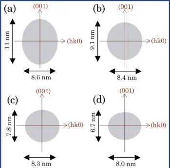
9 From anisotropic peak profile refinements, it is possible to extract directly the crystallite
shape of the as-prepared Zn1-xCoxO crystallites with great accuracy. The crystal shapes are
drawn in Figure 5. The pure ZnO compounds, already studied using same polyol route and
using same experimental condition exhibits the same crystal shape and size than the first ZC05
compound (i.e. the impact of the doping for such low cobalt concentration is negligible).
However, a significant evolution of the crystal size (volume) and shape (needle-like anisotropic
distortion) is shown with increasing cobalt concentration. From an olive shape with a large
diameter along the (001) axis, a length of 11 nm/ a width of 8.6 nm, the particles with increasing
the cobalt concentration tend to become more spherical and with a smaller crystallite size. Even,
a reversal of the anisotropy is observed for the ZC3 compound, with a width along (kk0)
direction becoming larger than the length along (001) direction. Besides the monotonic
evolution of the cell parameters versus the cobalt concentration, the monotonic modification in
the crystallite shapes supports the efficient and homogeneous introduction of cobalt ions inside
the würtzite crystallographic network.
Figure 5. Crystallite’s average morphology obtained from X-ray diffraction pattern refinements
using anisotropic peak profile function.
(001) (hk0) (hk0) (hk0) (hk0) (001) (001) (001) 11 nm 8.6 nm 9.1 nm 7.8 nm 8.4 nm 8.3 nm 8.0 nm 6.7 nm
(a)
(b)
(d)
(c)
10 SEM images were also made on various ZCX powders. The images reveal Co2+-doped ZnO
particles are made of spherical aggregates with sub-micron diameter in good agreement with
our previous reports on the synthesis of Ga3+-doped ZnO particles from polyol synthesis
(Figure 6) [19, 20]. The obtaining in a reproducible way of spherical aggregates is shown, i.e.
whatever the cobalt concentration very similar powder architecture is observed. Nevertheless,
an accurate study of the aggregate (particle) size distribution from image treatment has allowed
a significant evolution versus the cobalt concentration to be shown. Indeed, in the case of low
cobalt concentration a bimodal distribution for the particle size, with approximatively half-half
concentration, with centers located at 225 nm and 425 nm, is evidenced whereas, for high cobalt
concentration, a single mode centered on 225 nm is observed (i.e. the biggest aggregates have
disappeared) (Figure 7). Maybe, the change of the aggregate size can be correlated positively
with the change of crystallite size versus cobalt distribution, since for both scales a slight
tendency to the size decrease is noted. Considering that the aggregates have an average diameter
of about 200-400 nm and are constituted of a random pack (with about 40% of porosity) [19]
of olive-like crystallites (about 10 nm size), the aggregates are constituted of about 104
11
Figure 6. SEM photograhs of the various Co2+-doped ZnO spherical particles obtained by
hydrolysis-condensation in DEG (SEM microscopy). a) ZC0, b) ZC05, c) ZC1, d) ZC2 and e)
ZC3.
a)
b)
c)
d)
e)
12
Figure 7. Particle size distribution for ZC05 (red lines) and ZC2 (blue lines) particles
III.2. Co2+-doped ZnO@SiO2 core-shell powder
Three different amounts of TEOS were used in order to vary the shell thickness deposited
via sol-gel process on the Co-doped ZnO powder, the experiments being performed on the
ZC20 sample. The three as-prepared core@shell samples, from the thinner to the thicker silica
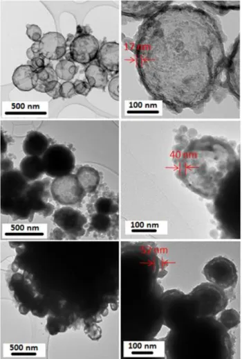
shell, are noted ZC20@SiO2-1, ZC20@SiO2-2 and ZC20@SiO2-3. TEM images for the
ZC20@SiO2-1 sample, before and after acid attack are reported on Figure 8. The silica shells
are evidenced by the acid attack which leads to a total dissolution of the ZC20 cores. It can be
seen, that quite homogeneous shells are obtained with just few silica nuclei due to the
competition between shell growth and secondary germination. TEM pictures on the three
ZC20@SiO2-1, ZC20@SiO2-2 and ZC20@SiO2-3 samples are compared in Figure 9, all the
pictures being taken after acid attack. Three main results can be extracted from these TEM
observations: (i) the silica shell thickness well increases from ZC20@SiO2-1 to ZC20@SiO2-2
and ZC20@SiO2-3, (ii) the agglomeration of the spherical aggregates increases with the amount 0 10 20 30 40 50 60 70 n umber of pa rt icles particle size
13 of TEOS used, certainly in correlation with the appearing of more and more silica colloids, (iii)
the silica shell becomes resistant to the acid attack and protect the core particle while the
thickness of the shell increases.
Figure 8. TEM photographs on the 12 mol% Co2+-doped ZnO core-shell compound
(ZC2@SiO2-1) before acid attack (a) after acid attack (b).
Figure 9. TEM photographs on the ZC2@SiO2 compounds with various average shell
14 III.3. Optical properties
In a first step, the optical properties were studied on the Co-doped ZnO powders. The
diffuse reflectivity K/S Kubelka-Munk transforms are plotted in Figure 10. For tetrahedral
symmetry, with regard to Tanabe−Sugano diagrams established for a 3d7 electronic configuration, three transitions corresponding to ν1 (4A2(F)→4A1(F)), ν2(4A2(F)→4T1(F)), and
ν3(4A2(F)→4T1(P)) are allowed [21]. For cobalt(II) ions the first two transitions are known to be located in the infrared with a wavelength of about 1400 and 1600 nm, respectively. The third
one is known to appear as a triplet due to the L−S Russel−Sanders coupling for which the maximal absorptions, for instance, in CoAl2O4 where the punctual symmetry of the tetrahedral
site is Td, are around 540, 590, and 640 nm.12 The visible triple transition, centered in the orange
region, leads to blue coloration when the material color is only due to the cobalt (II) ion located
in a tetrahedral site as a unique chromophore. The reported diffuse reflectance spectra for the
different cobalt rates clearly show that the visible range triple transition as well as a large
infrared peak between 1200 and 1700 nm associated to the convolution of ν1(4A2(F)→4A1(F))
(located here at 1640 nm: Figure 10b), ν2 (4A2(F) → 4T1(F) (located here at 1310 nm) low energy transitions, both characteristic of cobalt in a tetrahedral site, are well taking place
whatever the cobalt rate studied. Furthermore, the positions of the triple ν3 transition, calculated
from raw the K/S transform, remain about the same whatever the cobalt content at 565, 610,
and 660 nm (Figure 10b). Hence, one can assume that from the remarkable stability of the 3 (ν1,
ν2, ν3) absorption bands positions versus cobalt concentration, that cobalt adopts in all cases, at room temperature, its thermodynamically stable environment; i.e., as already assumed from
X-ray diffraction analyses, cobalt is located in a nearly isotropic tetrahedral site. The evolution of
the K/S intensity of the cobalt d-d absorption bands (focusing on ν1,and ν3 transitions) are plotted
in Figure 11 (evolution of the 660 nm peak intensity reported in Figure 11a , evolution of the
15
Figure 10. Diffuse reflection spectra (Rdiff%) (a) and K/S absorption spectra (b) of the various
Co2+-doped ZnO oxides.
.
a)
b)
wavelength (nm)
wavelength (nm)
K/
S
tr
an
sf
or
m
D
if
fu
se
re
flect
ivit
y
(%
)
565 610 660 1310 164016
Figure 11. Intensity at the maximum of the two absorption triplets in the visible range (c) and
the infrared range (d) associated with the d-d Co2+ transition versus cobalt concentration in the
various Co-doped ZnO compounds.
In a remarkable manner, the intensity of both transitions in visible range as well as in
infrared range evolves in a linear way versus cobalt concentration. That is a new evidence for
the efficient introduction inside the würtzite crystal of the cobalt previously titrated by ICP in
the powder. Thanks to the control of the d-d transitions intensity versus cobalt concentration a
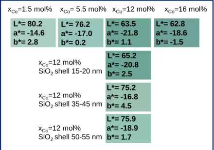
large scale of green compounds can be obtained. The L*a*b* parameters, extracted from the
diffuse reflectance curve, using x, y, z color space system defined from CIE (1961), are
regrouped in Figure 12. Obviously, for low cobalt concentration the green pigment exhibits a
a)
b)
K/S in ten sity (vi s. p eak ) K/S in ten sity (IR p eak ) cobalt concentration cobalt concentration17 less saturated green color that for higher doping rates. Hence, the L* luminosity parameter
gradually decreases versus the cobalt concentration from 80.2 for 1.5 mol% of cobalt efficiently
introduced in ZnO down to 62.8 for the 16 mol% Co-doped ZnO. Interestingly, the green hue,
which is directly characterized by the a* value (red hue - green hue axis), shows a saturation
maximum for intermediate cobalt concentration: the most saturated green color is obtained for
the 12mo% Co-doped ZnO pigment (ZC2 sample). That is why the coatings of the pigment
particles by silica shell were made on this sample.
Figure 12. L*a*b* parameters and coloration of the different Co-doped ZnO compounds and
Co-doped ZnO@SiO2 core-shell compounds prepared in this publication (corresponding to the
ZC2@SiO2-1, ZC2@SiO2-2, ZC2@SiO2-3 samples).
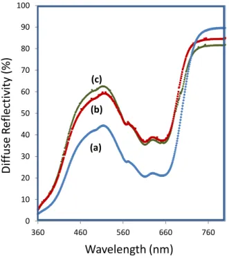
The diffuse reflectance focusing on the UV-visible range of the various core@shell
materials are joined in Figure 13. It can be seen an increase in the visible reflectivity while the
silica shell thickness increase. The comparison of the obtained colours of the three core@shell
compounds and the core compound are also reported in Figure 12. Consequently to the addition
of silica shells, the Luminosity parameter tends to increase versus the shell thickness. However,
the green hue is only slightly impacted by the shell, and very beautiful green hue: a* = -18.9,
L*= 80.2 a*= -14.6 b*= 2.8 L*= 76.2 a*= -17.0 b*= 0.2 L*= 63.5 a*= -21.8 b*= 1.1 L*= 62.8 a*= -18.6 b*= -1.5 L*= 75.2 a*= -16.8 b*= 4.5 L*= 75.9 a*= -18.9 b*= 1.7
xCo=1.5 mol% xCo= 5.5 mol% xCo=12 mol% xCo=16 mol%
xCo=12 mol% SiO2 shell 15-20 nm xCo=12 mol% SiO2 shell 35-45 nm xCo=12 mol% SiO2 shell 50-55 nm L*= 65.2 a*= -20.8 b*= 2.5
18 b* = 1.7 remains for the ZC2@SiO2-3 sample. For this sample, the combination of optimal
color properties and of a chemical stability (for illustration besides acid attack) is so reached.
Figure 13. Diffuse reflection spectra (Rdiff%) of the ZC2@SiO2 compounds with various
average shell thickness : a)ZC2@SiO2-1, b) ZC2@SiO2-2, c)ZC2@SiO2-3.
IV. Conclusion
In this paper, the polyol synthesis route has allowed the obtaining of Zn1-xCoxO
compounds, with nanometric crystallites (olive shape) / micrometric spherical aggregates, a
large range of cobalt concentration associated to a large deep green – pale green scale of
colorations. The color of the as-prepared powders is attributed to the efficient substitution of
the zinc ions by cobalt (II) ions inside the tetrahedral sites of the würtzite phase. The spherical
aggregates can be easily coated via sol-gel process by silica shells with tuned thickness in order
to limit pigment’s toxicity; this, without a significant decrease of the colorimetric properties.
Acknowledgements 0 10 20 30 40 50 60 70 80 90 100 360 460 560 660 760 (a) (b) (c) Wavelength (nm) Dif fus e refle xi o n (%) Wavelength (nm) D if fu se R ef lect ivit y (% )
19 The authors are thankful to L’Agence Nationale de la Recherche for financial support, under the ChoCoComp project (No. ANR-13-RMNP-0011)
References
[1] Šulcova P, Trojan M. New green pigments; ZnO–CoO. Dyes & Pigments.1999; 40:83-86.
[2] Zhou N, Zhang Y, Nian S, Li W, Li J, Cao W, Wu Z. Synthesis and characterization of
Zn1-xCoxO green pigments with low content cobalt oxide. J Alloys Compds 2017;711:406-413.
[3] Karasu B, Turan S. Effects of cobalt, copper, manganese and titanium oxide additions on
the microstructures of zinc containing soft porcelain glazes. J. Eur. Ceram. Soc.
2002;22:1447-1455.
[4] Cui H, Zayat M, Levy D. Nanoparticle Synthesis of Willemite Doped with Cobalt Ions
(Co0.05Zn1.95SiO4) by an Epoxide-Assisted Sol-Gel Method. Chem. Mater. 2005;17:5562-5566.
[5] Gaudon M, Toulemonde O, Demourgues A. Green Coloration of Co-Doped ZnO Explained
from Structural Refinement and Bond Considerations. Inorg. Chem. 2007;46: 10996-11002.
[6] Milao TM, Oliveira JFA, Araujo VD, Bernardi MIB. Zn0.97M0.03O (M = Co, Fe, and V)
pigments: thermal, structural, and optical characterization. J Therm Anal Calorim
2011;103:873–877.
[7] Siang Yeo B, Bell AT. Enhanced Activity of Gold-Supported Cobalt Oxide for the
Electrochemical Evolution of Oxygen. J. Am. Chem. Soc. 2011;133:5587–5593.
[8] Chanda A, Gupta S, Vasundhara M, Joshi SR, Mutta GR, Singh J. Study of structural,
optical and magnetic properties of cobalt doped ZnO nanorods. RSC Adv., 2017;7:50527–
50536.
[9] Hammad TM, Salem JK, Harrison RG. Structure, optical properties and synthesis of
20 [10] Gandhi V, Ganesan R, Hameed Abdulrahman Syedahamed H, Thaiyan M. Effect of Cobalt
Doping on Structural, Optical, and Magnetic Properties of ZnO Nanoparticles Synthesized by
Coprecipitation Method. J. Phys. Chem. C 2014;118:9715−9725.
[11] Woo HS, Kwak CH, Chung JH, Lee JH. Co-Doped Branched ZnO Nanowires for
Ultraselective and Sensitive Detection of Xylene. ACS Appl. Mater. Interfaces
2014;6:22553−22560.
[12] Senapati S, Kar Nanda K. Designing Dual Emissions via Co-doping or Physical Mixing of
Individually Doped ZnO and Their Implications in Optical Thermometry. ACS Appl. Mater.
Interfaces 2017;9:16305-16312.
[13] Hyun Kim J, Kim H, Kim D, EonIhm Y, Choo WK. Magnetoresistance in laser-deposited
Zn1–xCoxO thin films. Physica B: Condensed Matter, 2003;327:304-306.
[14] Bouloudenine M, Viart N, Colis S, Dinia A. Bulk Zn1−xCoxO magnetic semiconductors
prepared by hydrothermal technique. Chem. Phys. Lett. 2004;397:73-76.
[15] Xu X, Cao C, Chen Z. Effects of temperature and atmosphere on the magnetic properties
of Co-doped ZnO rods. J Magn Magn Mater. 2011;323:1886-1889.
[16] Birajdar SD, Khirade PP, Bhagwat VR, Humbe AV, Jadhav KM. Synthesis, structural,
morphological, optical and magnetic properties of Zn1−xCoxO (0 ≤ x ≤ 0.36) nanoparticles synthesized by sol-gel auto combustion method. J Alloys Compds 2016;683:513-526.
[17] Liu S, Han M.-Y. Silica-coated metal nanoparticles. Chem. Asian J. 2010:5:36–45.
[18] Chia SL, Leong DT, Reducing ZnO nanoparticles toxicity through silica coating. Heliyon
2016:2:e00177.
[19] Trenque I, Gaudon M, Duguet, E, Mornet S. Visible-transparent and UV/IR-opaque colloidal
dispersions of Ga-doped zinc oxide nanoparticles. New J. Chem., 2016;40:7204-7209.
[20] DengPan N, Tao X, Yu Z, XiangJun L. Synthesis and structure analysis of aluminum doped
21 [21] Robertson L, Duttine M, Gaudon M, Demourgues A. Cobalt/Zinc Molybdates as New Blue
Pigments Involving Co2+ in Distorted Trigonal Bipyramids and Octahedra. Chem. Mater.