BUSINESS MODEL AND STRATEGY ANALYSIS FOR RADIOLOGISTS TO USE
ACIVES
ELECTRONIC HEALTH RECORDS (EHR) - H T - UT
by
Palani Perumal
B.E., Electrical and Electronics Engineering Madras University, 1996
Submitted to the System Design and Management program in partial fulfillment of the requirements for the degree of
Master of Science in Engineering and Management at the
Massachusetts Institute of Technology
February 2012
@2012 Palani Perumal. All rights reserved.
The author hereby grants to MIT permission to reproduce and to distribute publicly paper and electronic copies of this thesis document in whole or in part in
any medium now known or hereafter created.
Signature of Author:
System Design and Management Program
Febr 10, 2012
Certified by: LDr.
John D. Halamka, MD, MS 'Professor and CIO, Harvard Medical School,
CIO, Beth Israel Deaconess Medical Center
Thesis Supervisor
Certified by: V
Dr. Max P. Rosen, MD, MPH Associate Professor of Radiology, Harvard Medical School, Vice Chairman of Radiology, Beth Israel Deaconess Medical Center Thesis Supervisor
Accepted by:
Prof. Frank Levy Professor of Urban Economics Department of Urban Studies and Planning
Massachusetts Institute of Technology Thesis Reader
Patrick Hale Director, Syte ign and Management Program Massachusetts Institute of Technology Accepted by:
BUSINESS MODEL AND STRATEGY ANALYSIS FOR RADIOLOGISTS TO USE
ELECTRONIC HEALTH RECORDS (EHR) by
Palani Perumal
Submitted to the System Design and Management Program in February, 2012 in Partial Fulfillment of the Requirements for the
Degree of Master of Science in Engineering and Management
ABSTRACT
Radiology is a medical specialty that employs imaging to diagnose and treat disease. It has long been an advance user of technology to capture, store, share, and use images electronically. In 2009, President Obama signed into law a measure, the HITECH Act
(part of the stimulus package), that incentivizes healthcare providers to use electronic health records (EHR) in care delivery to improve quality, efficiency, safety, and reduce cost.
The meaningful use (MU) program's Stage 1 requirements (part of HITECH Act) did not include imaging requirements, leading to confusion among radiologists and other specialties with regard to what MU offers to and requires of them. This thesis attempts to clarify the contribution radiology can make to MU by understanding radiology as a system, including its surrounding issues and its drivers, using Stage 1 MU
requirements, data from qualitative research, and results from analysis. It answers the following question:
Should Radiologists be considered part of the care team, leveraging EHR for meaningful use and hence eligible for incentive payments?
It does so via the following methods:
a) Discussing in detail current issues surrounding radiology systems from quality, safety, efficiency, and cost perspectives;
b) Discussing MU in the context of radiology and reviewing what is missing in it for
radiologists;
c) Providing deeper systems analysis of current behaviors and why they have this form at this time; and
d) Explaining how MU objectives can help to overcome many current issues and
ultimately help to improve health outcomes. Specific changes to MU criteria to achieve these benefits are recommended.
This thesis employs systems concepts and tools including system architecture and system dynamics for research and analysis to understand the system and derive hypotheses. A system dynamics model is used to analyze current drivers in imaging and to clarify the impact MU can have on these drivers. Thesis conclusions are supported by the analysis performed using the model as well as information gathered
through industry interviews, online articles, academic and industry journals, and blogs.
Thesis Supervisor 1: Dr. John D. Halamka, MD, MS
Title: Professor and CIO, Harvard Medical School, CIO, Beth Israel Deaconess Medical Center
Thesis Supervisor 2: Dr. Max P. Rosen, MD, MPH
Title: Associate Professor of Radiology, Harvard Medical School, Vice Chairman of Radiology, Beth Israel Deaconess Medical Center
ACKNOWLEDGEMENTS
This thesis is the culmination of my three-year long venture as part of the System Design and Management (SDM) program at MIT, which I commenced in January 2009. With a full-time job at Microsoft Corporation (based in Seattle, Washington), and two young kids, completing this program (based in Cambridge, Massachusetts) has been
nothing short of challenging. In the process of undertaking this daunting initiative, I have incurred innumerable debts to teachers, colleagues, institutions, friends and family.
For their guidance, support and patience, I am most grateful to my thesis advisors
-Dr. John D. Halamka and -Dr. Max P. Rosen. Their invaluable insights, constructive criticism and positive encouragement have been key to the completion of this thesis. Their efforts in helping me connect with various industry references and contacts for data gathering have been tremendous. I am also very thankful to Prof. Frank Levy for graciously offering to review my thesis and going beyond his realm of duty in providing me with timely feedback to steer the thesis in the right direction. Additionally, I wish to thank Dr. David J. Hartzband (Lecturer, Engineering Systems Division at MIT) who was my initial advisor and had offered significant insights on Healthcare IT. However, upon acknowledging, at an early stage, the challenges in access to data and difference in area of interests, he graciously accepted my decision to change the direction of my research.
Many thanks to the staff at Beth Israel Deaconess Medical Center (BIDMC) for their time and contribution to my research. I would specially like to mention Laurie Pascal, Oleg Pianykh, Donna Tobey Hallett, Dr. Katherine Dallow, Jesse L. Wei, Jim Brophy, James Hamilton, Patricia M. Gardner and Loma Puerto. I am also thankful to Inland Imaging, especially Jon Copeland, Dr. Tasneem Lalani, and Kevin Kirk. Amongst the many in Radiology and Technology industry to whom I am most appreciative for their generosity in taking the time to converse with me and share their thoughts on the subject of my research, I would like to extend my sincere thanks to Dr. Keith J. Dreyer (Massachusetts General Hospital), Steve Fox (Blue Cross Blue Shield, MA), Donald Rucker (Siemens Healthcare), and Jacques Gilbert (GE Healthcare).
I would like to thank Dr. Patrick Hale, Director of SDM program, for his continual support throughout the duration of my association with the program. My sincere thanks to William F Foley who has been my primary contact at the SDM program office, for distance education support, registration, thesis submission and more. In addition, I would like to thank the staff at SDM program office, namely, Christine L. Bates, Jeff Shao and Alam Elalam, for their valuable assistance.
I would like to thank my Managers at Microsoft - Fuyau Lin, Elsie Nallipogu and Mario Garzia -for their generous support and flexibility during my tenure with the program.
I wish to express my most sincere appreciation to Larry Clifford Gilman, who made
himself available at short notice to patiently review every chapter of this thesis and make substantial editorial changes.
For her ongoing friendship, discussions, encouragement and support over the years, I wish to convey my heartfelt thanks to Najma Huq, my colleague and friend for more than a decade. My gratitude to her for proof-reading every chapter at short notice. My special appreciation to her and her husband Erfan Ahmed for providing me with a home away from home whenever I had to be on campus.
Finally, I wish to thank the various members of my family who have endeavored in their own special ways, to allow me the opportunity to educate myself. I am eternally grateful to my Uncle P.K. Adhikesavan and Aunt A. Lakshmi who have played the vital role of adoptive parents in my life. I would not be where I am today without their generous initiative, support and guidance. My heartfelt thanks and gratitude to my parents P.K. Perumal and Malliga Perumal for their continual encouragement and support throughout my life. I am especially thankful to my parents-in-law D.
Prakasam and P. Sulochana for their generosity of spirit in helping my wife at home while I had to be away on campus. Last but not least, I would like to thank my wife Jayabarathy Palani without whose patience, love and support, I would not have been able to complete this program. She has been my staunch ally from the very start, and has endured a second and difficult pregnancy mostly on her own. For her unwavering support and initiative to run the household with two kids in order to allow me to focus on my studies, I shall be forever in her debt. I am thankful to my children Vishwesh
and Deethashree for providing me with laughter and joy just when I needed it most.
My sincere thanks to my brother Saravanan Perumal and sister Santhi Suresh for the
TABLE OF CONTENTS
1. INTRODUCTION ... 15
1.1. Healthcare in the US ... 15
1.2. Thesis Scope and Objectives... 16
1.3. Research M ethodology ... 17
2. RADIOLOGY ECOSYSTEM ... 18
2.1. Advances in Radiology...18
2.2. Hospital Inform ation System ... 18
2.2.1. Single-Vendor Enterprise System ... 19
2.2.2. Best-Of-Breed Architecture... 20
2.2.3. Hybrid Hom egrow n System ... 22
2.2.4. HIS at BIDIMC ... 23
2.3. Radiology System Com ponents... 25
2.3.1. Radiology Inform ation System ... 25
2.3.2. M odalities...25
2 .3 .3 . D IC O M ... 2 5 2 .3 .4 . P A C S ... 2 7 2.3.5. Dictation and Transcription... 27
2.3.6. Users and Needs...28
2 .4 . M o d a litie s ... 2 9 2 .4 .1 . X -R a y ... 2 9 2.4.2. Ultrasound...29
2.4.3. Com puted Tom ography ... 30
2.4.4. M agnetic Resonance Im aging ... 30
2.4.5. Positron Em ission Tom ography ... 31
2.5. W orkflow at BIDM C...31
2.6. Reim bursem ent M odel ... 3 4 27. Trends in Radiology ... 38
2.7.1. Rising Im aging Spending ... 38
2.8. System s Issues...42
2.8.1. DICOM Standards Issues ... 43
2.8.2. Inform ation System (IT) Issues...44
3. M EANINGFUL USE AND RADIOLOGY ... 47
3.1. Electronic Health Record...47
3.2. HITECH Act and M eaningful Use ... 47
3.2.1. Overview ... 47
3.2.2. M eaningful Use ... 48
3.2.3. CM S Incentive Program s ... 48
3.2.3.1. Overview of the Two Program s... 48
3.2.3.2. M edicare EHR Incentive Program ... 50
3.2.3.3. M edicaid EHR Incentive Program ... 51
3.2.3.4. Differences between M edicare and M edicaid Program s ... 53
3.2.4. Process Overview ... 53
3.3. Stage 1 Objectives and M easures ... 55
3.3.1. Core Objectives...55
3.3.2. M enu Set Objectives ... 56
3.3.3. Clinical Quality M easures ... 57
3.4. Certified EHR Technology...59
3.4.1. Standards... ... 60
3.4.2. Certifications ... 61
3.4.3. Certified EHR Products... 61
3.5. State rofRadiology in M U ... 63
3.5.1. RadiologyinStage ... 63
3.5.2. Perspectives of Radiologists ... ... 64
3.5.3. Perspectives of EHR Vendors... 67
4. BUSINESS ANALYSIS FOR RADIOLOGISTS... 69
4.1. Analysis of Drivers in Im aging ... 69
4 .2 . R e su lts ... ... 7 5 4.2.1. Radiology for M U ... ... 75
4.2.1.2. Im aging Data Sharing and Exchange ... 77
4.2.1.3. Clinical Decision Support System ... 78
4.2.1.4. M U Im pact on Im aging Drivers ... 80
4.2.2. Revisions to Reim bursem ent Policy ... 83
4.2.3. Changes to M U for Radiology ... 85
5. Conclusions ... 88
LIST OF TABLES
Table 1. Radiology system stakeholders and needs. ... 28
Table 2. CM S 2010 M edicare physician fee schedule ... 35
Table 3. Code specific impacts of CMS 2010 Medicare physician fee schedule ... 36
Table 4. Three stages of Meaningful Use and its objectives...49
Table 5. M edicare EHR Incentive Program details...50
Table 6. Medicare incentive payment schedule based on first CY of payment...51
Table 7. M edicaid Incentive Program details... 51
Table 8. Medicaid patient volume thresholds by provider type... 52
Table 9. Medicaid EHR incentive payments by calendar year. ... 52
Table 10. Differences between Medicare and Medicaid her incentive programs... 53
T a b le 11 . O N C A T C B List...6 2 Table 12. Current radiology EHR product list... 62
LIST OF FIGURES
Figure 1. HIS Architecture: single-vendor enterprise system. ... 19
Figure 2. HIS architecture: best-of-breed. ... 21
Figure 3. HIS architecture: hybrid system. ... 23
Figure 4. HIS system at BIDMC...24
Fig u re 5 M o d a lity : X -ray...29
Figure 6 Modality: ultrasound...29
Figure 7 Modality: computed tomography... 30
Figure 8 Modality: magnetic resonance imaging... 30
Figure 9 Modality: positron emission tomography... 31
Figure 10. Radiology workflow at BIDMC. ... 32
Figure 11. Number of MRI units/million persons for 2006... 39
Figure 12. Spending under Medicare for different services. ... 39
Figure 13. Radiology system drivers. ... 72
Figure 14. Radiology in the care delivery cycle... 76
Figure 15. Likely MU impact on imaging dynamics... 77
Figure 16. M U impact on imaging study ordering process ... 80
Figure 17. M U influence on imaging drivers... 82
1. INTRODUCTION 1.1. Healthcare in the US
The United States spends more per capita on healthcare than any other industrialized nation, but lags most other countries in outcome and healthcare coverage.
Government and private payers reimburse for quantity, not quality, of healthcare. There is little coordination among providers in care delivery, and data are siloed in hospitals, clinics, labs, and pharmacies, locked away in proprietary systems.
Since its inception, radiology has gone through dramatic technological advances that have significantly improved the physician's capability to diagnose disease conditions earlier and more accurately, and in some cases treat them radiologically, saving thousands of lives. Radiology services are utilized for diagnosis and treatment across multiple specialty areas, including orthopedics, cardiology, cancer treatment, and many others. This tremendous growth has also brought unintended changes in physician and vendor behavior, leading to increased inappropriate utilization of imaging services. The effects range from increased healthcare spending to patients getting exposed to unnecessary radiation. In 2006, 13.3% of diagnostic imaging tests conducted in US was redundant or unnecessary [46]. Thus, both quality and safety issues plague the system.
On February 17, 2009, President Obama signed the American Recovery and Reinvestment Act of 2009 (a.k.a. "the Stimulus Bill") into law. This includes a provision to spend up to $19.2 billion to increase use of electronic health records (EHR) to increase healthcare quality and reduce cost. This part of the Act, referred as the HITECH (Health Information Technology for Economic and Clinical Health) Act, provides guidelines on EHR standards and certifications as well as incentives for healthcare players to adopt certified EHR technology. The government strongly believes that use of EHR will lead to several benefits, and accordingly there is an unprecedented drive to adopt EHR across the US.
As specified by the HITECH Act, the Center for Medicare and Medicaid Services (CMS) manages the Meaningful Use incentive program, whose current Stage 1 requirements focus on primary care. This has created confusion among radiologists (and other
specialties within medicine) about whether they are eligible for the incentive payments or will be penalized later for not meeting Stage 1 requirements. Also, it is questionable whether MU program's primary objective of improving quality, safety, and efficiency is
achievable in current form, i.e., as part of the healthcare delivery process, and thus not directly addressing radiologists' needs.
This thesis examines the dynamics of the radiology specialty, including driving factors and causal relationships of technology, business, and policy conditions at this time. The following subsection explains in detail its scope, objectives, and methods.
1.2. Thesis Scope and Objectives
First, this thesis studies the current radiology system to understand its ecosystem, stakeholders, and technological subsystems, as well as issues in the areas of
behaviors (i.e., usage, environmental), how radiologists are perceived in the healthcare ecosystem, technology, and policy. A study was conducted, primarily in Beth Israel Deaconess Medical Center, to characterize the current workflow, systems in use
(including information technology [IT] systems), a literature review, and interviews with industry experts.
Second, MU Stage 1 requirements are reviewed in detail in the context of radiology, with discussion of core requirements, eligibility standards, incentives, and penalties. The impact of MU Stage l's omission of imaging and other concerns relevant to radiology is discussed.
Third, using a system dynamics model provides insight into drivers in current imaging behaviors and possible MU impact could be on those drivers.
This thesis answers the following question:
Should Radiologists be considered part of the care team, leveraging EHR for meaningful use and hence eligible for incentive payments?
It does so via the following methods:
a) Discussing in detail current issues surrounding radiology systems from quality, safety, efficiency, and cost perspectives;
b) Discussing the HITECH Act in the context of radiology and reviewing what is
missing in it for radiologists;
c) Providing deeper systems analysis of current behaviors and why they have this form at this time; and
d) Explaining how MU objectives can help to overcome many current issues and
ultimately help to improve health outcomes. Specific changes to MU criteria to achieve these benefits are recommended.
The scope of this thesis is limited to radiology systems within United States.
1.3. Research Methodology
To understand the current radiology system and its issues, a literature review has encompassed industry and online journals, online articles, blogs, and academic
journals. In addition, extensive interviews conducted with personnel at Beth Israel and Deaconess Medical Center (BIDMC), including radiologists, the PACS administrator, the Operations Director, the Medical Director, personnel in Business Planning and Strategy, and personnel in Information Systems.
To understand MU requirements, an extensive literature study was performed on current legislation, which was discussed with select industry experts in this area. Participation in relevant conferences, interview with IT vendors, and other activities supported understanding of MU requirements.
This thesis employs systems concepts and tools such as system architecture and system dynamics models to analyze and understand the dynamics of the system under consideration and to formulate recommendations.
This thesis is primarily a qualitative study, but cites quantitative information where applicable. Recommendations are made on qualitative aspects of the radiology system, supported by (a) qualitative data collected through research as already described, (2) results from system analysis, and (3) considering the entire setup from a systems perspective.
2. RADIOLOGY ECOSYSTEM 2.1. Advances in Radiology
Medical imaging has revolutionized the practice of medicine. Medical imaging technology continues to advance rapidly, continually offering new life-saving
capabilities and new hope for winning the war against many devastating diseases. The radiology field already made great leaps in using advanced information technologies, especially as compared to other medical specialties.
Technologies such as computed tomography (CT) revolutionized the field by enabling physicians to look inside people without having to subject them to anesthesia and sharp blades. Imaging has enabled physicians to dissect patients without harming them. One could see things that one could not see before. The beauty of imaging is that one can visualize the anatomy before doing anything to the patient [5].
Advanced imaging such as CT and magnetic resonance imaging (MRI, which eliminates ionizing radiation exposure to patients), and, most recently, positron
emission tomography (PET) scans, which have brought cancer detection and treatment to a higher level, have given doctors whole new sets of data to work with. Small tumors that were often overlooked can be seen before they become big tumors. Vascular
lesions that might escape the trained eye of a surgeon can be found on a
high-resolution digital image. Cardiac imaging and circulation mapping have become more precise, disease can be diagnosed long before a patient becomes terminal, and
orthopedic surgeries can be avoided or at least confined in scope by using scans. In principle, costs could be controlled or even reduced because less surgery and hospital stays may be needed [5]. However, as shall be reviewed below in detail, this has not been the sole result of improvements in imaging technology.
2.2. Hospital Information System
A Hospital Information System (HIS) encompasses multiple subsystems ranging from
specialty departments to administrative, financial, clinical, laboratory, and pharmaceutical departments, all networked to function as a unified system in hospital-based healthcare delivery. Before delving specifically into the radiology
HIS and where radiology fits in. The overall architecture of HISs in US hospitals can
be grouped into three high-level categories based on number of subcomponents and interface type. Below the details of the three prominent architectures, and advantages and disadvantages of each type, are discussed. Subsequently, radiology information systems (RISs) and other technical components of radiology are discussed. This chapter concludes with discussion of workflow, reimbursement policy, and current issues in radiology.
2.2.1. Single-Vendor Enterprise System
A single-vendor system is a large-scale HIS that integrates core clinical functions and
operations in a hospital environment. The HIS stores all patient data, treatment
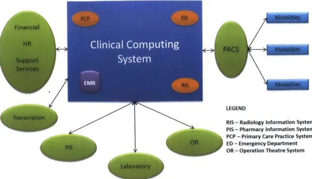
information, and other related data in a common database that is accessed by internal personnel directly. Information is (ideally) always consistent and up-to-date. Figure 1 represents a typical architecture in which the HIS comprises subsystems to manage primary care, intensive care unit, emergency department, pharmacy, radiology, pathology, etc. A few external systems are interfaced with the HIS for specialized functions, such as a PACS (Picture Archive Communication System), laboratory
systems, operation theater, and Enterprise Resource Planning (ERP) for financial/
human-resources/administration support services. HL7 is the global industry-standard protocol used to integrate software systems in healthcare informatics.
LEGEND
RIS - Radiology Information System PIS - Pharmacy Information System PCP - Primary Care Practice System ED - Emergency Department OR - Operation Theatre System
ICU - Intensive Care Unit System
Figure 1. HIS Architecture: single-vendor enterprise system.
Hospital Information
Some advantages of this type of HIS are:
e Clinical systems are integrated closely within the context of a single HIS
system, enabling consistent user interfaces across subsystems and an easier learning curve.
* Common data model: data and processes are integrated together, ensuring data integrity, consistency. Data are updated in real time from all integrated
systems.
* Fewer interface points: fewer integration issues, lower integration and operating costs.
* One vendor, one contract for most applications, thus fewer operational issues. Some disadvantages of this type of HIS are:
e Higher implementation cost at the beginning due to larger scope and
complexity; higher cost to get organizational consensus to re-engineer internal processes, which is often required for implementation.
e Risks of single-vendor lock-in (e.g., higher maintenance/update charges during
life time of usage) [161.
* High switching cost to change to different vendor or architecture.
* Some of vendor subsystems within HIS are top quality; others are average or below-average quality.
Major vendors in this space are Epic, Cerner, and Meditech.
2.2.2. Best-Of-Breed Architecture
Best-of-breed (BOB) HIS architecture seeks to employ the best products in each application specialty typically needed in a clinical environment for improved clinical and business performance. Figure 2 is an example where applications from multiple vendors are interfaced together in hub-and-spoke model using an Enterprise
Application Integration Engine (EAI) to connect them together. The role of an EAI is to simplify and automate business processes to the greatest extent possible [17] within
among connected applications or data sources. Some characteristics of the BOB model are a proprietary data model, owned by each application; a highly encapsulated
system boundary, limiting exposure to outside world know-how; lack of direct communication between applications; and a significantly larger number of interface points. An EAI controls the number of interface points required, thus reducing complexity of integrating subsystems; keeps information consistent across subsystems; and provides a common fagade interface to end-users.
LEGEND
RIS - Radiology Information System PIS - Pharmacy Information System PCP - Primary Care Practice System ED - Emergency Department OR - Operation Theatre System ICU - Intensive Care Unit System
Figure 2. HIS architecture: best-of-breed.
Advantages of BOB systems include:
e Providers get best-in-class clinical applications, which often improve clinical
performance, safety, and quality.
" Multi-vendor modularity of subsystems allows flexibility in business-process redesign to suit organizational requirements (unlike in single-vendor systems)
r 7fl-icationIntegration Engine
* Risks associated with long-term support for the system are distributed across multiple vendors; i.e., if a particular vendor falls out of the market, the whole system is not necessarily affected.
Disadvantages of BOB systems include:
* The higher number of interface points entails extensive integration costs,
resources, and time. With constant changes in the business environment, it is a challenge to keep the system updated.
* It is difficult to manage system integration of this complexity due to a shortage of EAI experts; the dynamic nature of an EAI requires a special skill set to manage such implementations. In 2003, it was reported that 80% of all EAI projects failed, primarily due to management issues [17].
* Significant integration issues arise from the complexity involved. Non-standard applications may lead to loss of productivity and to inefficiencies, hence to increased operational cost. Access to information is crucial at critical times in healthcare, and an inefficient operational environment poses a challenge to cost-effective management.
There are many EAI vendors; a few are Intersystems Ensemble, Microsoft BizTalk, and IBM Web Sphere.
2.2.3. Hybrid Homegrown System
This HIS type, essentially a combination of single-vendor and BOB system Figure 3, is prevalent in hospitals that originally developed home-grown EHR systems over the years. Third-party vendor applications usually outnumber single-vendor systems in
this architecture, but are not as numerous as in the BOB model. This architecture primarily relies on a single-vendor model and there is no EAI. The goal is to use the fewest vendors to get the job well done; its complexity resides in integrating third-party systems using internal IT-department resources. Resources are always limited and priority is given to crucial tasks that directly affect or benefit day-to-day clinical operations. System integration issues are prevalent in this environment for the reasons mentioned earlier, but complexity and frequency of issues are lower than is
I
Clinical Computing
1A E M R
Syste
m
Figure 3. HIS architecture: hybrid system.
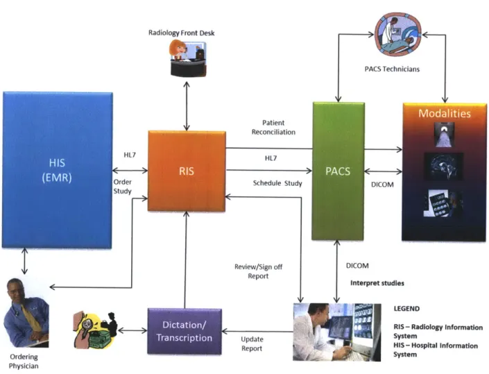
2.2.4. HIS at BIDMC
The current information system at BIDMC is an example of the hybrid homegrown HIS. It is the backbone of operations support; in particular, every step of the radiology workflow relies on it extensively. Figure 4 represents the current high-level IT systems
that support radiology practice in the BIDMC HIS, primarily for the outpatient setting. The radiology system as a whole includes various stakeholders, beneficiaries (primary
and indirect), and the Information systems that support its functions. Stakeholders are individuals or groups who may be affected by decisions or actions taken in the radiology system in any setting. The stakeholders in the radiology system at BIDMC are as follows:
* Physician community " Radiologists
* Information Systems division * Patients (customers)
* Regulators
LEGEND
RIS- Radiology Information System PIS - Pharmacy Information System PCP - Primary Care Practice System ED - Emergency Department OR - Operation Theatre System
" BIDMC (business)
* Software vendors
" Device (modality) vendors
" Employees (non-physicians)
Radiology Front Desk
k0
%if
HL7 Drder Study Dit .0n Trncrp ion PACS Technicians Patient Reconciliation HL7 Schedule Study Review/Sign off Report Update Report DICOMIJ
DICOM Interpret studies LEGENDRIS - Radiology Information System
HIS - Hospital Information System
Radiologists
Figure 4. HIS system at BIDMC.
The primary beneficiaries (utilizers) of the radiology system are the "ordering
physicians," doctors who seek to diagnose and/or treat their patient's conditions by leveraging imaging and other radiology services. Patients are indirect beneficiaries in this sense, in those cases where the imaging studies are of help to them. All other stakeholders also have vested interests in the system of one type or another.
'I
Ordering Physician
2.3. Radiology System Components
The radiology system consists, from an IT perspective, of several subsystems, as reviewed below. HL7 governs subsystem interfaces-for example, data interchange between RIS and HIS, between RIS and PACS, etc.-to assure successful functioning of entire system.
2.3.1. Radiology Information System
A Radiology Information System (RIS) is a complete IT system supporting operation of
radiology practice either within a hospital or private group-practice setting. The RIS is the primary system used by everyone in a radiology department for performing their
day-to-day jobs. The primary functions of RIS are as follows:
" Scheduling
" Billing (at BIDMC, RIS sends information to billing system in HIS, which
forwards billing to relevant payers)
* Coding
" Front Desk (check-in, demographics, etc.)
" Dictation
* Transcription
" Reporting (both study-related and for executive reporting)
2.3.2. Modalities
Each imaging technology used to conduct a study is referred as an imaging "modality" in medical terminology. This merely indicates the type of device involved, such as X-ray, CT, MRI, PET, or other. Section 0 covers this subject in more detail.
2.3.3. DICOM
Digital Imaging and Communications in Medicine (DICOM) is a standard developed by the American College of Radiology (ACR) and National Electrical Manufacturers
Association to promote standardized communication of medical across devices (both diagnostic and therapeutic) manufactured by various vendors in a standardized
manner. Other goals are to facilitate the development and expansion of picture archiving and communication systems (PACs) that can also interface with other hospital information systems [34] and allow creation of diagnostic information databases that can be interrogated by a wide variety of devices distributed geographically.
The DICOM standard in its current version (3.0, evolved from version 1.0 [1985] and 2.0 [1988]) is structured as a multi-part document to facilitate evolution of the standard in a rapidly changing technological environment. The DICOM standard facilitates interoperability of medical imaging equipment for networked communication and off-line media communication and spells out conformance requirements,
syntax/semantics of commands, and associated information (e.g., patient information, reports, study information) that can be exchanged using the protocols.
DICOM automatically associates image with metadata, demographic information, and
other contextual data [39], but is primarily about study rather than patient. Also,
DICOM provides flexibility to vendors to include both optional and proprietary
information through use of these tags. Ideally, all vendors would store content in optional and private tags in consistent format, using the same tag (or attribute) key names so that images generated in one vendor's PACS system can be transferred to another without loss of information. Private tags can be used to store vendor-specific information which may be used for diagnosis, product development, or research.
Today the standard has expanded to support various fields such as radiology, cardiology, dentistry, ophthalmology, and others, and many vendors provide devices conforming to this standard. Interchange of images between devices and PACS systems are typically seamless thanks to the DICOM standard. In addition to the
PACS, modalities workstations are equipped with software which conforms to DICOM.
This has made possible an entire field of "tele-radiology" where geographically
independent devices communicate using this standard. Such connectivity is necessary for cost effectiveness in today's integrated healthcare system.
The standard has also been continually developed to match technological advances. For example, the most recent version of standard specifies media formats for Blu-ray,
dose information summaries in radiology reports, and WADO (Web Access to DICOM persistent Objects) via web services.
2.3.4. PACS
A PACS (Picture Archive Communication System) is a client server system for centrally
storing and retrieving images generated by various imaging modalities, allowing clients pull the images for the clinical use. In a hospital setting (or in a group of hospitals) a
single PACS is deployed with connectivity to multiple sites. At each site all modalities link to the PACS, which maintain a "work list" or list of imaging studies at various stages. A PACS exposes a native application interface that is primarily used by
technicians and radiologists to perform their primary functions (e.g., conduct studies, review/validate images, interpret and dictate reports for studies). Most PACS vendors also provide a web interface for accessing images remotely, eliminating the need for expensive PACS client workstation software.
PACS is well matured now in industry, and PACS systems provided by many vendors
are able to communicate with each other using the DICOM message format. A PACS communicates with each modality following DICOM standards; indeed, any exchange of images between two radiology systems, either in software or hardware, obeys
DICOM.
Most device manufacturers have their own PACS system, which is integrated
particularly well with their modalities and devices. In addition there is a large selection of PACS/RIS systems from IT vendors that also work with all radiology devices from major device manufactures, including GE Healthcare, Siemens, Philips, and Fujitsu. Radiologists derive big value from the system because they can be much more
productive reading digitized images via a PACS rather than putting hardcopy film onto a light board: PACS-enabled capabilities include remote instant access, viewing large set of images in a single screen, and annotation.
2.3.5. Dictation and Transcription
Dictation is a primary method for radiologists to record their reports about studies they have interpreted. Dictation is transcribed offline into a text report which is stored in an RIS. There are many ways to generate radiology reports, including conventional
typing, offline transcription systems, and on-demand systems. Each has its pros and cons; these revolve mostly around cost, quality, and speed. Given number of cases a radiology department deals with day to day in a hospital setting, high-accuracy, on-demand, near-real-time transcription is needed necessary for operational efficiency. BIDMC has been using eScription, an industry leading medical transcription system, and is now moving to Mmodal, a more real-time transcription system specifically targeted for radiology dictation and reporting.
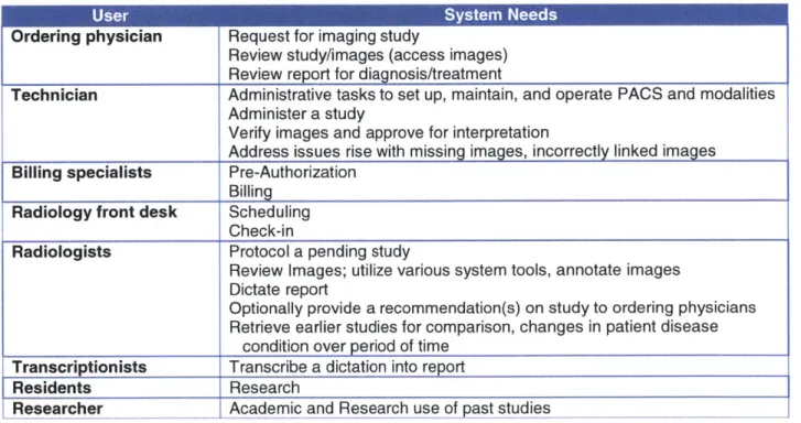
2.3.6. Users and Needs
Table 1 provides a summary of needs of primary users in the radiology system.
Ordering physician Request for imaging study
Review study/images (access images) Review report for diagnosis/treatment
Administrative tasks to set up, maintain, and operate PAUS and modalities Administer a study
Verify images and approve for interpretation
Address issues rise with missing images, incorrectly linked images Billing specialists Pre-Authorization
Billing Radiology front desk Scheduling
Check-in
Radiologists Protocol a pending study
Review Images; utilize various system tools, annotate images Dictate report
Optionally provide a recommendation(s) on study to ordering physicians Retrieve earlier studies for comparison, changes in patient disease
condition over period of time
Transcriptionists Transcribe a dictation into report
Residents Research
Academic and Research use of past studies Table 1. Radiology system stakeholders and needs.
Technician
2.4. Modalities
2.4.1. X-Ray
X-ray technology enables doctors to see through human tissue to examine broken bones, cavities, and swallowed objects. Modified X-ray procedures can be used to
examine softer tissues, such as Figure 5 Modality: X-ray.
the lungs, blood vessels, or
intestines. An X-ray machine uses the same film technology as an ordinary camera, but X-ray light sets off the chemical reaction instead of visible light [44]. It is
increasingly common to acquire X-ray images digitally, without film. 2.4.2. Ultrasound
Diagnostic ultrasound, also known as medical sonography or ultrasonography, uses high-frequency sound waves to create images of structures inside the body. By directing sound waves into the body
and measuring their echoes, Figure 6 Modality: ultrasound
the ultrasound machine is
able to build up a picture in a manner closely analogous to radar. In addition to producing an image, ultrasound can also produce audible sounds of blood flow, enabling medical professionals to use both sound and visuals to assess health. While most commonly identified with use during pregnancy, ultrasound is also used widely by virtually all medical specialties, including cardiology and surgery, to
visualize muscles, tendons, and other structures. Ultrasound is now established as a critical tool both for routine and urgent-care diagnostics [44].
2.4.3. Computed Tomography
Computed [axial] tomography (CT), also commonly referred to as CAT scanning, is an imaging technique that provides detailed 3-D images of volumes inside the body. CT uses a thin beam of X-rays to take a series of cross-sectional pictures of specific organs or areas inside the body from multiple different angles. The CT's computer then analyzes the pictures and
constructs a three
dimensional image of the area of interest. During some CT scans, a contrast medium or "dye" is used to outline Figure 7 Modality: computed tomography. blood vessels or highlight
Source [49]
organs of the body so that they can be seen more easily [441.
2.4.4. Magnetic Resonance Imaging
Magnetic resonance imaging (MRI) is a medical imaging technology that uses radio waves and a powerful magnet
organs, blood vessels, muscles, joints, tumors, areas of infection, and more. These very high quality pictures can show the difference between normal and diseased soft
tissues of the body, making MRI especially useful for a wide range of different types of imaging, including neurological and musculoskeletal. A contrast agent may be used to make the MRI image more informative [441.
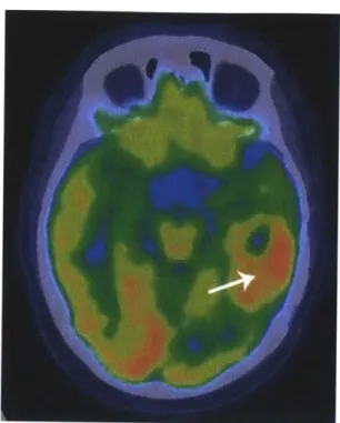
2.4.5. Positron Emission Tomography
Positron emission tomography (PET) technology, often used in combination with CT, uses a scanner and a small amount of radioactive glucose (sugar) which is injected into a patient to make detailed, computerized pictures of areas inside the body where the glucose is used.
A PET-CT scan consists of two parts: first,
a CT scan to pinpoint the location for the PET, then the PET scan itself. During a PET scan, a ring of detectors picks up radiation signals from the patient's body coming from previously injected
radiopharmaceuticals. The computer then
Figure 9 Modality: positron emission tomography.
analyzes the information and constructs Source [48]
an image of the targeted area. During some PET/CT scans, a contrast medium
or "dye" is used to make the image more informative [441.
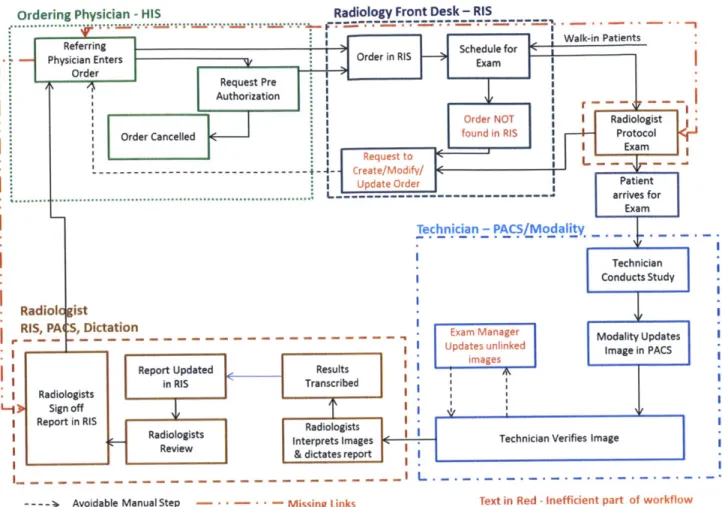
2.5. Workflow at BIDMC
Figure 10 depicts the current workflow of the BIDMC radiology department. Workflow setup plays an important role in day-to-day operations, and is crucial to the
Ordering Physician - HIS Radiology Front Desk - RIS
Physician Enters Order in RIS Schedu for
qRequest
Pre Authorization
-Order NOT I Radiologist
Order Cancelled found in RIS Protocol
- ~Request to iEa
--- - Create/Modify/
Update Order Patient
... ... L--- arrives for Technician - PACS/Modality Technician Conducts Study Radiol ist RIS, PA :, Dictation
r - - - - - - - - - - - - - - - - - - - - - - - - - - Exam Manager Modality Updates
I I Updates unlinked Image in PACS
Report Updated Results iae
in RIS Transcribed
I Radiologists
L-) Sign off
I Report in RIS Radiologists
Radiologists Interprets images-
Technician Verifies Image Review& dictates report
---- > Avoidable Manual Step - - - - Missing Links Text in Red -Inefficient part of workflow Figure 10. Radiology workflow at BIDMC.
The workflow begins with a physician ordering an imaging study for a patient through the ordering portion of the HIS. BIDMC has an advanced, rule-based Clinical Decision Support (CDS) system to aid the ordering process. The CDS is integrated with ACR appropriateness criteria. After patient-data inputs are taken from the ordering physician, the information is uploaded to a cloud-based web service hosted by
ANVITA. The service returns best-practices information to assist in ordering the right tests for the particular case. In addition, for selective insurance providers (Blue Cross Blue Shield of Massachusetts, Tufts, and soon Harvard Pilgrim) it automatically authorizes the test as part of the initial ordering workflow. Both pre-authorization approved studies and those not requiring require pre-authorization are entered into RIS as imaging orders. The exam may be scheduled in several different ways, e.g. patient walk-in, call-in through call center to schedule in advance, or physician office
system-typically due to the physician office forgetting to call in while patients come directly from the office to Radiology-the front-desk assistant calls the physician office to get the order into the system (which can happen almost immediately).
Once an exam is scheduled, insurance details are verified and the order is converted into a requisition to conduct a study. At this time patient information and order data are propagated to both the PACS and the Modality (e.g., MRI, CT), where the exam is scheduled. In advance of the exam, a radiologist protocols the study to specify that the correct test is ordered and what information is requested from the study. If this results in requesting additional information, this is discussed with the physician's office. Sometimes this involves changing test type if the radiologist strongly feels that the original request is not optimal for the patient's condition. This happens very rarely in the hospital outpatient setting, where patients regularly come to same hospital. In the case of private-practice groups, there is little or no opportunity for radiologists to interact with the physician's office.
Once the protocol is complete, a technician performs the actual exam using the
appropriate modality and the image is transferred to the PACS. The technician verifies the image using the PACS console and marks it as verified. However, if patient
information (e.g., date of birth) is altered between when the original order is sent to Modality/PACS and the exam is conducted, the changes are propagated to the PACS server but not the Modality. The result is that images are not linked to the right patient in PACS and are left in an unlinked state. The Exam Manager periodically reviews such unlinked images and relinks them to right study request in PACS. During interviews, BIDMC personnel estimated that this happens for up to 5% of studies conducted.
Once an exam is updated, as verified in RIS by the technician, the image (or set of images) is ready for interpretation by the radiologists and appears on their list for review. A radiologist use PACS workstations to view and analyze the images and often to add annotations that get stored in the PACS along with the images. After review, the radiologist dictates results which get transcribed into the RIS. Upon receipt of the report in the RIS, the radiologist reviews the final report and signs off in the RIS. This
is the final step of the radiology workflow, after which physician gets to review the image through a web-based interface with PACS and report through HIS.
2.6. Reimbursement Model
The reimbursement component for radiology has two parts, the Professional
Component (PC) and the Technical Component (TC). Payers pay for the PC of radiology services furnished by a physician to an individual patient in all settings under the fee schedule for physician services, regardless of the specialty of the physician who performs the service. The interpretation of a diagnostic procedure includes a written report. The TC is billed according to equipment, supplies, technicians, and facility, but does not include interpretation. Different rules apply whether facility is inpatient,
outpatient, or a free-standing imaging center.
The payment model is based on number of studies conducted and interpreted and is thus essentially a type of Pay for Service (a.k.a. Fee for Service, FFS) , a very common payment model in US health care. The greater the number of services provided, the more providers are reimbursed (as long as there is medical necessity). This has introduced several unintended behaviors into the radiology field, driving up cost, utilization, and spending. The same behavior has led to increases in self-referral in imaging services from private-group practitioners.
Congress responded to continued increases in imaging utilization and cost by making payment cuts through several legislation channels. Summarized below are initiatives targeting radiologists in the recent past:
a) The Deficit Reduction Act (DRA) of 2005, signed into law by President Bush in
2006, included payment cuts of up to $2.8 billion for imaging services over 5
years [10]. The law equalizes Medicare reimbursement rates between outpatient imaging procedures and independent imaging centers by capping
reimbursement for the TC of physician-office imaging to the lesser of the Hospital Outpatient Prospective Payment System (HOPPS) or Medicare Fee Schedule (MPFS) payment [101. The provision also cut reimbursement for the technical portion of MR imaging, CT, and ultrasound exams on contiguous body parts by 25% in 2006 and an additional 25% in 2007.
b) The 2010 Medicare Physician Fee schedule (MPFS) changes equipment
utilization rate for imaging systems priced at more than $1 million, from 50% to
90% [11]. By increasing equipment utilization, it essentially cuts per-service
technical fees drastically. Many contend that a utilization rate increase to 90% is impractical and is therefore far from reality for most practices; it is also unclear whether a 8 hour work period is used for the calculation or a 24-hour period. For example, imaging centers in rural areas don't get enough patient volume to maintain a 90% utilization rate. And where an imaging modality is operating at 90% utilization, it is unable to handle urgent cases.
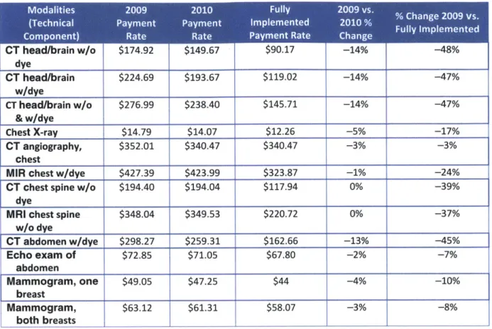
c) Table 2 and Table 3 outline the impact to reimbursement as result of this initiative.
Cumulative impacts of CMS's 2010 Medicare physician fee schedule Final Rule, excluding the potential change in the conversion factor of -21.2%.
Impact of Allowed Impact of Impact of
Resourced Combined
Specialty Charges WokRU Practice Based Ipc
(mil) WokRU Expense RVU Malpractice ipc
(% change)
Interventional radiology $225 -1 -9% 0% -10%
Nuclear Medicine $74 -5% -15% -2% -23%
Radiation Oncology $1,809 0% -3% -2% -5%
radiology $5,056 0% -14% -2% -16%
Table 2. CMS 2010 Medicare physician fee schedule Source: [10]
d) The June, 2011 MedPAC Recommendations to Congress targets three out of its
four recommendations to payment adjustments related to imaging services:
e Congress should direct the Secretary to apply a multiple procedure payment
reduction to the professional component of diagnostic imaging services provided
by the same practitioner in the same session.
e Congress should direct the Secretary to reduce the physician work component
of imaging and other diagnostic tests that are ordered and performed by the same practitioner.
* Congress should direct the Secretary to establish a prior authorization program for practitioners who order substantially more advanced diagnostic imaging services than their peers.
CT head/brain w/o dye 5174.92 5149.67 -14% -48% CT head/brain $224.69 $193.67 $119.02 -14% -47% w/dye CT head/brain w/o $276.99 $238.40 $145.71 -14% -47% & w/dye Chest X-ray $14.79 $14.07 $12.26 -5% -17% CT angiography, $352.01 $340.47 $340.47 -3% -3% chest
MIR chest w/dye $427.39 $423.99 $323.87 -1% -24%
CT chest spine w/o $194.40 $194.04 $117.94 0% -39%
dye
MRI chest spine $348.04 $349.53 $220.72 0% -37%
w/o dye CT abdomen w/dye $298.27 $259.31 $162.66 -13% -45% Echo exam of $72.85 $71.05 $67.80 -2% -7% abdomen Mammogram, one $49.05 $47.25 $44 -4% -10% breast Mammogram, $63.12 $61.31 $58.07 -3% -8% both breasts
Table 3. Code specific impacts of CMS 2010 Medicare physician fee schedule
The radiology community has raised several concerns about these payment cuts, including their possible impact on quality of care. For example, as reimbursement declines, providers have to make up for the loss in some other manner, such as reducing personnel or outsourcing administrative functions such as billing, contract renegotiation, etc. [11]. This may eventually lead to limiting the physician's ability to order the best possible test for a given patient, thus affecting the quality of diagnosis. Thus, although the initiatives may reduce utilization and curtail total spending, concern on quality of the care remains. Other concerns of the community include:
* Reduction in technical fees can have significant impact in rural areas where volume of service is low and hence it is difficult to offset equipment costs while
maintaining service.
* The law does not address the self-referral issue, which many think the root cause of increased Medicare imaging expenditures [10].
* From a radiology perspective, "CMS and Congress are attacking the utilization problem in a counter-productive way where they will stifle investment, research and development and potentially force closure of a lot of centers, impairing access-especially in rural areas" (Alan Kaye, MD) [11].
On the private payer side, efforts to curtail spending range from integrated Clinical Decision Support (CDS) systems to provide relevant information to physicians when ordering and to Imaging Management programs in which the physician calls third-party administrators to get pre-authorization.
One example (obtained through personal interview) of these cost-control efforts in action is a program implemented by Blue Cross Blue Shield of Massachusetts
(BCBSMA) for physicians (under their HMO) to get pre-authorization before ordering an imaging study. Unlike any other pre-auth program, this one does not deny a request, but directs the physician to consider all alternatives before submitting an order. The program focuses not on denying procedures but on "educating" physicians about trends, usage statistics, and other information relevant to making better
decision to order a right study atfirst time. BCBSMA measured the progress of the program and found it has a physician acceptance rate of >90% with no measurable degradation in quality. After the program started in 2006, at which time spending on imaging was increasing by high single-digit percentages yearly basis, costs moderated, moving to <1% increase/year in 2009 and negative growth in 2010. Based on the success of this program, BCBSMA is looking to fold a similar program into their PPO program.
2.7. Trends in Radiology
Radiology has contributed valuable services over the decades, but some aspects of system setup have led to unintended behaviors that have contributed to quality and safety issues, inefficiencies, and higher spending. This section discusses key trends in radiology related to spending and utilization.
2.7.1. Rising Imaging Spending
Proliferation of imaging technology has resulted in improved diagnostic capability in almost every sub-specialty of medicine, where imaging is used as an effective
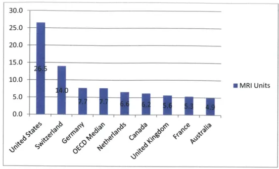
diagnostic tool for early detection and for treatment. Indeed, radiology workflow has been IT-enabled for a longer time now than any other specialty. At the same time, cost of imaging services has grown significantly due to capital investment in equipment and the attractiveness of the industry from a business perspective. Higher return on investment in this area has resulted in mushrooming of the number of free-standing imaging centers and in-office imaging equipment ownership by private group practice physicians. Figure 11 compares number of MRI units/million persons as of 2006 across a number of countries. Not surprisingly, the US has largest MRI units/million persons among these industrialized countries.
Moreover, total spending in US healthcare for diagnostic imaging has been steadily increasing over the years and currently is growing at twice the rate of rise in total healthcare costs. Per MedPAC's June, 2011 report [6], imaging-related expenditures are the fastest growing of all types of services in Medicare claims. From 2000 to 2009, volume of imaging services grew by over 60%. In terms of payments, imaging services makes up 19% of total Medicare payments for 2009; by volume its share is 14% [6]. The two primary reasons for this spending increase are increased cost and increasing utilization. Advances in imaging technologies such as PET and MRI greatly increased the capital investments in equipment, software, and resources required to acquire and operate them. This in turn has increased average cost per imaging study at almost twice the rate of other technologies (e.g., lab procedures, pharmaceuticals) [2].
30.0 25.0 20.0 15.0 10.0 5.0 0.0 U MRI Units d, .'%
Figure 11. Number of MRI units/million persons for 2006. Source: [8]
2000 2001 2002 2003 2004 2005 2006 2007 2008 2009
Note: Volume is units of service multiplied by relative value units from the physician fee schedule. Volume for all years is measured on a common scale, with relative value units for 2009.
Figure 12. Spending under Medicare for different services. 100 90 80 70 60 50 40 30 20 , cm e 8. 0 10 0
2.7.2. Reasons for Growing Utilization
Growing utilization is the result of multiple factors and is a primary contributor to spending increase. Interviews and literature research reveal that several reasons are often cited for the trend of growing utilization trend in radiology. Summarized below are the most common reasons cited for growing utilization. (A detailed analysis of primary drivers in imaging and the behaviors they are causing is given in Chapter 4.)
Self Referral. Self-referral is referral for a procedure in which the referring physician is
also the service provider or has an ownership interest and benefits financially by providing the service [2]. Up to 3.2 times as many scans are ordered in such cases; self-referral leads to approximately $16 billion a year in unnecessary imaging procedures [2], according to a study by Levin and Rao [3]. These behaviors, often in private group practice, are clearly profit-driven.
A study conducted by Center for Studying Health System Change, the 2008 Health
Tracking Physician Survey, reveals the severity of the self-referral issue [7]. The study finds that advanced medical imaging equipment is likely to be owned by 30.3% of surgeons, 10.6% of primary care physicians treating adults, 13.5% of non-procedure-based medical specialists such as neurologists, and 15.7% of procedure-non-procedure-based medical specialists such as cardiologists.
Reimbursement Models. The current fee-for-service payment model is cited as a major motivator to order more imaging studies, as this directly contributes to rise in profit. This is universal problem in the US healthcare system, not just in radiology.
Ordering Physician Behavior. Each specialty has different imaging ordering behavior
patterns. Non-radiologist physicians often lack knowledge of alternative options available to study a condition. For example, a PCP may take a conservative approach in most cases, trying out more basic diagnostic options before considering an imaging study, whereas specialty doctors have tended to go straight to imaging because they may assume that a patient coming to them does not require basic level of diagnosis, rather, imaging for a more precise diagnosis. However, this behavior has got better recently; today, most specialty providers know what the right study for a given condition is. Another aspect of physician ordering behavior is that higher patient
may choose imaging as a short-cut. Also, the ordering physician's lack of knowledge regarding what study would be appropriate, and/or lack of access to such guidelines, may contribute to the ordering of more unnecessary studies.
Patient Expectations. Patient expectations are high today in terms of getting imaging done early, as patients understandably want a definite diagnosis as quickly as
possible. For example, in urban areas patients are more knowledgeable than in rural areas, and demand more from providers as they actively participate in personal healthcare. Given that insured patients are not the primary payers for such services, there is little incentive to think twice before asking for an imaging study.
Defensive Medicine. Medical liability concerns drive physicians to be defensive in diagnosis. Physicians may order studies even in cases where they aren't sure whether a particular study will help, just to avoid getting sued for not following certain
protocols. No law penalizes physicians for ordering wrong or irrelevant tests, only when they don't order a test per clinical guidelines for a given clinical condition. A study in Massachusetts found that 25% of high-tech imaging studies were ordered principally for defensive purposes, at a cost of $1.4 billion per year [2]. Though it is difficult to exactly quantify the contribution of defensive medicine to inappropriate use of imaging services, most professionals agree that it is at least 5% of total health costs
[2].
Access to Older Studies and Medical History. Patients often see different physicians during the treatment cycle, and may not carry complete medical histories with them during such visits. In the absence of previous reports or studies, and or a complete medical history, or due to technical incompatibility between systems used by different providers, a new physician often choose to order study even if it was conducted before to diagnose the patient. Within a given hospital environment this is not typically an issue, rather, this problem arises primarily between different providers.
Lack of Care Coordination. Physicians and radiologists coordinate very little today in care delivery. This is true across most of the areas within medicine, where the patient goes through a fragmented care process and there is little exchange of information across providers treating same patient. There are two noteworthy issues here: (1) radiologists never get to know the end result of diagnosis they have provided to


![Table 2. CMS 2010 Medicare physician fee schedule Source: [10]](https://thumb-eu.123doks.com/thumbv2/123doknet/14192171.478299/35.918.110.800.501.679/table-cms-medicare-physician-fee-schedule-source.webp)