HAL Id: hal-02952983
https://hal.inrae.fr/hal-02952983
Submitted on 5 Oct 2020HAL is a multi-disciplinary open access archive for the deposit and dissemination of sci-entific research documents, whether they are pub-lished or not. The documents may come from teaching and research institutions in France or abroad, or from public or private research centers.
L’archive ouverte pluridisciplinaire HAL, est destinée au dépôt et à la diffusion de documents scientifiques de niveau recherche, publiés ou non, émanant des établissements d’enseignement et de recherche français ou étrangers, des laboratoires publics ou privés.
Distributed under a Creative Commons Attribution| 4.0 International License
Usurp the Ubiquitin-Dependent Degradation Pathway of
Host Cells
Frederic Taieb, Jean-Philippe Nougayrède, Eric Oswald
To cite this version:
Frederic Taieb, Jean-Philippe Nougayrède, Eric Oswald. Cycle Inhibiting Factors (Cifs): Cyclomod-ulins That Usurp the Ubiquitin-Dependent Degradation Pathway of Host Cells. Toxins, MDPI, 2011, 3 (4), pp.356-368. �10.3390/toxins3040356�. �hal-02952983�
toxins
ISSN 2072-6651www.mdpi.com/journal/toxins Review
Cycle Inhibiting Factors (Cifs): Cyclomodulins That Usurp the
Ubiquitin-Dependent Degradation Pathway of Host Cells
Frédéric Taieb 1,2,3,4,*, Jean-Philippe Nougayrède 1,2,3,4 and Eric Oswald 1,2,3,4,5
1
INRA, USC Molecular and Cellular Pathogenesis of Escherichia coli Infections, Toulouse, F-31300, France; E-Mail: jp.nougayrede@envt.fr (J.-P.N.); e.oswald@envt.fr (E.O.)
2
Inserm, U1043, Toulouse, F-31300, France
3
University of Toulouse, UPS, Centre de Physiopathologie de Toulouse Purpan (CPTP), Toulouse, F-31300, France
4
CNRS, U5282, Toulouse, F-31300, France
5
CHU Toulouse, Hôpital Purpan, Service de Bactériologie-Hygiène, Toulouse, F-31300, France
* Author to whom correspondence should be addressed; E-Mail: f.taieb@envt.fr; Tel.: +33-5-6119-3286; Fax: +33-5-6119-3975.
Received: 18 February 2011; in revised form: 16 March 2011 / Accepted: 16 March 2011 / Published: 29 March 2011
Abstract: Cycle inhibiting factors (Cifs) are type III secreted effectors produced by diverse
pathogenic bacteria. Cifs are “cyclomodulins” that inhibit the eukaryotic host cell cycle and also hijack other key cellular processes such as those controlling the actin network and apoptosis. This review summarizes current knowledge on Cif since its first characterization in enteropathogenic Escherichia coli, the identification of several xenologues in distant pathogenic bacteria, to its structure elucidation and the recent deciphering of its mode of action. Cif impairs the host ubiquitin proteasome system through deamidation of ubiquitin or the ubiquitin-like protein NEDD8 that regulates Cullin-Ring-ubiquitin Ligase (CRL) complexes. The hijacking of the ubiquitin-dependent degradation pathway of host cells results in the modulation of various cellular functions such as epithelium renewal, apoptosis and immune response. Cif is therefore a powerful weapon in the continuous arm race that characterizes host-bacteria interactions.
Keywords: bacterial toxin; type III secretion system; eukaryotic cell cycle; NEDD8;
ubiquitin; cullin-RING E3 ubiquitin ligase
1. Discovery and Distribution of the Cycle Inhibiting Factors (Cifs) That Triggers an Original Cytopathic Effect in Host Cells
The effect of Cif was first observed in 1997 by De Rycke et al. in HeLa cells transiently infected with enteropathogenic Escherichia coli (EPEC) strains isolated from weaning rabbits or human infants with diarrhea [1]. This so-called cytopathic effect (CPE) was first characterized by the progressive formation of actin stress fibers together with focal adhesions, and a dramatic cell enlargement (Figure 1). It was further shown that these large cells were irreversibly impaired for cell proliferation, with a complete lack of mitotic figures [2] (Figure 1). This effect is dependent on the locus of enterocyte effacement (LEE), the cluster of genes responsible for the attaching and effacing lesion, the hallmark of EPEC and enterohemorraghic E. coli (EHEC) infection [3,4]. The protein responsible for the CPE identified by Marches et al. was called Cif (cycle inhibiting factor). The gene coding for Cif is located outside the LEE, on a lambdoid prophage [5].
Figure 1. Cycle inhibiting factor (Cif) induces cell enlargement, actin stress fibers and
proliferation arrest. HeLa cells were infected with enteropathogenic Escherichia coli (EPEC) producing Cif (left panel) or Cif mutant EPEC (right panel). F-actin is stained with phalloidin (red) and DNA is stained with DAPI (blue). Arrows indicate mitotic/dividing cells. Bars represent 20 µM.
Two epidemiological studies showed that 60 to 70% of EPEC and EHEC strains are cif-positive [6,7]. However, in one of these studies, it was shown that two thirds of these cif-positive E. coli strains do not induce a typical CPE due to frameshift mutations or insertion of an IS element in the cif gene. Strikingly, cif is always found as a pseudogene in EHEC strains; this could be due to the small size of the strain collection, or could suggest an evolutionary counter-selection of functional Cif in Shiga-like producing EHEC strains [6]. Nonetheless, plasmid-mediated expression of wild type Cif in EHEC confers the CPE phenotype [5].
The E. coli Cif protein is 282 amino-acids long, showing no similarity with any protein except several homologues in the human pathogens Yersinia pseudotuberculosis (56% of identity) and beta-proteobacterium Burkholderia pseudomallei (26%). Cif homologues are also found in the entomopathogens Photorhabdus luminescens (23%) and Photorhabdus asymbiotica (26%). The Cif proteins are thus named CifEc, CifYp, CifBp, CifPl and CifPa respectively. In all these bacteria the cif
genes are located in prophage or prophage-like region (Escherichia and Photorhabdus species), bordered by two vestigial transposase genes (Burkholderia) and in a locus frequently rearranged nearby the yrs region homologous to a recombination target of bacteriophages (Yersinia) [8]. Thus
Cif-encoding regions are found in DNA domains prone to rearrangement, suggesting that the cif genes were acquired by horizontal gene transfer. Consistent with this hypothesis, the Cif-producing EPEC strain E22 was shown to produce cif-carrying infectious phage particles supporting that the cif gene was disseminated by horizontal gene transfer among EPEC and enterohemorrhagic (EHEC) strains, as shown for other non LEE-encoded effectors [6,9,10].
Analysis of the Cif xenologues showed that they are all functional as they reproduced the typical CifEc-associated CPE when transfected into HeLa cells, or lipofected following purification [8].
Importantly, CifPl was shown to be expressed in vivo in an insect model, the natural host of
Photorhabdus, and reproduced the CPE in the non-mammalian insect Sf9 cell line in vitro [11]. Similarly, CifBp is also active in vitro when strains of E. coli (EPEC) or Burkholderia thailandensis are
transformed with a plasmid coding for CifBp and used toinfect mammalian cell lines [8,12]. Such
conservation of an effector function among very distant pathogens belonging to -protobacteria and
-protobacteria that infect invertebrate and mammal hosts is unique and remarkable. It also supports the role of Cifs as virulence factors and could reveal an original strategy of pathogenesis. However, Photorhabdus spp. are symbiotic of the nematode vector Heterorhabditis (that releases the bacteria into the insect) suggesting that Cif might also act as a fitness or symbiotic factor in the nematode host.
2. Cif Proteins are Type III Effectors That Traffic to the Nucleus of the Host Cells
Initial experiments showed that the CPE triggered by cif-positive EPEC strains was dependent on the type III secretion system (T3SS) encoded by the LEE as mutants of the secretion and translocation apparatus (EscN and EspA, EspB, EspD, respectively) were unable to induce the CPE [1,2,5,13]. This T3SS injects an arsenal of effector proteins into the host cell that hijack cellular functions for the pathogen’s benefit [3,4,14]. Using a reporter system based on a translational fusion of the effector protein with β-lactamase that allows detection of translocation in living host cells, Charpentier and Oswald showed that an exchangeable N-terminal translocation signal comprising the first 16 amino acids of CifEc is necessary and sufficient to mediate its translocation by the EPEC T3SS [15]. Using a
lipid-based cell delivery system bypassing T3SS requirement, purified CifEc induced a CPE similar to
that observed upon EPEC infection. This result indicated that CifEc is necessary and sufficient and does
not require other bacterial molecules to induce the CPE [16]. Once injected into the host cell cytoplasm by the T3SS, CifEc is rapidly sorted to the peri-nuclear area and the nucleus, as observed by
confocal imaging and cell fractionation [17]. As CifEc lacks classical nuclear localization sequences,
the mechanism of nuclear targeting of Cif remains to be elucidated.
While CifBp has been shown to be injected by the T3SS of Escherichia and Burkholderia
species and successfully reproduces the CPE [8,12], we and other have been unable to translocate CifYp
and CifPl by the E. coli T3SS into the host cells. However, we have indirect evidence that they are
bona fide type 3 effectors as both Yersinia and Photorabdhus strains possess T3SS. Moreover, none of the Cifs could induce the CPE without electroporation, transfection or lipofection suggesting that Cif activity depends on its injection into the host cell [8,11,16].
3. Crystal Structure Studies of Cif Homologs Reveals a Family of Proteins Sharing a Conserved Active Site
Despite a low level of sequence similarity between Cif homologs, the crystal structure determination of CifEc, CifPl and CifBp showed that these proteins are structurally well-conserved
(Figure 2) [18–20]. The overall structure of these Cif can be divided in two lobes with head and tail domains where the N-terminal part corresponds to the tail and contains the potential substrate binding site (see below) whereas the C-terminal part forms the head domain hosting the enzymatic site. The enzymatic site consists in a catalytic triad composed of residues Cys, His and Gln strictly conserved in all Cifs homologs (Figure 2).
Figure 2. Cif homolog proteins are structurally related and share a conserved catalytic site.
CifPl, CifBp and CifEc are represented as composites of ribbon and surface plot diagrams.
Note that the crystallized CifEc protein lacked 99 amino acids in its N-terminal part. The
side chains of the critical residues of the catalytic triad (Cys, His, Gln) are shown. The α-helices α4 and α5 of CifBp are indicated. The right panel represents a superimposition of
the three Cifs magnified at the level of the catalytic pocket.
The conserved catalytic triad is located in a negative charged cleft with a putative occluding loop near the active site that might control the substrate accessibility to the catalytic site [19]. The structural organization of this triad reveals that Cifs are divergent members of the superfamily of enzymes that comprises papain-like cysteine proteases, acetyl transferases, deamidases and transglutaminases. In particular, Cif shares homology with the avirulence effector AvrPphB from Pseudomonas syringae that belongs to the same superfamily and with the cysteine protease YopT from Yersinia spp. [19]. Mutation of one of the residues of the catalytic triad abolishes CifPl, CifBp, CifEc and CifYp activity,
corroborating the implication of an enzymatic-dependent activity [8,16,19,20]. Further analysis of the tail domain showed that deletion of the N-terminal protruding α4 and α5 helix of CifBp
(Figure 2) and of the corresponding 4 domain of CifEc suppressed the toxin activity suggesting that
this domain could mediate substrate recognition [17,20].
Based on the structure and function conservation, Cif homologs constitute a family of cyclomodulins that likely target a molecule/pathway conserved in a wide range of eukaryotic cells from invertebrates to mammals [8,21].
4. Cifs Are Cyclomodulins That Trigger Host Cell Cycle Arrest
Upon injection into host cells by the T3SS (or by transfection or lipofection of purified proteins) Cif proteins induce an irreversible cell cycle arrest with complete inhibition of mitotis entry (Figure 1: absence of mitotic figures in cells injected with Cif). This property makes Cifs as members of the cyclomodulin family defined as bacterial molecules that hijack host cell cycle functions [22,23].
The cell cycle arrest induced by CifEc is associated to the inhibitory phosphorylation of the cyclin
dependent kinase (CDK) 1-CyclinB complex whose activation is necessary for the cell cycle G2/M
transition [1,2,5]. Other cyclomodulins, such as CDT or Colibactin, also block the G2/M transition
through inhibition of CDK1 activity. However, in contrast to CDT and Colibactin, which have been shown to be bona fide genotoxins, CifEc does not induce DNA damage nor activate the DNA damage
response pathway (ATM/ATR, CHK1/2, CDC25 sequestration) that leads to the inhibitory phosphorylation of CDK1 [16]. Therefore, CifEc induces the cell cycle arrest by activating a pathway
independent from the canonical activation of the G2/M checkpoint. Further studies, using synchronized
cells, have shown that CifEc also blocks S-phase entry. Depending on the stage of the cell cycle during
infection with cif-positive EPEC, cells arrest either at the G1/S or G2/M transition. The analysis of host
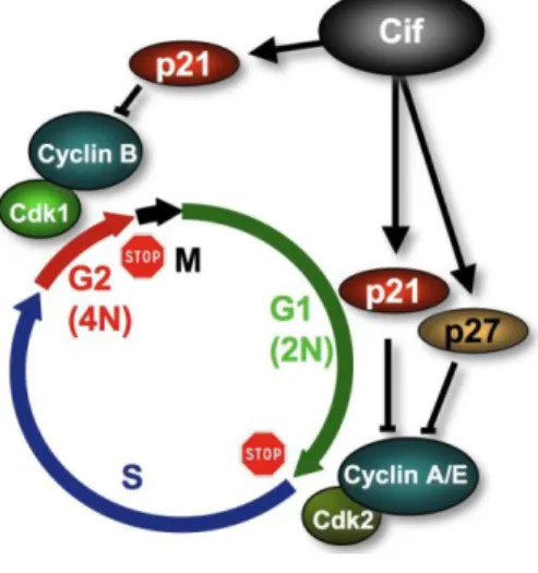
cell proteins regulating both S- and M- phases entries demonstrated a Cif-dependent accumulation of p21Waf1/Cip1 and p27kip2 (hereafter called p21 and p27) [24]. These proteins belong to the Cip/Kip family of CDK inhibitors (CKI). p21 is an inhibitor of the CDK1/CyclinB complex. This complex is the eukaryotic universal inducer of mitosis entry and p21 accumulation is known to inhibit G2/M
transition. Both p21 and p27 inhibit CDK2-CyclinE and A complexes whose activation are also required for G1/S and S-phase progression. p21 also binds to PCNA, and the latter is required for S-phase progression [25]. Therefore, Cif-dependent accumulation of CKI is consistent with the observed G1/S and G2/M cell cycle arrests (Figure 3). The inhibitory phosphorylation of CDK1
observed upon CifEc injection could be explained by the disruption of the CDK1 positive feed-back
loop leading to CDK1 dephosphorylation via CDC25. Further investigation revealed that CKI accumulation does not depend on transcriptional activation but relies on their stabilization in infected cells [24]. Since p21 and p27 are degraded by the ubiquitin-proteasome system, these results strongly suggested that CifEc controls this proteolytic pathway. However, suppression of p21 and/or p27
accumulation did not alleviate Cif-induced inhibition of G1/S or G2/M transitions implying an
alternative mechanism for blockage of the cell cycle [24]. Whether this arrest is associated to stress fibers induction was excluded since disruption of stress fiber formation in HeLa cells by C3 exoenzyme-mediated inhibition of RhoA, did not alleviate cell cycle inhibition [2]. Morevover, only HeLa and RK13 cells exhibit stress fibers following cif+ EPEC infection suggesting that these two phenotypes are not related [16]. Nonetheless, these results indicated that accumulation of CKI participates in a multi-step process leading to the Cif-induced arrest of cell proliferation.
This cell cycle inhibition and CKI accumulation was observed with all Cif homologues [8] and in various cell lines derived from colonic epithelium (differentiated or not differentiated Caco-2 cells, DLD1, HCT116 wild-type, p21−/− or p53−/−) showing the independency of cell-cycle arrest from the p53 transcriptional program [16,24]. The Cif-induced cystostatic effect is also observed in non-transformed IEC-6 cells derived from rat intestinal crypt epithelium. However, the outcome of this arrest is cell type dependent as it persists for more than 3 days in HeLa cells without evident cell death,
whereas it culminates in apoptosis 48 h after infection of IEC-6 cells [26]. Whether this Cif-induced cell death is a consequence of the cytostatic effect or relies on an independent pathway is still unclear, but it is tempting to propose that the functionality of p53 pathway, intact in non-transformed cells, might be decisive for the fate of Cif-arrested cells.
5. Cif Effector Proteins Target NEDD8 to Inhibit Specific Ubiquitin-Dependent Degradation Pathways
Three independent teams recently deciphered the molecular mode of action of CifEc and
CifBp [12,17,27]. Despite few differences mainly related to the species origin of Cif and diverse
approaches, these complementary studies showed a common mechanism of action between the two xenologs.
Figure 3. Cif inhibits host cell cycle progression. Cif induces the accumulation of p21 and
p27 that inhibits CDK1-CyclinB and CDK2-CyclinA/E. Since these complexes are responsible for G2/M and G1/S transitions, Cif provokes an accumulation of cells arrested in G1 and G2 phases of the cell cycle.
5.1. CifEc Interacts with Host Protein NEDD8
As mentioned earlier, Cif-dependent accumulation of p21 and p27 results from the inhibition of their degradation by the ubiquitin-proteasome system [28]. Ubiquitylation is a well-known and highly controlled post-translational protein modification that involves three successive steps. First, ubiquitin is processed by the ubiquitin activating enzyme (E1) and then transferred to the ubiquitin conjugating enzyme (E2). The last step requires the ubiquitin ligase (E3) that determines the substrate to be ubiquitylated and promotes a direct transfer of ubiquitin from E2 to the substrate (RING-type E3s) or an indirect transfer via formation of an ubiquitin thioester-bound E3 prior ubiquitin transfer to the substrate (HECT-type E3s). Repetitions of this cycle lead to the attachment of a polyubiquitin chain to the target protein, resulting in modulation of its function or localization, or its sorting for degradation, depending on the length and the type of linkage within the polyubiquitin chain [29–31]. Yeast two-hybrid screens and pull-down assays showed that CifEc interacts with the ubiquitin-like protein
NEDD8 [17,27]. The main known role of NEDD8 is the conjugation of the cullin subunit of the complex cullin-RING E3 ubiquitin ligase (CRL) and activation of its ligase activity [32–34]. CRL are
composed of the scaffold protein cullin (at least five different cullins CUL1/2/3/4A/4B are known), the RING protein that associates with E2, and different substrate recognition modules that bind the target protein (Figure 4) [35,36]. Inactivation of CifEc by mutation of the catalytic cysteine increased its
binding to NEDD8 [17]. In contrast, deletion of the 4 domain of CifEc, which results in loss of
activity, abolished the interaction with NEDD8 implying the role of this domain to mediate Cif effect. CifEc was unable to pull-down ubiquitin showing the specificity of interaction with NEDD8. Moreover,
NEDD8 co-compartmentalizes with CifEc in the nuclei of infected cells supporting the functional
binding of these 2 proteins [17]. Co-immunoprecipitation experiments showed that CifEc interacts only
with the neddylated forms of cullins (CUL1 to 4B) associated with the RING protein [17] and with the recognition module Skp1 and Skp2 proteins [27]. It thus appeared that CifEc interacts with the whole
CRL complex through binding to NEDD8 (Figure 4).
Figure 4. Model of CifEc inhibition of CRL activation cycle. Neddylated active CRL (left)
ubiquitylates the substrate (sub) and substrate recognition module (SRM) leading to their degradation and generating inactive CRL (top). Deneddylation of CRL by the COP9 signalosome allows binding of CAND1 (right). SRM fixation induces CAND1 release and subsequent neddylation of cullin to generate an active CRL [35,36]. Cif binding to and deamidation of NEDD8 impairs the action of COP9, blocking the activation cycle and locking the CRL in an inactive neddylated state.
5.2. Cif Inhibits CRL Activity
Since Cif proteins induce the stabilization of p21 and p27, which are substrates of CRL associated to CUL1, in vitro studies were conducted to investigate the inhibitory effect of Cif on CRL. Jubelin et al. showed that addition of CifEc to reconstituted CRL inhibited its ubiquitin ligase
activity [17]. Moreover, in cellulo ubiquitylation of various CRL substrates such as p27 or RhoA was severely impaired by CifEc and CifBp, respectively [12,27]. Stabilization of various CUL1 to
4B-associated CRL substrates including β-catenin, RhoA, IBα, Cdt1, CyclinD1 reveals that CifEc can
How CifEc inhibits CRL activity is not fully elucidated. However, Cui et al. showed that the CRL
inhibition is associated to CifEc-dependent NEDD8 deamidation of Gln40, converting glutamine
residue in glutamic acid (Glu). Replacement of NEDD8 with deaminated NEDD8 (NEDD8Q40E) abolished the in vitro stimulatory effect on Nrf2 ubiquitylation by a CUL3/ROC1/Keap1 complex. Similarly, ectopic expression of NEDD8Q40E in HeLa cells resulted in the stabilization of CRL substrate and recapitulated the effects of Cif [12]. On the other hand, CifEc was shown to promote
cullin neddylation of CUL1 to 4B [17] that correlates with the inhibition of deneddylation of cullin by the CSN complex (COP9 signalosome) [27]. This phenotype is similar to the effect observed using CSN5 null cells that also accumulate CRL substrates such as p27 [37]. Together, these results suggest that modification of NEDD8 by Cif inhibits cullin deneddylation by the CSN resulting in a decrease of CRL activation. Indeed, to be fully active, CRLs undergo an activation cycle in which cullins oscillate between neddylated and non-neddylated states [35,36]. Based on these results, we propose a model in which CifEc interferes with this cycle and thus locks CRLs in a neddylated but inactive state (Figure 4).
A notable difference between CifBp and CifEc is the capacity of CifBp to deamidate not only NEDD8,
but also the Gln40 of ubiquitin. CifBp generates an E2-ubiquitinQ40E thioester complex that impairs
ubiquitin chain synthesis and the ubiquitylation pathway in vitro and in vivo [12]. This supplementary affinity to ubiquitin extends the inhibitory activity of CifBp to all ubiquitylation process independently
of the E3 ligase. The relevance of this difference is still unclear and requires further studies on the range of action of other Cif homologs and bacterial deamidases toward ubiquitin and ubiquitin-like molecules. In this line, it is noteworthy that the Mycobacterium deamidase of Pup (Dop), required for the full virulence of M. tuberculosis, targets the prokaryotic ubiquitin-like molecule Pup to convert the terminal glutamine residue into glutamate [38,39]. Also, while acting through a different pathway to stimulate RhoA activity, it is remarkable that the cyclomodulin CNF-1 directly deamidates RhoA to induces actin stress fibers [40,41].
6. Concluding remarks and perspectives
The role of NEDD8 on CRL activation offers to the bacteria injecting Cif into the host cells an “Achilles heel” to hijack various cellular signaling functions. Indeed, CRLs represent the largest subfamily of E3s and therefore play regulatory roles in numerous and diverse cellular functions. The diversity of substrate recognition modules associated to the different cullin subunits gives to the CRL the capacity to ubiquitylate and thus to control the stability of hundreds of proteins [35,36]. Cif capacity to induce stress fibers in certain eukaryotic cells could be related to Cif-stabilization of RhoA [12,17], which is targeted by CUL3-associated CRL [42]. Prevention of inappropriate replication depends on the degradation of the licensing factor Cdt1 during S-phase [43]. As CUL4-associated CRL targets Cdt1 for ubiquitylation-dependent degradation [44,45], inhibition of CRL by Cif provides a satisfactory explanation for occurrence of re-replication observed 3 days following infection as Cdt1 is stabilized in infected HeLa cells [2,16,17]. Moreover, Cif was shown to prevent the degradation of IB, a central inhibitor of NF-B pro-inflammatory response [12,17]. Down-regulation of the NF-B pathway has been proposed to mediate the inflammatory tolerance of commensal bacteria in the mammalian intestinal epithelia [46,47]. In addition, stabilization of β-catenin by Cif might participate in dendritic cells tolerogenicity as it was recently shown that the WNT signaling pathway regulates immunosuppressive responses [48].
In conclusion, Cif might represent a fitness factor that facilitates bacterial evasion from the host immune response, via inhibition of inflammatory pathways in dendritic cells of the gut-associated lymphoid tissue. Cif-inhibition of the host cell cycle might slow down multiplication of intestinal progenitors to delay epithelial cells renewal, thus favoring gut colonization (Figure 5). We could also speculate about the role of Cif as a stabilizer of other type III co-injected effectors, to modulate their effect on host cells and boost the virulence of cif-expressing strains. Further studies of the impact of Cif in the pathogen-host interactions will undoubtedly contribute to our knowledge of bacterial pathogenic strategies. It also appears that the continuous “arm race” that characterizes host-pathogen relationship has lead the bacteria to develop sophisticated “weapons” (virulence factors) capable of controlling host’s functions for their own benefit. Therefore, much can be learned from eukaryotic functions using bacterial toxins such as Cif. While most pharmacological inhibitors or bacterial/viral proteins inhibit CRL by decreasing cullin neddylation [49–52], Cif is the first example of a bacterial effector that inhibits CRL functions by stabilizing cullin neddylation. Thus Cif represents a unique tool to study the regulation of the ubiquitin proteasome system. Lately, it was reported that inhibitors of NEDD8 represent a promising approach for cancer treatment [52] opening a potential development of innovative therapeutics using Cif-producing bacteria.
Figure 5. Cif hijacks several and various signaling pathways by inhibiting the ubiquitin
ligase activity of CRL. Cif is injected in the host cells and impairs CRL activity via binding to NEDD8. This inhibition results in stabilization of (i) p21 and p27 leading to cell cycle inhibition; (ii) Cdt1 allowing inappropriate replication; (iii) RhoA inducing actin stress fibers; (iv) IkB leading to alteration of inflammatory response; (v) β-catenin promoting differentiation process disruption and tolerogenicity; (vi) unidentified protein(s) inducing apoptosis.
References
1. de Rycke, J.; Comtet, E.; Chalareng, C.; Boury, M.; Tasca, C.; Milon, A. Enteropathogenic Escherichia coli O103 from rabbit elicits actin stress fibers and focal adhesions in HeLa epithelial cells, cytopathic effects that are linked to an analog of the locus of enterocyte effacement. Infect. Immun. 1997, 65, 2555–2563.
2. Nougayrede, J.P.; Boury, M.; Tasca, C.; Marches, O.; Milon, A.; Oswald, E.; de Rycke, J. Type III secretion-dependent cell cycle block caused in HeLa cells by enteropathogenic Escherichia coli O103. Infect. Immun. 2001, 69, 6785–6795.
3. Kaper, J.B.; Nataro, J.P.; Mobley, H.L. Pathogenic Escherichia coli. Nat. Rev. Microbiol. 2004, 2, 123–140.
4. Nataro, J.P.; Kaper, J.B. Diarrheagenic Escherichia coli. Clin. Microbiol. Rev. 1998, 11, 142–201. 5. Marches, O.; Ledger, T.N.; Boury, M.; Ohara, M.; Tu, X.; Goffaux, F.; Mainil, J.; Rosenshine, I.; Sugai, M.; de Rycke, J.; Oswald, E. Enteropathogenic and enterohaemorrhagic Escherichia coli deliver a novel effector called Cif, which blocks cell cycle G2/M transition. Mol. Microbiol. 2003, 50, 1553–1567.
6. Loukiadis, E.; Nobe, R.; Herold, S.; Tramuta, C.; Ogura, Y.; Ooka, T.; Morabito, S.; Kerouredan, M.; Brugere, H.; Schmidt, H.; Hayashi, T.; Oswald, E. Distribution, functional expression, and genetic organization of Cif, a phage-encoded type III-secreted effector from enteropathogenic and enterohemorrhagic Escherichia coli. J. Bacteriol. 2008, 190, 275–285. 7. Creuzburg, K.; Middendorf, B.; Mellmann, A.; Martaler, T.; Holz, C.; Fruth, A.; Karch, H.;
Schmidt, H. Evolutionary analysis and distribution of type III effector genes in pathogenic Escherichia coli from human, animal and food sources. Environ. Microbiol. 2011, 13, 439–452. 8. Jubelin, G.; Chavez, C.V.; Taieb, F.; Banfield, M.J.; Samba-Louaka, A.; Nobe, R.;
Nougayrede, J.P.; Zumbihl, R.; Givaudan, A.; Escoubas, J.M.; Oswald, E. Cycle inhibiting factors (CIFs) are a growing family of functional cyclomodulins present in invertebrate and mammal bacterial pathogens. PLoS ONE 2009, 4, e4855.
9. Konczy, P.; Ziebell, K.; Mascarenhas, M.; Choi, A.; Michaud, C.; Kropinski, A.M.; Whittam, T.S.; Wickham, M.; Finlay, B.; Karmali, M.A. Genomic O island 122, locus for enterocyte effacement, and the evolution of virulent verocytotoxin-producing Escherichia coli. J. Bacteriol. 2008, 190, 5832–5840.
10. Coombes, B.K.; Wickham, M.E.; Mascarenhas, M.; Gruenheid, S.; Finlay, B.B.; Karmali, M.A. Molecular analysis as an aid to assess the public health risk of non-O157 Shiga toxin-producing Escherichia coli strains. Appl. Environ. Microbiol. 2008, 74, 2153–2160.
11. Chavez, C.V.; Jubelin, G.; Courties, G.; Gomard, A.; Ginibre, N.; Pages, S.; Taieb, F.; Girard, P.A.; Oswald, E.; Givaudan, A.; Zumbihl, R.; Escoubas, J.M. The cyclomodulin Cif of Photorhabdus luminescens inhibits insect cell proliferation and triggers host cell death by apoptosis. Microbes Infect. 2010, 12, 1208–1218.
12. Cui, J.; Yao, Q.; Li, S.; Ding, X.; Lu, Q.; Mao, H.; Liu, L.; Zheng, N.; Chen, S.; Shao, F. Glutamine Deamidation and Dysfunction of Ubiquitin/NEDD8 Induced by a Bacterial Effector Family. Science 2010, 329, 1215–1218.
13. Nougayrede, J.P.; Marches, O.; Boury, M.; Mainil, J.; Charlier, G.; Pohl, P.; de Rycke, J.; Milon, A.; Oswald, E. The long-term cytoskeletal rearrangement induced by rabbit enteropathogenic Escherichia coli is Esp dependent but intimin independent. Mol. Microbiol.
1999, 31, 19–30.
14. Frankel, G.; Phillips, A.D.; Rosenshine, I.; Dougan, G.; Kaper, J.B.; Knutton, S. Enteropathogenic and enterohaemorrhagic Escherichia coli: more subversive elements. Mol. Microbiol. 1998, 30, 911–921.
15. Charpentier, X.; Oswald, E. Identification of the secretion and translocation domain of the enteropathogenic and enterohemorrhagic Escherichia coli effector Cif, using TEM-1 beta-lactamase as a new fluorescence-based reporter. J. Bacteriol. 2004, 186, 5486–5495.
16. Taieb, F.; Nougayrede, J.P.; Watrin, C.; Samba-Louaka, A.; Oswald, E. Escherichia coli cyclomodulin Cif induces G2 arrest of the host cell cycle without activation of the DNA-damage checkpoint-signalling pathway. Cell Microbiol. 2006, 8, 1910–1921.
17. Jubelin, G.; Taieb, F.; Duda, D.M.; Hsu, Y.; Samba-Louaka, A.; Nobe, R.; Penary, M.; Watrin, C.; Nougayrede, J.P.; Schulman, B.A.; Stebbins, C.E.; Oswald, E. Pathogenic Bacteria Target NEDD8-Conjugated Cullins to Hijack Host-Cell Signaling Pathways. PLoS Pathog. 2010, 6, e1001128.
18. Crow, A.; Race, P.R.; Jubelin, G.; Varela Chavez, C.; Escoubas, J.M.; Oswald, E.; Banfield, M.J. Crystal structures of Cif from bacterial pathogens Photorhabdus luminescens and Burkholderia pseudomallei. PLoS ONE 2009, 4, e5582.
19. Hsu, Y.; Jubelin, G.; Taieb, F.; Nougayrede, J.P.; Oswald, E.; Stebbins, C.E. Structure of the cyclomodulin Cif from pathogenic Escherichia coli. J. Mol. Biol. 2008, 384, 465–477.
20. Yao, Q.; Cui, J.; Zhu, Y.; Wang, G.; Hu, L.; Long, C.; Cao, R.; Liu, X.; Huang, N.; Chen, S.; Liu, L.; Shao, F. A bacterial type III effector family uses the papain-like hydrolytic activity to arrest the host cell cycle. Proc. Natl. Acad. Sci. USA 2009, 106, 3716–3721.
21. Samba-Louaka, A.; Taieb, F.; Nougayrede, J.P.; Oswald, E. Cif type III effector protein: A smart hijacker of the host cell cycle. Future Microbiol. 2009, 4, 867–877.
22. Nougayrede, J.P.; Taieb, F.; de Rycke, J.; Oswald, E. Cyclomodulins: Bacterial effectors that modulate the eukaryotic cell cycle. Trends Microbiol. 2005, 13, 103–110.
23. Oswald, E.; Nougayrede, J.P.; Taieb, F.; Sugai, M. Bacterial toxins that modulate host cell-cycle progression. Curr. Opin. Microbiol. 2005, 8, 83–91.
24. Samba-Louaka, A.; Nougayrede, J.P.; Watrin, C.; Jubelin, G.; Oswald, E.; Taieb, F. Bacterial cyclomodulin Cif blocks the host cell cycle by stabilizing the cyclin dependent kinase inhibitors p21(waf1) and p27(kip1). Cell Microbiol. 2008, 10, 2496–2508.
25. Besson, A.; Dowdy, S.F.; Roberts, J.M. CDK inhibitors: Cell cycle regulators and beyond. Dev. Cell 2008, 14, 159–169.
26. Samba-Louaka, A.; Nougayrede, J.P.; Watrin, C.; Oswald, E.; Taieb, F. The enteropathogenic Escherichia coli effector Cif induces delayed apoptosis in epithelial cells. Infect Immun. 2009, 77, 5471–5477.
27. Morikawa, H.; Kim, M.; Mimuro, H.; Punginelli, C.; Koyama, T.; Nagai, S.; Miyawaki, A.; Iwai, K.; Sasakawa, C. The bacterial effector Cif interferes with SCF ubiquitin ligase function by inhibiting deneddylation of Cullin1. Biochem. Biophys. Res. Commun. 2010, 401, 268–274.
28. Kitagawa, K.; Kotake, Y.; Kitagawa, M. Ubiquitin-mediated control of oncogene and tumor suppressor gene products. Cancer Sci. 2009, 100, 1374–1381.
29. Ciechanover, A.; Finley, D.; Varshavsky, A. Ubiquitin dependence of selective protein degradation demonstrated in the mammalian cell cycle mutant ts85. Cell 1984, 37, 57–66.
30. Hershko, A.; Ciechanover, A. The ubiquitin system for protein degradation. Annu. Rev. Biochem.
1992, 61, 761–807.
31. Hershko, A.; Ciechanover, A. The ubiquitin system. Annu. Rev. Biochem. 1998, 67, 425–479. 32. Huang, D.T.; Ayrault, O.; Hunt, H.W.; Taherbhoy, A.M.; Duda, D.M.; Scott, D.C.; Borg, L.A.;
Neale, G.; Murray, P.J.; Roussel, M.F.; Schulman, B.A. E2-RING expansion of the NEDD8 cascade confers specificity to cullin modification. Mol. Cell 2009, 33, 483–495.
33. Saha, A.; Deshaies, R.J. Multimodal activation of the ubiquitin ligase SCF by Nedd8 conjugation. Mol. Cell 2008, 32, 21–31.
34. Duda, D.M.; Borg, L.A.; Scott, D.C.; Hunt, H.W.; Hammel, M.; Schulman, B.A. Structural insights into NEDD8 activation of cullin-RING ligases: Conformational control of conjugation. Cell 2008, 134, 995–1006.
35. Merlet, J.; Burger, J.; Gomes, J.E.; Pintard, L. Regulation of cullin-RING E3 ubiquitin-ligases by neddylation and dimerization. Cell Mol. Life Sci. 2009, 66, 1924–1938.
36. Bosu, D.R.; Kipreos, E.T. Cullin-RING ubiquitin ligases: Global regulation and activation cycles. Cell Div. 2008, 3, 7.
37. Tomoda, K.; Yoneda-Kato, N.; Fukumoto, A.; Yamanaka, S.; Kato, J.Y. Multiple functions of Jab1 are required for early embryonic development and growth potential in mice. J. Biol. Chem.
2004, 279, 43013–43018.
38. Cerda-Maira, F.A.; Pearce, M.J.; Fuortes, M.; Bishai, W.R.; Hubbard, S.R.; Darwin, K.H. Molecular analysis of the prokaryotic ubiquitin-like protein (Pup) conjugation pathway in Mycobacterium tuberculosis. Mol. Microbiol. 2010, 77, 1123–1135.
39. Striebel, F.; Imkamp, F.; Sutter, M.; Steiner, M.; Mamedov, A.; Weber-Ban, E. Bacterial ubiquitin-like modifier Pup is deamidated and conjugated to substrates by distinct but homologous enzymes. Nat. Struct. Mol. Biol. 2009, 16, 647–651.
40. Flatau, G.; Lemichez, E.; Gauthier, M.; Chardin, P.; Paris, S.; Fiorentini, C.; Boquet, P. Toxin-induced activation of the G protein p21 Rho by deamidation of glutamine. Nature 1997, 387, 729–733.
41. Schmidt, G.; Sehr, P.; Wilm, M.; Selzer, J.; Mann, M.; Aktories, K. Gln 63 of Rho is deamidated by Escherichia coli cytotoxic necrotizing factor-1. Nature 1997, 387, 725–729.
42. Chen, Y.; Yang, Z.; Meng, M.; Zhao, Y.; Dong, N.; Yan, H.; Liu, L.; Ding, M.; Peng, H.B.; Shao, F. Cullin mediates degradation of RhoA through evolutionarily conserved BTB adaptors to control actin cytoskeleton structure and cell movement. Mol. Cell 2009, 35, 841–855.
43. Xouri, G.; Dimaki, M.; Bastiaens, P.I.; Lygerou, Z. Cdt1 interactions in the licensing process: A model for dynamic spatiotemporal control of licensing. Cell Cycle 2007, 6, 1549–1552.
44. Kim, Y.; Kipreos, E.T. The Caenorhabditis elegans replication licensing factor CDT-1 is targeted for degradation by the CUL-4/DDB-1 complex. Mol. Cell Biol. 2007, 27, 1394–1406.
45. Nishitani, H.; Sugimoto, N.; Roukos, V.; Nakanishi, Y.; Saijo, M.; Obuse, C.; Tsurimoto, T.; Nakayama, K.I.; Nakayama, K.; Fujita, M.; Lygerou, Z.; Nishimoto, T. Two E3 ubiquitin ligases, SCF-Skp2 and DDB1-Cul4, target human Cdt1 for proteolysis. EMBO J. 2006, 25, 1126–1136. 46. Collier-Hyams, L.S.; Sloane, V.; Batten, B.C.; Neish, A.S. Cutting edge: Bacterial modulation of
epithelial signaling via changes in neddylation of cullin-1. J. Immunol. 2005, 175, 4194–4198. 47. Kumar, A.; Wu, H.; Collier-Hyams, L.S.; Hansen, J.M.; Li, T.; Yamoah, K.; Pan, Z.Q.;
Jones, D.P.; Neish, A.S. Commensal bacteria modulate cullin-dependent signaling via generation of reactive oxygen species. EMBO J. 2007, 26, 4457–4466.
48. Manicassamy, S.; Reizis, B.; Ravindran, R.; Nakaya, H.; Salazar-Gonzalez, R.M.; Wang, Y.C.; Pulendran, B. Activation of beta-catenin in dendritic cells regulates immunity versus tolerance in the intestine. Science 2010, 329, 849–853.
49. Munro, P.; Flatau, G.; Lemichez, E. Bacteria and the ubiquitin pathway. Curr. Opin. Microbiol.
2007, 10, 39–46.
50. Rytkonen, A.; Holden, D.W. Bacterial interference of ubiquitination and deubiquitination. Cell Host Microbe 2007, 1, 13–22.
51. Angot, A.; Vergunst, A.; Genin, S.; Peeters, N. Exploitation of eukaryotic ubiquitin signaling pathways by effectors translocated by bacterial type III and type IV secretion systems. PLoS Pathog. 2007, 3, e3.
52. Soucy, T.A.; Smith, P.G.; Milhollen, M.A.; Berger, A.J.; Gavin, J.M.; Adhikari, S.; Brownell, J.E.; Burke, K.E.; Cardin, D.P.; Critchley, S.; et al. An inhibitor of NEDD8-activating enzyme as a new approach to treat cancer. Nature 2009, 458, 732–736.
© 2011 by the authors; licensee MDPI, Basel, Switzerland. This article is an open access article distributed under the terms and conditions of the Creative Commons Attribution license (http://creativecommons.org/licenses/by/3.0/).