Characterization of a Bifunctional Cell Wall Hydrolase in
the Mobile Genetic Element ICEBsJ
By Tyler A. DeWitt Sc.B., Biology Brown University, 2004 ,AASSACHUSE TS INSTITUTE or T L
u-iE
BRA R IE S
Submitted to the Microbiology Graduate Program in Partial Fulfillment of the Requirements for
the Degree of Doctor of Philosophy at the Massachusetts Institute of Technology
August 2013 C 2013 Tyler DeWitt
All Rights Reserved
The author hereby grants to MIT permission to reproduce and to distribute publicly paper and
electronic copies of this thesis document in whole or in part in any medium now known or
hereafter created.
Signature of Author:
. Y lr- \j V-1 Tyler A. DeWitt
Microbiology Graduate Program August 29, 2013
Alan D. Grossman
Professor of Biology
Thesis Supervisor
Accepted by:
Michael T. Laub
Certified by:
I . F - - %AHM---Characterization of a Bifunctional Cell Wall Hydrolase in
the Mobile Genetic Element ICEBsJ
By Tyler A. DeWitt
Submitted to the Microbiology Graduate Program on August 29, 2013 in Partial Fulfillment of the Requirements for the Degree of Doctor of Philosophy.
Abstract
Integrative and conjugative elements (ICEs) are mobile genetic elements that are normally found
stably integrated into bacterial chromosomes, but under certain situations can excise and transfer
to a recipient cell through conjugation. ICEs contain a set of genes that encode the molecular machinery needed for conjugative transfer. Most, if not all ICEs encode an enzyme with peptidoglycan hydrolase function that is involved in transfer. While these hydrolases are widespread, they are some of the least-studied components of conjugative transfer systems, and very little is known about their function. The integrative and conjugative element ICEBsJ encodes a two-domain cell wall hydrolase, CwlT, that has both muramidase and peptidase activities. I examined the role of CwlT in ICEBs 1 transfer.
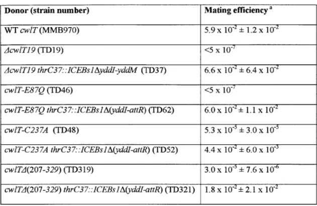
CwlT is required for transfer of ICEBs1. I found that deletion of cwlT completely abrogates ICEBs] conjugation. To my knowledge, all other characterized hydrolases from conjugative systems are at least partially dispensable.
The muramidase domain of CwlT is absolutely required for transfer, while the
peptidase domain is partially dispensable. I determined the effect of both of CwlT's hydrolytic activities on ICEBsJ transfer, using point mutations of the catalytic domains.
In order to function in conjugation, CwlT must be exported from the cytoplasm and must be able to dissociate from the cell membrane. I investigated the effect of cellular localization on CwlT activity in conjugation by using a variety of signal sequence mutants to alter CwlT's subcellular localization. Contrary to previous predictions that CwlT is a lipoprotein, I found that a deletion of its putative lipid anchor site has no effect on its role in conjugation.
CwlT acts on the cell wall of the donor and not on that of the recipient. It is unclear whether hydrolases in conjugative transfer systems work on the donor cell, the recipient cell, or both. ICEBs] was able to transfer with high efficiency into species with cell wall that is
indigestible to CwlT, indicating that the protein does not function on the recipient. In conjugative
systems, the enzymatic specificity of the hydrolase may play an important role in determining
Acknowledgements
This work would not have been possible without the support of every single one of my labmates. The other graduate students in the lab, Tracy Washington, Charlotte Seid, Laurel Wright, and Kayla Menard have offered camaraderie, support, and humor when I most needed it.
My benchmates Laurel Wright and Dr. Jacob Thomas created a fabulous dynamic in 68-552D that put a smile on my face almost every day.
I am particularly grateful to C. Lee and Janet Smith, whose technical advice, patient
explanations, and dedicated mentorship have helped me almost every single day of my graduate
career.
My friends in the Microbiology program, Ben Vincent and Ana Oromendia, have offered me encouragement and support when I needed it, and merciless teasing when I deserved it. As the administrator for the whole Microbiology program, Bonnie Lee Whang has been a
constant sunny, helpful, patient, and hilarious presence during my whole time at MIT. I know that my graduating will reduce her workload significantly, and I thank her for all that she has
done to help me navigate the institute.
The community at Simmons Hall: GRTs, students of 3/4C, and especially Housemasters Steve Hall and John and Ellen Essigmann, have provided me with a loving, supportive second family
during my time at MIT, a perfect complement to my experiences in the lab.
Last, but of course first, I am tremendously grateful to my advisor, Alan Grossman, a brilliant mentor, who sees a teachable moment in every interaction. Alan has put up with me for five years, taught me so much along the way, and always respected my sometimes unconventional choices. I am deeply appreciative for his support.
Table of Contents Abstract Acknowledgements List of Figures List of Tables Chapter 1 Chapter 2 Appendix A Appendix B Chapter 3
Introduction
A bifunctional cell wall hydrolase is needed in donor cells
for transfer of an integrative and conjugative element
ICEBs] genes yddl and yddJ function in conjugation
and may show genetic interaction with wiT
Expression of the extracellular sigma factor SigV
inhibits transfer of ICEBs1
Discussion
2 3 5 6 7 50 80 85 91List of Figures Chapter I
Figure 1
Modes of Horizontal Gene Transfer
9
Figure 2
Lifecycle of Integrative and Conjugative Elements (ICEs)
9
Figure 3
Structure of Peptidoglycan from E. coli and B. subtilis
12
Figure 4
Variation in Peptide Composition and Crosslinking
14
Figure 5
Modifications to the Glycan Strands
15
Figure 6
Hydrolase Cleavage of Bonds in Peptidoglycan
17
Figure 7
Comparison of Lysozyme and Lytic Transglycosylase
17
Cleavage Products
Figure 8
Transformation Machinery in Gram-negative and
22
Gram-positive Organisms
Figure 9
Comparison of Models for Gram-negative and
30
Gram-positive Type IV Secretion
Figure 10
Genetic Map of ICEBs1
39
Figure 11
Map of Features in cwlT
40
Figure 12
Biochemical Activities of CwlT
40
Chapter 2
Figure 1
Map of ICEBs] and its derivatives
74
Figure 2
CwlT degrades cell wall peptidoglycan
75
of Bacillus subtilis but not of Bacillus anthracis
Appendix A
Figure 1
Conjugation effects of ayddlJ deletion
83
List of Tables Chapter 2
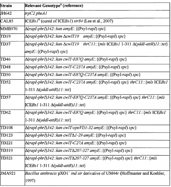
Table 1 Strains used
Table 2 Effects of cwlT mutations on conjugative transfer of ICEBsJ Table 3 Effects of CwlT signal peptide modification on transfer of ICEBs] Table 4 Bacillus anthracis receives ICEBs! as effectively
as does Bacillus subtilis
Appendix A
Table 1 Strains used
Appendix B
Table 1 Strains used
70 71 72 73 82 88
Chapter 1
Introduction:
Introduction: Peptidoglycan Hydrolases in Horizontal Gene Transfer
Whereas all organisms inherit genes "vertically" from their parents, many prokaryotes are also able to acquire new genes "horizontally" from their immediate environment. This process is known as "horizontal gene transfer" (HGT), and researchers are increasingly discovering the broad impact that HGT has on bacterial evolution.
The amount of horizontally acquired DNA varies widely within bacterial species. Some organisms such as Mycoplasma genitalium appear to have none. Others, like vancomycin-resistant Enterococcusfaecalis V583 and Synechocystis PCC6803 have acquired nearly 25% and
17% of their genomes respectively from horizontal sources (Ochman et al., 2000; Paulsen et al., 2003). In many cases, regions of horizontally acquired DNA encode traits that confer a survival advantage, such as antibiotic resistance, increased pathogenicity, ability to colonize hosts, or new metabolic capabilities (Wozniak and Waldor, 2010). Interestingly, the realization that many prokaryotic organisms contain a considerable amount of DNA from other sources could
complicate certain traditional concepts such as organism and species (Goldenfeld and Woese, 2007).
There are three main mechanisms of HGT (Figure 1): transformation, transduction, and conjugation. During transformation, bacteria develop a physiological state of competence, enabling them to uptake DNA directly from the extracellular environment. In transduction, phage particles that have accidentally packaged genomic DNA from a bacterial host deliver that DNA into a new cell during infection. In conjugation, a sequence of DNA is transferred by cell-cell contact between a donor cell and a recipient cell through a process often referred to as "mating." Two types of elements commonly transfer by conjugation: conjugative plasmids, and integrative and conjugative elements (ICEs). ICEs are mobile genetic elements that are typically found
Recipient
Conjugative
plasmid or
transposon
Donor
transformation
transduction
conjugation
Figure 1. Modes of Horizontal Gene Transfer
In transformation, bacteria uptake DNA directly from the extracellular environment. In
transduction, phage particles that have accidentally packaged genomic DNA from a bacterial
host deliver that DNA into a new cell during infection. In conjugation, a sequence of DNA is
transferred by cell-cell contact between a donor cell and a recipient cell. (Figure from
Grossman Lab)
recipient
chromosome
ICE
cell
Excision
Nicking
Transfer
Integration
Figure 2. Lifecycle of Integrative and Conjugative Elements (ICEs)
ICEs are mobile elements typically found integrated into the chromosome of a host cell. Under
certain circumstances, they can excise to form a circular plasmid intermediate, one strand of
which is nicked and then transferred to a recipient cell through a multiprotein mating pore
complex. After transfer, the element can recircularize and integrate into the chromosome of the
recipient. (Figure from Grossman Lab)
integrated into a host cell's chromosome, but that can excise and transfer under certain
circumstances (Figure 2).
Despite their differences, all three of these HGT processes comprise two fundamental DNA
transfer events: the DNA leaves a donor cell and then enters a recipient cell. Each time transfer
occurs, the DNA must cross the bacterial cell wall, a strong, rigid sacculus that maintains the
cell's shape and resists internal osmotic pressure. The cell wall is made of peptidoglycan, a
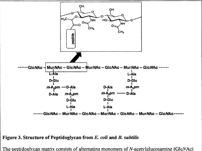
polymer of long carbohydrate chains crosslinked by short peptides (Figure 3).
Different horizontal transfer mechanisms mediate DNA passage across the cell wall in a
variety of ways, but all rely to some degree on hydrolase enzymes that digest peptidoglycan. For
example, in conjugation and competence, large multiprotein complexes are assembled across the
cell wall, and they mediate the secretion (Bhatty et al., 2013) and uptake (Chen et al., 2005) of
DNA, respectively. By contrast, phage particles carrying transducing DNA are often released
from host cells by cell wall lysis that rapidly kills the host (Oliveira et al., 2013). In some cases,
HGT mechanisms are thought to make use of native hydrolases in the cell, while in others, gene
cassettes involved with horizontal transfer encode specialized peptidoglycan hydrolases.
In the work described here, I investigate the function of a two-domain cell wall hydrolase,
CwlT, that is encoded by the integrative and conjugative element ICEBsJ. To provide
background, this introduction discusses how cell wall hydrolases help DNA to cross the cell wall
in the different mechanisms of HGT.
The introduction begins with a description of peptidoglycan structure, its various chemical
modifications, and the native host cell hydrolases that digest and remodel it during normal cell
growth. Then, I discuss each of the three main mechanisms of horizontal gene transfer and
explain the role hydrolases are thought to play in each. As ICEBs] is a conjugative element, I
emphasize aspects of conjugation, particularly its mechanisms, and associated molecular
machinery and hydrolases. Finally, to give immediate context for my own work, I provide an
overview of ICEBs1 and its cell wall hydrolase CwlT.
Peptidoglycan
Structure. Most bacteria are surrounded by a cell wall made of peptidoglycan. The cell wall
forms a rigid capsule around cells, giving them shape, and resisting the high internal turgor
pressure to prevent osmotic lysis (Vollmer et al., 2008a). In Gram-negative organisms, the cell
wall peptidoglycan is sandwiched between the cytoplasmic membrane and the outer membrane.
Gram-positive organisms do not have an outer membrane: a comparatively thicker cell wall is in
direct contact with the extracellular environment.
Peptidoglycan is a covalent matrix, consisting of long glycan (carbohydrate) chains
crosslinked to each other by short peptides (Figure 3). The glycan chains are made of alternating
glucose derivatives, N-acetylglucosamine (GlcNAc) and N-acetylmuramic acid (MurNAc),
joined by P(1-4) glycosidic bonds. Crosslinks are formed between the strands from short peptide
chains attached to the sugars, either by direct or interpeptide bridges, which can be between 1 to
7 amino acids long.
The peptidoglycan layer varies in thickness dramatically depending on organism. E. coli has
a peptidoglycan layer approximately 6 nm thick, which is relatively standard for the
Gram-negative organisms (Vollmer et al., 2008a). By contrast, the peptidoglycan of B. subtilis is
approximately 40-50 nm thick (Hayhurst et al., 2008; Matias and Beveridge, 2005).
H
0 OH
H3C NHO '_
CH3
CH3
--- GIcNAc MurNAc - G cNAc urNAc - GIcNAc - MurNAc - GIcNAc
---L-AWa L-Ala
I I
D-Glu D-Glu
I I
,mA pm -D-Ala II D-Ala m-AI2pm
D-Ala m -Apm m-Apm-- D-Ala
I I
D-Glu D-Glu
I I
L-Aa L-Ala
I I
--- GIcNAc - MurNAc - GIcNAc - MurNAc - GIcNAc - MurNAc -
GIcNAc----Figure 3. Structure of Peptidoglycan from E. coli and B. subtilis
The peptidoglycan matrix consists of alternating monomers of N-acetylglucosamine (GlcNAc) and N-acetylmuramic acid (MurNAc). The inset shows the structure of MurNAc, GlcNAc, and the
p(1-4)
bond between them. The amino acid composition and crosslinking structure of the peptide stems are representative of peptidoglycan seen in both E co/i and B. subtilis. (Figure wasadapted from Vollmer et al., 2008b, using public domain images available from Wikimedia
Commons.)
Peptidoglycan as a Barrier to Transport. The peptidoglycan layer serves as a barrier to transport across the cell envelope, containing small holes 2-4 nm in diameter (Dijkstra and Keck, 1996) that allow the passage of low molecular-weight compounds but exclude those larger. Globular proteins larger than 25 to 50 kDa cannot pass through the peptidoglycan (Demchick and Koch, 1996; Dijkstra and Keck, 1996). Dedicated secretion systems are normally required to allow larger substrates through the cell wall, and localized peptidoglycan digestion is often required for their assembly; these will be discussed in more detail below.
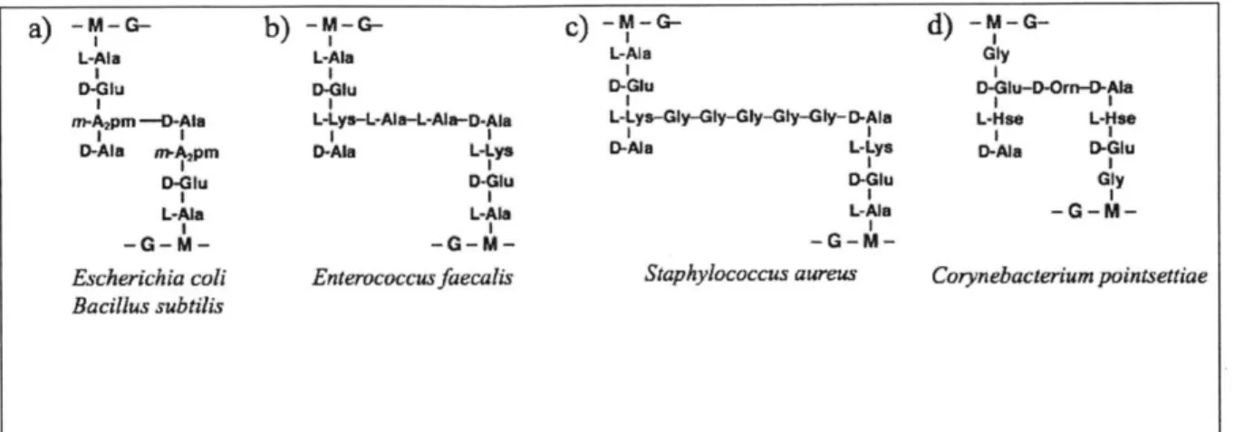
Variations in Peptidoglycan Structure and Composition. Peptidoglycan structure can vary
somewhat between species, and these variations can have important functional ramifications
(reviewed in Schleifer and Kandler, 1972; Vollmer, 2008; Vollmer et al., 2008a). The most common variations are in the amino acid compositions and crosslinking patterns of the peptide side-chains. Figure 4 shows some representative examples of peptidoglycans from different species. The third residue in the peptide side-chains is the most variable, though the first also shows some diversity.
Peptide Stems and Crosslinking. There are two main types of crosslinkage between the
side-chains: in the most common (3-4 linkage), the crosslinking extends from the residue at position 3
of one chain to the alanine at position 4 of the other. The second form (2-4 linkage) is found only in Corynebacteria, and it involves a crosslink between the second and fourth residues of
connected side-chains. Crosslinks can be either direct, or they can involve a cross-bridge containing from one to seven residues. Bacillus subtilis contains a direct 3-4 crosslink, though direct crosslinking tends to be more common in Gram-negative species, and crossbridges are found more predominantly in Gram-positive ones.
The amount of crosslinking also varies significantly between species, from approximately 20% in K coli to over 90% in S. aureus (Vollmer et al., 2008a). Generally, peptidoglycan from Gram-positive species tends to be more highly crosslinked. The amount of crosslinking can also change within a specific organism depending on growth phase, with the peptidoglycan tending to become more heavily crosslinked during late-exponential and stationary phases (Fordham and Gilvarg, 1974; Pisabarro et al., 1985).
a) - M-G- b) -Pm-G- c) -I-G d) --
G-L-Ai L. L-Aa Gly
D Gtu D4Iu G-Glu D-lu--Or-O-O-A
m- pm -- D-Ala L-Lys-L-L-Ala--D-Ala L-ys-Gy-Gly-GlGy-Gly-D-Ala L-Hse L-Hs
t I I I I I I
0-Ala m.Agpm 0-Ala l ys 0-Al. L-ys D-Ala DfWu
I- L_ IP I
D-Glu D.Glu D-Glu Gly
I I I I
L-Ala L-A a L-A-- ---
-I I I
Escherichia coli Enterococcus faecalis Staphylococcus aureus Corynebacterium pointsettiae Bacillus subtilis
Figure 4. Variation in Peptide Composition and Crosslinking
The structure of peptide composition and crosslinking varies between species. Peptide stems are
shown from a) E. coli and B. subtilis, b) E. faecalis, c) S. aureus, and d) C. pointsettiae. The
amino acid in the third position is most variable, and crosslinks between stems can be direct, or
involve bridges ranging in length from one to seven residues. Orn and L-Hse represent
D-ornithine and L-homoserine, respectively. (Figure is adapted from Vollmer et al., 2008a, and
Schleifer and Kandler, 1972).
Modifications to Glycan Strands. The monomers of the glycan strands are also sites of two
common modifications involving acetyl groups: N-deacetylation and O-acetylation (Figure 5)
(reviewed in Vollmer, 2008). Both of these modifications confer resistance to peptidoglycan
digestion by lysozyme, and O-acetylation has been shown to also confer resistance to nearly all
muramidases, members of the broader class of carbohydrate-digesting hydrolases to which
lysozyme belongs. Since lysozyme is an important factor in the innate immune system of
humans and other animals, these modifications that reduce sensitivity to its action are often
linked to pathogenicity (Boneca et al., 2007; Vollmer and Tomasz, 2002).
In N-deacetylation, the acetyl group is removed from the amine at C-3 of the carbohydrate
monomers, and this modification can be made to both acetylglucosamine and
cereus, L. monocytogenes, and S. pneumoniae. In O-acetylation, the hydroxyl group normally
attached to C-6 of N-acetylmuramic acid is replaced with an acetyl group. Species with highly
0-acetylated peptidoglycan include B. anthracis, B. cereus, S. aureus, S. pneumoniae, and N.
gonorrhoeae. Both N-deacetylation and O-acetylation occur after the peptidoglycan has been
synthesized, and these modifications are mediated by specific enzymes.
a)
00
)c)
F2H *C H0tH3 Oi N 0
CH3 Cs,
Unmodified
N-deacetylation
O-acetylation
Figure 5. Modifications to the Glycan Strands
Carbohydrate monomers can be N-deacetylated and O-acetylated, two modifications that confer
resistance to lysozyme and other muramidases. a) Unmodified MurNAc-GlnNAc disaccharide.
b) N-deacetylation of Mur and Gln, with deacetylated amine groups indicated by shading. c)
0-acetylation at MurNAc, with acetyl group indicated with shading. (Figure created using public
domain images available from Wikimedia Commons.)
Cell Wall Hydrolases
There are a wide variety of hydrolase enzymes that can cleave the covalent bonds in
peptidoglycan. Autolysins are hydrolases encoded by bacterial genomes whose main role is to
digest portions of the cell wall to allow its remodeling during the bacterial lifecycle. These will
be discussed in some detail below. Hydrolases encoded by horizontal transfer systems and phage
Specificities of Cell Wall Hydrolases. There are two main categories of hydrolases:
glycosidases, which act on the carbohydrate chains of peptidoglycan, and peptidases, which act on the peptides. There are hydrolases that can cleave every type of bond found in peptidoglycan, and they are characterized accordingly (Figure 6). Only a subset of these hydrolase types is relevant for discussion in the context of this work.
Of the glycosidases, lytic transglycosylases (LT) and lysozymes both cleave the
P(1-4)
glycosidic bond between MurNAc and GlcNAc. However, their products are different. Lytic transglycosylases create a 1,6-anhydro ring at MurNAc by an intramolecular transglycosylationreaction, while lysozymes create free hydroxyl groups on both MurNAc and GlnNAc (Figure 7). Some of the energy of the broken glycosidic bond is retained in this ring, and it is thought that cleavages by lytic transglycosylases can be reversed relatively easily because of this (Moak and Molineux, 2000). Within the peptidase class, endopeptidases cleave peptide bonds between amino acids, either in the peptide side-chains or in the crosslinking bridges. Amidases cleave the amide bonds that link peptide side-chains to MurNAc residues.
Function ofBacterialAutolysins. Bacterial species generally have a large number of
peptidoglycan hydrolases (reviewed in Smith et al., 2000; Vollmer et al., 2008b). The genomes of . coli and B. subtilis each encode approximately 35 identified hydrolases (Sudiarta et al., 2010; Uehara and Bemhardt, 2011). Hydrolases often have overlapping functions, making it difficult to assign specific functions to certain hydrolases, as individual knockouts often do not show a phenotype.
Autolytic activity is important for many cell processes. For example, some of the covalent bonds of peptidoglycan must be broken so that new subunits can be added and the cell wall expanded. At the end of cell division, autolysins digest cell wall at the septum to allow
separation of daughter cells. During spore formation, the asymmetric septum must be digested to ---- GIcNAc - MurNAc GItcrAc - Mur.OVAc - GIcNAc - MurNAc - GIcNAc
----Amidase -Ala
1 0-v--wEndopepfidase
N-a ceImuramidases:
m-A pm-DA Lytic Transglycosylase and
Lysozyme
hr wv nFAla 1
y~~7~ D-Gli N-acetylglucosaminidase
L Ala
---GIONAc - MurNAc - GleNAC - MurNAc - Gti-,NAc - MurNAc -
GIcNJAc----Figure 6. Hydrolase Cleavage of Bonds in Peptidoglycan
Hydrolases cleave specific bonds within peptidoglycan, and they are classified accordingly. The peptidoglycan structure shown is from E. co/i or B. subtilis. Amidases cleave the amide bond between MurNAc and the peptide chain, endopeptidases cleave peptide bonds between residues
within the peptide chain, and carboxypeptidases cleave peptide bonds to remove terminal amino acids. N-acetylglucosaminidases and N-acetyhnuramidases both cleave glycosidic bonds within the glycan chains. Lytic transglycosylases and lysozymes both cleave the same bond, though they create different products (see Figure 5). (This diagram is adapted from Vollmer et al., 2008b).
ON N CH CN CH, H 011<' N CH3 CHS 04r-O OH b) O
cleavage by lytic transglycosylase
- ci. c) OH 0 OH 0- NH C ON H) NH "O k o C#4 cleavage by lysozyme
Figure 7. Comparison of Lysozyme and Lytic Transglycosylase Cleavage Products. Lysozymes and lytic transglycosylases are both N-acetylmuramidases and cleaving the same 0(1-4) bond, though their products are different. a) Representative GlcNAc-MurNAc-GlyNAc fragment. b) Lytic transglycosylases create a 1,6 anhydro ring at MurNAc by an intermolecular transglycosylation reaction. c) Lysozymes create a free hydroxyl group on C-1 of MurNAc and C4
allow prespore engulfment. Later, autolysin-mediated lysis of the mother cell frees the spore.
Digestion of spore peptidoglycan allows germination.
Peptidoglycan hydrolases are also thought to mediate localized digestion of cell wall to allow the insertion and assembly of large protein complexes that cannot fit through the naturally occurring channels in the matrix (Dijkstra and Keck, 1996; Vollmer et al., 2008b). The clusters of genes that encode proteins for structure and assembly of these complexes also typically contain a peptidoglycan hydrolase. For example, in . co/i there are hydrolases are associated with Type IV pilus formation and flagellum assembly (Koraimann, 2003; Nambu et al., 1999). Hydrolases are also involved in assembly of systems that secrete conjugative DNA and other
substrates, and these are discussed below.
Regulation of Cell Wall Hydrolases. Autolytic activity can kill cells if it proceeds
uncontrolled. Regulation of autolysins is not well understood, but it appears to occur at a number of different levels. The first is genetic. Some hydrolases are controlled by temporally specific regulatory factors that only allow expression at points in the lifecycle when specific autolytic activity is needed. In B. subtilis for example, most activity of the two major autolysins, LytC and LytD occurs as cells enter stationary phase (Foster, 1992; Margot and Karamata, 1992).
Spatial localization also plays an important role. For example, S. aureus Atl (Yamada et al., 1996) and E coli AmiC (Bernhardt and de Boer, 2003; Heidrich et al., 2001) and EnvC
(Bernhardt and de Boer, 2004) are involved in cell division, and they all show localization to the
septum. Cell wall binding domains on some autolysins are thought to anchor them to certain
regions of peptidoglycan and prevent diffusion and indiscriminate lysis (Catalao et al., 2013). In B. subtilis, the autolysin LytE associates with MreB, an actin-like protein that directs its
Proteolytic cleavage seems to play an important role in a number of different autolysin
regulatory events. In S. aureus, the autolysin Alt is produced in a pro-form. Its signal peptide is cleaved, and then it is further processed to yield separate glucosaminidase and amidase domains
(Komatsuzawa et al., 1997). In B. subtilis, cell surface and extracellular proteases maintain
steady-state levels of LytF and LytE by degrading unnecessary protein at the cell poles (Yamamoto et al., 2003). In E coli, a soluble transglycosylase (Slt35) is released by the proteolytic cleavage of a membrane-bound lipoprotein precursor, MltB (Ehlert et al., 1995).
The energized bacterial membrane also plays a role in autolysin regulation, and this may
inhibit autolysis close to the cell membrane and allow a gradient of hydrolase activity that
increases with distance from it (Vollmer et al., 2008b). Disruption of membrane polarization can cause rapid autolysis of bacterial cells (Jolliffe et al., 1981).
Phage-encoded hydrolases that cause whole-cell lysis at the end of viral infection are regulated by a variety of unique mechanisms that are discussed below.
Transformation and Competence
In natural transformation (Figure 8), cells take up DNA from the extracellular environment. In order to do so, organisms must be in a physiological state of competence. Some species are always competent, while most develop competence in response to environmental conditions such as nutrient limitation or cell density. Cells may take up DNA in order to acquire genetic
diversity, to repair damaged sequences in their own genome, or to obtain nutrients. Approximately 1% of described bacterial species are known to be naturally competent (Thomas and Nielsen, 2005). Competence has been studied most extensively in Bacillus subtilis,
Streptococcus pneumoniae, Neisseria gonorrhoeae, and Haemophilus influenza (Chen et al., 2005).
Uptake Machinery. With the exception of Helicobacter pylori, which uses a conjugation-like apparatus for DNA uptake, both Gram-negative and Gram-positive organisms show a good deal of similarity in their mechanisms of and machinery for DNA uptake. B. subtilis serves as a representative organism for Gram-positive competence, and N. gonnorhea, for Gram-negative. (Mechanisms of transformation are reviewed in Chen et al., 2005.; Chen and Dubnau, 2004)
In Gram-negative organisms, DNA is thought to transit the outer membrane through a channel composed of secretin proteins (PilQ, in N. gonnorhea), which assemble into doughnut-like multimers that can serve as aqueous channels (Collins et al., 2004). Most species take up DNA of different sequences with relatively similar affinity, though some, such as H. influenzae and members of the genus Neisseriacae, preferentially take up DNA with specific uptake sequences that show similarity to elements in their own genomes (Smith et al., 1995). In Gram-positive organisms, DNA binds to a receptor protein on the outside of the cytoplasmic membrane (ComEA in B. subtilis) (Provvedi and Dubnau, 1999).
Crossing the Wall and Membranes. A competence pseudopilus is a prominent structure in
both Gram-negative and Gram-positive competence (Chen et al., 2006). This is a structure that shows similarity to type 4 pili, which are long, thin, hair-like appendages that play a role in cell adhesion and twitching motility. The pseudopilus spans the cell envelope: in Gram-positive
organisms, it extends through the cell wall, and in Gram-negative organisms it extends through the cell wall and outer membrane. The proposed function of the competence pseudopilus is to
bind DNA and drive its translocation across the outer membrane and cell wall through
binding proteins (ComE in N. gonnorhea [Hamilton and Dillard, 2006], ComEA in B. subtilis
[Provvedi and Dubnau, 1999]), and then it is translocated through a channel in the cytoplasmic
membrane, composed of polytopic membrane proteins (ComA in N. gonnorhea [Facius et al.,
1996], ComEC in B. subtilis [Inamine and Dubnau, 1995]). Gram-positive organisms also appear
to have a membrane-associated ATP-binding protein (ComFA in B. subtilis [Londono-Vallejo
and Dubnau, 1994]) that may be involved in DNA transport across the membrane.
Transforming DNA enters the cytoplasm as a single strand, and it is thought that a nuclease
activity may be coupled to the transport process (Chen et al., 2005; Chen and Dubnau, 2004).
Once in the cytoplasm of the cell, the transforming DNA is protected from degradation by
various proteins. It can integrate into the bacterial chromosome in a RecA-dependent manner, or
if a plasmid, it can recircularize and be maintained extrachromosomally.
Hydrolases in Competence and Transformation. A variety of somewhat scattered evidence
suggests that hydrolases participate in the assembly of competence machinery. Specialized lytic
transglycosylases have been shown to participate in the formation of type IV pili, which are
closely related to conjugative psuedopili. These hydrolases include CofT in enterotoxigenic E.
coli (Taniguchi et al., 2001), and the PilT proteins from plasmids R64 (Sakai and Komano, 2002)
and pO 113 (Srimanote et al., 2002).
Ordinary cell wall turnover mediated by the autolysins may also play a role in competence
development. Readily transformable strains of B. subtilis contain a higher level of autolysin
activity than poorly transformable ones (Young et al., 1963; Young et al., 1964), a relationship
also seen in Streptococcus (Ranhand, 1973). Similarly, the development of competence in B.
subtilis is co-regulated with the activity of autolysins (Guillen et al., 1989).
a) Gram-negative (N. gonnorhea)
Transduction
In transduction, foreign DNA is delivered into cells by bacteriophages (reviewed in Lang et
al., 2012). This occurs because phages sometimes accidentally package sequences from the
genomes of their cellular hosts, as opposed to, or in addition to, their own phage nucleic acid.
This host DNA is then introduced into a new cell during infection by a transducing phage.
Transduction can be classified as either generalized or specialized, depending on the identity
of the packaged host DNA. Phages such as P1 of E. coli perform generalized transduction,
P Q
Figure 8. Transformation Machinery in Gram-negative and Gram-positive Organisms.
Not all components are represented
a) In Gram-negative organisms (represented by N. gonnorhea, DNA passes through an outer
membrane channel formed by the secretin PilQ, assisted by the pilot protein PilP. ComE,
located in the periplasm, is involved in uptake of the DNA, and delivers it to the ComA
channel in the cytoplasmic membrane. The competence psuedopilus is composed of major
pilin PilE (orange) and minor pilin ComP (red).
b) In Gram-negative organisms (represented by B. subtilis), membrane-bound receptor
ComEA delivers DNA to the ComEC channel at the cytoplasmic membrane. The competence
pseudopilus is composed of major pilin ComCG (orange), and minor pseudopilins ComGD,
ComGE, and ComGG (red). (Figures adapted from Chen and Dubnau, 2004)
packaging essentially random fragments of host genome. On the other hand, lysogenic phages
such as Lambda may perform specialized transduction, packaging host DNA attached to the
phage DNA that is adjacent to the phage attachment site on the host chromosome. The amount of
DNA that can be transferred via transduction is limited by the size of the phage head, but for
some phages, it is nearly 100 kb (Ochman et al., 2000).
Like the DNA delivered into cells by transformation, transduced DNA usually integrates into
the genome via site-specific recombination. Occasionally, plasmids can also be transferred by
transduction (Mahan et al., 1993), where they recircularize and replicate in the new host.
Transduction is unique from conjugation and transformation, because it is the only form of
HGT that does not require the donor and recipient to be in close proximity, or that the foreign
DNA come from the immediate environment. Thus, in both terrestrial and marine ecosystems,
transducing phages are often seen as unique reservoirs of widely diverse exogenous genes
(Anderson et al., 2011; Jiang and Paul, 1998; Zeph et al., 1988).
Phage Hydrolase Systems
Phage-encoded hydrolases digest cell wall peptidoglycan at different stages in the phage
lifecycle, and they fall into two classes: virion-associated hydrolases that locally degrade the
peptidoglycan to allow DNA insertion, and endolysins, which cause large-scale lysis of host cells
to release phage particles at the end of the infection cycle. Aspects of both of these phage
hydrolase systems may be similar to mechanisms in hydrolases from secretion systems and
conjugative elements.
Entry into the Cell: Virion-Associated Hydrolases. Virion-associated peptidoglycan
hydrolases (VAPH) are widespread in phages that infect both Gram-positive and Gram-negative bacteria (Moak and Molineux, 2004). They are usually found associated with the phage tail, and they possess many types of hydrolytic domains, including lysozymes, endopeptidases, and transglycosylases (Rodriguez-Rubio et al., 2012). Some VAPD, particularly those that infect Staphylococcus and Mycobacteria, have been shown to have multiple lytic domains, usually a muramidase and a peptidase (Oliveira et al., 2013).
Function in Infection. It is thought that virion-associated hydrolases create a small, localized opening in the cell wall peptidoglycan that allows the insertion of DNA. However, few specifics are known about their functional roles. In Gram-negative organisms, they tend not to be essential for infection, though the hydrolase (gp16) in T7 was shown to be required when host cells were grown at 20C (Moak and Molineux, 2000). DNA internalization is less efficient and delayed in their absence, and they appear to be most important in situations such as stationary phase growth when the peptidoglycan is more heavily cross-linked (Moak and Molineux, 2000; Piuri and Hatfull, 2006; Rydman and Bamford, 2002).
A number of hydrolases have been identified in Gram-positive phages, though phenotypes of their deletions have not been examined. In the B. subtilis phage 029, the hydrolase gpl3 is an essential morphogenic factor that is required for tail assembly (Xiang et al., 2008), but its functions as a hydrolase have not been explored. It is possible that phages infecting Gram-positive bacteria are more dependent on hydrolases to bypass the thick cell walls of their hosts, and that the functions of these enzymes would be essential.
Other Putative Functions of Virion-Associated Hydrolases. Virion-associated hydrolases
tail fiber of phage T5 has been shown to also have membrane fusogenic properties and may be
involved in creating a pore between the two membranes for DNA translocation (Boulanger et al.,
2008). In T7, gp16 digestion allows three injected proteins to pass the cell wall and create a
channel for DNA in the inner membrane. This hydrolytic enzyme may be actively involved in
translocating DNA, and it is thought that while its N-terminus is anchored to peptidoglycan, its
C-terminal may ratchet along DNA using a "hand-over-hand" motion, similar to how kinesin
moves along actin filaments. Its C-terminus may have molecular motor ability (Boulanger et al.,
2008).
Exit from the Cell: Phage Endolysins.
At the end of the phage infection cycle, progeny virions are usually released by host cell
lysis, which is most often mediated by phage-encoded hydrolases known as endolysins. A
variety of hydrolytic activities are found in phage endolysins: lysozymes, lytic transglycosylases,
endopeptidases, and amidases (Catalao et al., 2013). The lethal activity of these enzymes is
tightly regulated. Mechanisms of regulation vary somewhat, though a general strategy centers on
sequestration of the hydrolase, either in the cytoplasm or in the membrane, followed by its
release and rapid cell lysis.
Canonical Endolysin-Holin Systems.
The most common regulation paradigm is exemplified
by the lysis system in phage Lambda, and it is universal in almost all dsDNA phages, albeit with
some exceptions (Catalao et al., 2013; Loessner, 2005; Wang et al., 2000). In this mechanism,
phage hydrolases are synthesized and accumulate in the cytoplasm, as they lack signal sequences
that would allow their secretion. Small hydrophobic accessory proteins known as holins create
pores in the cytoplasmic membrane, causing depermeabilization at a specific time; this event
allows the endolysins to escape from the cytoplasm, reach the cell wall, and cause host lysis. Signal-Arrest-Release Endolysins. In contrast to those mentioned above, members of a small class of phage endolysins are exported out of the cytoplasm, but remain anchored in the
cytoplasmic membrane by a non-cleavable N-terminal transmembrane sequence known as a Signal-Arrest-Release (SAR) domain (Xu et al., 2004). This anchor is only metastable, and holin-mediated membrane permeabilization can release the endolysin. The holins associated with
SAR endolysins tend to create much smaller channels than those in canonical holin-endolysin systems because endolysins do not need to pass through them, and they are referred to as pinholins to reflect this fact (Pang et al., 2009). Release of the SAR domain from the membrane often causes a refolding and conformational rearrangement of the enzyme that allows residues of the catalytic site to move into an active formation (Catalao et al., 2013). Thus far, only a few SAR endolysins have been identified, and the best-characterized examples are Lyz of P1 (Xu et al., 2004), R protein of coliphage 21 (Sun et al., 2009), and Lyz103 of the Erwinia amylovora phage ERA103 (Kuty et al., 2010). It is estimated that approximately 25% of phages contain a SAR endolysin (Park et al., 2007).
Holins are not completely required for SAR endolysin release. Since the N-terminal anchor is only metastable, SAR endolysins can slowly escape from the membrane and cause lysis on their own. The observation that SAR endolysins do not require any accessory factors has led to the proposal that they may represent the most evolutionarily primitive form of endolysins (Park et al., 2007).
Endolysins with Signal Sequences. Finally, a very small class of endolysins exemplified by the Lys44 protein from Oenococcus Oeni phage fOg44 have signal sequences that direct their
unhindered export from the cell (Catalao et al., 2013; Nascimento et al., 2008; Sao-Jose et al.,
2000). Contrary to those of SAR endolysins, the N-terminal signal sequence of Lys44 plays an inhibitory role and its cleavage is required for activity. Additionally, depolarization of the cytoplasmic membrane appears to activate enzymatic function. In this unique case, holins do not mediate endolysin access to the cell wall, but rather are involved in creating activating conditions for it.
Conjugation
In conjugation, a sequence of DNA is transferred by cell-cell contact between a donor cell
and a recipient cell through a process often referred to as "mating." Two types of genetic elements can transfer autonomously by conjugation: conjugative plasmids and integrative and conjugative elements (ICEs). Conjugative plasmids are maintained extrachromosomally, whereas ICEs are integrated into a host cell's chromosome and propagated passively along with the host genome. Under some conditions, an ICE can excise, circularize, and transfer into a recipient cell
where it then reintegrates.
Both ICEs and conjugative plasmids encode the conjugation machinery needed for their own transfer. The conjugation genes are often organized into closely related modules clustered by biological function, and these clusters show evidence of frequent exchange between other mobile elements, phages, and host genomes (Burrus et al., 2002; Osborn and Boltner, 2002; Toussaint and Merlin, 2002). Usually, the transfer functions of conjugative plasmids and ICEs are repressed, but these elements can be stimulated to transfer by exposure to a wide range of inducers, including DNA damage, pheromones, antibiotics to which the elements confer
resistance, and other metabolites (Bellanger et al., 2009; Grohmann et al., 2003; Thomas and
Nielsen, 2005).
Type IVSecretion Systems. Similar mechanisms appear to be involved in the transfer of both conjugative plasmids and ICEs (Alvarez-Martinez and Christie, 2009; Lee et al., 2010; Toussaint and Merlin, 2002; Zechner et al., 2012). Translocation of the transferred DNA (T-DNA) is mediated by protein machinery that comprises a type IV secretion system (T4SS). The T4SS is
an elaborate, multiprotein apparatus that allows substrates to pass out of the cell through the cell membrane and the cell wall. T4SS can transport a wide range of substrates: some are involved in the conjugation of plasmids and ICEs, while others secrete virulence factors, effector proteins and other nucleoprotein complexes into either host cells or culture supernatant (Abajy et al., 2007; Cascales and Christie, 2003; Christie et al., 2005).
Most of our knowledge about type IV secretion mechanisms comes from systems in Gram-negative organisms. Some of the best-studied systems are plasmids from Escherichia coli, including F (Wong et al., 2012), RP4 (Rabel et al., 2003), and R388 (Vecino et al., 2011). Also, the Legionella pneumophila Dot/lcm (Nagai and Kubori, 2011) and Agrobacterium tumefaciens Ti plasmid transfer systems have been well studied. In particular, the current paradigm model for Type IV secretion is based largely on studies of the conjugation apparatus involved in the
transfer of the Ti plasmid from A. tumefaciens, and transfer genes in other systems are often named after similar ones from A. tumefaciens. This model will be presented below. Much less is known about transfer in Gram-positive organisms, though there are a number of similarities to the T4SS in Gram-negative organisms.
Modelfor Agrobacterium Ti Plasmid Transfer. A. tumefaciens is an agricultural pathogen
(reviewed in Pitzschke and Hirt, 2010). The plasmid integrates into the plant genome, and it
encodes enzymes that promote both tumor growth and the production of opines,
amino-acid-sugar conjugates that provide nutrients to the bacterium. The Ti plasmid has an extraordinarily
broad host range: in vitro, it is able to transfer into almost any eukaryotic species, from fungi to
human cells (Lacroix et al., 2006). The conjugation system can also mobilize other plasmids
such as RSF 1010 that do not encode their own conjugation functions (Stahl et al., 1998).
The products from at least 12 genes on the Ti plasmid (virBi-virBi], and virD4) comprise
the T4SS that mediates DNA and protein transfer into plants and other organisms (Figure 9), and
this is often referred to as the VirB/D4 system (reviewed in Alvarez-Martinez and Christie, 2009;
Bhatty et al., 2013; Fronzes et al., 2009; Zechner et al., 2012). The VirB proteins are involved in
creating the secretion channel that extends across-and allows substrates to pass through-the
inner membrane, cell wall, and outer membrane of the donor cell.
Substrate Processing and Recruitment. A number of proteins are involved with the
processing of the transferred DNA (T-DNA) and its delivery to the secretion channel. The
relaxase VirD2 nicks one strand of the Ti plasmid at a specific repeated sequence, becoming
covalently attached to its 5' end and forming the core of a complex known as the relaxosome.
VirD2 is thought to remain associated with the T-DNA during its passage through the secretion
channel and into the plant host. The protein has a nuclear localization signal and is involved with
targeting the T-DNA to the plant nucleus (Pitzschke and Hirt, 2010). The coupling protein VirD4
interacts with the relaxosome, and transfers the T-DNA to the membrane-bound ATPases VirB4
and VirBl l.
Gram Positive
7\
Substrate Secretion Pathway
Substrate Secretion Pathway
Figure 9. Comparison of Models for Gram-negative and Gram-positive Type IV Secretion
A subset of the Gram-negative secretion apparatus components are conserved in Gram-positive
systems. Gram-positive secretion systems lack the core complex formed by VirB7, VirB8, and
Viri 0, and it has been proposed that VirB8 forms a channel across the peptidoglycan. A VirB 11
homolog is absent in Gram-positive systems, though ATPases VirD4 and VirB4 are conserved.
While some Gram-positive systems encode surface adhesins, none are known to make
conjugative pili. As the cell wall is considerably thicker in Gram-positive organisms, the
hydrolase VirB1 may play a more significant role in complex assembly. (Figures adapted from
Bhatty et al., 2013).
Passage Through the Cell Envelope. From VirB4 and VirB 11, the substrate likely passes
through a complex of membrane-bound proteins (primarily VirB6 and VirB8, as well as VirB3
and VirB 10) that forms a channel in the inner membrane. VirB7, VirB9, and VirB 10 create a
core complex that crosses the peptidoglycan and spans the entire cell envelope. Proteins in this
complex may also make a cap that protrudes from the cell surface and creates a channel
through the outer membrane. A peptidoglycan hydrolase, VirB 1, is thought to locally digest the
cell wall peptidoglycan to allow the assembly of this multiprotein complex; VirB1 will be
discussed in greater detail below in the section on hydrolases in conjugative elements.
VirB4 and Vir 1I as well as the coupling protein VirD4 all exhibit ATPase activity, and although the mechanisms are not entirely clear, it is thought that they are involved in powering substrate translocation through the secretion channel.
Interactions with Recipients: The Conjugative Pilus. A conjugative pilus extends from the
cell surface and is composed primarily of subunits of the pilin protein VirB2. The protein VirB5 is found at the end of the pilus, and it has been proposed that VirB5 mediates contacts between
donor and recipient cells. Although the lumen of the pilus appears wide enough to accommodate
single-stranded DNA and unfolded proteins, it is unclear whether it acts as a conduit for substrates (Alvarez-Martinez and Christie, 2009). The hydrolase VirBI is also essential for the biogenesis of the pilus, though it is unclear what role it plays in this process (Zupan et al., 2007).
Mechanisms of Gram-Positive Conjugative Transfer
Much less is known about mechanisms of conjugation in Gram-positive species. These systems need only translocate substrate across one membrane, but they must also mediate transfer across a cell wall that is much thicker than those in Gram-negative organisms. Some of the best-studied, representative Gram-positive elements are the conjugative plasmids pCW3, pIP501, pCF1O, and the ICEs Tn96 and ICEBs] (Alvarez-Martinez and Christie, 2009), though none of these has yet emerged as a clear prototypical system. A core set of genes in Gram-positive systems show similarity to those in better-characterized Gram-negative systems, and comparison can provide a general picture of how DNA translocation might occur (Figure 9).
Substrate Processing and Recruitment Gram-positive conjugation systems encode both a
relaxase and a VirD4-like coupling protein similar to those in Gram-negative systems, and it is likely that substrate processing and recruitment occur in much the same ways. Studies in both pCW3 and pIP501 have shown an interaction between the relaxase and coupling protein (Abajy et al., 2007; Chen et al., 2008).
Passage Through the Cell Envelope Of the components involved in secretion channel formation, VirBi, VirB3, VirB4, VirB6, and VirB8 appear to be at least partially conserved in
Gram-positive systems (reviewed in Bhatty et al., 2013). The membrane pore structure is likely quite comparable to that in Gram-negative organisms, formed by proteins similar to VirB3, VirB6, and VirB8, (Porter et al., 2012). Other small membrane proteins may also associate with this core complex.
The structure of the envelope-spanning secretion channel must be very different from that in Gram-negative species, because none of the components that make the main core complex, VirB7, VirB9, or VirBlO, are conserved in Gram-positive species. Multimerized VirB8 may create a structure that spans the cell wall (Bhatty et al., 2013; Porter et al., 2012), forming a channel to the outside of the cell through which the secretion substrates travel. Alternatively, some of the small membrane proteins found in these systems may polymerize into long fibers to create a channel across the wall (Alvarez-Martinez and Christie, 2009).
Substrate secretion is likely energized by the ATPase activities of the coupling protein and VirB4-like protein (Berkmen et al., 2010), as the VirB11 ATPase is not conserved in Gram-positive species (Bhatty et al., 2013).
Interactions with Recipients: Surface Adhesins. No Gram-positive elements are known to
produce conjugative pili. The plasmid pCF10 encodes an adhesin that is anchored to the cell and promotes donor-recipient aggregation (Waters and Dunny, 2001). Surface adhesins may also be
encoded by pIP501 and pCW3, but there are no signs of genes with such functions in either ICEBsJ or Tn916 (Alvarez-Martinez and Christie, 2009).
Role ofthe Hydrolase. In Gram-positive systems, the VirBI-like hydrolase has an
N-terminal transmembrane domain, and may form part of the translocation complex structure.
These hydrolases have been shown to interact with the coupling protein and VirB6-like subunits
in the conjugal plasmids pIP501 and pCW3, (Abajy et al., 2007; Steen et al., 2009), and they
may play a more active role in substrate recruitment than in Gram-negative systems. The role of hydrolases will be discussed in more detail below.
Hydrolases in conjugation systems
Specialized cell wall hydrolases are found in most, if not all, Type IV Secretion Systems, from both Gram-positive and Gram-negative backgrounds (Zahrl et al., 2005). The hydrolases are some of the least-studied components of T4SS, and their function is not well characterized. However, it is generally assumed that at least one of their roles is to create localized openings in the cell wall peptidoglycan to allow the assembly of the secretion apparatus. The peptidoglycan mesh normally contains gaps averaging approximately 2-4 nm in diameter (Demchick and Koch, 1996; Dijkstra and Keck, 1996), and it would need to be significantly remodeled to
accommodate the passage of a large protein complex. Structural studies on the T4SS in A. tumefaciens have estimated that the core, cell-envelope spanning complex has a diameter of
Hydrolase Structure. These hydrolases have some common features. VirB 1 from
Agrobacterium is the best-characterized hydrolase from a Gram-negative transfer system. At its N-terminus is a single catalytic domain with a lysozyme-like structure fold that also shows similarity to bacteriophage lytic transglycosylases (Mushegian et al., 1996). This motif is also found in P19 of plasmid RI, TraL of IncN plasmid pKMlOl, and TrbN of IncP plasmid RP4. In Gram-positive conjugative systems, representative hydrolases include Orfl4 of Tn916, TcpG of pCW3, CwlT of ICEBs], and Orf7 of pIP501. All of these have two catalytic domains: an N-terminal glycosidase and a C-terminal peptidase. The hydrolase PrgK from pCF1O appears to represent a slightly different class of Gram-positive hydrolase: it also has glycosidase and muramidase functions, but has an additional N-terminal peptidoglycan-binding LytM domain (Bhatty et al., 2013). Approximately 700 residues in length, PrgK is nearly as twice as large as the other Gram-positive hydrolases.
BiochemicalActivities. For the most part, the biochemical activities of these hydrolases have not been thoroughly characterized. VirB1, P19, and TcpG have all been shown to degrade peptidoglycan in vitro (Bantwal et al., 2012; Bayer et al., 2001; Zahrl et al., 2005), though the
enzymatic mechanisms and cleavage sites of these and most other T4SS hydrolases have been inferred only from homology. An exception is CwlT, which was shown to have
N-acetylmuramidase (lysozyme-like) activity and DL-endopeptidase activity (Fukushima et al., 2008). Interestingly, experimental observations of lysozyme-like activity contradicted previous homology comparisons, which had predicted that the N-terminal region had lytic
transglycosylase activity. In light of these findings, it is possible that many of the other T4SS hydrolases predicted to contain lytic transglycosylase domains may actually act as lysozymes.
Throughout this paper, carbohydrate-digesting domains of hydrolases will be referred to as
muramidases, to acknowledge the uncertainty in the identity of these domains.
Phenotypes of Hydrolase Disruptions. Hydrolase function is not completely required for
conjugation in any of the systems studied thus far. For Gram-negative organisms, disruption of
the hydrolase seems to reduce transfer by 10-100 fold. Transfer of RI is decreased
approximately 10-fold with the deletion of its hydrolase P19 (Bayer et al., 2001), disruption of
TraL from pKMlOl decreased transfer by about 10-100 fold (Winans and Walker, 1985), and a
VirBI deletion reduces Ti plasmid transfer by 10-100 fold, depending on the assay used
(Mushegian et al., 1996; Berger and Christie, 1994; Bohne et al., 1998).
In the Gram-positive conjugative plasmid pCW3, deletion of the hydrolase TcpG caused
transfer to decrease by 1000-fold (Bantwal et al., 2012), a larger decrease than is seen in
Gram-negative systems. A catalytic mutant of the peptidase domain was also constructed, which
showed approximately a 10-fold decrease in transfer. This heightened requirement for hydrolytic
activity may be due in part to the much thicker, more heavily crosslinked cell wall in
Gram-positive organisms (Abajy et al., 2007). It may also have to do with interactions that the
hydrolase makes with essential components of the secretion system; these will be discussed
below.
Hydrolase Complementation. It is generally assumed that element-encoded hydrolases are
not completely required for transfer because other hydrolases can substitute for their activity
(Abajy et al., 2007; Bantwal et al., 2012; Baron et al., 1997; Hoppner et al., 2004). The cells'
native autolysins or hydrolases from other secretion systems may be able to locally enlarge the
peptidoglycan in the absence of a dedicated hydrolase.
In general, the hydrolases from many Gram-negative elements show a low degree of
specificity in cross-complementation assays and are able to substitute for each other. Deletion of the hydrolase P19 from plasmid R1 can be complemented by TpgF from the T3SS of Shigella connei, and by TrbN from the conjugative plasmid RP4 (Zahrl et al., 2005). The conjugation deficiencies of a VirBI deletion from Agrobacterium can be complemented nearly to wild-type levels by the VirB1 protein from Brucella suis, and partially by TraL from pKM101 (Hoppner et
al., 2004). Thus far, there have been no cross-complementation studies for hydrolases in positive conjugative elements. This low level of specificity may indicate that, at least in Gram-negative organisms, the hydrolase may be a relatively late evolutionary addition to secretion
systems (Zahrl et al., 2005).
Interactions with Other Secretion Channel Components. More information about the roles
and other functions of these hydrolases could be inferred from studies of the other subunits that they interact with. The interactions that the peptidoglycan hydrolases make suggest that they may fulfill other roles, in addition to, or in concert, with their roles in peptidoglycan degradation. VirBI has been shown to associate with core secretion channel proteins both in Brucella suis (VirB8, VirB9, and VirB 11) (Hoppner et al., 2005) and in Agrobacterium (VirB4, VirB8, VirB9, VirB0, and VirBI1) (Ward et al., 2002). It has been suggested that these interactions may restrict hydrolytic activity to the site of T4SS assembly (Hoppner et al., 2005) and/or that VirB1 may actively recruit elements of the secretion channel (Ward et al., 2002). Additionally, VirBi's peptidoglycan degradation likely also causes indirect stabilization of the channel components; as more space opens up, members of the secretion pore can promote stabilizing interactions
al., 2002). However, it is unlikely that these interactions are extremely specific, since hydrolases
from different systems are able to functionally substitute for one another.
In Gram-positive systems, interaction studies suggest that the hydrolase may play a more
active role in substrate recruitment. In the Gram-positive conjugative plasmids pCW3 and
pIP501, the hydrolases (TcpG in pCW3; Orf7 into pIP501) have been shown to interact with the
coupling protein (TcpA in pCW3; Orfl0 into pIP501) (Abajy et al., 2007; Steen et al., 2009).
This interaction may aid in transporting the coupling protein to the secretion channel
components. In pIP501, the hydrolase (Orf7) has also been shown to interact with the VirB4
homolog Orf5, and with another protein, Orfl4, whose function is unclear (Abajy et al., 2007).
As has been suggested for Gram-negative systems, the hydrolase may play a role in recruitment
of these proteins, or help to stabilize interactions between them by locally opening the cell wall.
Additional Roles of Peptidoglycan Hydrolases. At least one characterized hydrolase has