HAL Id: hal-03043263
https://hal.archives-ouvertes.fr/hal-03043263
Submitted on 7 Dec 2020HAL is a multi-disciplinary open access archive for the deposit and dissemination of sci-entific research documents, whether they are pub-lished or not. The documents may come from teaching and research institutions in France or abroad, or from public or private research centers.
L’archive ouverte pluridisciplinaire HAL, est destinée au dépôt et à la diffusion de documents scientifiques de niveau recherche, publiés ou non, émanant des établissements d’enseignement et de recherche français ou étrangers, des laboratoires publics ou privés.
Genome analysis of Marinobacter phage AS1 suggests
its close interactions with host Marinobacter sp
Sreekumar Aparna, Ammini Parvathi, Angia Sriram Pradeep Ram,
Télesphore Sime-Ngando
To cite this version:
Sreekumar Aparna, Ammini Parvathi, Angia Sriram Pradeep Ram, Télesphore Sime-Ngando. Genome analysis of Marinobacter phage AS1 suggests its close interactions with host Marinobacter sp. Aquatic Microbial Ecology, Inter Research, 2019. �hal-03043263�
1
Genome analysis of Marinobacter phage AS1 suggests its close interactions with host 1
Marinobacter sp. 2
Aparna S.1, Parvathi A.1*, Pradeep Ram A. S.2 and Sime-Ngando T.2 3
4 1
CSIR-National Institute of Oceanography, Regional Centre (CSIR), Kochi-682 018, India 5
2
Laboratoire Microorganismes : Génome et Environnement, UMR CNRS 6023, Université Clermont-6
Auvergne, 1 Impasse Amélie Murat, 63178 Aubière Cedex, France 7 8 9 10 Corresponding author 11 Dr. Parvathi A. 12
CSIR-National Institute of Oceanography
13
Regional Centre, Dr. Salim Ali Road,
14
Post Box No. 1913, Kochi-682 018, India
15 Ph: 91-(0) 484-2390814, Fax: 91-(0) 484-2390618 16 Email: parvathi@nio.org 17 18 19
Running title: Whole genome characterization of Marinobacter phage AS1 20 21 22 23 24 25
2 Abstract
1
Marinobacter comprises a widespread and ecologically significant genus of marine bacteria
2
with vast metabolic capabilities. However, very few reports on phages infecting this genus exist. In
3
this study, we isolated and characterized a linear dsDNA genome of Marinobacter phage AS1,
4
infecting Marinobacter sp. strain, D1S9. The phage is a member of Podoviridae family having a
5
genome size of ~37 kb and a 57 % GC content with no significant overall similarity to any of the
6
available viral whole genome sequences in the database. The genome which encodes for 52 protein
7
coding genes with no tRNA genes, contains 21 protein coding genes with assigned putative functions.
8
High degree of recombination events with other members of the family Caudovirales is evident from
9
the mosaic pattern of inheritance of genes such as major capsid protein, terminase large sub unit,
10
portal protein and integrase. Presence of integrase gene, DNA methyl transferases and the occurrence
11
of AS1 sequences within the whole genome sequences of some members of the host taxon were
12
suggestive of its intense association with the host.
13 14
Keywords: Marinobacter, phage, complete genome sequence, Podovirus, Arabian Sea 15 16 17 18 19 20 21 22 23
3 1. Introduction
1
Viruses, primarily bacteriophages are the most prolific and diverse obligate parasites in the
2
marine biosphere where they outnumber bacteria on an average by one order of magnitude (Suttle,
3
2007, Sime-Ngando 2014). Bacteriophages have indispensable involvement in the ecological and
4
biogeochemical processes by regulating bacterial abundance, bacterial diversity, nutrient fluxes,
5
structuring the microbial food web dynamics and to a great extent in influencing host diversification
6
and speciation through horizontal gene transfer (Middelboe and Lyck, 2002, Bouvier and Del Giorgio
7
2007, McDaniel et al. 2010, Winget et al. 2011). A considerable fraction of bacterial genetic diversity
8
is acquired through horizontal gene transfer, imparting ecological and pathogenic attributes like
9
antibiotic resistance, resistance to phage infection, toxins and heavy metals (Stern and Sorek, 2010,
10
Xia and Wolz, 2014). Phage genomes are also coevolving concomitantly with their hosts. Compared
11
to other host-parasite systems, bacterial-phage coevolution is faster owing to their large population
12
sizes and short generation times (Buckling and Brockrust 2012). Studies with coevolving populations
13
of hosts and their infective phages revealed two modes of coevolution: arms race dynamics (ARD)
14
and fluctuating selection dynamics (FSD). These two were found to operate at different times in the
15
same population with arms race dynamics giving way to fluctuating selection dynamics in the long
16
run (Hall et al. 2011, Gokhale et al. 2013).
17
Most of the dsDNA phages share a large pool of genetic elements (Hendrix et al. 1999).
18
Phage genomes exhibit prevalent mosaic architecture as a result of extensive horizontal gene transfer
19
and in fact different genomic segments may have different ancestral origins. So, it is challenging to
20
deduce whole genome similarities and to assign phylogenetic relationships to newly sequenced
21
phages (Hatfull and Hendrix 2011). Isolation and genome characterization of additional phages would
22
help to resolve this problem to some extent.
4
Despite of the profound knowledge on abundance and role of marine viruses in
1
biogeochemical cycles and host population dynamics, genome level information on phage-host
2
interactions of ecologically significant bacteria are very limited. Sequencing and whole genome
3
characterization of phages infecting these bacteria might provide insights into the phage-host
4
interactions and how they shape the ecological roles of the hosts. Marinobacter, an ecologically
5
significant genus of bacteria, and its first representative, M. hydrocarbanoclasticus, was proposed by
6
Gauthier in 1992. Since then, new members belonging to this genus have been isolated from diverse
7
marine habitats ranging from oil producing wells to deep-sea benthic sediments with fifty-eight
8
species reported so far (Martin et al. 2003, Yoon et al. 2007, Xu et al. 2008, Zhang et al. 2008, Wang
9
et al. 2009, Wang et al. 2012, Chua et al. 2018). Several species have been associated with marine
10
sponges and microalgae (Green et al. 2006, Kaeppel et al. 2012, Lee et al. 2012, Lupette et al. 2016,
11
Sandhya et al. 2017). Members of this genus are known to have substantial impact on marine snow
12
formation and on various biogeochemical cycles with quite a few strains possessing a repertoire of
13
activities like degradation of various hydrocarbon compounds, denitrification, Fe (II) oxidation, Mn
14
(II) oxidation, Fe (III) reduction and redox cycling of arsenic and fumarate (Kaye et al. 2010, Kaeppel
15
et al 2012, Wang et al. 2012, Handley and Lloyd 2013, Bonis and Gralnick 2015). Although their
16
versatile functional capabilities enable them to survive in diverse environmental conditions and to
17
exploit variable resources (Singer et al. 2011), viruses infecting this genus are reported rarely (Zhu et
18
al. 2018), thus hindering an integrated understanding of the life cycle of these microbes in the marine
19
environments. In this study, we report the isolation of a bacteriophage infecting the genus
20
Marinobacter from surface waters of the Arabian Sea (southwest coast of India) and provide
first-21
hand information on the sequencing and whole genome analysis of it.
22 23
5 1
2. Materials and Methods 2
2.1. Isolation and purification of Marinobacter phage AS1 and its host 3
Marinobacter phage AS1 was isolated from the surface water of the tropical Arabian Sea, off
4
the coast of Kochi, southwest coast of India (9.9586º N and 76.0825º E, Fig. 1A) in December 2014
5
as described by Middelboe et al. (2010). Briefly, 25 mL water sample was amended with 1 mL 10×
6
Zobell marine broth and incubated at room temperature for 2 days in order to propagate potential
7
bacterial hosts and thereby the specific viruses in the sample (Himedia, Mumbai, India). The viral
8
enriched seawater was screened against lawns of bacterial isolates obtained from the same water
9
sample by means of soft agar overlay technique. Culturing of bacteria was done by spread plating
10
water samples on Zobell Marine Agar. The plaques formed on the bacterial lawn after overnight
11
incubation (37 ºC) were picked and transferred to phage buffer (SM buffer: 450 mM NaCl, 50 mM
12
MgSO4, 50 mM Tris, 0.01 % gelatin, pH 8), vortexed well and kept at 4°C. Centrifugation (10,000
13
×g, 10 min) was done the next day to remove the soft agar and host cell remnants which was followed
14
by filtration through 0.2µm pore size Acrodisc syringe filters (Sigma-Aldrich, India). The filtrate was
15
then serially diluted and plated by soft agar overlay method. Single plaque from the plate was isolated
16
and the purification was repeated for five consecutive times. The phage stock was stored at 4°C until
17
use.
18
The bacterial isolate for which the phage was isolated was identified using 16S rRNA gene
19
polymerase chain reaction (Amann et al. 1995). The PCR products were sequenced and identified by
20
BLAST search against non-redundant database of NCBI.
21
2.2. Transmission Electron microscopy (TEM) 22
6
For morphometric characterization of phage by TEM analysis, the purified phage particles
1
were collected onto 400-mesh carbon coated formvar grids by centrifugation (70,000 ×g, 20 minutes
2
at 4ºC) using a SW 40Ti rotor and stained at room temperature for 30 s with uranyl acetate stain (2 %
3
w/v) (Pradeep Ram et al. 2010). Excess stain on grids was removed by rinsing twice with 0.02 µm-
4
filtered distilled water and dried on filter paper. Grids were examined using a JEOL 1200Ex
5
transmission electron microscope (JEOL, Akishima, Japan) operated at 80 kV at a magnification
6
between × 60,000 and × 100,000.
7
2.3. Phage DNA isolation 8
Phage particles were precipitated with 8% polyethylene glycol (PEG) 8000 containing 1M
9
NaCl after overnight incubation at 4°C (Lawrence and Steward 2010). The mixture was centrifuged at
10
10000×g for 20 min at 4°C and the pellet was suspended in SM buffer to disaggregate overnight at
11
4°C. To remove the PEG, KCl was added to the viral suspension to a final concentration of 1M and
12
kept on ice for 30 min. PEG was pelleted by centrifugation at 12000×g for 10 min at 4°C and the
13
supernatant containing viral particles were transferred to a fresh tube. The viral suspension was
14
concentrated using Amicon Ultra-15 centrifugal filter unit with a molecular weight cut off of 30 kDa
15
(Millipore, India). The concentrated phage preparation was treated with nucleases (DNase I and
16
RNase A at final concentrations of 1 mg mL-1) to remove any host DNA and RNA. Phage DNA was
17
extracted using the phenol:chloroform (24:1) extraction method as described previously (Yang et al.
18
2017). Briefly, the nuclease treated phage preparation was treated with proteinase K (100 mg mL-1),
19
10% (w/v), SDS and 0.5 M EDTA (pH 8) and incubated in water bath at 55 °C for 1 hr. This was
20
followed by two rounds of phenol:chloroform:isoamyl alcohol (25:24:1) extraction at 12,000 rpm at
21
4°C for 10 min . Again, the aqueous phase was purified by chloroform:isoamyl alcohol (24:1)
22
extraction at 12,000 rpm at 4°C for 10 min. The supernatant was mixed with isoamyl alcohol and
7
incubated overnight at -20 °C. The pellet was washed with 70% ethanol and air dried. DNA was
1
resuspended in TE buffer (10 mM Tris-HCl, 1 mM EDTA, pH 8.0) and stored at 4 °C.
2 3
2.4. Genome sequencing and bioinformatic analyses 4
Whole genome sequencing library was prepared with Illumina-compatible NEXTflex rapid
5
DNA sequencing kit (BIOO Scientific, Inc., USA) at Genotypic Technology Pvt. Ltd., Bangalore,
6
India. 100 ng of Qubit quantified DNA was sheared using Covaris S220 sonicator (Covaris, Inc.,
7
USA) to generate specific fragments with size range of 300-400 bp. The fragment size distribution
8
was verified on Agilent 2200 TapeStation and subsequently purified using HighPrep magnetic beads
9
(MagBio Genomics, Inc., USA). Purified fragments were end-repaired, adenylated and ligated to
10
Illumina multiplex barcode adapters as per NEXTflex rapid DNA sequencing kit protocol.
11
Illumina universal adapters used in the study are:
12
5’-AATGATACGGCGACCACCGAGATCTACACTCTTTCCCTACACGACGCTCTTCCGATCT-13
3’ and Index Adapter: 5’-GATCGGAAGAGCACACGTCTGAACTCCAGTCAC [INDEX]
14
ATCTCGTATGCCGTCTTCTGCTTG-3’.
15
Adapter-ligated DNA was purified using HighPrep beads. Resultant fragments were amplified
16
for 5 cycles of PCR using Illumina-compatible primers provided in the NEXTflex rapid DNA
17
sequencing kit. Final PCR product (sequencing library) was purified with HighPrep beads, followed
18
by library quality control. Sequencing library was quantified by Qubit fluorometer (Thermo Fisher
19
Scientific, MA, USA) and its fragment size distribution was analyzed on Agilent 2200 TapeStation.
20
The library was sequenced using Illumina MiSeq (275×2 chemistry) platform. The Illumina
21
paired-end raw reads were quality checked using FastQC (Andrews 2010). The high-quality reads
22
were then assembled using SPAdes assembler (Bankevich et al. 2012). Gene calling and primary
8
annotation of the assembled genome was done using the RAST server (Aziz et al. 2008). The proteins
1
thus predicted were similarity searchedagainst Uniprot Virus and bacterial protein databases with an
2
E-value cut off of <10-5. The conserved domains on proteins were identified using NCBI conserved
3
domain database (Marchler-Bauer et al. 2016). The tRNAs were predicted in the assembled genome
4
using tRNAscan-SE (Lowe and Chan 2016). ARNold12 was used for the identification of
rho-5
independent transcriptional terminators (Naville et al. 2011). The search procedure used two
6
complementary programs Erpin and RNAmotif. Potential promoter sequences and the
trans-7
membrane helices in the proteins were predicted using Martin Reese’s Neural Network Promoter
8
Prediction (Reese 2001) and TMHMM2.014 server (http://www.cbs.dtu.dk/services/TMHMM/),
9
respectively. The SignalP 4.1 server (http://www.cbs.dtu.dk/services/SignalP/) was used to detect
10
presence and location of signal peptide in amino acid sequences. Phylogenetic trees were constructed
11
using MEGA version 7 (Kumar et al. 2016) by maximum likelihood method based on the LG+G+I
12
model with ClustalW aligned amino acid sequences of major capsid protein, phage portal protein,
13
terminase large subunit, major tail protein and integrase. Bootstrap values were based on 100
14
replications. Genome sequence of Marinobacter phage AS1 was deposited in GenBank database
15
under accession number, MK088078.
16
3. Results and Discussion 17
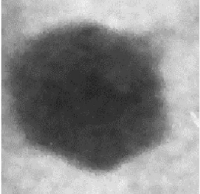
3.1. Growth and morphometric characteristics of Marinobacter phage AS1 18
The host, Marinobacer sp. strain D1S9 is a Gram negative, rod shaped bacterium (Fig 1B)
19
isolated from the surface waters of the Arabian Sea. The host present in the water sample was
20
enriched by the addition of nutrients which in turn amplified the specific viruses for the host. The
21
isolated phages formed clear, round plaques of 1-2 mm diameter with regular edges on lawns of the
22
host after 10-12 h of incubation (Fig 1C). Morphometric characteristics of phage AS1 examined by
9
transmission electron microscopy analysis indicated it belonged to family Podoviridae of the order
1
Caudovirales. The phage resembled morphotype C1 with an icosahedral head (isometric) having a
2
capsid diameter of ~50nm (Bradley, 1967). With a presence of short, stubby, non-contractile tail,
3
characteristic of this group (Fig. 2). Host range experiments with 9 laboratory isolates of
4
Marinobacter did not result any infection, indicating its narrow host range.
5
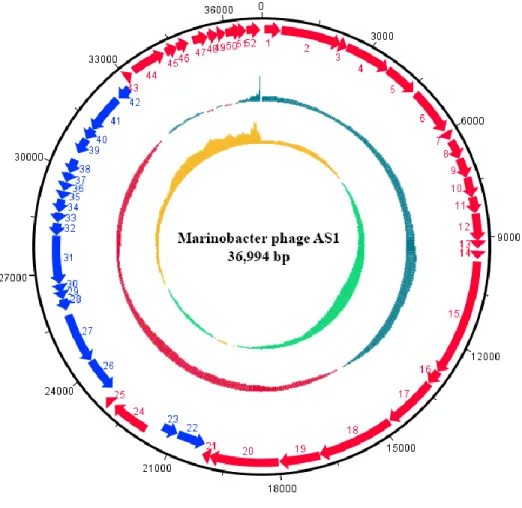
3.2. Genome features and annotation 6
Illumina sequencing and assembly of AS1 genome generated a single contig with a coverage
7
of 92.5%. The assembled genome of Marinobacter phage AS1 was linear with a sequence length of
8
37 kb with a GC content of 57% (Fig. 3). The general features of AS1 genome are listed in Table 1.
9
An initial whole genome similarity search against the NCBI non-redundant database using basic local
10
alignment search tool (BLAST) showed no close relatives to Marinobacter phage AS1. AS1 genome
11
showed no significant similarity to any of the three already reported Marinobacter phages, PS3
12
(GenBank accession, MF959999), PS6 (GenBank accession, MF959998) or B23 (Zhu et al. 2018;
13
GenBank accession, KY939598). However, the AS1 phage represents the first Marinobacter phage
14
belonging to the family Podoviridae. When phage AS1 genome was searched against the whole
15
genome shotgun sequences of the taxon Marinobacter (taxid: 2742) using NCBI- BLAST, it showed
16
an identity of 96 % with Marinobacter manganoxydans isolate UBA5690_contig_21412
17
(DIHS01000048) and 94 % with Marinobacter sp. N4 KEHDKFFH_1 (PSSX01000001) with query
18
coverages of 85% and 73 % respectively. This observation indicates the existence of fragments of
19
AS1 genome within Marinobacter population and its possible interactions with the hosts. A total of
20
52 protein coding genes were predicted in the genome, of which 21 have assigned putative functions
21
(Table 2). Genes related to phage structure and assembly, DNA modification, transcriptional
22
regulation and host cell lysis were arranged in distinct functional clusters along the genome (Table 3).
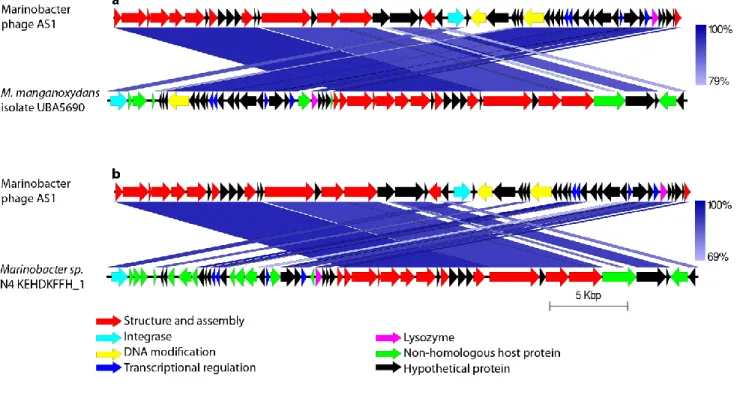
10
The similar sequences of AS1 found within the genomes of M. manganoxydans isolate
1
UBA5690_contig_21412 (in reverse orientation) and Marinobacter sp. N4 KEHDKFFH_1 are
2
depicted in Fig. 4. They include genes encoding the small and large subunits of terminase, capsid
3
proteins, portal protein, tail proteins, integrase, transcriptional regulators, methyl transferase,
4
endonuclease, lysozyme and hypothetical proteins. Phage sequence was broken up and various
5
functional clusters were shuffled within a particular region of the bacterial genomes.
6
The structural and assembly module encodes both small and large subunits of terminase,
7
portal protein, serine peptidase, major capsid protein, Gp6, Gp3, major tail protein, tail fiber proteins
8
and tail tape measure protein. The terminase large subunit protein (Gene 2) contains a P-loop having
9
nucleoside triphosphate hydrolase activity and belongs to Terminase_1 family (PF03354). Gene 3
10
encodes for a protein with a signal peptide (residues 1-21) and a transmembrane helix (residues
29-11
51) and is homologous to protein Gp3 of Klebsiella phage PhiKO2. Besides, a prohead maturation
12
protease (Gene 5) belonging to the MEROPS peptidase family S49 (protease IV family) was also
13
identified in phage AS1. It has a conserved catalytic Serine-Lysine dyad typical of a serine protease
14
domain (cd07022). The proteolytic activity is essential for the phage capsid maturation. The major
15
capsid protein of AS1 (Gene 6) belongs to HK97 family and it forms coiled coil structure at amino
16
acid positions 4-34 and 39-59. The delta domain of HK97 major capsid protein was shown to have
17
coiled coils involved in prohead assembly and maturation and it was also found to be removed by
18
proteases after the assembly (Oh et al. 2014). Gene 8 of the assembled genome codes for a protein
19
containing gp6 domain (cd08054) with 36 conserved oligomerization interface amino acid residues of
20
the domain. Gp6 of bacteriophage HK97 encodes head tail connector protein which forms an
21
oligomeric ring and serves as an interface for head and tail attachment (Cardarelli et al. 2010).
11
Phage AS1 codes for an integrase gene having 97.2 % homology with that of Marinobacter
1
sp. N4. It possess a C-terminal conserved domain similar to Shufflon-specific DNA recombinase Rci
2
and bacteriophage Hp1_like integrase (cd00796) belonging to the superfamily of DNA
breaking-3
rejoining enzymes. These enzymes contain a catalytic domain with six conserved amino acid
4
residues. The presence of integrase gene indicates the potential for a temperate lifestyle which is
5
further strengthened by the absence of any tRNA genes in the genome. It can be assumed that phage
6
AS1 utilizes its host’s tRNAs for translating the mRNA. Presence of tRNA genes provide phage with
7
competitive advantage over other phages through more efficient expression of their own genes
8
independent of their host’s tRNAs. Whereas, their absence results in a narrow host range, making the
9
phage more particular about selecting the host with similar codon usage bias (Bailly-Bechet et al.
10
2007).
11
Two sequence specific DNA methyl transferase genes, DNA adenine methyltransferase
12
(DAM) and DNA (Cytosine-5) methyltransferase (DCM), present in AS1 genome control
DNA-13
protein interactions by methylating adenine and cytosine residues of the DNA sequence. Considering
14
this fact, phage encoded DNA adenine methyl transferase may be interpreted as a coevolutionary
15
adaptation of the phage to protect itself from the host restriction enzymes (Murphy et al. 2014).
16
Normally, specific DNA methyl transferases are part of host restriction-modification system which
17
methylate host DNA at specific sites. This protects the host DNA from cleavage by its own restriction
18
endonucleases which on the other hand destroy the incoming foreign phage DNA. These enzymes
19
were also reported to have functions like controlling the expression of host virulence genes and
20
selective silencing of genes that they methylate (Low et al. 2001, Oakey et al. 2002). In bacteriophage
21
lambda, methylation by DNA adenine methyltransferase is associated with packaging of the phage
22
genome (Sternberg and Coulby 1990), whereas, DNA (Cytosine-5) - methyltransferases are found
12
rarely in bacteriophage genomes and their functions in the context of viral lifecycle are unknown.
1
Interestingly, a gene coding for the protein HNH endonuclease (gene 52) having homology to
5-2
methylcytosine specific restriction endonuclease McrA family (COG1403) with two highly conserved
3
histidine and one asparagine residues was identified in AS1 genome nearby the lysozyme gene within
4
the lysis module. McrA is a member of the superfamily HNHc (cI00083) which includes many
5
homing endonucleases, bacterial colicins, pyocins etc. and are rarely reported in Podoviruses.
6
Modified cytosine restriction (Mcr) systems capable of restricting phage λ modified by sequence
7
specific cytosine methylases has been earlier reported in Escherichia coli strain K12 (Raleigh and
8
Wilson 1986, Raleigh et al. 1989). The protein Gp74 from lambda-like phage HK97 was reported to
9
possess HNH endonuclease activity and mediate the cleavage of phage DNA (Moodley et al. 2012).
10
Later, Kala et al. (2014) discovered that these endonucleases were associated with the DNA
11
packaging terminase proteins in HK97 and majority of the large terminase subunits linked with HNH
12
endonucleases belonged to the Terminase_1 family (PF03354). The position of AS1 HNH
13
endonuclease, adjacent to terminase and other morphogenetic genes, suggests its potential
14
involvement in DNA packaging.
15
Genome of phage AS1 encodes pyocin activator protein (gene 36) with a conserved domain
16
belonging to the PrtN family (PF11112) and is involved in the transcriptional activation of the
17
polypeptide endonuclease toxin, pyocin. Other than lysozyme and HNH endonuclease, the lysis
18
module encodes a Cro/CI family transcriptional regulator containing a HTH motif and a phage
anti-19
termination Q type I family protein. The decision of the phage to enter either lytic or lysogenic cycle
20
is determined by Cro/C1 regulatory system which is well characterized and studied in phage λ
21
infecting the bacterium Escherichia coli (Ptashne 1967, Eisen et al. 1970, Schubert et al. 2007). The
22
antitermination Q Type I protein positively regulates the phage early and late genes by modifying the
13
host RNA polymerase and making it to proceed transcription past the terminator sequences. A total of
1
nine trans-membrane helices were predicted in seven proteins. 53 promoter sequences and 20
rho-2
independent terminator sequences were identified in the assembled genome.
3
Phylogenetic analysis revealed a mosaic pattern of inheritance of various proteins encoded by
4
phage AS1 (Fig. 5). Major capsid protein, terminase large subunit and portal protein of AS1 had a
5
Siphoviral lineage, whereas, integrase and major tail proteins were closely related to other Podoviral
6
homologs. Recombination driven exchange of genetic material and the resulting genetic mosaicism is
7
wide-spread among Caudovirales. Even though prominent morphological differences exist between
8
the three families (specifically tail morphology), the differentiation is not clear cut due to striking
9
sequence level similarities among several members of these different families. A well-known
10
example for this is the genetic relatedness between phages lambda and P22 which are, however,
11
classified under Siphoviridae and Podoviridae, respectively, based on their tail morphology. No
12
lineage for the tail proteins of AS1 were deduced due to the limited availability of homologs. 13
Conclusion 14
Marinobacter phage AS1, isolated in this study, belongs to the family Podoviridae, infect
15
marine bacteria belonging to genus Marinobacter, strain D1S9. The presence of integrase gene,
16
occurrence of DNA methyltransferases and the existence of a significant percentage of phage AS1
17
genes within various Marinobacter genomes are suggestive of their intense association with the host.
18
From the genome analysis, it is evident that phage AS1 relies greatly on its host’s replication and
19
translation machinery. Its codon usage bias must be similar to that of the host, since the phage
20
genome does not encode any tRNA genes. Another striking feature of the genome is its mosaicism
21
evident from the inheritance pattern of some of the important proteins with high similarity to their
22
siphoviral counterparts. It also contains most of the morphogenetic, assembly, and lysis genes
14
involved in the lytic induction. AS1 genome also encodes a gene which positively regulates the
1
expression of the bacterial toxin, pyocin; an interaction which attributes competitive advantage to the
2
host. All the above arguments indicate the possible coevolutionary interactions between the host and
3
the virus. As the members of genus Marinobacter are organisms with both phenotypic and metabolic
4
versatility and reported to have many significant ecological functions like their contribution in
5
biogeochemical cycles and marine snow formation, extensive studies on the involvement of these
6
viruses in impacting host metabolism as well as recruitment of this virus to metagenome need to be
7
carried out. The present study is a detailed analysis of the genomic properties of Marinobacter phage
8
AS1, guiding future research on intricate virus-host interactions and the role of phages in the
9
ecological functioning of the genus Marinobacter.
10
References 11
1. Suttle CA (2007) Marine viruses—major players in the global ecosystem. Nat Rev Microbiol
12
5:801-812.
13
2. Sime-Ngando T (2014) Environmental bacteriophages: viruses of microbes in aquatic
14
ecosystems. Front Microbiol 5:355.
15
3. Middelboe M, Lyck PG (2002) Regeneration of dissolved organic matter by viral lysis in
16
marine microbial communities. Aquat Microb Ecol 27:187-194.
17
4. Bouvier T, Del Giorgio PA (2007) Key role of selective viral‐induced mortality in
18
determining marine bacterial community composition. Environ microbiol 9:287-297.
19
5. McDaniel LD, Young E, Delaney J, Ruhnau F, Ritchie KB, Paul JH (2010) High frequency of
20
horizontal gene transfer in the oceans. Science 330:50.
21
6. Winget DM, Helton RR, Williamson KE, Bench SR, Williamson SJ, Wommack KE (2011)
22
Repeating patterns of virioplankton production within an estuarine ecosystem. Proc Natl Acad
23
Sci USA 108:11506-11511.
24
7. Stern A, Sorek R (2011) The phage‐host arms race: shaping the evolution of microbes.
25
Bioessays 33:43-51.
26
8. Xia G, Wolz C (2014) Phages of Staphylococcus aureus and their impact on host evolution.
27
Infection, Genet Evol 21:593-601.
15
9. Buckling A, Brockhurst M (2012) Bacteria–virus coevolution. In: Soyer O (ed) Evolutionary
1
systems biology, Springer, New York, NY pp. 347-370.
2
10. Hall AR, Scanlan PD, Morgan AD, Buckling A (2011) Host–parasite coevolutionary arms
3
races give way to fluctuating selection. Ecol Lett. 2011 Jul;14(7):635-42.
4
11. Gokhale CS, Papkou A, Traulsen A, Schulenburg H (2013) Lotka–Volterra dynamics kills the
5
Red Queen: population size fluctuations and associated stochasticity dramatically change
6
host-parasite coevolution. BMC Evol Biol 13:254.
7
12. Hendrix RW, Smith MC, Burns RN, Ford ME, Hatfull GF (1999) Evolutionary relationships
8
among diverse bacteriophages and prophages: all the world’sa phage. Proc Natl Acad Sci U S
9
A 96:2192-2197.
10
13. Hatfull GF, Hendrix RW (2011) Bacteriophages and their genomes. Curr Opin Virol
1:298-11
303.
12
14. Gauthier MJ, Lafay B, Christen R, Fernandez L, Acquaviva M, Bonin P, Bertrand JC (1992)
13
Marinobacter hydrocarbonoclasticus gen. nov., sp. nov., a new, extremely halotolerant,
14
hydrocarbon-degrading marine bacterium. Int J Syst Evol Microbiol 42:568-576.
15
15. Martin S, Márquez MC, Sánchez-Porro C, Mellado E, Arahal DR, Ventosa A (2003)
16
Marinobacter lipolyticus sp. nov., a novel moderate halophile with lipolytic activity. Int J Syst
17
Evol Microbiol 53:1383-1387.
18
16. Yoon JH, Lee MH, Kang SJ, Oh TK (2007) Marinobacter salicampi sp. nov., isolated from a
19
marine solar saltern in Korea. Int J Syst Evol Microbiol 57:2102-2105.
20
17. Zhang DC, Li HR, Xin YH, Chi ZM, Zhou PJ, Yu Y (2008) Marinobacter psychrophilus sp.
21
nov., a psychrophilic bacterium isolated from the Arctic. Int J Syst Evol Microbiol
58:1463-22
1466.
23
18. Xu XW, Wu YH, Wang CS, Yang JY, Oren A, Wu M (2008) Marinobacter pelagius sp. nov.,
24
a moderately halophilic bacterium. Int J Syst Evol Microbiol 58:637-640.
25
19. Wang CY, Ng CC, Tzeng WS, Shyu YT (2009) Marinobacter szutsaonensis sp. nov., isolated
26
from a solar saltern. Int J Syst Evol Microbiol 59:2605-2609.
27
20. Wang H, Li H, Shao Z, Liao S, Johnstone L, Rensing C, Wang G (2012) Genome sequence of
28
deep-sea manganese-oxidizing bacterium Marinobacter manganoxydans MnI7-9. J Bacteriol
29
194:899-900.
16
21. Chua MJ, Campen RL, Wahl L, Grzymski JJ, Mikucki JA (2018) Genomic and physiological
1
characterization and description of Marinobacter gelidimuriae sp. nov., a psychrophilic,
2
moderate halophile from Blood Falls, an antarctic subglacial brine. FEMS Microbiol Ecol
3
94:fiy021.
4
22. Green DH, Bowman JP, Smith EA, Gutierrez T, Bolch CJ (2006) Marinobacter algicola sp.
5
nov., isolated from laboratory cultures of paralytic shellfish toxin-producing dinoflagellates.
6
Int J Syst Evol Microbiol 56:523-527.
7
23. Lee OO, Lai PY, Wu HX, Zhou XJ, Miao L, Wang H, Qian PY (2012) Marinobacter
8
xestospongiae sp. nov., isolated from the marine sponge Xestospongia testudinaria collected
9
from the Red Sea. Int J Syst Evol Microbiol 62:1980-1985.
10
24. Huu NB, Denner EB, Ha DT, Wanner G, Stan-Lotter H (1999) Marinobacter aquaeolei sp.
11
nov., a halophilic bacterium isolated from a Vietnamese oil-producing well. Int J Syst Evol
12
Microbiol 49:367-375.
13
25. Shieh WY, Jean WD, Lin YT, Tseng M (2003) Marinobacter lutaoensis sp. nov., a
14
thermotolerant marine bacterium isolated from a coastal hot spring in Lutao, Taiwan. Can J
15
Microbiol 49:244-252.
16
26. Kaeppel EC, Gärdes A, Seebah S, Grossart HP, Ullrich MS (2012) Marinobacter adhaerens
17
sp. nov., isolated from marine aggregates formed with the diatom Thalassiosira weissflogii.
18
Int J Syst Evol Microbiol 62:124-128.
19
27. Lupette J, Lami R, Krasovec M, Grimsley N, Moreau H, Piganeau G, Sanchez-Ferandin S
20
((2016) Marinobacter dominates the bacterial community of the Ostreococcus tauri
21
phycosphere in culture. Front Microbiol 7:1414.
22
28. Sandhya SV, Preetha K, Vijayan KK (2017) Phylogenetic diversity of culturable bacteria in
23
Chaetoceros gracilis mass culture system of a marine finfish hatchery. J Mar Biol Ass India
24
59:12-18.
25
29. Handley KM, Lloyd JR (2013) Biogeochemical implications of the ubiquitous colonization of
26
marine habitats and redox gradients by Marinobacter species. Front Microbiol 4:136.
27
30. Kaye JZ, Sylvan JB, Edwards KJ, Baross JA (2011) Halomonas and Marinobacter ecotypes
28
from hydrothermal vent, subseafloor and deep-sea environments. FEMS Microbiol Ecol
29
75:123-133.
17
31. Bonis BM, Gralnick JA (2015) Marinobacter subterrani, a genetically tractable neutrophilic
1
Fe (II)-oxidizing strain isolated from the Soudan Iron Mine. Front Microbiol 6:719.
2
32. Singer E, Webb EA, Nelson WC, Heidelberg JF, Ivanova N, Pati A, Edwards KJ (2011) The
3
genomic potential of Marinobacter aquaeolei– A biogeochemical opportunitroph. Appl
4
Environ Microbiol 77:2763-2771.
5
33. Zhu M, Wang M, Jiang Y, You S, Zhao G, Liu Y, Yang Q, Liu Q, Liu Z, Gong Z, Shao H
6
(2018) Isolation and Complete Genome Sequence of a Novel Marinobacter Phage B23. Curr
7
microbiol 75:1619-1625.
8
34. Middelboe M, Chan A, Bertelsen SK (2010) Isolation and life cycle characterization of lytic
9
viruses infecting heterotrophic bacteria and cyanobacteria. In: Wilhelm SW, Weinbauer MG,
10
Suttle CA (ed) Manual of aquatic viral ecology, American Society of Limnology and
11
Oceanography, pp. 118-133.
12
35. Amann RI, Ludwig W, Schleifer KH (1995) Phylogenetic identification and in situ detection
13
of individual microbial cells without cultivation. Microbiol Mol Biol Rev 59:143-169.
14
36. Pradeep Ram AS, Arnous B, Danger M, Carrias JF, Lacroix G, Sime-Ngando T (2010) High
15
and differential viral infection rates within bacterial ‘morphopopulations’ in a shallow sand pit
16
lake (Lac de Créteil, France). FEMS Microbiol Ecol 74:83-92.
17
37. Lawrence JE, Steward GF (2010) Purification of viruses by centrifugation. In: Wilhelm SW,
18
Weinbauer MG, Suttle CA (ed) Manual of aquatic viral ecology, American Society of
19
Limnology and Oceanography, pp. 166-181.
20
38. Yang Y, Cai L, Ma R, Xu Y, Tong Y, Huang Y, Jiao N, Zhang R (2017) A novel
21
roseosiphophage isolated from the oligotrophic South China Sea. Viruses 9:109.
22
39. Andrews S (2010) FastQC: a quality control tool for high throughput sequence data.
23
40. Bankevich A, Nurk S, Antipov D, Gurevich AA, Dvorkin M, Kulikov AS, Lesin VM,
24
Nikolenko SI, Pham S, Prjibelski AD, Pyshkin AV (2012) SPAdes: a new genome assembly
25
algorithm and its applications to single-cell sequencing. J Comput biol 19:455-477.
26
41. Aziz RK, Bartels D, Best AA, DeJongh M, Disz T, Edwards RA, Formsma K, Gerdes S,
27
Glass EM, Kubal M, Meyer F (2008) The RAST Server: rapid annotations using subsystems
28
technology. BMC genomics 9:75.
18
42. Marchler-Bauer A, Bo Y, Han L, He J, Lanczycki CJ, Lu S, Chitsaz F, Derbyshire MK, Geer
1
RC, Gonzales NR, Gwadz M (2016) CDD/SPARCLE: functional classification of proteins via
2
subfamily domain architectures. Nucleic Acids Res 45:D200-D203.
3
43. Lowe TM, Chan PP (2016) tRNAscan-SE On-line: integrating search and context for analysis
4
of transfer RNA genes. Nucleic Acids Res 44:W54-W57.
5
44. Naville M, Ghuillot-Gaudeffroy A, Marchais A, Gautheret D (2011) ARNold: a web tool for
6
the prediction of Rho-independent transcription terminators. RNA Biol 8:11-13.
7
45. Reese MG (2001) Application of a time-delay neural network to promoter annotation in the
8
Drosophila melanogaster genome. Comput Chem 26:51-56.
9
46. Kumar S, Stecher G, Tamura K ((2016) MEGA7: molecular evolutionary genetics analysis
10
version 7.0 for bigger datasets. Mol Biol Evol 33:1870-1874.
11
47. Bradley DE (1967) Ultrastructure of bacteriophage and bacteriocins. Bacteriol Rev 31:230.
12
48. Oh B, Moyer CL, Hendrix RW, Duda RL (2014) The delta domain of the HK97 major capsid
13
protein is essential for assembly. Virology 456:171-178.
14
49. Cardarelli L, Lam R, Tuite A, Baker LA, Sadowski PD, Radford DR, Rubinstein JL, Battaile
15
KP, Chirgadze N, Maxwell KL, Davidson AR (2010) The crystal structure of bacteriophage
16
HK97 gp6: defining a large family of head–tail connector proteins. J Mol Biol 395:754-768.
17
50. Bailly-Bechet M, Vergassola M, Rocha E (2007) Causes for the intriguing presence of tRNAs
18
in phages. Genome Res 17:1486-1495.
19
51. Murphy J, Klumpp J, Mahony J, O’Connell-Motherway M, Nauta A, van Sinderen D (2014)
20
Methyltransferases acquired by lactococcal 936-type phage provide protection against
21
restriction endonuclease activity. BMC genomics 15:831.
22
52. Low DA, Weyand NJ, Mahan MJ (2001) Roles of DNA adenine methylation in regulating
23
bacterial gene expression and virulence. Infect Immun 69:7197-7204.
24
53. Oakey HJ, Cullen BR, Owens L (2002) The complete nucleotide sequence of the Vibrio
25
harveyi bacteriophage VHML. J Appl Microbiol 93:1089-1098.
26
54. Sternberg N, Coulby J (1990) Cleavage of the bacteriophage P1 packaging site (pac) is
27
regulated by adenine methylation. Proc Natl Acad Sci U S A 87:8070-8074.
28
55. Raleigh EA, Wilson G (1986) Escherichia coli K-12 restricts DNA containing
5-29
methylcytosine. Proc Natl Acad Sci U S A 83:9070-9074.
19
56. Raleigh EA, Trimarchi R, Revel H (1989) Genetic and physical mapping of the mcrA (rglA)
1
and mcrB (rglB) loci of Escherichia coli K-12. Genetics 122:279-296.
2
57. Moodley S, Maxwell KL, Kanelis V (2012) The protein gp74 from the bacteriophage HK97
3
functions as a HNH endonuclease. Protein Sci 21:809-818.
4
58. Kala S, Cumby N, Sadowski PD, Hyder BZ, Kanelis V, Davidson AR, Maxwell KL (2014)
5
HNH proteins are a widespread component of phage DNA packaging machines. Proc Natl
6
Acad Sci U S A 111:6022-6027.
7
59. Ptashne M (1967) Specific binding of the λ phage repressor to λ DNA. Nature 214:232.
8
60. Eisen H, Brachet P, Da Silva LP, Jacob F (1970) Regulation of repressor expression in λ. Proc
9
Natl Acad Sci U S A 66:855-862.
10
61. Schubert RA, Dodd IB, Egan JB, Shearwin KE (2007) Cro’s role in the CI–Cro bistable
11
switch is critical for λ’s transition from lysogeny to lytic development. Genes Dev
21:2461-12
2472.
20
Table 1. Genome features of Marinobacter phage AS1
Feature Marinobacter phage AS1
Genome size 36,994 bp
GC content 57 %
Total no. of proteins 52 No. of proteins with putative function 21
tRNA genes None
21 Table 2. Genome annotation of Marinobacter phage AS1
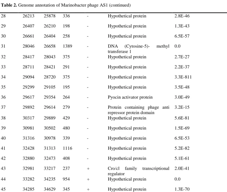
Gene Start (bp) Stop (bp) Length (bp)
Strand Putative function E-value
1 70 531 462 + Phage terminase small subunit P27 family
3.2E-111
2 531 2216 1686 + Terminase large subunit 0.0
3 2210 2404 195 + Gp3 2.8E-19
4 2404 3669 1266 + Putative portal protein 0.0
5 3657 4589 933 + Serine peptidase 0.0
6 4663 5946 1284 + Major capsid head protein 0.0
7 6000 6224 225 + Hypothetical protein 1.8E-48
8 6262 6795 534 + Gp6 6.4E-116
9 6797 7348 552 + Hypothetical protein 4.5E-128
10 7348 7902 555 + Hypothetical protein 5.6E-127
11 7899 8348 450 + Hypothetical protein 6.2E-98
12 8352 9107 756 + Major tail protein 8.3E-8
13 9172 9321 150 + Hypothetical protein -
14 9385 9600 216 + Hypothetical protein 1.3E-30
15 9665 12883 3219 + Tail tape measure protein 2.0E-28
16 12880 13278 399 + Hypothetical protein 8.3E-88
22
18 14779 16893 2115 + Tail fiber 1.4E-125
19 16893 18011 1119 + Hypothetical protein 1.1E-82
20 18016 19926 1911 + Hypothetical protein 0.0
21 19913 20146 234 + Hypothetical protein 5.2E-56
22 20955 20143 813 - Putative lipoprotein 9.2E-76
23 21434 20979 456 - Hypothetical protein 2.4E-96
24 21810 22895 1086 + Integrase 0
25 23007 23207 201 + Hypothetical protein 4.0E-07
26 24260 23265 996 - DNA methyl transferase 9.7E-131
27 25730 24270 1461 - Hypothetical protein -
Table 2. Genome annotation of Marinobacter phage AS1 (continued)
28 26213 25878 336 - Hypothetical protein 2.8E-46
29 26407 26210 198 - Hypothetical protein 1.3E-43
30 26661 26404 258 - Hypothetical protein 6.5E-57
31 28046 26658 1389 - DNA (Cytosine-5)- methyl transferase 1
0.0
32 28417 28043 375 - Hypothetical protein 2.7E-27
33 28711 28421 291 - Hypothetical protein 2.2E-37
34 29094 28720 375 - Hypothetical protein 3.3E-811
35 29299 29105 195 - Hypothetical protein 3.5E-48
36 29617 29354 264 - Pyocin activator protein 3.0E-49
37 29892 29614 279 - Protein containing phage anti repressor protein domain
3.2E-15
38 30317 29889 429 - Hypothetical protein 5.6E-81
39 30981 30502 480 - Hypothetical protein 1.5E-69
40 31316 30978 339 - Hypothetical protein 6.5E-53
41 32428 31313 1116 - Hypothetical protein 5.2E-82
42 32880 32473 408 - Hypothetical protein 5.1E-61
43 32981 33217 237 + Cro/cl family transcriptional regulator
2.0E-41
44 33282 34235 954 + Hypothetical protein 0.0
23
46 34619 34969 351 + Phage anti termination Q type I family- like protein
1.5E-15
47 35096 35527 432 + Lysozyme 3.6E-91
48 35524 35802 279 + Hypothetical protein 5.5E-53
49 35786 36016 231 + Hypothetical protein 6.0E-57
50 36013 36462 450 + Hypothetical protein 1.6E-43
51 36464 36592 129 + Hypothetical protein 2.8E-13
52 36592 36975 384 + HNH endonuclease 2.3E-87
Table 3. Functional categorization of phage AS1 proteins
Function No. of
proteins
Phage structure and assembly 13 Phage defense/DNA modification 2 Transcriptional regulation 4
Host lysis 1
Phage integration 1 Hypothetical proteins 31
24
c
b
Fig. 1 Isolation of phage AS1. (a) Sampling location in the Arabian Sea, (b) rod shaped Marinobacter host cells after staining with crystal violet, (c) clear round plaques of phage AS1 formed on a lawn of host cells.
25
27
Fig. 3 Circular representation of the double stranded linear genome of Marinobacter phage AS1 featuring (from outside to inside) coding DNA sequences with predicted functions in the forward strand (red), reverse strand (neon blue), GC content (maroon & blue) and GC skew (green & yellow).
28
Fig. 4 Comparison of similar sequences found within the sequences of host genus. Comparison with (a) M. manganoxydans isolate UBA5690 and (b) Marinobacter sp. N4 KEHDKFFH_1. The intensity of blue colour indicates the percentage of similarity between the two sequences.
29
a
b
c
d
e
Fig. 5 Phylogenetic trees of AS1 proteins. (a) Integrase, (b) major tail protein, (c) major capsid protein, (d) portal protein and (e) terminase, large subunit.