Asymmetrical Membranes and Surface Tension
Mounir Traı¨kia,* Dror E. Warschawski,* Olivier Lambert,†Jean-Louis Rigaud,†and Philippe F. Devaux* *Institut de Biologie Physico-Chimique, Unite´ Mixte de Recherche Centre National de la Recherche Scientifique 7099, Paris 75005 France; and†Institut Curie, Section de Recherche, Unite´ Mixte de Recherche-Centre National de la Recherche Scientifique 168 and Laboratoire de Recherche Correspondant-Commissariat a` l’Energie Nucle´aire 8, 75231 Paris cedex, France
ABSTRACT The31P-nuclear magnetic resonance chemical shift of phosphatidic acid in a membrane is sensitive to the lipid
head group packing and can report qualitatively on membrane lateral compression near the aqueous interface. We have used high-resolution 31P-nuclear magnetic resonance to evaluate the lateral compression on each side of asymmetrical lipid
vesicles. When monooleoylphosphatidylcholine was added to the external monolayer of sonicated vesicles containing dioleoylphosphatidylcholine and dioleoylphosphatidic acid, the variation of31P chemical shift of phosphatidic acid indicated
a lateral compression in the external monolayer. Simultaneously, a slight dilation was observed in the inner monolayer. In large unilamellar vesicles on the other hand the lateral pressure increased in both monolayers after asymmetrical insertion of monooleoylphosphatidylcholine. This can be explained by assuming that when monooleoylphosphatidylcholine is added to large unilamellar vesicles, the membrane bends until the strain is the same in both monolayers. In the case of sonicated vesicles, a change of curvature is not possible, and therefore differential packing in the two layers remains. We infer that a variation of lipid asymmetry by generating a lateral strain in the membrane can be a physiological way of modulating the conformation of membrane proteins.
INTRODUCTION
Lipid asymmetry in biomembranes is often viewed solely as a difference in chemical composition between the two monolayers. For example, it is well known that the plasma membrane of eukaryotic cells contains essentially phos-phatidylcholine and sphingomyelin in the outer monolayer, whereas aminophospholipids are predominantly located in the inner monolayer (Devaux, 1991). However, lipid asym-metry can also mean a difference in surface density result-ing from a difference in the number of lipids between the two coupled monolayers. Such a difference in lipid surface density can be achieved artificially by the addition of lyso-phosphatidylcholine (LPC) to the external surface of a lipid vesicle or of a biological membrane. Because of the very slow flip-flop of lipids, the asymmetrical surface density is generally stable. However, the mismatch causes a difference in lateral pressure or tension between the two leaflets that often, but not always, leads to vesicle shape change. In biological membranes, the lateral pressure formed by an asymmetrical lipid distribution could affect the structure and function of intrinsic membrane proteins.
We have used nuclear magnetic resonance (NMR) to probe the local variations of the lateral pressure in various types of unilamellar vesicles. High-resolution 31P-NMR spectra of phospholipid vesicles containing a mixture of dioleoylphosphatidylcholine (DOPC) and dioleoylphospha-tidic acid (DOPA) were recorded and the chemical shift of
DOPA was monitored. The potentiality of31P-NMR spec-troscopy for the assessment of lateral compressibility in a membrane was brought to light by Swairjo et al. (1994a). They showed that the31P chemical shift of DOPA is dif-ferent in the inner and outer monolayers of sonicated unila-mellar vesicles (SUVs). They inferred that the difference in chemical shifts was due to the difference in local curvature of the two monolayers, which is certainly true in sonicated vesicles. In fact, via its sensitivity to the local pKa, the chemical shift effectively reports on the packing of the lipid head groups and can be considered as a qualitative indicator of any change of surface tension.
NMR spectroscopy can be used to monitor qualitatively the surface tension of vesicles of various sizes as long as they contain phosphatidic acid and that high-resolution NMR measurements are possible. We have investigated successively asymmetrical SUVs, LUVs (large unilamellar vesicle) and FTVs (freeze-thaw vesicles). FTVs are unila-mellar vesicles obtained by repetitive cycles of freeze thaw-ing, a process that generates unilamellar vesicles with a distribution of sizes but with a significant contribution of vesicles with a diameter above 200 nm (Traı¨kia et al., 2000). With giant unilamellar vesicles (GUVs) the lipid concen-tration would be too low to allow NMR experiments to be performed. In the case of LUVs and FTVs magic angle spinning was necessary to achieve high resolution. Shape changes were visualized by cryoelectron microscopy.
In agreement with a former theoretical article (Farge et al., 1990), we have found that the addition of lysophosphati-dylcholine to one side of LUVs or FTVs triggers an overall change of curvature that is accompanied by an increased lateral compression in both leaflets and not only on the external one. However, with sonicated vesicles, which are
Submitted April 17, 2001, and accepted for publication April 26, 2002.
Address reprint requests to Philippe F. Devaux, Institut de Biologie Physico-Chimique, UMR CNRS 7099, 13 rue Pierre et Marie Curie, F75005 Paris, France. Tel.: 33-1-58-41-51-05; Fax: 33-1-58-41-50-24; E-mail: [email protected].
phospholipid bilayer, there is no possibility of changing the curvature and the surface pressure increases solely in the outer monolayer.
MATERIALS AND METHODS Chemicals
Lipids (DOPC, DOPA, and monooleoylphosphatidylcholine (MOPC)) and chemicals (HEPES, KCl, and EDTA) were purchased from Sigma Chem-ical Co. (Saint Quentin Fallavier, France) and were used without further purification. The purity of all lipids was verified by thin layer chromatog-raphy and by high resolution NMR in chloroform.
Vesicles preparation
All samples were prepared as an initial dry lipidic film of DOPC/DOPA (80:20 mol%) and the buffer was: 0.1 M HEPES, 0.1 M KCl, and 0.005 M EDTA at pH 8.0. SUVs (30 mg/mL) were prepared by sonication of a lipid dispersion in buffer under a stream of argon using a probe type sonicator (model VC50, Bioblock Scientific, Paris, France) at 40 W in an ice bath until a clear solution was obtained. The sample was afterwards centrifuged at 11,000⫻ g for 15 min to ensure the removal of metallic particles. Unless otherwise mentioned, LUVs were prepared by the reverse phase evapora-tion technique followed by filtraevapora-tion of the preparaevapora-tion (50 mg/mL) through a polycarbonate membrane, successively at 1, 0.4, and at 0.2m (Szoka et al., 1980; Traı¨kia et al., 2000). The LUVs were concentrated to 100 mg/mL by ultracentrifugation at 395,000⫻ g for at least 2 h (Beck-man, TLC100). FTVs were prepared at 100 mg/mL. Characterization of their lamellarity and size distribution was described in Traı¨kia et al. (2000). Stock solution of MOPC was prepared at 10 mg/mL in the buffer. Because of the large amount of lipids necessary (several hundred microliters of 100 mM phospholipids in one sample), a rather accurate determination of the ratio of DOPC/DOPA or DOPC/DOPA/MOPC could be achieved from the weight of the dry lipids and was always controlled by integration of the high resolution NMR spectra recorded with magic angle spinning (MAS). In a few instances, for SUVs or LUVs, the ratio of DOPC/DOPA was also checked by NMR in organic solvent but not for all samples.
NMR
NMR experiments were performed and processed on a Bruker AVANCE DMX400-WB NMR spectrometer (1H resonance at 400 MHz,31P reso-nance at 162 MHz) using a Bruker 4-mm MAS probe with an external lock for the LUVs, FTVs, and MLVs (multilamellar vesicles). A few experi-ments were carried out with an insert-containing rotor, which allows better field homogeneity but because of the low volume contained in such rotor longer accumulations were necessary. The spinning speed of the 4 mm ZrO2MAS rotor was controlled to within 5 Hz at 8 kHz and the temper-ature (fixed at 8°C unless otherwise specified) was calibrated as described in Traı¨kia et al. (1997). A 10-mm broadband liquid state NMR probe was used for the SUVs experiments. The31P-90° pulse lengths were 4.6 and 16
s for the MAS and the liquid state probes, respectively. The recycle delay
was 2 s and typical 31P spectral width was 20 ppm (3.24 kHz). 1H decoupling was not applied during acquisition of the31P magnetization. In all experiments, 4096 complex points were acquired. Before Fourier trans-formation, the data were zero filled to 8192 points, exponentially multi-plied with 10 Hz line broadening and treated with automatic baseline correction. The theoretical frequency reference, 0 ppm, corresponds in MAS spectra to phosphoric acid. As reported by Swairjo et al. (1994a), we found that DOPC resonance at 8°C consist of a single peak at⫺0.64 ppm that can be used as an internal reference. For broadband spectra, 0 ppm
corresponds to the isotropic peak of DOPC micelles. Other conditions for broadband31P-NMR spectroscopy were those of Traı¨kia et al. (2000).
Cryoelectron microscopy
A few microliters of the lipid mixtures were applied to a holey carbon film on a copper grid that was held by tweezers mounted on a shaft above a liquid ethane bath cooled by liquid nitrogen (Leica EM CPC). The sample was blotted with filter paper and immediately plunged into the liquid ethane (Dubochet et al., 1988). The vitreous specimens were transferred under liquid nitrogen to the cryo-transmission electron microscope cold stage (model 626 Gatan), which was inserted into the Philipps CM 120 electron microscope and maintained at⫺170°C throughout specimen ob-servation. Specimens were imaged at 120 kV by low dose technique, and micrographs were recorded on Kodak SO163 films with a magnification of 45,000⫻ and a 1-m defocus.
RESULTS
Sonicated unilamellar vesicles
Fig. 1 shows 31P-NMR spectra of DOPC/DOPA (80:20 mol%) SUVs with and without MOPC added externally. The top spectra in Fig. 1 were recorded at 45°C. At this temperature DOPC gives rise to two resonance peaks par-tially overlapping. When at 45°C MOPC is added, the lines slightly broaden and the two phosphatidylcholine peaks collapse in agreement with the results of Kumar et al. (1989), who found that 31P-T2 of POPC decreases upon addition of LPC to SUVs. As reported previously by Swairjo et al. (1994a), DOPA in SUVs gives rise to a doublet with two well-separated peaks, the low field line corresponds to the phosphorus on the external side of the vesicles and the high field line to the inner leaflet molecules. We have carried out NMR experiments (not shown) in the presence of 10 mM Pr3⫹ions, which selectively shifted the low field peak of DOPA and allowed us to confirm the conclusion of Swairjo et al. (1994a). Integration of DOPA peaks indicated roughly a ratio of 2 to 1, which was con-sistent with the proportion of outer to inner surfaces of SUVs.
The two DOPA peaks are still well separated in the presence of MOPC. The chemical shift of DOPA has been used to investigate the influence of MOPC on the surface pressure of both leaflets. Because the position and splitting between the two DOPA peaks are sensitive not only to the presence of MOPC but also to the temperature, the pH and the ratio of DOPA to DOPC (Swairjo et al., 1994a), it was important to maintain the latter parameters fixed in all further experiments. A relatively low temperature (8°C) was adopted to allow comparison with experiments involving LUVs that required a low temperature for reasons that will be explained below.
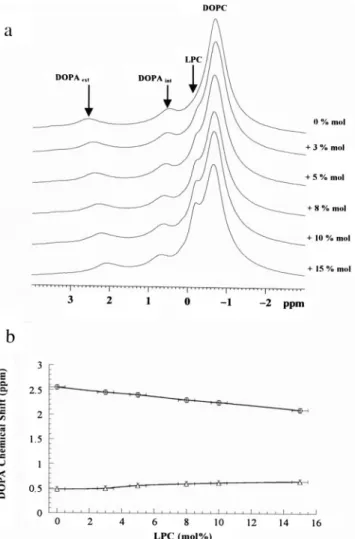
Fig. 2 shows the effect on the31P-NMR spectra of in-creasing concentration of MOPC at 8°C. The two chemical shifts associated with DOPA molecules are modified:
down-field. At this temperature there is no obvious change in the line width of the DOPC peak and no obvious redis-tribution between the spectral intensities of the two DOPA peaks. This suggests that addition of MOPC neither induced SUVs fusion nor caused their solubilization. Changes in␦PA would simply reveal changes in lateral pressure of each monolayer without important overall shape changes.
Large unilamellar vesicles
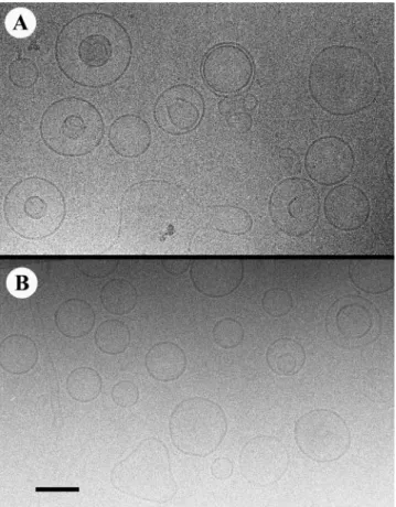
By cryoelectron microscopy, we found that the presence of an important percentage of DOPA (20 mol%) in the vesicles made it difficult to obtain an homogeneous population of LUVs when prepared by direct lipid extrusion under pres-sure according to the protocol designed by Cullis and col-laborators (Hope et al., 1985). Fig. 3 shows some examples of extruded DOPC/DOPA vesicles with odd shapes. Fur-thermore, we found that the ratio of DOPA/DOPC was
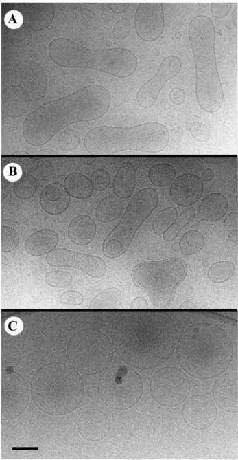
generally modified with a loss of DOPA. On the other hand, when LUVs were prepared by the phase reverse technique (Szoka etal. 1980), we obtained more homogeneous popu-lations of unilamellar vesicles with many spherical vesicles and almost no tubular ones (Fig. 4). Besides, the proportion of DOPC/DOPA was not modified. Addition of MOPC to the latter vesicles provoked the formation of more elongated vesicles (Fig. 5, A and B). Budding was observed but required the addition of 5% to 10% of MOPC.
Because glycerol was used in some NMR experiments described below, we have attempted to determine LUVs shapes with and without lysophosphatidylcholine addition in the presence of glycerol. Unfortunately when glycerol is present in samples examined by cryoelectron microscopy, the contrast is weak. Nevertheless, we found that all vesicles FIGURE 1 31P-NMR spectra (162 MHz) of SUVs containing a mixture
of DOPC/DOPA in 4:1 molar ratio prepared by sonication in buffer at pH 8. The doted spectra were obtained with MOPC added to in the outer monolayer: (a) 45°C, ( ) no MOPC; (- - - -) 30% MOPC; (b) 8°C, ( ) no MOPC; (- - - -) 15% MOPC.
FIGURE 2 Effect of MOPC addition to SUVs on the chemical shifts of the two DOPA resonance in DOPC/DOPA (4:1) SUVs. Temperature is 8°C. (a)31P-NMR spectra (162 MHz) of SUVs with an increasing con-centration of MOPC added externally. Percentages indicated on the right hand side of a are the molar percentages of MOPC added in relation to the total lipids present. (b) Dependence of the31P-DOPA chemical shifts in the outer (E) and inner (‚) leaflets, respectively. The uncertainty of the mol% of MOPC was estimated from lipid analysis in organic solvent.
seemed to be more spherical in the presence of glycerol than without and that the addition of MOPC tended on the contrary to give homogeneous populations of perfectly spherical vesicles instead of generating elongated or eight-shapes vesicles (see above). Fig. 5 C was obtained with 25% of glycerol and 10% MOPC added externally. With 50% glycerol the vesicles were very difficult to see but seemed to be perfect spheres (data not shown).
When31P-NMR spectra of LUVs or MLVs are recorded without sample spinning, the resolution is poor and does not allow one to separate the various phospholipid head groups, even if the spectrum is recorded at 45°C. Consequently, we have performed all further 31P-NMR experiments with 8
kHz sample spinning at the magic angle (MAS). The31 P-NMR spectrum of MLVs containing DOPA/DOPC exhibits a single narrow peak for DOPA at 2.2 ppm (Fig. 6 a). This is consistent with the lipid surface density being the same in both leaflets for lipid vesicles with large diameters. In the case of LUVs with a diameter of⬃200 nm, vesicle tumbling and phospholipid lateral diffusion on the surface of LUVs create a nonnegligible incoherent averaging that, in practice, diminishes the efficiency of magic angle spinning (Traı¨kia et al., 1997). In that case, it is preferable to reduce the averaging due to thermal motion and thus, paradoxically, to reduce the temperature to obtain narrow lines (Fig. 6 b). In fact, the best31P-NMR spectra with LUVs were obtained at 8°C after addition of 50% glycerol to minimize vesicle tumbling (Fig. 6 c). However, besides the resolution en-hancement reported above, glycerol can modify the inter-face between phospholipids and the aqueous environment due to the well known dehydration effect of glycerol (Fen-ske and Cullis, 1993). This may explain why, for example in the presence of 50% glycerol, ␦PA is moved up field by
⬃0.5 ppm in the absence of any MOPC. For that reason, we
have recorded spectra of DOPA/DOPC-LUVs with and without glycerol despite the fact that in the latter case the resolution was poorer and required the use of an insert-FIGURE 3 (A–C) Examples of cryoelectron micrographs showing LUVs
prepared by MLVs extrusion under N2pressure through 200-nm pores. DOPC/DOPA LUVs in 4:1 molar ratio. Scale bar⫽ 100 nm.
FIGURE 4 (A and B) Typical shapes of LUVs prepared by phase rever-sion followed by filtration as described in Materials and Methods. No glycerol. Scale bar⫽ 100 nm.
containing rotor. In all instances, we obtained a single line associated with DOPA (Fig. 6, b and c). When MOPC was added, the DOPA peak was shifted up field as shown by the dotted line spectra in Fig. 6, b and c.
Freeze-thawed vesicles
Cycles of freeze-thawing on lipid dispersions generate a population of unilamellar vesicles (MacDonald et al., 1994; Traı¨kia et al., 2000). Although these FTVs are not homogeneous in size, the contribution of the larger ves-icles (200 –500 nm) is dominant in the NMR spectrum
(Traı¨kia et al., 2000). We have recorded31P-MAS-NMR spectra of FTVs obtained after 10 freeze-thaw cycles of a DOPC/DOPA mixture (80:20 mol%) with or without MOPC. In the absence of MOPC a single line was as-signed to DOPA (dotted curve in Fig. 6 a), indicating that the packing of the outer and inner monolayers were very similar, thereby confirming that the contribution of very FIGURE 5 Cryoelectron micrographs of LUVs prepared by phase
rever-sion, containing a mixture of DOPC/DOPA (4:1 molar ratio) to which 5% of MOPC was added externally. (A and B) In the absence of glycerol. (C) 25% glycerol present. Scale bar⫽ 100 nm.
FIGURE 6 31P-MAS-NMR spectra of a mixture of DOPC/DOPA in 4:1 molar ratio, 100 mg/mL (162 MHz; spinning speed, 8 kHz; temperature 8°C). (a) ( ) MLVs obtained by vortexing the lipid suspension, no MOPC present; (- - - -) FTVs obtained with the same lipids and 10 cycles of freeze thawing. Notice in the latter case the increased line width, which is due to incoherent averaging produced by the Brownian motion of smaller vesicles. (b) LUVs without glycerol: ( ) in the absence of MOPC; (- - - -) with 8 mol% MOPC added externally. (c) LUVs in the presence of 50% glycerol: ( ) in the absence of MOPC, (- - - -) with 8 mol% MOPC. An insert-containing rotor was used for spectra a and c.
small vesicles (with a diameter analogous to that of sonicated vesicles) was not dominant in the spectra. When MOPC was added externally to preformed FTVs, the phosphorus peak of DOPA moved up field and there was still no indication of a line splitting, which would correspond to DOPA located, respectively, in the inner and the outer leaflets (Fig. 7). Thus, as in the case of 200-nm LUVs, the chemical shift of DOPA present in the inner leaflet of FTVs is modified by addition of MOPC in the outer leaflet.
Finally we have carried out experiments with FTVs for which MOPC was mixed with DOPC/DOPA before ves-icle formation. In that case, MOPC is likely to be present in both leaflets. These “symmetrical” vesicles, as ex-pected, give rise to a single DOPA peak (not shown). The position of this peak moved slightly up field when MOPC concentration was increased. However, the variation of DOPA peak position with MOPC concentration in sym-metrical vesicles was much smaller than in asymsym-metrical vesicles as shown in Fig. 8. The obvious difference between the influence of MOPC on the chemical shift of DOPA in asymmetrical versus symmetrical vesicles con-firms that MOPC, when added externally, does not
equil-ibrate rapidly between both leaflets. Nevertheless, the change in chemical shift observed at high MOPC con-centration in symmetrical vesicles is indicative of a lat-eral strain in both leaflets. This requires some comments that will be given in the Discussion section.
Broadband spectra of LUVs in the presence of MOPC
The quantitative interpretation of the data summarized in Fig. 8 would require a precise knowledge of the fraction of MOPC added that effectively is localized within the outer monolayer of each type of vesicles. Because of the limited expansitivity of a lipid monolayer, the addition of MOPC to only one monolayer in a lipid vesicle is likely to be limited. To determine what proportion of MOPC added actually resides in the membrane, we have examined broadband 31
P-NMR spectra of DOPC/DOPA-LUVs in the presence of increasing concentrations of MOPC (Fig. 9). If a fraction of MOPC remains in micelles, it can be identified in broad-FIGURE 7 Up field shift of DOPA peak position in31P-MAS-NMR
spectra of FTVs containing a mixture of DOPC/DOPA (4:1 molar ratio; 100 mg/mL) with increasing percentage of MOPC added externally. NMR spectra at 162 MHz, spinning speed, 8 kHz, temperature 8°C, no glycerol present. The insert corresponds to the full spectrum with 10% MOPC.
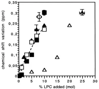
FIGURE 8 Absolute values of the variation of DOPA chemical shift for different type of DOPC/DOPA vesicles as a function of the percentage of MOPC added (mol%). (E) LUVs with MOPC added externally, no glyc-erol; (Œ) LUVs with MOPC added externally and with 50% glycglyc-erol; (䡺) FTVs with MOPC added externally, no glycerol; (f) FTVs with MOPC added externally, 25% glycerol; (‚) FTVs with MOPC in both monolayers. To facilitate comparisons, the variation of chemical shifts as a function of percentage MOPC added are represented instead of chemical shifts because glycerol itself causes a change of chemical shift (see text). The dotted line shows the average slope of the chemical shift variation as a function of MOPC concentration added externally (asymmetrical vesicles). The error bars are shown only for one point for each type of vesicles. Vertical error bars, associated with the variation of chemical shift, were evaluated from the line shape of the spectra and from the dispersion of repetitive experi-ments. The uncertainty on the concentration is evaluated by integration of the various peaks in the vesicle spectra and by a few controls involving lipid extraction at the end of the experiments.
band NMR spectroscopy by a narrow peak in the central part of the spectrum. Ideally, such experiments should be done with unilamellar vesicles with a diameter of several hundred nanometers to avoid narrowing of the NMR spec-trum by vesicle tumbling. Because the31P-NMR spectra of LUVs with a diameter in the 100- to 200-nm range is partially narrowed (Traı¨kia et al., 2000), we have used for these experiments LUVs prepared by phase reversion and filtered with 400 nm pores. Fig. 9, a to e, are broadband 31
P-NMR spectra of 400-nm DOPC/DOPA-LUVs recorded in the presence of increasing concentrations of MOPC added externally. Fig. 9 f is the spectrum of MOPC in buffer recorded under the same conditions and corresponds to micelles of MOPC. Fig. 9 a was recorded in the absence of
MOPC. It is not the typical spectrum of a homogeneous population of very large liposomes. Indeed, the central part of the spectrum reveals the existence of a distribution of sizes (Traı¨kia et al., 2000). This confirms that the extrusion of liposomes with pore sizes of 400 nm does not lead to a homogeneous population of unilamellar vesicles. In fact, these vesicles probably contain bilamellar vesicles as re-ported by Mayer et al. (1986). Nevertheless, such vesicles permit an easier evaluation of the micellar contribution in a composite NMR spectrum than 200-nm LUVs do.
Fig. 9 (b to e) shows that the addition of MOPC gives rise to a central narrow peak, the intensity of which grows with the proportion of MOPC added. At or above⬃5% MOPC, the contribution of this peak is nonnegligible, which sug-gests that a significant fraction of MOPC does not incorpo-rate in the membrane. Fig. 9, b, c, d, and e, cannot be simulated by a linear combination of spectra a (spectrum of liposomes) and f (spectrum of micelles) because the peak at 0 ppm is too broad (for example, see Fig. 9 e). Most likely, the broadening is due to rapid exchange of MOPC between micelles and membranes. By subtracting a fraction of spec-trum a from spectra b, c, d, and e respectively, we have estimated the contribution of MOPC nonincorporated into the vesicles and deduced the percentage of MOPC, which was effectively in the membrane (Fig. 9 B). For sake of simplicity the hypothesis used to draw the solid curve in Fig. 9 B was that all vesicles were unilamellar. If these “LUVs” actually contain a nonnegligible proportion of bi-lamellar vesicles (Mayer et al., 1986), then the proportion of MOPC per phospholipid in the external monolayer is higher than indicated in Fig. 9 B. Hence, the curve corresponds to a lower limit of MOPC percentage that is effectively incor-porated into unilamellar vesicles.
DISCUSSION
Asymmetrical insertion of lysophosphatidylcholine
MOPC was used here to create asymmetrical membranes. Long chain lysophosphatidylcholine molecules have a crit-ical micellar concentration in water in the micromolar range (Kumar and Baumann, 1991; Needham et al., 1997). Add-ing lysophosphatidylcholine to a very concentrated suspen-sion of unilamellar vesicles (several millimolar of phospho-lipids) allows one to rapidly intercalate new molecules in the outer surface. NMR experiments shown in Fig. 9 indicate nevertheless that a fraction of MOPC is rapidly exchanging between micelles and membranes, when the percentage of MOPC compared with total phospholipids present is above 2% to 3%. At least 50% of MOPC added penetrate the vesicle surface. There was no saturation under our experimental conditions (Fig. 9 B).
NMR data (Fig. 9 A) and electron microscope micro-graphs (Fig. 5) obtained with LUVs show that LUVs are not destroyed, neither do they form smaller vesicles when 5% FIGURE 9 (A) Broadband31P-NMR spectra of LUVs extruded through
400-nm pores with variable amounts of MOPC added externally. (a) No MOPC; (b) 1% MOPC; (c) 2.4% MOPC; (d) 4.7% MOPC; (e) 8% MOPC; (f) MOPC in buffer. (B) Percentage of MOPC incorporated in the vesicles as a function of the percentage of MOPC added as determined by subtrac-tion of the narrow component (see text for more details). Temperature is 8°C.
MOPC is added. This is a strong indication that MOPC has no lytic activity. Actually, more than 30% lysophosphati-dylcholine can be mixed with phosphatilysophosphati-dylcholine without preventing bilayer formation (van Echteld et al., 1981; Bhamidipati and Hamilton, 1995).
There are several direct reports in the literature about LPC flip-flop rate in liposomes and in biological mem-branes showing that the transmembrane diffusion of long chain LPC is extremely slow, even slower than that of phosphatidylcholine. De Kruyff et al. (1977), van den Besselaar et al. (1977), and Bhamidipati and Hamilton (1995) reported that the rate of LPC flip-flop is so small in SUVs that it is barely measurable. A half time of LPC flip-flop at 37°C above 100 h was estimated for SUVs and 46 h for hand-shaken liposomes (van den Besselaar et al., 1977). Experiments showing shape changes induced by the addition of LPC to erythrocytes (Sheetz and Singer, 1974) or to unilamellar vesicles (Farge and Devaux, 1992; Mathivet et al., 1996; Mui et al., 1995; this paper) can be understood in the framework of the bilayer couple hypoth-esis if, and only if, LPC does not equilibrate rapidly be-tween the two monolayers. Needham and Zhelev (1995) have reported that LPC flips rapidly with a half time of a few minutes. However, they investigated the reorientation of LPC in giant vesicles that were stretched by aspiration in a micropipette. This constraint may accelerate lipid flip-flop (Raphael and Waugh, 1996).
Vesicles shape and surface tension
The shapes of giant unilamellar vesicles with an asymmet-rical lipid distribution has been documented in previous articles (Farge and Devaux, 1992; Mui et al., 1995; Mathivet et al., 1996). Complete phase diagrams of vesicles morphologies have been calculated by bending energy min-imization as proposed originally by Helfrich (1973; Svetina and Zeks, 1989; Berndl et al., 1990; Seifert et al., 1991). A more recent and elaborate model takes into account the lateral elasticity of each lipid monolayers (Miao et al., 1994). Whereas theoretical shapes can be confronted easily to experiments, the surface tension that is responsible for shape changes is difficult if not impossible to measure directly.
If one assumes that the surface tension or lateral pressure of a lipid bilayer in the resting state is zero, lateral pressure within each monolayer manifests itself directly only in the lateral compressibility when the membrane is subject to a lateral stress (Marsh, 1996). Each layer of a lipid bilayer can be described as a two-dimensional elastic surface with its own elastic moduli (Kin and Kout, respectively). Typical values for the area compressibility modulus of fluid bilayers are KA⬇ 0.14 N/m (Kwok and Evans, 1981). In the case of asymmetrical constraints applied to the two leaflets, bilayer bending allows a membrane to partially relax the compres-sion or dilation of each leaflet. An elastic membrane should
continue to bend as long as the forces exerted on the two membrane halves form a nonzero torque. Thus, at equilib-rium, the stress of each layer should be the same but not necessarily zero. The mechanical response of a bilayer to a net addition of surface area due to the insertion of␦N lipids in one monolayer comprising initially N0 lipids has been previously analyzed (Farge and Devaux, 1993; Mui et al., 1995). If h is the thickness of the bilayer, R0the average radius of the vesicle budding out, the areal strain for a mechanically symmetrical membrane is approximately:
␣ ⬇Rh
0
␦N
N0
as long as h⬍⬍ R0. When MOPC is added to a membrane formed essentially of DOPC, this formula applies if one assumes as a first approximation that the cross-section of a MOPC molecule is identical to that of a DOPC molecule. h/R0 appears as a scaling parameter that modulates the influence of lipid asymmetry on the lateral tension of a vesicle. As noted also by Mui et al. (1995), it may have a dramatic effect on the tension caused by the insertion of new molecules. In the case of bud visible by optical mi-croscopy (R⬇ 2m) on a giant liposome, h/R0 is of the order of 2.5⫻ 10⫺3. Vesicle budding in GUVs is triggered when ␦N/N0 is of the order of 10⫺2 (Farge and Devaux, 1992) or less (Berndl et al., 1990). Then␣ is ⬃2.5 ⫻ 10⫺5 and the surface tension due to the compression of the outer monolayer is of the order of: T⫽ KA␣ ⬇ 0.35 ⫻ 10⫺5N/m, which is negligible. On the other hand, in the case of LUVs with a diameter of⬃200 nm, the bud may have a radius of
⬃25 nm (see Fig. 5 B), and h/R0⬇ 0.2. In the latter case, to induce such shape change by addition of MOPC,␦N/N0has to be of the order of 5% to 10%. Then␣ ⬇ 2 ⫻ 10⫺2and T⬇ 3 ⫻ 10⫺3N/m. A tension of that order of magnitude is not far to the tension that can produce vesicle lysis by dilation of the membrane (Kwok and Evans, 1981). It is sufficient to inhibit surface undulations (Farge and Devaux, 1993). In other words, an asymmetrical lipid distribution can generate an important surface tension in LUVs. In those vesicles, tension builds up before shape change takes place. The extreme situation is that of SUVs, which cannot undergo shape change because their curvature is the highest possible for a lipid bilayer. Indeed, it is difficult to decrease or increase, by whatever means, the average area per lipid molecules in a single leaflet by more than 2% to 4% because of the limited range of elastic deformations. The inner layer of sonicated vesicles is likely to be compressed to the limit of what is possible before a change of phase or membrane collapse.
DOPA as a lateral pressure sensor
A basic assumption for the interpretation of the NMR ex-periments is that the phosphorus chemical shift of
phospha-tidic acid allows one to detect a lateral compression of the lipids near the aqueous interface and not only membrane bending. A water/lipid interface represents an interphase between a bulk aqueous phase of high dielectric constant (⑀ ⬇ 80) and the hydrophobic phase of a membrane of very low dielectric constant. One consequence of the lower di-electric constant is that the adsorption of cations and anions to the ionizable groups of lipids is facilitated (Tocanne and Teissie´, 1990). However, the actual ionization state of phos-phatidic acid (PA) at the aqueous interface is also strongly dependent upon the phosphate accessibility to the buffer. Close packing by reducing the area available for the head-group phosphate allows the monoanionic form of PA to be more favorable than the dianionic form. Titration curves at various pH with sonicated dispersions of phosphatidylcho-line/PA mixtures allowed Swairjo et al. (1994a) to show that the difference in chemical shift of PA in the two monolayers of SUVs can be explained by a difference in the apparent pKa of the phosphate moiety on the two sides of SUVs. They calculated a pKavalue around 12 in the inner leaflet instead of 7 for a free phosphate. The pKashift in the inner leaflet would arise from the tight packing associated with the inner leaflet curvature. On the other hand, the PA head-group in the outer-leaflet would be in a more relaxed geometry that allows the phosphorus to sense the buffer pH. A priori, the expansion (or contraction) of each mono-layer is associated with a stress profile through the mem-brane (Gruner and Shyamsunder, 1991; Cantor, 1997). Lip-ids labeled with13C or2H could be used to investigate by NMR the entire stress profile (Chiu and Wu, 1990; Koenig et al., 1997).31P, on the other hand, is a natural probe that enables one to explore easily at least the two interfaces between phospholipids and water. Although it appears dif-ficult to relate theoretically in a quantitative and rigorous manner the variation of DOPA chemical shift (␦PA) to the lateral pressure or surface tension, the modification of␦PAis a convenient indicator of lateral stress modifications. Pro-viding the two membrane leaflets have the same lipid com-position and are facing the same buffer, identical values of
␦PA for the inner and outer leaflets reveal identical stress. The actual modification of the stress profile in a given experiment depends on what causes the membrane to bend. An external constraint, for example the aspiration of the membrane in a micropipette or actin polymerization push-ing the membrane are processes that are likely to stretch one monolayer and compress the other monolayer (Marsh, 1996). But when a new lipid distribution, caused by the insertion of drugs or by a flippase activity, is sole respon-sible for the bending, the constraint is internal, i.e., the deformation must reach a final equilibrium with zero torque as long as the membrane remains elastic.
Our NMR experiments and those of Kumar et al. (1989) indicate that the addition of LPC to SUVs has practically no influence on the line width, hence on vesicles size and finally on the average curvature. Yet,␦PAvaried
consider-ably in the outer leaflet upon addition of MOPC, whereas it remained almost constant in the inner one. Because it is hard to imagine how the curvature could change on one side of a membrane and remain constant on the opposite side, we suggest that a change of ␦PA should not be associated systematically with a change of membrane curvature but rather to compression/dilation near each interface accessible to the buffer.
Interpretation of data obtained with sonicated unilamellar vesicles
Compression of SUVs outer monolayer after the addition of MOPC is indicated by the up-field shift of␦PA(ext). This is not surprising; inserting MOPC in the outer monolayer should allow less area per lipid (Fig. 10 A). Perhaps more surprising is the slight downfield shift of ␦PA(int), which suggests membrane dilation, hence a small increase of area per head group in the inner monolayer. However, because SUVs are spherical, there is no alternative to a slight ex-pansion of the outer surface upon insertion of new mole-cules (elastic response). Because the inner monolayer ad-heres to the outer monolayer, it can expand slightly and release some pressure in the head-group region, which is probed by the phosphate chemical shift.
A similar observation was made by Swairjo et al. (1994b). The authors reported that the addition of annexin V to SUVs resulted in the same modification as the one reported here. They proposed a protein-induced change in vesicle morphology that corresponds to reduced curvature. Their data could be explained as well by implying a slight expansion of the outer leaflet due to the interaction of annexin V with the lipid head groups, involving perhaps a partial protein intercalation within the outer monolayer. Simultaneously the inner leaflet would be slightly relaxed as indicated by the sign of the change of␦PA(int) reported by these authors. Kumar et al. (1989) concluded from 31 P-NMR-T2 measurements that the addition of LPC to soni-cated POPC vesicles tightens phospholipid packing but only in the outer monolayer. In the present study, we see a small variation of ␦PA(int), it may simply tell that T2 and ␦PA, which are measurements of two different magnetic reso-nance properties, are not simply correlated and may have different sensitivity domains.
Large unilamellar vesicles and freeze-thawed vesicles
Comparison between Figs. 4 and 5 reveal that the addition of MOPC to the outer leaflet of LUVs, triggers an overall shape change. Although we have not seen the formation of protrusions and pseudopods as reported by Mui et al. (1995), we saw the formation of elongated vesicles and even eight shaped vesicles, at least in the absence of glycerol
(Fig. 5, A and B). The vesicles shapes shown in Fig. 5, A and B resemble giant vesicles with an asymmetrical lipid distri-bution (Farge and Devaux, 1992), it should be stressed that it was necessary to add 5% LPC to achieve these transfor-mations, whereas less than 1% mismatch suffice to obtain these shapes with GUVs. These observations are consistent with the scaling factor discussed above.
When LUVs were formed in the presence of 25% glyc-erol, their shapes before addition of MOPC were rather spherical. Upon addition of MOPC, there was no significant elongation. On the contrary they formed homogeneous pop-ulations of perfect spheres (Fig. 5 C). Such shape change implies an average change of volume: perhaps glycerol that has a disordering effect on lipid acyl chains (Fenske and Cullis, 1993) makes the bilayer more permeable to water.
In any case, when MOPC is added to large vesicles, it can be inferred from the high resolution31P-NMR spectra that there is an increased surface pressure not only in the outer leaflet but also in the inner leaflet because the signal cor-responding to DOPA remains a single peak and moves up-field. The high spectral resolution in Fig. 6 excludes the possibility that ␦PA(ext) could move up-field, whereas
␦PA(int) would move down-field or even remain constant. Thus, for LUVs (and FTVs) the insertion of MOPC in the outer leaflet triggers both bending and surface tension. The important point we want to emphasize here is that the lateral pressure not only increases in the outer leaflet but also in the
inner leaflet. Thus, without necessitating a transfer of mol-ecules the two monolayers are subjected to a lateral stress. If the membrane was a limited sheet without boundary conditions (Fig. 10 B), it would simply bend until both layers were fully relaxed. However, the equilibrium confor-mation for a closed membrane is that for which the strains in both layers are identical and correspond to the same lateral pressure.
Symmetrical membranes containing MOPC
Fig. 8 shows that if MOPC is included in both monolayers of FTVs, the variation of DOPA resonance position is very small at least as long as the percentage of MOPC included in each monolayer is less than 10%. Thus, symmetrical and asymmetrical distributions of MOPC have very different effects. Clearly␦PAvariation after addition of MOPC to the external monolayer cannot be explained by the dilution of the DOPA head group. For symmetrical membranes, there is no curvature expected from MOPC addition. On the other hand, adhesion of the two monolayers, which have opposite spontaneous curvatures, generates a “frustrated” flat bilayer with a constrained packing near the phospholipid head groups of both leaflets (Fig. 10 C) (Charvolin and Sadoc, 1988; Marsh, 1996).
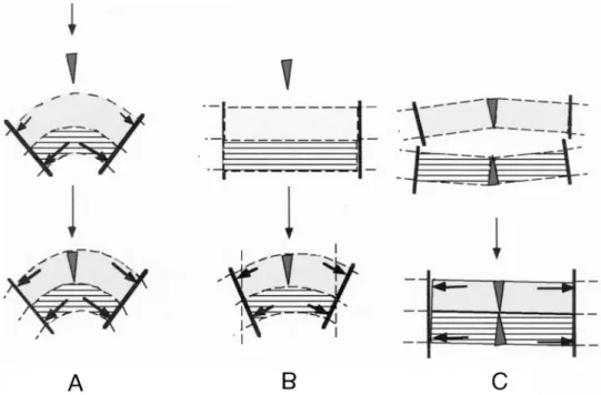
FIGURE 10 Schematic models representing the consequence of the insertion of MOPC in various types of unilamellar vesicles in the absence of glycerol. (A) SUVs. The incorporation of MOPC in the outer leaflet increases the surface pressure in the external surface. Because the membrane cannot bend anymore, the vesicle swells slightly and consequently surface pressure in the inner monolayer decreases slightly. (B) LUVs. Incorporation of MOPC provokes bending, which stops when both leaflets experience the same lateral pressure, hence the momentum is zero. (C) Symmetrical FTVs. Both monolayers have a positive spontaneous curvature due to the presence of wedge shape LPC. The adhesion of the two monolayers is only possible with formation of some surface tension (lateral pressure) but no overall membrane curvature is generated by the presence of LPC.
Biological relevance
We have shown that an excess of lipids as small as 2% to 3% on one side of a vesicles can generates surface tension. Such situation can be triggered in a biological membrane by the synthesis and release of new lipids on one side of a membrane or by the activity of a phospholipid translocase. For small organelles, with a diameter⬃200 nm, a surface tension should appear even before a significant shape change (budding) takes place.
The shape change caused by the aminophospholipid translocase activity in human erythrocytes (Seigneuret and Devaux, 1984) has led to propose that the accumulation of phospholipids on the inner leaflet of the plasma membrane of eukaryotic cells by the aminophospholipid translocase could be involved at the early stage of endocytosis (Devaux, 1991; Mu¨ller et al., 1994; Farge et al., 1999; Devaux, 2000; Rauch and Farge, 2000). The hypothesis that modulation of lipid asymmetry could also modulate surface pressure and, by this process, conformation of membrane protein has also been suggested (Bogdanov et al., 1993). There are experi-mental observations actually demonstrating the role of sur-face pressure on the modulation of membrane protein func-tion. Martinac et al. (1990) have shown that the addition of amphiphiles such as lysolecithin or chlorpromazine to giant Escherichia coli spheroplasts resulted in the opening of mechanosensitive ion channels. The work of Martinac et al. (1990) shows that compounds intercalated in the outer or in the inner leaflet have the same physiological effect. This is a strong indication that the mechanism involved is not the local curvature per se but rather the lateral compression. The influence of lateral pressure on phospholipase activity has been illustrated in monolayers (Bougulavsky et al., 1994) and in large unilamellar vesicles (Lehtonen and Kinnunen, 1995). The lytic activity of melittin is also strongly influ-enced by the tension of vesicles under osmotic stress (Benachir and Lafleur, 1996). Using osmotic stress, Goulian et al. (1998) have shown that the tension of giant vesicles can modulate the dimerization of gramicidin and hence the opening of the channel.
Finally, we might speculate that the influence of the flippase family on the lateral pressure of a membrane could be a way to regulate the activity of transmembrane proteins and not only of mechanosensitive channels. The fact that in reconstituted vesicles with a diameter of ⬃200 nm the P-glycoprotein activity was found quasi null (Rothnie et al., 2001) or very small (Romsicki and Sharom, 2001) is per-haps the consequence of the lateral pressure generated by the unidirectional transport of lipids.
This work was supported by grants from the Center National de la Recherche Scientifique (UMR 7099 and UMR 168), the Center d’Etude Atomique (LRC 8), and from the European Community (HPRN-CT-2000-00077).
REFERENCES
Benachir, T., and M. Lafleur. 1996. Osmotic and pH transmembrane gradient control the lytic power of melittin. Biophys. J. 70:830 – 840. Berndl, K., J. Ka¨s, R. Lipowsky, E. Sackmann, and U. Seifert. 1990. Shape
transformations of giant vesicles: extreme sensitivity to bilayer asym-metry. Europhys. Lett. 13:659 – 664.
Bhamidipati, S. P., and J. A. Hamilton. 1995. Interactions of lyso 1-palmi-toylphosphatidylcholine with phospholipids: a13C and31P NMR study.
Biochemistry. 34:5666 –5677.
Bogdanov, A., B. Verhoven, R. A. Schlegel, and P. Williamson. 1993. Asymmetry in transbilayer lateral pressure may drive expansion of the secretion fusion pore. Biochem. Soc. Trans. 21:271–275.
Bougulavsky, V., M. Rebecchi, A. J. Morris, D. Y. Jhon, S. G. Rhee, and S. McLaughlin. 1994. Effect of monolayer surface pressure on the activities of specific phospholipases C-beta 1,-gamma1 and delta 1.
Biochemistry. 33:3032–3037.
Cantor, R. S. 1997. The lateral profile in membranes; a physical mecha-nism of general anesthesia. Biochemistry. 36:2339 –2344.
Charvolin, J., and J.-F. Sadoc. 1988. Films of amphiphiles: packing con-straints and phase diagrams. J. Phys. Chem. 92:5787–5792.
Chiu, L.-M., and W.-G. Wu. 1990. Effective bilayer expansion and eryth-rocyte shape change induced by monopalmitoyl phosphatidylcholine.
Biophys. J. 57:1225–1232.
De Kruyff, B., A. M. H. P. van den Besselaar, and L. L. M. van Deenen. 1977. Outside-inside distribution and translocation of lysophosphatidyl-choline in phosphatidyllysophosphatidyl-choline vesicles as determined by 13C-NMR using (N-13CH
3)-enriched lipids. Biochim. Biophys. Acta. 465:443– 453. Devaux, P. F. 1991. Static and dynamic lipid asymmetry in cell
mem-branes. Biochemistry. 30:1163–1173.
Devaux, P. F. 2000. Is lipid translocation involved during endo- and exocytosis? Biochimie. 82:497–509.
Dubochet, J., M. Adrian, J.-J. Chang, J.-C. Hosuo, J. Lepault, and A. W. McDowall. 1988. Cryo-electron microscopy of vitrified specimens. Q.
Rev. Biophys. 21:129 –228.
Farge, E., M. Bitbol, and P. F. Devaux. 1990. Biomembrane elastic response to intercalation of amphiphiles. Eur. Biophys. J. 19:69 –72. Farge, E., and P. F. Devaux. 1992. Shape change of giant liposomes
induced by an asymmetric transmembrane distribution of phospholipids.
Biophys. J. 61:347–357.
Farge, E., and P. F. Devaux. 1993. Size dependent response of liposomes to phospholipid transmembrane redistribution: from shape change to induced tension. J. Phys. Chem. 97:2968 –2961.
Farge, E., D. M. Ojcius, A. Subtil, and A. Dautry-Varsat. 1999. Enhance-ment of endocytosis due to aminophospholipid transport across the plasma membrane of living cells. Am. J. Physiol. 276:725–733. Fenske, D. B., and P. R. Cullis. 1993. Acyl chain orientational order in
large unilamellar vesicles: comparison with multilamellar liposomes: a 2H and31P nuclear magnetic resonance study. Biophs. J. 64:1482–1491. Goulian, M., O. N. Mesquita, D. K. Fygenseon, C. Nelsen, O. S. Andersen, and A. Libchaber. 1998. Gramicidin channel kinetics under tension.
Biophys. J. 74:328 –337.
Gruner, S. M., and E. Shyamsunder. 1991. Is the mechanism of general anesthesia related to lipid membrane spontaneous curvature. Ann. N. Y.
Acad. Sci. 625:685– 697.
Helfrich, W. 1973. Elastic properties of lipid bilayers: theory and possible experiments. Z. Naturforsch. 28c:693–703.
Hope, M. J., M. B. Bally, G. Webb, and P. R. Cullis. 1985. Production of large unilamellar vesicles by rapid extrusion procedure: characterization of size distribution, trapped volume and ability to maintain a membrane potential. Biochim. Biophys. Acta. 812:55– 65.
Koenig, B. W., H. H. Strey, and K. Gawrisch. 1997. Membrane lateral compressibility determined by NMR and X-ray diffraction: effect of acyl chain polyunsatuartion. Biophys. J. 73:1954 –1966.
Kumar, V. V., and W. J. Baumann. 1991. Lanthanide-induced phospho-rus-31 NMR downfield chemical shifts of lysophosphatidylcholines are sensitive to lysophospholipid critical micelle concentration. Biophys. J. 59:103–107.
Kumar, V. V., B. Maleiwicz, and W. J. Baumann. 1989. Lysophosphati-dylcholine stabilizes small unilamellar phosphatiLysophosphati-dylcholine vesicles.
Biophys. J. 55:789 –792.
Kwok, R., and E. Evans. 1981. Thermoelasticity of large lecithin vesicles.
Biophys. J. 35:637– 652.
Lehtonen, J. Y. A., and P. K. J. Kinnunen. 1995. Phospholipase A2 as a mechanosensor. Biophys. J. 68:1888 –1894.
MacDonald, R. C., F. D. Jones, and R. Qiu. 1994. Fragmentation into small vesicles of dioleoylphosphatidylcholine bilayers during freezing and thawing. Biochim. Biophys. Acta. 1191:362–370.
Marsh, D. 1996. Lateral pressure in membranes. Biochim. Biophys. Acta. 1286:183–223.
Martinac, B., J. Adler, and C. Kung. 1990. Mechanosensitive ion channels of E. coli activated by amphipaths. Nature. 348:261–263.
Mathivet, L., S. Cribier, and P. F. Devaux. 1996. Shape change and physical properties of giant phospholipid vesicles prepared in the pres-ence of an AC electric field. Biophys. J. 70:1112–1121.
Mayer, L. D., M. J. Hope, and P. R. Cullis. 1986. Vesicles of variable sizes produced by rapid extrusion procedure. Biochim. Biophys. Acta. 858: 161–168.
Miao, L., U. Seifert, M. Wortis, and H.-G. Do¨bereiner. 1994. Budding transitions of fluid– bilayer vesicles: the effect of area-difference elas-ticity. Phys. Rev. E. 49:5389 –5407.
Mui, B. L.-S., H.-G. Do¨bereiner, T. D. Madden, and P. R. Cullis. 1995. Influence of transbilayer area asymmetry on the morphology of large unilamellar vesicles. Biophys. J. 69:930 –941.
Mu¨ller, P., T. Pomorski, and A. Herrmann. 1994. Incorporation of phos-pholipid analogues into the plasma membrane affects ATP-induced vesiculation of human erythrocyte ghosts. Biochem. Biophys. Res.
Com-mun. 199:881– 887.
Needham, D., N. Stoicheva, and D. Zhelev. 1997. Exchange of monoo-leoylphosphatidylcholine as monomer and micelle with membranes con-taining poly(ethylene glycol)-lipid. Biophys. J. 73:2615–2629. Needham, D., and D. Zhelev. 1995. Lysolipid exchange with lipid vesicle
membranes. Ann. Biomed. Eng. 23:287–298.
Raphael, R. M., and R. E. Waugh. 1996. Accelerated interleaflet transport of phosphatidylcholine molecules in membranes under deformation.
Biophys. J. 71:1374 –1388.
Rauch, C., and E. Farge. 2000. Endocytosis switch controlled by trans-membrane osmotic pressure and phospholipid asymmetry. Biophys. J. 78:3036 –3047.
Romsicki, C. Y., and F. J. Sharom. 2001. Phospholipid flippase activity of the reconstituted P-glycoprotein multidrug transporter. Biochemistry. 40:6937– 6947.
Rothnie, A., D. Theron, L. Soceneantu, C. Martin, M. Traı¨kia, G. Berridge, C. F. Higgins, P. F. Devaux, and R. Callaghan. 2001. The importance of
cholesterol in maintenance of P-glycoprotein activity and its membrane perturbing influence. Eur. Biophys. J. 30:430 – 442.
Seifert, U., K. Berndl and Lipowsky R. 1991. Shape transformations of vesicles: phase diagrams for spontaneous– curvature and bilayer cou-pling models. Phys. Rev. A. 44:1182–1202.
Seigneuret, M., and P. F. Devaux. 1984. ATP-dependent asymmetric distribution of spin-labeledphospholipids in the erythrocyte membrane.
Proc. Natl. Acad. Sci. U.S.A. 71:3751–3755.
Sheetz, M. P., and S. J. Singer. 1974. Bilayer membranes as bilayer couples: a molecular mechanism of drug-erythrocyte interactions. Proc.
Natl. Acad. Sci. U. S. A. 71:4457– 4461.
Svetina, S., and B. Zeks. 1989. Membrane bending energy and shape determination of phospholipid vesicles and red blood cells. Eur.
Bio-phys. J. 17:101–111.
Swairjo, M. A., M. F. Roberts, M.-B. Campos, J. R. Dedman, and B. Seaton. 1994b. Annexin V binding to the outer leaflet of small unila-mellar vesicles leads to altered inner leaflet properties:31P and1H-NMR studies. Biochemistry. 33:10944 –10950.
Swairjo, M. A., B. A. Seaton, and M. F. Roberts. 1994a. Effect of vesicle composition and curvature on the dissociation of phosphatidic acid in small unilamellar vesicles-A31P-NMR study. Biochim. Biophys. Acta. 1191:354 –361.
Szoka, F. C., F. Olson, T. Heath, W. J. Vail, E. Mayhew, and D. Papah-adjopoulos. 1980. Preparation of unilamellar liposomes of intermediate size (0.1– 0.2 microns) by combination of reverse phase evaporation and extrusion through polycarbonate membranes. Biochim. Biophys. Acta. 601:559 –571.
Tocanne, J.-F., and J. Teissie´. 1990. Ionization of phospholipids and phospholipid-supported interfacial lateral diffusion of protons in mem-brane model systems. Biochim. Biophys. Acta. 1031:111–142. Traı¨kia, M., D. B. Langlais, G. M. Cannarozzi, and P. F. Devaux. 1997.
High resolution spectra of liposomes using MAS NMR: the case of intermediate size vesicles. J. Magn. Res. 125:140 –144.
Traı¨kia, M., D. E. Warschawski, M. Recouvreur, J. Cartaud, and P. F. Devaux. 2000. Formation of unilamellar vesicles by repetitive freeze-thaw cycles: characterization by electron microscopy and 31P-NMR.
Eur. Biophys. J. 29:184 –195.
van den Besselaar, A. M. H. P., H. van den Bosch, and L. L. M. van Deenen. 1977. Transbilayer distribution and movement of lysophos-phatidylcholine in liposomal membranes. Biochim. Biophys. Acta. 465: 454 – 465.
van Echteld, C. J. A., B. De Kruijff, J. G. Mandersloot, and J. De Gier. 1981. Effects of lysophosphatidylcholines on phosphatidylcholine and phosphatidylcholine-cholesterol liposomes systems as revealed by 31P-NMR, electron microscopy and permeability studies. Biochim. Biophys.