HAL Id: inserm-02506954
https://www.hal.inserm.fr/inserm-02506954
Submitted on 12 Mar 2020
HAL is a multi-disciplinary open access
archive for the deposit and dissemination of
sci-entific research documents, whether they are
pub-lished or not. The documents may come from
teaching and research institutions in France or
abroad, or from public or private research centers.
L’archive ouverte pluridisciplinaire HAL, est
destinée au dépôt et à la diffusion de documents
scientifiques de niveau recherche, publiés ou non,
émanant des établissements d’enseignement et de
recherche français ou étrangers, des laboratoires
publics ou privés.
development and protection from intestinal
inflammation in mice
Ute Bank, Katrin Deiser, Carlos Plaza-Sirvent, Lisa Osbelt, Amelie Witte,
Laura Knop, Rebecca Labrenz, Robert Jänsch, Felix Richter, Aindrila Biswas,
et al.
To cite this version:
Ute Bank, Katrin Deiser, Carlos Plaza-Sirvent, Lisa Osbelt, Amelie Witte, et al.. c-FLIP is crucial
for IL-7/IL-15-dependent NKp46+ ILC development and protection from intestinal inflammation in
mice. Nature Communications, Nature Publishing Group, 2020, 11 (1), pp.1056.
�10.1038/s41467-020-14782-3�. �inserm-02506954�
c-FLIP is crucial for IL-7/IL-15-dependent NKp46
+
ILC development and protection from intestinal
in
flammation in mice
Ute Bank
1,13
, Katrin Deiser
1,13
, Carlos Plaza-Sirvent
1,2
, Lisa Osbelt
3,4
, Amelie Witte
1
, Laura Knop
1
,
Rebecca Labrenz
1
, Robert Jänsch
1
, Felix Richter
1
, Aindrila Biswas
5
, Ana C. Zenclussen
6
, Eric Vivier
7,8,9
,
Chiara Romagnani
10,11
, Anja A. Kühl
12
, Ildiko R. Dunay
5
, Till Strowig
4
, Ingo Schmitz
1,2,14
&
Thomas Schüler
1,14
✉
NKp46
+innate lymphoid cells (ILC) modulate tissue homeostasis and anti-microbial immune
responses. ILC development and function are regulated by cytokines such as Interleukin
(IL)
−7 and IL-15. However, the ILC-intrinsic pathways translating cytokine signals into
developmental programs are largely unknown. Here we show that the anti-apoptotic
mole-cule cellular FLICE-like inhibitory protein (c-FLIP) is crucial for the generation of
IL-7/IL-15-dependent NKp46
+ILC1, including conventional natural killer (cNK) cells, and ILC3.
Cytokine-induced phosphorylation of signal transducer and activator of transcription
5 (STAT5) precedes up-regulation of c-FLIP, which protects developing NKp46
+ILC from
TNF-induced apoptosis. NKp46
+ILC-speci
fic inactivation of c-FLIP leads to the loss of all
IL-7/IL-15-dependent NKp46
+ILC, thereby inducing early-onset chronic colitis and
subse-quently microbial dysbiosis; meanwhile, the depletion of cNK, but not NKp46
+ILC1/3,
aggravates experimental colitis. In summary, our data demonstrate a non-redundant function
of c-FLIP for the generation of NKp46
+ILC, which protect T/B lymphocyte-sufficient mice
from intestinal in
flammation.
https://doi.org/10.1038/s41467-020-14782-3
OPEN
1Institute of Molecular and Clinical Immunology, Medical Faculty, Otto-von-Guericke University, Magdeburg, Germany.2Systems-Oriented Immunology and
Inflammation Research Group, Department of Immune Control, Helmholtz Centre for Infection Research, Braunschweig, Germany.3Institute of Medical Microbiology and Hospital Hygiene, Medical Faculty, Otto-von-Guericke University, Magdeburg, Germany.4Microbial Immune Regulation Research Group, Helmholtz Centre for Infection Research, Braunschweig, Germany.5Institute of Inflammation and Neurodegeneration, Medical Faculty, Otto-von-Guericke University, Magdeburg, Germany.6Experimental Obstetrics and Gynecology, Medical Faculty, Otto-von-Guericke University, Magdeburg, Germany.7Centre d’Immunologie de Marseille-Luminy, Aix Marseille Université, Inserm, CNRS, Marseille, France.8Service d’Immunologie, Hôpital de la Timone, Assistance Publique-Hôpitaux de Marseille, Marseille, France.9Innate Pharma Research Labs., Innate Pharma, Marseille, France.10Innate Immunity, German Rheumatism Research Center (DRFZ), Leibniz Association, Berlin, Germany.11Medical Department I, Charité - University Medical Center Berlin,
Berlin, Germany.12Charité– Universitätsmedizin Berlin, corporate member of Freie Universität Berlin, Humboldt Universität zu Berlin, and Berlin Institute of
Health, iPATH, Berlin, Germany.13These authors contributed equally: Ute Bank, Katrin Deiser.14These authors jointly supervised this work: Ingo Schmitz,
Thomas Schüler. ✉email:thomas.schueler@med.ovgu.de
123456789
I
nnate lymphoid cells (ILC) modulate immune responses and
tissue homeostasis at multiple levels. Reminiscent to the
classification of helper T cell subsets, different types of ILC can
be distinguished based on their cytokine and transcription factor
expression profiles
1. Type 1 NKp46
+T-bet
+(ILC1) produce
Interferon-γ (IFN-γ) and contribute e.g. to Toxoplasma gondii
clearance
2. Conventional NK cells (cNK) represent a
sub-population of ILC1, which is characterized by the co-expression
of T-bet and Eomes
1. Distinct from T-bet
+Eomes
−helper-like
ILC1, cNK synthesize cytotoxic molecules, such as perforins and
granzymes as well as death ligands (DLs) like tumor necrosis
factor
α (TNF), TNF-related apoptosis inducing ligand (TRAIL)
and FAS ligand (FASL) enabling them to induce target cell
apoptosis
3. On the contrary, NKp46
−GATA3
+ILC2 produce
Interleukin (IL)−5 and IL-13 and promote helminth rejection
4,
while NKp46
+/−RORγt
+ILC3 support anti-bacterial responses
in mice lacking T and B lymphocytes
5. Furthermore, ILC3
reg-ulate tissue homeostasis and regeneration, e.g., in the intestine
and lung
6. For example, ILC3-derived IL-22 promotes intestinal
epithelial stem cell regeneration
7, providing an explanation for
the tissue-protective effect of IL-22 in the course of dextran
sodium sulfate (DSS)-induced colitis
8.
Most studies defining immune modulatory and tissue
protec-tive functions of NKp46
+ILC3 were performed with T and B
lymphocyte (T/B)-deficient mice. However, recent evidence
sug-gests that T lymphocytes can compensate for the lack of NKp46
+ILC3
9–11. Whether the degree of compensation is determined by
the experimental system or whether ILC3 are generally of limited
importance in a T/B-competent host is still unclear.
Developing and mature ILC express a multitude of cytokine
receptors including those for IL-15 and IL-7
12. Both cytokines
signal via signal transducer and activator of transcription 5
(STAT5)
13and are crucial for ILC development and survival
2,14.
For example, IL-15 withdrawal activates the intrinsic apoptosis
pathway in cNK
15. This is associated with the failure to repress
expression of the pro-apoptotic molecule Bcl2-interacting
med-iator of cell death (Bim), the loss of anti-apoptotic myeloid cell
leukemia-1 (Mcl-1) and the subsequent death of cNK
15. IL-7 and
IL-15 are produced by e.g., stromal
fibroblasts
16,17and intestinal
epithelial cells (IEC)
6,18, which create the cytokine environment
required for the maintenance of ILC homeostasis and function
12.
However, ILC-intrinsic signaling molecules required for the
conversion of environmental cues into developmental programs
are largely unknown. In order to identify such signaling
mole-cules and to define unique functions of intestinal NKp46
+ILC,
we made use of NKp46
iCremice
19. These mice are T/B-competent
and allow targeted gene inactivation in NKp46
+ILC.
Here, we show that the anti-apoptotic molecule cellular
FLICE-like inhibitory protein (c-FLIP), a master regulator of the extrinsic
apoptosis pathway
20, is a target of STAT5-dependent cytokine
signaling in NKp46
+ILC. The NKp46-specific inactivation of
c-FLIP leads to the loss of NKp46
+ILC1, including cNK, and ILC3
without affecting T and B lymphocyte homeostasis. Furthermore,
we provide evidence that cytokine-dependent c-FLIP induction
prevents a TNF-induced suicide program in developing NKp46
+ILC. Finally, we define a non-redundant function of
c-FLIP-dependent NKp46
+cNK in the intestine. Specifically, different
from mice only lacking NKp46
+ILC3, mice lacking NKp46
+cNK
fail to recover from acute colitis and develop early signs of chronic
disease. Furthermore, although not affecting the establishment of a
normal commensal microbiota in the steady state, NKp46
+ILC
counteract inflammation-associated commensal dysbiosis. In
sum-mary, we show that the anti-apoptotic molecule c-FLIP is crucial for
the cytokine-dependent development of NKp46
+cNK/ILC1 and
ILC3 and subsequent protection from intestinal inflammation by
cNK, a function that cannot be compensated by other immune cells.
Results
c-FLIP is essential for the development of NKp46
+ILC. c-FLIP
is a master regulator of the extrinsic apoptosis pathway
20and
protects immune and non-immune cells from caspase
8-dependent apoptosis
21,22. The expression of c-FLIP is crucial for
T cell development
23and is up-regulated by activated human and
mouse T cells
24. IL-15 is crucial for the development of T-bet
+ILC1 including cNK
25,26. Whether and how the external
apop-tosis pathway is involved in IL-15-dependent ILC development
and function was unclear. In order to identify a functional link
between IL-15 signaling and Cflar (encoding c-FLIP) gene
reg-ulation, CD3
−NK1.1
+NKp46
+ILC were purified from spleens of
C57BL/6 (B6) mice and cultured for 16 h in the presence or
absence of IL-15. The murine Cflar gene encodes for two isoforms
of c-FLIP, the long form c-FLIP
Land the short Raji isoform
c-FLIP
R27. Quantitative real-time PCR (RT-qPCR) revealed that
NKp46
+ILC significantly up-regulate the mRNA of the long
isoform c-FLIP
L, but not c-FLIP
R, in response to IL-15 (Fig.
1
a).
Elevated Cflar gene activity correlated with the phosphorylation of
STAT5 and elevated c-FLIP protein levels (Fig.
1
b). The blockade
of STAT5 phosphorylation by pimozide led to impaired
IL-15-dependent c-FLIP up-regulation (Fig.
1
b). Hence, c-FLIP is a target
of STAT5-dependent IL-15 signaling in mature NKp46
+ILC.
Next, we investigated whether c-FLIP is required for the
development of IL-15/STAT5-dependent NKp46
+ILC. For this
purpose, conditional STAT5 (STAT5
fl/fl) and c-FLIP (c-FLIP
fl/fl)
knockout mice were crossed to NKp46
iCre-transgenic mice to
generate NK
ΔSTAT5and NK
Δc-FLIPmice, respectively. NKp46
iCre-transgenic mice harboring wildtype alleles of the respective target
gene served as NK
WTcontrols. Similar to IL-15-deficient (IL-15
−/−)
and IL-15 receptor
α-deficient (IL-15R
−/−) mice, numbers of
splenic NKp46
+ILC were strongly reduced in NK
ΔSTAT5and
NK
Δc-FLIPmice (Fig.
1
c). IL-15 deprivation causes cNK apoptosis
28.
Accordingly, residual splenic IL-15
−/−and IL-15R
−/−NKp46
+ILC
showed increased rates of caspase-3/7 activity (Fig.
1
d). The
frequencies of NKp46
+ILC expressing active caspase-3/7 were also
elevated in NK
ΔSTAT5and NK
Δc-FLIPmice (Fig.
1
d).
Furthermore, we observed increased frequencies of immature
double negative (DN) NK1.1
−NKp46
−and single positive (SP)
NK1.1
+NKp46
−cells in conjunction with a reduced abundance
of double positive (DP) NK1.1
+NKp46
+ILC in spleens and bone
marrow (BM) of IL-15
−/−, IL-15R
−/−, NK
ΔSTAT5, and NK
Δc-FLIPmice (Fig.
1
e, f). Of note, the development of other immune cells
remained unaltered in NK
Δc-FLIPmice (Supplementary Fig. 1).
Altogether, our results demonstrate that c-FLIP is crucial for the
IL-15/STAT5-dependent development of NKp46
+ILC in the BM
and their survival in the periphery.
c-FLIP protects ILC precursors from TNF-induced apoptosis.
ILC1 produce effector molecules such as IFN-γ and DLs like TNF
and TRAIL
29. In the absence of c-FLIP, human cNK become
sensitive to their own effector molecules and undergo apoptosis
30.
We therefore hypothesized that the loss of NKp46
+ILC in
NK
Δc-FLIPmice resulted from death receptor (DR)-induced
apoptosis during ILC1 development. To elucidate whether
effector genes are activated in BM ILC, we
first analyzed IFN-γ
reporter mice expressing eYFP under control of the Ifng
pro-moter. As shown in Fig.
2
a, Ifng promoter activity increased from
the NK1.1
−NKp46
−DN to the NK1.1
+NKp46
−SP stage and
reached its maximum at the NK1.1
+NKp46
+DP stage. Similarly,
TNF mRNA levels increased progressively from the DN to the DP
stage (Fig.
2
b). Of note, DR mRNAs for TRAIL-R2, FAS and
TNF-RI were also detectable throughout ILC development, the
latter being particularly high in DP cells (Fig.
2
c). In addition,
WT IL-15–/– IL-15R–/– NKΔSTAT5 NKΔc-FLIP Bone marrow 30.9 53 0.851 15.3 Spleen 20 50.3 0.352 29.3 34.1 52.3 3.72 9.82 17.6 51.5 13.8 17.1 4.45 64.6 17.1 13.9 16.1 43.6 3.06 37.2 18.9 53.6 3.57 23.7 NK1.1-APC-Cy7 5.54 92.9 0 1.57 8.17 63.9 12.8 15.1 0.435 99.2 0.0311 0.373 NK1.1-APC-Cy7 NKp46-V450
f
e
0 2 4 6 8 Untr. IL-15 c-FLIP (MFI ×10 3) IL-15 IL-15+ pimozidea
pSTAT5-PE c-FLIP-APCb
Relative cell number
Untr. IL-15 IL-15 IL-15 +pim. 0 102 103 104 105 0 20 40 60 80 100 0 20 40 60 80 100 0 5 10 15 0 0.5 1.0 1.5
Relative mRNA levels
Untr. IL-15 c-FLIPL c-FLIPR WT WT IL-15 –/– IL-15R –/– NK WT NK ΔSTAT5 NK WT NK Δc-FLIP 0 1000 2000 3000 ILC (×10 3) 0 500 1000 1500 2000 2500 0 500 1000 1500 0 500 1000 1500 2000 2500
c
Caspase3/7 + ILC (%) 0 10 20 30 40 50 0 10 20 30 0 5 10 15 0 2 4 6 8 10d
0 102 103 104 105 WT WT IL-15 –/– IL-15R –/– NK WT NK ΔSTAT5 NK WT NK Δc-FLIP 0 102 103 104 105 0 102 103 104 105 0 102 103 104 105 0 102 103 104 105 0 102 103 104 105 0 102 103 104 105 0 102 103 104 105 0 102 103 104 105 0 102 103 104 105 0 102 103 104 105 NKp46-V450 0 102 103 104 105 0 102 103 104 105 0 102 103 104 105 0 102 103 104 105 0 102 103 104 105 0 102 103 104 105 0 102 103 104 105 0 102 103 104 105 0 102 103 104 105 0 102 103 104 105Fig. 1 c-FLIP is crucial for IL-15-dependent ILC development. a NK1.1+NKp46+ILC were FACS-purified from spleens of C57BL/6 (B6) wild type (WT) mice and incubated for 16 h w/wo IL-15. c-FLIPLand c-FLIPRmRNA levels were quantified by RT-qPCR. Data are from four independent experiments.
b B6 splenocytes were cultured for 16 h in the presence (red) or absence of IL-15 (black; upper row). Alternatively, cells were cultured for 16 h in the presence of IL-15 with (blue) or without the STAT5 inhibitor pimozide (red; bottom row). Relative levels of c-FLIP and phosphorylated STAT5 (pSTAT5) were determined byflow cytometry. Shown are representative histograms (fluorescence minus one controls in gray) after gating on Lin−CD122+NK1.1
+NKp46+ILC. Graphs show meanfluorescence intensities (MFI) from 2–3 independent experiments with a total of 7–8 mice per group. c–e Cells from
spleen andf BM of the indicated mouse lines were analyzed byflow cytometry. c–f After gating on Lin−CD122+cells,c absolute cell numbers (n= 8–10) andd frequencies of active caspase3/7+apoptotic NK1.1+NKp46+ILC were determined (n= 6–10). e, f Shown are representative contour plots from at least two independent experiments with a total ofe 8–10 or f 6–10 mice per group. Numbers indicate percentages. a–d Data represent mean +/± SEM. Source data are provided as source datafile; *p ≤ 0.05; **p ≤ 0.005; ***p ≤ 0.001; ****p ≤ 0.0001. a, c, d Two-tailed Mann–Whitney U test; b two-tailed Wilcoxon matched-pairs signed rank test.
stages (Fig.
2
d). Interestingly, only c-FLIP
LmRNA showed
a maturation-associated increase (Fig.
2
d) similar to TNF
(Fig.
2
b), TRAIL-R2 and TNF-RI (Fig.
2
c).
In order to elucidate the impact of DLs on c-FLIP-dependent
ILC development, we established an in vitro culture system. DN
cells were enriched from BM of NK
WTand NK
Δc-FLIPmice and
cultured for 9 days. In accordance with our in vivo data, the
generation of DP cells was impaired in the absence of c-FLIP
(Fig.
2
e). Importantly, this effect could be recovered most
effectively by the antibody-mediated blockade of TNF (Fig.
2
f).
Together, our results demonstrate that TNF and TNF-RI
up-regulation are programmed events in the course of NKp46
+ILC
development. The partial recovery of NK
Δc-FLIPILC development
by
αTNF treatment strongly suggests that the cytokine-induced,
STAT5-dependent up-regulation of c-FLIP
L(Fig.
1
a, b) is crucial
for protection of developing ILC from TNF-induced apoptosis.
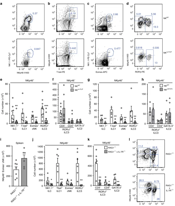
IL-7- and IL-15-dependent intestinal NKp46
+ILC require
c-FLIP. The small intestine (SI) harbors NKp46
+T-bet
+ILC1,
including T-bet
+Eomes
+cNK, as well as NKp46
−GATA3
+ILC2
and NKp46
+/−CD4
+/−RORγt
+ILC3
12. In order to analyze
whether c-FLIP affects intestinal ILC homeostasis, leukocytes
were isolated from the small intestinal lamina propria of NK
WTand NK
Δc-FLIPmice. The abundance of NKp46
+NK1.1
+ILC
(Fig.
3
a, e) including T-bet
+ILC1 (Fig.
3
b, e), NK1.1
+Eomes
+cNK (Fig.
3
c, e) and NKp46
+RORγt
+ILC3 (Fig.
3
d, e) were
strongly reduced in the SI of NK
Δc-FLIPmice. Importantly,
numbers
of
NKp46
−CD4
+/−RORγt
+ILC3
(Fig.
3
d,
f)
and NKp46
−GATA3
+ILC2 (Fig.
3
f) remained unaltered in
NK
Δc-FLIPmice. Hence, NK
Δc-FLIPmice lack NKp46
+ILC but
have normal numbers of NKp46
−ILC in the SI. Similar data were
obtained with NK
ΔSTAT5mice (Fig.
3
g, h) further emphasizing
the functional link between STAT5 and c-FLIP.
The development of cNK/ILC1 largely relies on IL-15
2,25,31(Supplementary Fig. 2) rather than IL-7 signaling. The latter
point is exemplified by the fact that cNK numbers in spleen and
SI did not differ significantly between RAG1
−/−and RAG1
−/−×
IL-7R
−/−mice (Fig.
3
i, j). On the contrary, numbers of all other
SI NKp46
+and NKp46
−ILC subsets were strongly reduced in
RAG1
−/−× IL-7R
−/−mice (Fig.
3
j, k). IL-15 can partially
compensate for the lack of IL-7R signaling
32providing an
explanation for the survival of some residual ILC in RAG1
−/−×
IL-7R
−/−mice (Fig.
3
j, k). Of note, this compensatory effect
was less efficient for NKp46
+RORγt
+ILC3, which were
more severely affected by the lack of IL-7R signaling than
NKp46
−RORγt
+ILC3 (Fig.
3
l; lower panel).
IL-7R and IL-15R signaling converge at the level of STAT5
phosphorylation which precedes c-FLIP up-regulation (Fig.
1
b).
0 2 4 6 0 2 4 6 8 0 2 4 6 DN SP DP DN SP DP DN SP DP
Relative mRNA levels
TRAIL-R2 FAS TNF-RI
c
1.5 2.0 0.5 1.0 0.0 DN SP DP DN SP DPRelative mRNA levels
d
c-FLIPL c-FLIPRe
DP fold induction 0 2 4 6 8 10f
1.5 2.0 2.5 0 0.5 1.0 Cell number DP (×10 3)ControlαFASLαTRAIL αTNF
NKΔc-FLIP
a
IFN-γ (eYFP)
Relative cell number
DN SP DP 0 20 40 60 80 100
b
0 1 2 3 4 DN SP DPRelative mRNA levels
TNF 0 200 400 600 800 1000 IFN-γ
Promoter activity (eYFP MFI)
DN SP DP 0 102 103 104 105 NK WT NK Δc-FLIP
Fig. 2 The generation of NKp46+ILC requires c-FLIP-mediated protection from TNF-induced apoptosis. a To determine Ifng promoter activity in unstimulated ILC precursors, freshly isolated BM cells from eYFP-transgenic IFN-γ reporter mice were analyzed by flow cytometry. Mean fluorescence intensities (MFI) were determined for eYFP after gating on Lin−CD122+DN, SP, and DP cells. Representative histograms and pooled results (bar diagrams, mean+ SEM) from two independent experiments with a total of five mice are shown (DN = 3 data points). b–d Lin−CD122+DN, SP, and DP cells were FACS-purified from BM of B6 mice and the indicated mRNAs were quantified by RT-qPCR. mRNA levels of DN cells were set to 1 and all other values were calculated in relation. Shown are pooled results (mean+ SEM) from b, d two and c three independent experiments. e, f Lin−CD122+DN cells frome NKWT
and NKΔc-FLIPmice orf NKΔc-FLIPmice alone were enriched by FACS-sorting.f To block DR-function,αFASL, αTRAIL, or αTNF antibodies were added to the indicated cultures.e, f After 9 days of culture, numbers of viable DP cells were calculated in relation to those at day 0. f Absolute cell numbers are shown. Data (mean+ SEM) were pooled from 3–5 independent experiments. a–f Source data are provided as source data file; e, f *p ≤ 0.05; **p ≤ 0.005; ***p≤ 0.001; ****p ≤ 0.0001 (paired Student’s t test).
NKWT NKΔc-FLIP
b
NKp46-V450 T-bet-PE NKp46-V450 NK1.1-PE-Cy7a
5.37 0.847 Eomes-APC NK1.1-PE-Cy7c
2.66 0.477 NKp46-V450 RORγt-PEd
5.56 19.3 5.01 0.335 13 0.618 9.19 0.442 RORγt-PE NKp46-V450 RAG1–/– x IL-7R–/–l
8.73 26.5 28 42.8 11.2 31.4 RAG1–/– NKp46 + Eomes + cNK (×10 3) 0 200 400 600 800 RAG1 –/– RAG1 –/– x IL-7R –/– Spleenj
k
i
0 200 400 600 800 1000 1200 1400 NK1.1+ ILC Tbet+ ILC1 Eomes+ cNK RORγt+ ILC3 Cell number (×10 3) NKp46+ GATA-3+ ILC2 CD4– CD4+ RORγt+ ILC3 0 200 400 600 800 NKp46– RAG1–/– RAG1–/– x IL-7R–/– 0 20 40 60 80 50 100 150 200 400 450 500 NK1.1+ ILC Eomes+ cNK T-bet+ ILC1 RORγt+ ILC3 CD4+ CD4– GATA-3+ ILC2 RORγt+ ILC3 Cell number (×10 3)e
NKp46+f
NKp46– NKWT NKΔc-FLIP 0 20 40 60 80 100 120 0g
NKp46+h
NKp46– NK1.1+ ILC Eomes+ cNK T-bet+ ILC1 RORγt+ ILC3 CD4– CD4+GATA-3+ ILC2 RORγt+ ILC3 0 50 100 150 200 NKWT NKΔSTAT5 Cell number (×10 3) 0 102 103 104 105 0 102 103 104 105 0 102 103 104 105 0 102 103 104 105 0 102 103 104 105 0 102 103 104 105 0 102 103 104 105 0 102 103 104 105 0 102 103 104 105 0 102 103 104 105 0 102 103 104 105 0 102 103 104 105 0 102 103 104 105 0 102 103 104 105 0 102 103 104 105 0 102 103 104 105 0 102 103 104 105 0 102 103 104 105 0 102 103 104 105 0 102 103 104 105Fig. 3 NKΔc-FLIPmice lack IL-7- and IL-15-dependent NKp46+ILC in the small intestine. a–h and j–l Leukocytes were isolated from the small intestinal (SI) lamina propria ofa–f NKΔc-FLIP,g, h NKΔSTAT5,j–l RAG1−/−× IL-7R−/−and respective control mice.i Spleen cells from RAG1−/−× IL-7R−/−and RAG1−/−mice were analyzed.a–h and j–l To discriminate between different subsets of NKp46+(ILC: NK1.1+; ILC1: T-bet+, cNK: Eomes+; ILC3: RORγt+) and NKp46−ILC (ILC3: RORγt+; ILC2: GATA3+), cells were analyzed byflow cytometry after gating on Lin−CD45+cells.e–k Absolute cell numbers are shown as mean+ SEM. a–d and l Shown are representative contour plots and e–k pooled results from 3 to 5 independent experiments with a total of e, f 10–11, g, h 6, i 6–7 and j, k 6 mice per group. e For NKp46+ILC, ILC1, cNK, and ILC3, 10-11, 7, 7–8, and 10–11 samples were analyzed, respectively. f For NKp46−CD4−ILC3, CD4+ILC3 and ILC2, 8–9, 8–9 and 7 samples were analyzed, respectively. g, h 5–6, i 6–7, j, k 5–6 samples were analyzed for the indicated ILC subsets.a–d and l Numbers indicate percentages. e–k Source data are provided as source data file; *p ≤ 0.05; **p ≤ 0.005; ***p ≤ 0.001; ****p≤ 0.0001 (two-tailed Mann–Whitney U test).
Hence, the simultaneous lack of IL-15- and IL-7-dependent
NKp46
+ILC1 and ILC3 in NK
Δc-FLIPand NK
ΔSTAT5mice
strongly suggests that both cytokines promote ILC development
and survival via the STAT5-dependent induction of c-FLIP.
c-FLIP-dependent NKp46
+ILC protect mice from acute
coli-tis. Inflammatory bowel disease (IBD) is associated with
pro-nounced changes in ILC1 and ILC3 frequencies and functions
29.
Whether and how the absence of NKp46
+ILC1 and ILC3 affects
the course of IBD was unclear. In order to address this subject,
NK
Δc-FLIPand control mice were treated with DSS for 5 days.
The administration of DSS causes IEC damage, loss of epithelial
integrity, innate immune cell activation, and results in IBD-like
symptoms
33. To monitor disease progression, relative body
weight (Fig.
4
a), feces consistency (Fig.
4
b), and the degree of
intestinal bleeding (Fig.
4
c) were determined and the overall
colitis score was calculated (Fig.
4
d). As shown in Fig.
4
a–d,
disease severity was strongly increased in NK
Δc-FLIPmice.
Accordingly, the specific colon weight was strongly increased in
NK
Δc-FLIPmice indicating edema formation and increased
cel-lularity (Fig.
4
e). Furthermore, the colon length was significantly
decreased in NK
Δc-FLIPmice (Fig.
4
f, g) and
inflammation-related alterations in tissue architecture were more pronounced
(Fig.
4
h). When colonic immune cell infiltrates were analyzed, we
observed the almost complete absence of NKp46
+NK1.1
+ILC
including T-bet
+ILC1, Eomes
+cNK and RORγt
+ILC3 (Fig.
4
i,
j) in NK
Δc-FLIPmice. On the contrary, numbers of NKp46
−ILC
did not differ significantly between NK
Δc-FLIPand NK
WTmice
(Fig.
4
k). Importantly, CD45
+CD11b
+granulocytes, in
parti-cular pro-inflammatory Ly6C
+Ly6G
+neutrophils, rather than
Ly6C
−Ly6G
+/−monocytes, accumulated more efficiently in the
inflamed colon of NK
Δc-FLIPmice (Fig.
4
l, m). Thus, the lack of
c-FLIP-dependent, NKp46
+ILC promotes acute colitis and
cannot be compensated by T or B lymphocytes.
NKp46
+ILC protect mice from early onset of chronic colitis.
Repeated DSS administration causes chronic colitis
33. Since it was
unclear whether this is also affected by NKp46
+ILC, NK
WTand
NK
Δc-FLIPmice were treated repetitively with DSS. In agreement
with the results from the acute colitis model (Fig.
4
a–d), disease
severity was significantly more pronounced in NK
Δc-FLIPthan in
NK
WTmice after the
first 5-day period of DSS treatment
(Fig.
5
a). After termination of the
first DSS treatment, NK
WTmice had recovered completely until day 15. This was not the case
for NK
Δc-FLIPmice. They failed to recover completely until day
19, when the second 5-day period of DSS administration was
initiated (Fig.
5
a). Importantly, NK
Δc-FLIPmice rapidly developed
maximum disease scores comparable to those following the
first
phase of DSS treatment (Fig.
5
a). NK
WTmice showed elevated
disease scores at day 25, which were above those observed at the
peak of the
first treatment cycle but still significantly below those
of NK
Δc-FLIPmice (Fig.
5
a).
Following the second DSS cycle, NK
WTmice did not recover
completely demonstrating the establishment of chronic colitis.
Still, recovery was significantly less efficient in NK
Δc-FLIPmice
(Fig.
5
a). After an additional phase of DSS treatment, disease
scores in NK
WTmice were slightly above those observed in the
previous phase but still below those in NK
Δc-FLIPmice (Fig.
5
a).
Analysis of colon samples at day 42 revealed a significant decrease
in colon length (Fig.
5
b) and an increase in specific colon weight
in NK
Δc-FLIPmice (Fig.
5
c). Furthermore, inflammation-related
histopathological changes were more pronounced in the colon of
NK
Δc-FLIPmice (Fig.
5
d–f) correlating well with their increased
disease severity (Fig.
5
a).
The cytokine milieu has a major impact on the course of IBD
34.
We therefore determined cytokine production in supernatants of
cultured colon samples isolated at days 3 and 10 after the initiation
of DSS treatment. Except reduced levels of IFN-γ and GM-CSF in
NK
Δc-FLIPmice, levels of IL-22, IL-10, IL-12p70, TNF, IL-17A,
IFN-β, IL-6 and MCP-1 did not differ significantly between mouse
strains at day 3 (Fig.
5
g; upper row). At day 10, IL-12p70, MCP-1,
and GM-CSF levels were significantly lower in NK
Δc-FLIPsupernatants. On the contrary, all other cytokines were produced
at comparable amounts (Fig.
5
g; lower row). It is important to
stress out that GM-CSF was the only cytokine that was reduced in
NK
Δc-FLIPsamples at both time points. Given that the relative
importance of a particular cytokine correlates positively with the
persistence of its production
35, we hypothesized that GM-CSF is
of particular importance for disease control in our model. In order
to test whether the number of GM-CSF-producing ILC is altered
in the inflamed colon of NK
Δc-FLIPmice, colonic lamina propria
leukocytes (LPL) were analyzed by
flow cytometry at day 5 of DSS
treatment. As shown in Fig.
5
h, numbers of GM-CSF
+NKp46
+,
but not NKp46
−, ILC were significantly reduced in NK
Δc-FLIPmice. GM-CSF promotes epithelial cell recovery after DSS
treatment and GM-CSF deficiency is associated with disease
aggravation
36. Hence, increased disease severity and impaired
regeneration in NK
Δc-FLIPmice (Fig.
5
a–f) correlated with (i) the
reduced production of GM-CSF in colon samples and (ii) lower
numbers of GM-CSF
+NKp46
+ILC in the colon of NK
Δc-FLIPmice (Fig.
5
g, h). In summary, our data (Fig.
5
a–h) provide
evidence for a function of c-FLIP-dependent NKp46
+ILC1/3 in
T/B-sufficient mice. These cells are crucial to limit intestinal
inflammation, facilitate recovery and thereby prevent the early
onset of chronic intestinal inflammation.
NKp46
+ILC counter inflammation-related commensal
dys-biosis. Intestinal homeostasis is maintained by a multitude of
interactions between immune cells, IEC and the commensal
microbiota
6. Disruption of this complex network is frequently
associated with aberrant immune responses, subsequent
inflam-mation and microbial dysbiosis
37,38. Particularly, we have shown
that the severity of DSS colitis is influenced by specific
interac-tions between microbial communities and host immune pathways
with distinct changes in microbiota composition being sufficient
for exacerbation of the disease
39. We therefore asked next
whe-ther the presence or absence of NKp46
+ILC affects the relative
abundance of pro- and/or anti-inflammatory bacteria in NK
WTand NK
Δc-FLIPmice. For this purpose, feces samples were
col-lected before induction of chronic DSS colitis (before DSS) and at
the end of the observation period (after DSS). To minimize
potential cage-specific variations in microbiota composition,
NK
WTand NK
Δc-FLIPmice littermates were, whenever possible,
co-housed permanently. Feces samples were analyzed using 16S
rRNA gene sequencing of the V4 region
40and microbiota
com-position was compared between NK
WTand NK
Δc-FLIPmice
(Fig.
6
a–c). Our analyses revealed a complex pattern of
com-munity structures in NK
WTand NK
Δc-FLIPmice. The relative
contribution of factors including
“Genotype”, “Cage”, and “Time
point” to variability within the microbiota demonstrated time
point-dependent differences between both mouse lines (Fig.
6
a;
R
2= 0.214, p < 0.001) indicating that repeated colitis induction
had a lasting impact on the microbiome composition. Notably,
genotype-dependent differences in microbiome composition were
not visible before induction of DSS colitis (Fig.
6
a, b; ADONIS
values). Thus, NKp46
+ILC do not have a major impact on the
composition of the commensal microbiota under steady-state
conditions. Under inflammatory conditions, however, the
respectively, is associated with genotype-specific changes in the
microbiota (Fig.
6
c; R
2= 0.123, p < 0.01).
Additionally, we compared the bacterial taxa in the commensal
microbiota of NK
WTand NK
Δc-FLIPmice. In line with Fig.
6
a–c,
the composition of the commensal microbiota was very similar
prior to the induction of colitis (Fig.
6
d). In contrast, chronic DSS
colitis induced overt changes in microbiome composition in both
mouse strains and distinct changes between them (Fig.
6
e).
We used the linear discriminant analysis (LDA) effect size
(LEfSe) method considering only operational taxonomic units
NKΔc-FLIP NKWT 0 2 4 6 Days Colitis score 0 2 4 6 85 90 95 100 105
Relative body weight (%)
0 1 2 3 Intestinal bleeding 0 1 2 3 Feces consistency 0 2 4 6 0 2 4 6 0 2 4 6
b
d
a
c
e
20 25 30 35 40 45Specific colon weight (mg/cm)
f
5 6 7 8 9 10 Colon length (cm)i
g
NKΔc-FLIP NKWT NKp46-V450 NK1.1-PE-Cy7 NKp46-V450 RORγt-PE 0.231 2.70 0.466 1.59 2.6 0.061 1.05 0.249 NKΔc-FLIP NKWTl
10.1 21.0 19.5 13.3 14.6 33.0 Ly6C-FITC Ly6G-BV421 NKΔc-FLIP NKWTh
NKΔc-FLIP NKWTDistal Medial Proximal
Colon 0 2 4 6 8 10 5 10 15 0 NKp46+ T-bet+ NK1.1+ 0.0 0.5 1.0 1.5 RORγt + 0 2 4 6 8 0 2 4 6 RORγt + NKp46–
k
Cell number (×10 3) Eomes+ 0 10 20 30GATA-3 +j
0 10 20 30 40 50 PMN CD45 +CD11b +(%)m
Ly6C Ly6G – – + – + + 0 102 103 104 105 0 102 103 104 105 0 102 103 104 105 0 102 103 104 105 0 103 –103 104 105 0 103 –103 104 105 0 102 103 104 105 0 102 103 104 105 0 103 –103 104 105 0 103 –103 104 105 0 102 103 104 105 0 102 103 104 105 NKΔc-FLIP NKWT NKΔc-FLIP NKWT(OTUs) with the Kruskal–Wallis test < 0.05 and LDA scores > 3.0
to identify statistically significant differences
41between diseased
NK
WTand NK
Δc-FLIPmice (Fig.
6
f). In NK
WTmice the genera
Prevotella (OTU 8), Muribaculum (OTU 5; within the
Bacterio-dales S24-7), Alistipes (OTU 33; within the family Rikenellaceae)
and two undefined OTUs (OTU 94 and OTU 76; within the
family Ruminococcaceae) relatively expanded in the course of DSS
colitis (Fig.
6
f). In feces of NK
Δc-FLIPmice the frequencies of
bacteria belonging to the genera Turicimonas (OTU 75; within
the Alcaligenaceae), Turicibacter (OTU 2; within the
Erysipelo-trichaceae), Bacteroides (OTU 12; within Bacteroidaceae) and a
bacterium belonging to an ambiguous taxon (OTU 61; within the
Coriobacteriaceae) were significantly increased. Elevated levels of
Turicibacter and Bacteroides correlate positively with
inflamma-tion and tumor development in mice
42. Moreover, expansion of
Alcaligenaceae is frequently observed in Crohn’s disease patients
and drives systemic inflammation in mice
43. Hence, the lack of
NKp46
+ILC in NK
Δc-FLIPmice is associated with increased
disease severity (Fig.
5
a–f), altered cytokine expression (Fig.
5
g, h)
and the subsequent expansion of potentially pathogenic bacteria
(Fig.
6
a–f).
IL-7R-dependent NKp46
+ILC3 do not affect acute colitis. In
DSS-treated mice, NKp46
+ILC3 are an important source of
tissue-protective IL-22 and their deletion in T/B-deficient RAG
−/−mice
is associated with increased disease severity
8. We therefore
hypo-thesized that the lack of NKp46
+ILC3 was responsible for disease
aggravation in NK
Δc-FLIPmice (Figs.
4
a-h and
5
a-f). In order to
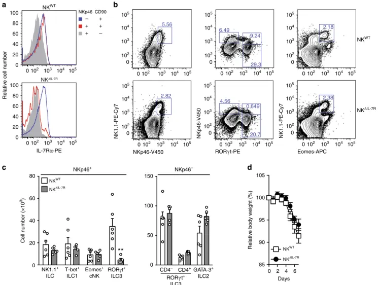
eliminate NKp46
+ILC3 from T/B-competent mice, we exploited
the strict IL-7R-dependence of NKp46
+RORγt
+ILC3 (Fig.
3
l). For
this purpose, conditional IL-7R (IL-7R
fl/fl) knockout mice
were crossed to NKp46
iCre-transgenic mice to generate NK
ΔIL-7Rmice. As compared to NK
WTmice, SI-derived helper-like
NKp46
+CD90
+ILC from NK
ΔIL-7Rmice hardly expressed any
IL-7Rα (CD127) while non-targeted NKp46
−CD90
+ILC still
expressed it at normal levels. As expected, NKp46
+CD90
−cNK
expressed least IL-7R in both mouse strains
32(Fig.
7
a). Next, we
determined the composition of the ILC pool in the SI of NK
WTand NK
ΔIL-7Rmice. NK
ΔIL-7Rmice were nearly completely
devoid of NKp46
+RORγt
+ILC3 (Fig.
7
b, c), while numbers of
NKp46
+Tbet
+ILC1 (Fig.
7
c), NKp46
+Eomes
+cNK (Fig.
7
b, c),
and all NKp46
−ILC subsets (Fig.
7
c) appeared normal compared
to NK
WTmice.
Next, we tested whether the lack of NKp46
+RORγt
+ILC3
affects the outcome of acute DSS colitis. For this purpose, NK
WTand NK
ΔIL-7Rmice were treated with DSS for 5 days and disease
parameters were determined. As shown in Fig.
7
d, relative body
weight did not differ between both mouse strains. Altogether, our
results demonstrate that the lack of NKp46
+RORγt
+ILC3 does
not affect the course of DSS-induced colitis.
cNK cells ameliorate acute intestinal inflammation. Since
NKp46
+ILC3 did not affect the course of DSS-induced colitis
(Fig.
7
d), we hypothesized that NKp46
+ILC1 deficiency
accounted for disease aggravation in NK
Δc-FLIPmice (Figs.
4
a–h
and
5
a–f). This assumption was supported by the fact that the
mild course of disease in NK
WTmice correlated with the
accu-mulation of c-FLIP-dependent Ly49C
+cNK (Supplementary
Fig. 3). In order to elucidate whether cNK are involved in disease
modulation, B6 mice were treated with cNK-depleting
anti-Asialo-GM1 (αAsialo) or control antibody. As shown in
Sup-plementary Fig. 4A,
αAsialo-treated mice suffered more from
DSS-induced colitis than control animals. Since the efficacy of
cNK depletion may vary between target tissues, we analyzed the
frequencies and absolute numbers of colonic ILC.
Anti-Asialo-treated mice contained significantly fewer NKp46
+NK1.1
+ILC
in the inflamed colon (Supplementary Fig. 4B, F, I). This
reduction was mainly due to the depletion of NKp46
+NK1.1
+
Eomes
+cNK (Supplementary Fig. 4C, G, J) and correlated with
elevated frequencies of neutrophils in peripheral blood
(Supple-mentary Fig. 4E), similar to what we had observed in the inflamed
colon of NK
Δc-FLIPmice (Fig.
4
l and m). On the contrary,
fre-quencies of NKp46
+Eomes
−ILC1 (Supplementary Fig. 4C) and
NKp46
+/−RORγt
+ILC3 (Supplementary Fig. 4D, H) remained
largely unaffected by
αAsialo treatment, whereas absolute
num-bers of the latter were significantly reduced (Supplementary
Fig. 4K). Hence, antibody-mediated side effects on other ILC
subsets could not be formally excluded.
As a result of NKp46-specific Eomes inactivation, cNK
development is strongly impaired while other ILC remain largely
unaffected
44–46. We therefore generated cNK-deficient NK
ΔEomes(NKp46
iCre× Eomes
fl/fl) mice to further validate the
anti-inflammatory function of cNK. NK
ΔEomesmice and control
animals were treated with DSS for 5 days. Similar to NK
Δc-FLIP(Fig.
4
a–d) and αAsialo-treated B6 mice (Supplementary Fig. 4A),
disease severity was significantly increased in NK
ΔEomesmice as
compared to NK
WTcontrols (Fig.
8
a–d). In accordance with a
more pronounced inflammatory response, colon weight of
NK
ΔEomesmice was increased (Fig.
8
e) while its length was
decreased (Fig.
8
f, g). Importantly, we confirmed the selective loss
of Eomes
+cNK, but not helper-like ILC1, in the inflamed colon
of NK
ΔEomesmice (Fig.
8
h, i). This correlated with the
accumulation of neutrophils (Fig.
8
j, k) similar to what we had
observed for NK
Δc-FLIPmice (Fig.
4
l, m). Thus, cNK depletion,
either by
αAsialo treatment (Supplementary Fig. 4) or
NKp46-specific Eomes inactivation (Fig.
8
), is sufficient to aggravate
disease. Hence, our results strongly suggest that cNK act as
immune
modulators
in
the
inflamed colon with
anti-inflammatory properties superior to all other NKp46
+ILC.
Discussion
Immune cells are equipped with a multitude of cytokine receptors
allowing them to sense changes in their environment and adapt
activation as well as survival thresholds accordingly
13,47. How
cytokine signals are translated into ILC-specific developmental/
survival programs is largely unknown. IL-15 is an environmental
Fig. 4 c-FLIP-dependent NKp46+ILC protect mice from acute intestinal inflammation. a–m NKWTand NKΔc-FLIPmice were treated with DSS for 5 days.a Relative body weight, b feces consistency and c the degree of intestinal bleeding were measured on a daily basis to calculate d the overall colitis score. At day 7,e the specific weight and f, g the length of the colon were determined. g, h Shown are representative photographs of g colon and cecum (scale unit= cm) as well as h H&E–stained tissue sections from NKWTand NKΔc-FLIPmice (n= 6/group) at day 7 (scale bars = 100 µm). i–m At day 7,
colonic lamina propria leukocytes were analyzed byflow cytometry. i–k After gating on Lin−CD45+cells, the different subsets of NKp46+(ILC: NK1.1+; ILC1: T-bet+, cNK: Eomes+; ILC3: RORγt+) and NKp46−ILC (ILC3: RORγt+; ILC2: GATA3+) were analyzed in eight mice per group.l, m Colon samples from two mice were pooled and Ly6C+/−Ly6G−monocyctes and Ly6C+Ly6G+neutrophils (PMN) were analyzed byflow cytometry (6–8 samples per group).i, l Representative contour plots are shown. Numbers indicate percentages. a–f, i–m Data were pooled from two independent experiments with a total of 15–16 mice per group. Data show means + SEM; *p ≤ 0.05; **p ≤ 0.005; ***p ≤ 0.001; ****p ≤ 0.0001 (two-tailed Mann–Whitney U test). Source data are provided as source datafile.
factor that is indispensable for ILC1/cNK development
25,26.
Previous studies demonstrated that IL-15 withdrawal impairs
cNK metabolism
48and activates the intrinsic apoptosis
path-way
15. In the absence of IL-15 cNK fail to downregulate the
pro-apoptotic molecule Bim. At the same time, the induction of the
anti-apoptotic Mcl-1 is impaired and IL-15-deprived cNK
undergo apoptosis
15.
Here, we provide evidence for an additional mode of
ILC-specific, non-redundant IL-15 action. We identify c-FLIP as a
target of STAT5-dependent IL-15 signaling. c-FLIP is a master
regulator of the extrinsic apoptosis pathway
20and protects
immune and non-immune cells from caspase 8-dependent
apoptosis
21,22. Usually, this type of apoptosis is triggered by
DLs such as TNF or TRAIL, which are produced at high levels by
b
g
a
0 5 10 15 20 25 30 35 40 45 50 0 1 2 3 4 5 6 7 8 DSS DSS DSS Colitis score NKΔc-FLIP NKWT Days 0 1 2 3 4 5 Histopathological score 0 50 100 150 MPO + cells (10hfp) NKΔc-FLIP NKWT 0 5 10 15 0.0 0.2 0.4 0.6 0 200 400 600 0 1 2 3 0 5 10 15pg/mg colonic tissue (day 3)
0.2 0.6 0.4 0.0 0.0 0.5 1.0 1.5 0.0 0.2 0.4 0.6 0.8 IFN-γ TNF 0.0 0.5 1.0 1.5 2.0 IL-10 IL-17A
IL-22 IL-12p70 IFN-β GM-CSF
0 100 200 300 400 500 IL-6
pg/mg colonic tissue (day 10)
0 1 2 3 4 5 IFN-γ 0 1 2 3 TNF 0 0 10 20 30 IL-10 0 2 4 6 8 10 IL-17A 0 5 10 15 20 IL-22 0.0 0.2 0.4 0.6 IFN-β 0.0 0.2 0.4 0.6 0.8 IL-12p70 0 50 100 150 200 GM-CSF MCP-1 MCP-1 200 400 600 0 0 200 400 600 IL-6 NKΔc-FLIP NKWT 5 6 7 8 9 Colon length (cm)
c
0 20 40 60 80Specific colon weight (mg/cm)
h
0 100 200 300 400 Number of GM-CSF + cells NKp46– CD90+ ILC NKp46+ CD90+ ILC CD90– cNK 0 200 400 600 800 1000 0 2000 4000 6000 ns NKΔc-FLIP NKWT NKΔc-FLIP NKWT NKΔc-FLIP NKWTe
f
d
Distal Medial Proximal Colon
mature ILC1
29. To prevent their DL-induced premature death in
the course of an immune response c-FLIP is required
30. Our
results demonstrate that immature ILC already require c-FLIP in
the very early phase of their development. This appears to be due
to the fact that developing ILC up-regulate effector genes such as
Ifng and Tnf already at the SP stage. This is paralleled by DR
up-regulation, particularly of TNF-R1, thus rendering BM ILC
sen-sitive to TNF-induced apoptosis. Consequently, only
c-FLIP-competent ILC precursors developed into DP ILC in vitro.
Importantly, the blockade of TNF partially restored the
genera-tion of c-FLIP-deficient DP ILC. Whether TNF producgenera-tion by
developing ILC is a cell autonomous process or whether it is
influenced by environmental factors remains to be shown.
T/B-deficient mice lacking NKp46
+ILC3 succumb to C.
rodentium infection. On the contrary, their T/B-competent
counterparts control infection
9,10. This demonstrates that
adap-tive immune cells and NKp46
+ILC have redundant functions
9,10,
a conclusion that is supported by human data
11. However,
NKp46
+ILC3 are indispensable for the maintenance of cecal
homeostasis
9. Hence, the relative contribution of NKp46
+ILC to
the modulation of immune responses appears to vary in a
context-dependent fashion. This is further exemplified by
apparently opposing results obtained in mouse models of
intestinal inflammation. While NKp46
+ILC3 protect
T/B-defi-cient mice from DSS-induced colitis
8, they promote
αCD40-induced colitis in T-deficient mice
10. Whether and how NKp46
+ILC affect the severity of DSS-induced colitis in T/B-competent
mice had not been studied in detail before. To address this issue,
we used ILC1/3-deficient, T/B-competent NK
Δc-FLIPmice. Our
data clearly demonstrate that NKp46
+ILC are indispensable for
disease control. Neither T or B lymphocytes nor NKp46
−ILC
were able to compensate for the lack of NKp46
+ILC in NK
Δc-FLIP
mice. Importantly, with the help of NK
ΔIL-7Rmice, we could
exclude NKp46
+ILC3 as major regulators of acute DSS-induced
colitis emphasizing the importance of NKp46
+ILC1. In
DSS-treated NK
Δc-FLIPmice, ILC-deficiency correlated with increased
disease scores, altered cytokine profiles and the accumulation of
neutrophils in the inflamed colon. Our data suggest that this
neutrophil-associated effect was due to the lack of cNK, which
can limit the recruitment and pro-inflammatory function of
neutrophils
49. This anti-inflammatory function of cNK is
NKG2A-dependent as shown by the fact that NKG2A
anti-bodies block cNK–neutrophil interactions in vitro and exacerbate
DSS-induced colitis in vivo. Based on these observations, a
dominant regulatory role of cNK in DSS-induced colitis was
postulated
49. However, NKG2A is not only expressed by cNK but
also by, e.g. ILC1
26and CD8
+T cells
50. Hence disease
exacer-bation in response to anti-NKG2A treatment
49may involve
multiple NKG2A
+immune cell types. However, as we have
shown here, (i) the mild course of disease in NK
WTmice
correlated with the accumulation c-FLIP-dependent Ly49C
+cNK, (ii)
αAsialo-GM-mediated cNK depletion aggravated
dis-ease in B6 mice and, above of all, cNK-deficient NK
ΔEomesmice
were by far more sensitive to DSS-induced colitis as compared to
controls. Of note, disease aggravation was comparable for
cNK-deficient NK
ΔEomesand NK
Δc-FLIPmice lacking all NKp46
+ILC.
Hence, our
findings support the view that cNK have a dominant
anti-inflammatory function in DSS-induced colitis, which results,
at least partially, from the restriction of neutrophil infiltration and
function in the inflamed colon
51. However, it is important to
emphasize that the cell surface molecules used to define
neu-trophils in our experimental system are used to define
myeloid-derived suppressor cells (MDSCs) in others
52. In addition to their
nearly identical cell surface phenotype, MDSCs and neutrophils
share functional features as well. For example, and contrary to
their well-known anti-inflammatory functions in tumor models
53,
MDSCs were shown to promote DSS-induced colitis
54similar to
neutrophils
49. Hence, our experimental approach does not allow
us to define the relative contribution(s) of MDSCs and
neu-trophils to disease progression. Based on the complex intercellular
interactions driving colitis
55we also cannot exclude that cNK
deficiency promotes pro-inflammatory functions of other
cell types.
NK
Δc-FLIPmice developed maximum disease scores after only a
single phase of DSS administration. Furthermore, they failed to
recover completely after the
first DSS cycle and showed maximum
disease scores after each additional DSS phase. This early
estab-lishment of chronic disease in NK
Δc-FLIPmice correlated with the
prolonged reduction of tissue-protective GM-CSF and lower
numbers of GM-CSF
+NKp46
+ILC including cNK. Hence, the
protective effect of cNK may not only rely on the suppression of
neutrophil effector functions but also on the production of
tissue-protective GM-CSF. It is important to stress that no other immune
cell type, including NKp46
+ILC1/3, is able to compensate for the
early protective and regenerative effects of NKp46
+cNK. This
may, at least partially, rely on the disease-related control of the
commensal microbiota. Of note, the lack of NKp46
+ILC in NK
Δc-FLIP
mice did not have a significant impact on the composition of
the commensal microbiota prior to DSS-induced colitis. This
argues against a direct impact of NKp46
+ILC on the
establish-ment of the microbiota under homeostatic conditions in
T/B-competent mice. Nevertheless, after establishment of chronic
colitis the composition of the commensal microbiota changed in a
NKp46
+ILC-dependent fashion. For example, Coriobacteriaceae,
Alcalignaceae, Erysipelotrichaceae, and Bacteroidaceae were more
abundant in NK
Δc-FLIPmice, while Rikenellaceae, Bacteriodales
S24-7, Prevotellaceae, and Ruminococcaceae were enriched in
NK
WTmice. Commensal dysbiosis is frequently observed in
dif-ferent disease models and the altered abundance of certain
bac-teria has disease-modulating effects
37,38. For example, the
Fig. 5 The lack of c-FLIP-dependent NKp46+ILC is associated with the early onset of chronic intestinal inflammation. a–h NKWTand NKΔc-FLIP
littermates were treated with DSS for 5 days followed by a 14-day recovery phase on normal drinking water. This treatment was repeated twice.a Shown are representative results (mean colitis score ± SEM) of one out of two independent experiments with 11–13 mice per group. b–f At day 42, b colon length andc specific colon weight were determined, d–f tissue samples from distal, medial, and proximal colon were collected and d, e stained with H&E (scale bars= 100 µm) to determine e histopathological scores (HPS). f For each colon segment, myeloperoxidase (MPO)+cells were enumerated in 10 high powerfields (magnification ×400). e, f Each symbol represents mean values for individual mice after pooling results from the three colon segments. b, c, e, f Data show mean+/± SEM for the respective experimental groups. b–f 5–7 mice were analyzed. g At experimental days 3 (upper row) and 10 (lower row), colon samples were isolated and incubated for 24 h. Cytokine levels were determined in culture supernatants and normalized to tissue mass (n= 11–13/group). h At day 5, frequencies of GM-CSF-producing cells isolated from the colonic lamina propria were determined by flow cytometry. After gating on Lin−CD45+cells, GM-CSF production was analyzed for NKp46+CD90−cNK as well as NKp46+/−CD90+helper-like ILC. Pooled results (mean+ SEM) from four independent experiments with a total of seven colon samples (pooled from 2 to 3 mice) per group are shown. Statistical significances were determined using a, e, f, g, h two-tailed Mann–Whitney U and b, c two-tailed unpaired Student’s t test (*p ≤ 0.05; **p ≤ 0.005; ***p≤ 0.001; ****p ≤ 0.0001; ns = not significant). a–c and e–h Source data are provided as source data file.
overabundance of Bacteroides has been associated with increased
rates of inflammation and tumor development
42. Moreover, the
expansion of bacteria from the family Alcaligenaceae drives
inflammation in ILC-depleted RAG1
−/−mice
43. On the contrary,
the enhanced relative abundance of Prevotella in diseased NK
WTmice may have beneficial effects, since it correlates inversely
with inflammation in a mouse model of inflammation-induced
carcinogenesis
42. Altogether, these results demonstrate that the
Fig. 6 The lack of c-FLIP-dependent NKp46+ILC is associated with disease-related alterations in microbiome composition. a–f Feces samples of NKWT
and NKΔc-FLIPlittermates (Fig.5) were collected before DSS administration (before DSS) and at the end of the observation period (after DSS, d49). Whenever possible, mice shared cages from birth on and throughout the experiments. Analyzed samples (n= 10–13/group and time point) had a minimum sequencing depth of 1000 reads and a mean sequencing depth of 19,298.3 ± 7781.349 reads (mean ± SD).a–c Non-metric multidimensional scaling (NMDS) ordination analysis of fecal microbiota composition was performed using Bray–Curtis distances grouped by a genotype and treatment or b, c genotype b before and c after DSS-induced colitis. Individual effect size of tested covariates is indicated. a–c To calculate the variance explained by individual factors such as genotype, cage effect, and treatment a permutational multivariate analysis of variance (ADONIS) was used. *p < 0.05; **p < 0.01; ***p < 0.001. A significant effect was dedicated when p < 0.05 and R2> 0.10 (equivalent to 10% of explained variance).d, e Relative abundance of the
average microbiome compositiond before and e after DSS administration was determined on the family level. Phylum and families are indicated. f Statistically significant differences on family levels in fecal microbiota composition between NKWTand NKΔc-FLIPmice. Data were analyzed using linear
discriminant analysis (LDA) effect size (LEfSe) method (Kruskal–Wallis test with p < 0.05 and LDA scores > 3.0). Data are displayed as bar plot ranked according to their effect size and associating them to the genotype of the mice.a–f Source data are provided as source data file.
expansion of disease-promoting microbes, such as Proteobacteria,
i.e. Alcaligenaceae, occurs selectively in DSS-treated NK
Δc-FLIPmice. This indicates that c-FLIP-dependent NKp46
+ILC
mod-ulate the composition of the commensal microbiota under
inflammatory conditions but not prior to disease. Whether
alterations of the commensal microflora are a cause or a result of
inflammation remains an important question for the future.
In summary, we identify the anti-apoptotic molecule c-FLIP as
a target of cytokine-induced STAT5 activation, which is
indis-pensable for the development of IL-15- and IL-7-dependent
NKp46
+ILC1 and ILC3, respectively. Furthermore, we provide
evidence that cNK, but not ILC1/3, restrict the degree of intestinal
inflammation correlating with specific alterations in the
com-mensal microbiota. Hence, c-FLIP-dependent NKp46
+cNK
protect T/B-sufficient mice from intestinal inflammation, a
function that cannot be replaced by any other immune cell.
Methods
Mice. NKp46iCre19, c-FLIPfl/fl23, STAT5fl/fl56(provided by L. Hennighausen),
IL-15−/−25, IFN-γ reporter mice (Great)57, RAG1−/−58, IL-7R−/−59, and
RAG1−/−× IL-7R−/−were maintained under specific pathogen-free conditions at
the central animal facility of the Medical Faculty of the Otto-von-Guericke-University Magdeburg. IL-7Rfl/fl(stock no. 022143)60, Eomesfl/fl(stock no.
017293)61, and IL-15Rα−/−mice (stock no. 003723)31including appropriate
control mice were purchased from The Jackson Laboratory. Whenever possible, control littermates were used and cohoused (age 8–16 weeks). Both sexes were considered equally. Experimental procedures were approved by the relevant animal experimentation committee and performed in compliance with international and local animal welfare legislations (Landesverwaltungsamt Sachsen-Anhalt, permit numbers AZ 42502-2-1202 and AZ 42502-2-1521 Uni MD).
Cell isolation. Mice were used euthanized by isoflurane inhalation overdose. Single-cell suspensions of spleen were prepared by forcing the organs through metal sieves. BM leukocytes wereflushed from tibia, femur, and coxal bone using PBS/2 mM EDTA (ROTH). To lyse erythrocytes, cell suspensions were incubated with ACK lysis buffer for 90 s and subsequent addition of RPMI 1640 (Biochrome) with 10% FCS (PAN Biotech). Cells resuspended in PBS/2 mM EDTA werefiltered through 40 µm cell strainers (Corning, Durham, NC, USA).
LPL were isolated from different parts of the intestine according to published protocols2,62. Briefly, residual mesenteric fat and Peyer’s Patches were carefully
removed. Intestinal tissue was opened longitudinally and washed two to three times in ice-cold PBS with 100 U/ml penicillin/streptomycin (P/S; GIBCO Life Technologies). The epithelial layer and containing intraepithelial leukocytes were removed by two incubation steps in Hank’s balanced salt solution without Ca2+
and Mg2+(HBSS; Biochrom) supplemented with 5 mM EDTA, 10 mM HEPES and P/S for 20 min at RT with slow rotation (100 rpm) and subsequent vortexing
CD90 NKp46 + + + – + – Relativ e cell n umber
a
Days 0 2 4 6 85 90 95 100 105 Relativ e body w eight (%)d
b
5.56 2.82 9.24 29.3 6.49 0.649 4.56 2.18 2.38RORγ t-PE Eomes-APC NKp46-V450 NK1.1-PE-Cy7 NKp46-V450 20.7 NK1.1-PE-Cy7 NKΔIL-7R IL-7Rα-PE 0 20 40 60 80 100 NKWT 0 20 40 60 80 100 0 20 40 60 80 Cell n umber (×10 3) NKp46+ NKp46–
c
0 50 100 150**
GATA-3+ ILC2 CD4– CD4+ RORγ t+ ILC3 NKWT NKΔIL-7R 0 102 103 104 105 0 102 103 104 105 0 102 103 104 105 0 102 103 104 105 0 102 103 104 105 0 102 103 104 105 0 102 103 104 105 0 102 103 104 105 0 102 103 104 105 0 102 103 104 105 0 102 103 104 105 0 102 103 104 105 0 102 103 104 105 0 102 103 104 105 NK1.1+ ILC Eomes+ cNK T-bet+ ILC1 RORγt+ ILC3 NKWT NKΔIL-7R NKWT NKΔIL-7RFig. 7 IL-7R-dependent ILC3 do not affect acute colitis. a–c Small intestinal lamina propria leukocytes were isolated from NKWTand NKΔIL-7Rmice. Cells
were analyzed byflow cytometry as described in Fig.3.a Overlays show relativefluorescence intensities of IL-7Rα (CD127) for the indicated ILC. Data are representative for four mice per genotype.b Shown are representative contour plots and c pooled results (means+ SEM) from two independent experiments with 5–6 mice per group. Four to six samples were analyzed. d To induce acute colitis, NKWTand NKΔIL-7Rmice were treated with DSS for
5 days as described in Fig.4. Relative body weights were determined on a daily basis. Pooled results (means ± SEM) from two independent experiments with 17–18 mice per group are shown. c, d Source data are provided as source data file; *p ≤ 0.05; **p ≤ 0.005; ***p ≤ 0.001; ****p ≤ 0.0001 (two-tailed Mann–Whitney U test).