HAL Id: hal-00423750
https://hal.archives-ouvertes.fr/hal-00423750
Submitted on 30 May 2020
HAL is a multi-disciplinary open access
archive for the deposit and dissemination of
sci-entific research documents, whether they are
pub-lished or not. The documents may come from
teaching and research institutions in France or
abroad, or from public or private research centers.
L’archive ouverte pluridisciplinaire HAL, est
destinée au dépôt et à la diffusion de documents
scientifiques de niveau recherche, publiés ou non,
émanant des établissements d’enseignement et de
recherche français ou étrangers, des laboratoires
publics ou privés.
Copyright
A 20-amino acid module of protein kinase Cepsilon
involved in translocation and selective targeting at
cell-cell contacts.
Barthélémy Diouf, Alejandra Collazos, Gilles Labesse, Françoise Macari,
Armelle Choquet, Philippe Clair, Cécile Gauthier-Rouvière, Nathalie C.
Guérineau, Philippe Jay, Frédéric Hollande, et al.
To cite this version:
Barthélémy Diouf, Alejandra Collazos, Gilles Labesse, Françoise Macari, Armelle Choquet, et al.. A
20-amino acid module of protein kinase Cepsilon involved in translocation and selective targeting at
cell-cell contacts.. Journal of Biological Chemistry, American Society for Biochemistry and Molecular
Biology, 2009, 284 (28), pp.18808-15. �10.1074/jbc.M109.004614�. �hal-00423750�
A 20-Amino Acid Module of Protein Kinase C
⑀ Involved in
Translocation and Selective Targeting at Cell-Cell Contacts
*
□SReceived for publication, December 29, 2008, and in revised form, April 9, 2009 Published, JBC Papers in Press, May 8, 2009, DOI 10.1074/jbc.M109.004614
Barthe´le´my Diouf‡1,2, Alejandra Collazos‡1,3, Gilles Labesse§, Franc¸oise Macari‡, Armelle Choquet‡, Philippe Clair¶, Ce´cile Gauthier-Rouvie`re储, Nathalie C. Gue´rineau**, Philippe Jay‡, Fre´de´ric Hollande‡4, and Dominique Joubert‡5 From the‡CNRS UMR5203, INSERM, U661, University of Montpellier I and II, and the Department of Cellular and Molecular Oncology, Institut de Ge´nomique Fonctionnelle, 141, rue de la Cardonille, F-34094 Montpellier Cedex 5, France, the **Department of Endocrinology, Institut de Ge´nomique Fonctionnelle, 141, rue de la Cardonille, F-34094 Montpellier Cedex 5, France,§Centre de Biologie Structurale, 29, rue de Navacelles, CNRS UMR5048, INSERM, U554, University of Montpellier I, F-34094 Montpellier Cedex 5, France, ¶Plateforme qPCR Haut De´bit, Universite´ Montpellier II, 34095 Montpellier, France, and储Centre de Recherche de Biochimie Macromole´culaire-CNRS Formation de Recherche 2593, 1919, route de Mende, 34293 Montpellier Cedex 5, France
In the pituitary gland, activated protein kinase C (PKC) iso-forms accumulate either selectively at the cell-cell contact (␣ and⑀) or at the entire plasma membrane (1 and␦). The molec-ular mechanisms underlying these various subcellmolec-ular locations are not known. Here, we demonstrate the existence within PKC⑀ of a cell-cell contact targeting sequence (3CTS) that, upon stim-ulation, is capable of targeting PKC␦, chimerin-␣1, and the PKC⑀C1 domain to the cell-cell contact. We show that this selective targeting of PKC⑀is lost upon overexpression of 3CTS fused to a (R-Ahx-R)4(where Ahx is 6-aminohexanoic acid) vec-torization peptide, reflecting a dominant-negative effect of the overexpressed 3CTS on targeting selectivity. 3CTS contains a putative amphipathic␣-helix, a 14-3-3-binding site, and the Glu-374 amino acid, involved in targeting selectivity. We show that the integrity of the␣-helix is important for translocation but that 14-3-3 is not involved in targeting selectivity. However, PKC⑀translocation is increased when PKC⑀/14-3-3 interaction is abolished, suggesting that phorbol 12-myristate 13-acetate activation may initiate two sets of PKC⑀ functions, those depending on 14-3-3 and those depending on translocation to cell-cell contacts. Thus, 3CTS is involved in the modulation of translocation via its 14-3-3-binding site, in cytoplasmic dese-questration via the␣-helix, and in selective PKC⑀targeting at the cell-cell contact via Glu-374.
Activation of cytoplasmic kinases often induces their target-ing to various subcellular locations where they phosphorylate their substrates and exert their biological functions. Represent-ative examples of proteins for which targeting involves complex
and various molecular mechanisms are provided by the protein kinase C (PKC)6family, which comprises 10 known isoforms, displaying ubiquitous, tissue- or cell type-specific expression and playing crucial roles in signal transduction (1, 2). Depend-ing on the cell type and the stimulus, various inactive cytoplas-mic PKC isoforms may, upon activation, associate with the plasma, Golgi, or nuclear membranes (3–5). Even within a given cell type, a particular isoform can be targeted and accu-mulated at various subcellular locations (6, 7), and these pro-cesses involve direct interaction with phospholipids or other proteins (8, 9).
In pituitary GH3B6 cells, PKC isoforms accumulate at differ-ent subcellular locations upon phorbol 12-myristate 13-acetate (PMA) stimulation or thyrotropin-releasing hormone (TRH) receptor activation (10, 11). Activated PKC␣ and -⑀ accumulate selectively at cell-cell contacts, whereas PKC1 and -␦ are detected along the entire plasma membrane. The selective par-titioning of specific PKC isoforms at cell-cell contacts is not restricted to the GH3B6 cell line. It was also observed in blas-tocysts (12), in the pituitary gland (11), at heterotypic contacts between fibroblasts and epithelial cells (13), at the interface between macrophages and IgG-coated beads (14), and at the immunological synapse (15–17). Although the molecular mechanism underlying this partitioning remains largely unknown, an interesting clue was provided by the discovery in human pituitary and thyroid tumors of a natural PKC␣ D294G mutant (18, 19), which is devoid of cell-cell contact targeting selectivity (20). A similar loss of selectivity is found when an E374G mutation is introduced in PKC⑀ (11), indicating that the Asp-294 and Glu-374 amino acids located within the V3 region of PKC␣ and ⑀, respectively, are essential for proper targeting after activation. Interestingly, the PKC␣ D294G mutant was also shown to be a loss-of-function mutant (21). However, because replacing Phe by Glu in the corresponding position does not induce the targeting of PKC␦ to the cell-cell contact, it is likely that other amino acids are required for cell-cell contact targeting selectivity.
*This work was supported by the Ministe`re de la Recherche et de la Tech-nologie and by Association pour la Recherche contre le Cancer Grant 5695. □S
The on-line version of this article (available at http://www.jbc.org) contains
supplemental “Experimental Procedures” and Figs. 1– 4.
1Both authors contributed equally to this work.
2Supported by a grant from the Association pour la Recherche contre le
Can-cer. Present address: Dept. of Pharmaceutical Sciences, St. Jude Children’s Research Hospital, 262 Danny Thomas Pl., Memphis, TN 38105.
3Present address: Protein Phosphorylation Laboratory, London Research
Inst., Cancer Research UK, London WC2A 3PX, UK.
4To whom correspondence may be addressed. Tel.: 33-467-668-144; Fax:
33-467-542-432; E-mail: [email protected].
5To whom correspondence may be addressed. Tel.: 33-467-142-818; Fax:
33-467-542-432; E-mail: [email protected].
6The abbreviations used are: PKC, protein kinase C; aa, amino acid(s); Ahx,
6-aminohexanoic acid; GFP, green fluorescent protein; HPLC, high pres-sure liquid chromatography; PMA, phorbol 12-myristate 13-acetate; RhoB, rhodamine B; TRH, thyrotropin-releasing hormone; aa, amino acid.
at INRA Institut National de la Recherche Agronomique on August 4, 2015
http://www.jbc.org/
In the present work, we sought to deepen our understanding of the requirements for efficient targeting to the cell-cell con-tact by focusing our analysis on the sequence surrounding posi-tion Asp-294 of PKC␣ and Glu-374 of PKC⑀. On the basis of isoform sequence comparison, we identified a 20-aa stretch in the V3 region of PKC⑀ that includes Glu-374 and contains one of the two 14-3-3-binding sites of PKC⑀ and a putative amphipathic␣-helix. This 20-aa module fulfills the criteria of a cell-cell contact targeting sequence, and we therefore propose to name this sequence 3CTS.
EXPERIMENTAL PROCEDURES
Materials—Plasmids containing the full-length PKC1, -⑀,
and -␦ cDNAs were provided generously by P. J. Parker (Protein Phosphorylation Laboratory, London Research Institute, Can-cer Research UK, London, WC2A 3PX, UK). The plasmid con-taining the full-length PKC␥ was generously provided by Dr. N. Saito (Laboratory of Molecular Pharmacology, Biosignal Research Center, Kobe University, Kobe, Japan). Monoclonal anti-pan14-3-3 was purchased from Santa Cruz Biotechnology, and polyclonal anti-PKC⑀ used in Western blotting was from Euromedex. Mouse monoclonal anti-PKC⑀ used for immuno-precipitation experiments was purchased from Pharmingen. Mouse monoclonal anti-green fluorescent protein (GFP) was purchased from Roche Applied Science. Anti-pSer antibody was purchased from Zymed Laboratories Inc.
Construction of Plasmids, Site-directed Mutagenesis, RNA Extraction and Reverse Transcription-PCR, Cell Culture, Transfection, and Western Blotting—Experimental procedures and primer sequences are described in the supplementary “Experimental Procedures”.
Vectorization of 3CTS and the Scramble Peptide—Two pep-tides were synthesized using standard automated solid-phase N-(9-fluorenyl)methoxycarbonyl (Fmoc)/tert-butyl chemistry and purified on a reverse phase HPLC C18 column to 98% purity. Their molecular weight was checked by mass spectrom-etry. The vector peptide H-Ahx-R-Ahx-RR-Ahx-RR-Ahx-RR-Ahx-R-NH2was coupled to the C terminus of 3CTS or a scram-ble peptide, with both peptides including 3 additional aa of the PKC⑀ sequence (to maintain a distance between Glu-374 and the vector peptide) and a cysteine to allow the chemical reac-tion for the linkage with the peptide vector. Rhodamine B (RhoB) was added to the N terminus of both constructs, yield-ing a RhoB-LKELENNIRKALSFDNRGEEHRA-C-vector pep-tide (for 3CTS) or a RhoB-GEHDFLKERLKNIREAESNRLAN-C-vector peptide complex (scramble peptide). Coupling of the vector peptide with the other peptides was achieved in a one-pot two-step reaction using the heterobifunctional linker N-succinimidyl 3-maleimidopropionate (Fluka 63179) in dry N,N-dimethylformamide, in the presence of N,N-diisopropyl-ethylamine. The N-terminal function of the vector peptide was first reacted, resulting in maleimidopropionyl-GPS1168. Addi-tion of either 3CTS or the control peptide resulted in the for-mation of a thioether linkage between the thiol of their cysteine and the maleimide moiety. The progression of each step of the coupling process was monitored by analytic reverse phase C18 HPLC. The resulting coupling products were purified to 98% on a reverse phase C18 HPLC column, aliquoted, and lyophilized.
Immunoprecipitation—For immunoprecipitation, anti-PKC⑀, anti-GFP, or anti-IgG was cross-linked on protein G Plus-aga-rose beads. Fourg of each antibody was cross-linked to 50 l of protein G Plus-agarose beads with dimethyl pimelimidate. GH3B6 cells were washed twice with phosphate-buffered saline, harvested, resuspended in lysis buffer (50 mMTris, pH
7.5, 150 mM NaCl, 2 mM EGTA, 1% Nonidet P-40, 0.25% sodium deoxycholate, 1 mMsodium orthovanadate, 10 mM
NaF, 10 mM -glycerophosphate) containing a mixture of
protease inhibitors, and incubated for 45 min at 4 °C. Cell lysates were prepared from cells incubated or not with PMA and centrifuged for 10 min at 13,000 rpm. Supernatants were collected. Aliquots of supernatants containing 500 g of proteins were precleared with 50l of protein G Plus-agar-ose beads for 30 min at 4 °C. Precleared supernatants were then incubated with 50l of PKC⑀-, GFP-, or anti-IgG-coupled beads overnight at 4 °C. Beads were then washed three times with lysis buffer and two times with 50 mMTris, pH 7.5, 150 mMNaCl. Bound material was eluted
with Laemmli buffer for SDS-PAGE.
Real-time Fluorescence Microscopy—Real time recording was performed as described (22). At the time of observation, the culture medium was replaced by a prewarmed buffer, at 37 °C, composed of 140 mMNaCl, 5 mMKCl, 2 mMCaCl2, 2 mMMgCl2, 10 mMHepes, 6 mMglucose, pH 7.4. Images were
acquired continuously, with intervals between frames of 0.533 s.
Modeling—Fold compatibility for the full-length and trun-cated sequences of the human PKC⑀ sequence was searched using the meta-server @TOME (23). Domain organization was refined using fold recognition results. Automatic modeling of recognized domains was performed with SCWRL (24) and MODELLER (25), and the validity of the resulting models was evaluated with PROSA (26) and Verify3D (27). Fold recognition was resumed for the variable regions. Improved three-dimen-sional models were built using MODELLER 7.0 with the loop optimization procedure.
RESULTS
Different V3 Domain Sequences and Localizations of PKC␣, -⑀, -␦, -1, and -␥
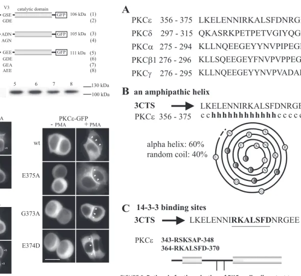
Previous studies showed that, in the V3 region of PKC␣ and PKC⑀, respectively, the GDE or GEE motif is essential but not sufficient for selective cell-cell contact targeting. Accordingly, replacing GFE by GEE in the PKC␦ V3 domain does not result in a selective cell-cell contact targeting of a PKC␦-GFP fusion protein (11). To improve our understanding of the molecular mechanisms underlying PKC targeting to the cell-cell contact, we compared the V3 sequences of PKC␣, ⑀, and ␦ (11) with those of PKC1 and -␥ (Fig. 1A) and analyzed PKC1 and -␥ localization when fused to GFP.
In TRH-stimulated GH3B6 cells, PKC1-GFP translo-cated to the entire plasma membrane, including the cell-cell contact region (Fig. 1C). Changing the GSE of PKC1 into the GDE of PKC␣ induced its selective localization at cell-cell contacts (Fig. 1C). We then analyzed the behavior of PKC␥ (Fig. 1A), an isoform that, when activated, selectively
PKC
⑀ Cell-Cell Contact Targeting
at INRA Institut National de la Recherche Agronomique on August 4, 2015
http://www.jbc.org/
localizes at cell-cell contacts but that is not naturally expressed in GH3B6 cells. Like PKC␣, it possesses an Asp in position 294, but, unlike PKC␣, the motif reads ADN instead of GDE. Here, we show that the targeting selectivity of PKC␥-GFP was lost when ADN was changed to AGN (Fig. 1C), suggesting that only the central Asp or Glu within the ADN, GDE, or GEE motif is essential for selectivity. Accord-ingly, activated PKC⑀ still localized at cell-cell contacts after changing the GEE motif into AEE (G373A) or GEA (E375A), and the Glu-374 of PKC⑀ can be replaced by an Asp (as in PKC␣) without affecting the selective targeting to cell-cell contacts (Fig. 1C). The genuine nature of PKC⑀ translocation at the actual cell-cell contact upon activation was then con-firmed by costaining with p120-catenin, a marker of
adher-ens junctions (supplemental Fig. 1A). Indeed, both proteins colocalized at the cell-cell contacts.
In Search of 3CTS, the Cell-Cell Contact Targeting Sequence
Identification of a Candidate 20-Amino Acid Sequence in the PKC⑀ V3 Region—Glu-374 and Asp-294 are necessary but not sufficient to target PKC⑀ and -␣ to cell-cell contacts (11), imply-ing that neighborimply-ing amino acids could be important. We first compared the amino acid sequence close to the Glu-374 or to the corresponding amino acid of various PKC isoforms (Fig. 2A) and detected putative structural motives very close to the Glu-374 of PKC⑀: a putative amphipathic ␣-helix (aa 358–369) (see the Network Protein Sequence Analysis server) (Fig. 2B) and a binding site for the 14-3-3 protein (aa 364 –370) (Fig. 2C) (see the ELM server). PKC⑀ possesses another 14-3-3-binding site, in position 343–348 (Fig. 2C), in agreement with the fact that 14-3-3 generally dimerizes to achieve efficient binding. The 356 –375 sequence of PKC⑀ thus includes the amino acid Glu-374 essential for cell-cell contact targeting selectivity, a putative amphipathic␣-helix, and one of the two 14-3-3-bind-ing sites present in PKC⑀ (28).
FIGURE 1. Targeting of PKC1, -␥, and -⑀ and their different mutants in
GH3B6 cells. Various point mutations were performed in PKC1 (GSE
mutated into GDE), PKC␥ (ADN mutated into AGN) and PKC⑀ (GEE mutated into GEA, AEE, or GDE). GH3B6 cells were transiently transfected with the various GFP-tagged constructs (A). All constructs were translated at their expected size (B). Gels were loaded with 15g of proteins. Two days after transfection, cells were treated or not with 100 nMPMA for 30 min, and obser-vations were performed with conventional microscopy with an Axiophot 2.0 from Zeiss (C). The scale bars represent 5m. Note that targeting is consid-ered to be selective for the cell-cell contact (e.g. wild-type (wt) PKC⑀) when there is no accumulation anywhere else along the cell membrane. The seem-ingly higher staining detected at the cell-cell contact for PKC isoforms that also translocate along the whole membrane (e.g. wile-type PKC1) is because these isoforms are present along both apposed cell membrane constituting the contact. A plot profile is shown insupplemental Fig. 1Bto describe a selective versus a nonselective targeting to the cell-cell contact, as we already reported (10).
FIGURE 2. Rationale for the selection of PKC⑀ cell-cell contact targeting
sequence. A, sequences of PKC⑀, ␦, -␣, -1, and -␥ upstream of the D(E) amino
acid. B, amphipathic␣-helix of PKC⑀, and as a comparison, its absence in PKC␣. The amphipathic ␣-helix of PKC⑀ is characterized by a hydrophobic residue every 3 or 4 amino acids and by the opposed distribution of the hydrophobic and charged amino acids in the helix. Dark circles represent hydrophobic residues. C, position of the 14-3-3-binding sites.
at INRA Institut National de la Recherche Agronomique on August 4, 2015
http://www.jbc.org/
The PKC⑀ 356–375 Sequence Is a 3CTS—To determine
whether the 356 –375 sequence of PKC⑀ contains the essential molecular determinants for selective cell-cell contact targeting, it was introduced into the V3 region of PKC␦, fused to the C1 region of PKC⑀, or inserted into chimerin-␣1, a non-PKC
pro-tein normally localized in the Golgi in the presence of PMA. These constructs were fused to GFP. Controls consisted of the 356 –375 sequence containing the E374G mutation (mut 3CTS) and of the 17 aa downstream of the GEE motif (cont) (Fig. 3A). Although PKC␦-GFP translocated to the entire plasma mem-brane upon activation, including the cell-cell contact, the chi-meric 3CTS-PKC␦-GFP selectively translocated to cell-cell contacts, suggesting that this 20-aa PKC⑀ sequence probably contains the essential molecular determinants for cell-cell con-tact targeting (Fig. 3B). Indeed, when the inserted 20-aa sequence was mutated (E374G,⫹mut 3CTS) or replaced by the 17 aa found downstream of the GEE motif in wild-type PKC⑀ (⫹cont), the resulting PKC␦ chimeras translocated to the entire plasma membrane upon activation, similar to wild-type PKC␦. It is of note that wild-type PKC␦ and PKC␦ bearing the control 3CTS also accumulated in the perinuclear region. In addition, the C1–3CTS-GFP fusion protein was found to translocate and accumulate selectively at the cell-cell contact upon PMA acti-vation, in contrast with the same fusion protein bearing the E374G mutation, the construct bearing the control sequence, or C1-GFP, which all accumulated indiscriminately along the entire cell membrane (Fig. 3B).
Finally, in the presence of PMA, chimerin-␣1 bearing the 20-aa sequence partially accumulated at cell-cell contacts, whereas the control sequence did not alter chimerin-␣1 local-ization (Fig. 3C). Thus, the 20-aa sequence does behave as a selective targeting sequence regardless of the protein it is fused to, and we consequently propose to call it 3CTS for cell-cell contact targeting sequence.
It is of note that, in agreement with data showing that the V3 region does not contain any phosphatidylserine-binding site, 3CTS is not by itself capable of accumulating at cell-cell con-tacts (29, 30). When fused to GFP alone, the resulting construct is only able to accumulate transiently and very weakly at cell-cell contacts (supplemental Fig. 2D).
Dominant-negative Effect of 3CTS Overexpression—To assess the potential role of 3CTS in the regulation of targeting selectivity, we analyzed whether exogenous 3CTS could disrupt the PKC⑀ translocation process. To deliver intracellularly the 3CTS sequence, RhoB-3CTS was chemically linked to the pep-tide vector sequence (R-Ahx-R)4already used for the vectoriza-tion of oligonucleotides (31). Cells were incubated in the pres-ence of 2.5MRhoB-3CTS-(R-Ahx-R)4for 30 min. A 3CTS
scramble peptide also linked to (R-Ahx-R)4was used as a con-trol. As shown in Fig. 4A, 3CTS entered the cells only when fused to (R-Ahx-R)4, and incubating cells with RhoB-3CTS-(R-Ahx-R)4abolished targeting selectivity but not translocation (Fig. 4B), meaning that 3CTS is indeed essential for specifying the location of activated PKC⑀ in pituitary cells. The control sequence did not affect targeting selectivity.
PKC⑀ and 3CTS Interact with 14-3-3 in Pituitary Cells
14-3-3 adaptor proteins are conserved polypeptides that mediate the cellular effects of many protein kinases through their ability to bind specific serine- or threonine-phosphoryla-ted peptide motifs (32). Recently, phosphorylation sites within PKC⑀, which control its association with 14-3-3, have been identified (28, 33). It was demonstrated that association of FIGURE 3. 3CTS is a targeting sequence. A, 3CTS bearing or not the
sub-stitution of Glu-374 by Gly (mut 3CTS) and a control sequence consisting of GEE plus the 17 amino acids downstream of the GEE (cont) were intro-duced in PKC␦-GFP, in C1-GFP, and in chimerin-␣1-GFP. The PshAI site gacagctgtc in PKC␦ was in positions 951–960; the Pm1I site cac/gtg in chimerin-␣1 was in positions 406 – 411. All proteins were expressed at their expected size (supplemental Fig. 3). Two days after transient trans-fection, GH3B6 cells were observed as in Fig. 1, in the presence or absence
of PMA. B, the translocation of PKC␦-GFP (upper) and C1-GFP (lower) with
3CTS, mut 3CTS, or the control sequence is shown. C, the translocation of
chimerin-␣1-GFP with 3CTS or the control sequence is shown.
PKC
⑀ Cell-Cell Contact Targeting
at INRA Institut National de la Recherche Agronomique on August 4, 2015
http://www.jbc.org/
PKC⑀ with 14-3-3 is essential for the completion of cytokinesis. To determine whether the 14-3-3-binding site located within 3CTS is involved in the translocation of PKC⑀, we first built an
in silicomodel of the interaction between 3CTS and 14-3-3 and then tested it by characterizing functionally the interaction between PKC⑀ or 3CTS and 14-3-3.
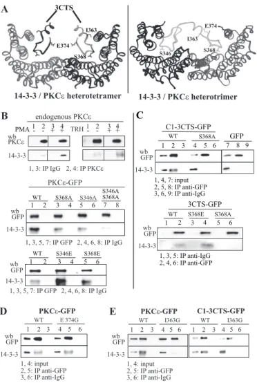
Modeling the Interaction between 3CTS and 14-3-3—The model was derived from the crystal structure Protein Data Bank code 1ib1 of the 14-3-3-serotonin N-acetyltransferase com-plex (34). This structure contains tetramers of 14-3-3-acetyl-transferase with one-to-one interactions involving mainly the phosphopeptide and its recognition site. This suggests that PKC could, similarly, bind as soon as one of its 14-3-3-binding motifs is phosphorylated. Alternatively, the presence of two putative 14-3-3-binding sites on the same polypeptide implies that one monomeric PKC could interact with two 14-3-3 polypeptides as part of a heterotrimer. A model of both types of complexes can thus be generated.
The first one, composed of two identical phosphosites, would be predicted to fit perfectly with the crystal structures revealed for various 14-3-3 complexes (Fig. 5A). The interactions with the 3CTS motif may extend to its helical region, including Ile-363. In this configuration, the model predicted that the 14-3-3 and 3CTS interface may span 10 residues of the 3CTS central region (from Ile-363 to Arg-372), whereas the N terminus of the helix and the motif GEE at the C terminus would not be expected to interact with 14-3-3. When unbound, and except for the 5 last C-terminal residues (including Glu-374), 3CTS was predicted to adopt a helical conformation. However, upon binding to 14-3-3, partial unfolding of the helix is expected to allow a perfect fit into the 14-3-3 groove.
In the alternative complex, both phosphoserines 346 and 368 would be interacting with the 14-3-3-binding pocket. The predicted helical segment of the central region of 3CTS
would bridge the two phosphosites. The resulting heterotri-meric model is represented in Fig. 5A. The precise orienta-tion of the amphipathic helix cannot be clearly determined using this model, and additional interactions of PKC⑀ with 14-3-3 could thus involve the hydrophobic region of the amphipathic helix, particularly via its Ile-363. The function-ality and significance of this PKC⑀/14-3-3 interaction are assessed below.
In addition, the model predicted that the remaining helical segment, which contains the hydrophobic Ile-363 residue, should not prevent direct interaction of Ile-363 with 14-3-3 at the edge of the binding groove. This model further implied
FIGURE 4. Peptide vectorization of 3CTS abolishes selectivity of
translo-cation. RhoB-3CTS and RhoB-scramble 3CTS were chemically linked to the
peptide vector sequence Ahx(R-Ahx-R)4. Cells were incubated for 30 min in the presence of either peptide (2.5M) followed by a 15-min incubation with 100 nMPMA. A, RhoB fluorescence was observed intracellularly only when the peptides were coupled to the vectorization peptide (left and right). B, in the presence of RhoB-3CTS alone, fluorescence was observed only at the plasma membrane. In these conditions, selectivity was abolished when cells were incubated in the presence of RhoB-3CTS-Ahx(R-Ahx-R)4(left).
FIGURE 5. PKC⑀ interaction with 14-3-3. A, molecular models of two com-plexes of a 14-3-3 protein dimer with two 3CTS (left) or with a sequence encompassing Ser-346 and Ser-368 (right) from PKC⑀. The models are derived from the crystal structure Protein Data Bank code 1ib1 of the 14-3-3 -sero-tonin N-acetyltransferase complex (34) using MODELLER 7.0. Left, two identi-cal phosphosites would be predicted to fit perfectly with the crystal struc-tures revealed for various 14-3-3 complexes. The interactions with the 3CTS motif may extend to its helical region including Ile-363. Right, both phospho-serines 346 and 368 would be interacting with the 14-3-3-binding pocket. The predicted helical segment of the central region of 3CTS would bridge the two phosphosites. B, endogenous PKC⑀ and PKC⑀-GFP interact with 14-3-3 upon PMA or TRH stimulation. Middle and bottom, effect of the S368A and S346A and of the S346E and S368E mutations on PKC⑀-GFP interaction with 14-3-3.
C, C1–3CTS-GFP bearing or not a S368A mutation and 3CTS-GFP bearing or
not a S368E or S368A mutation interact with 14-3-3. D and E, incidence of the E374G mutation (D) and of the I363G mutation (E) on the interaction of PKC ⑀-GFP or C1–3CTS-⑀-GFP with 14-3-3. Experiments shown in B–E have been per-formed in the presence of 100 nMPMA.
at INRA Institut National de la Recherche Agronomique on August 4, 2015
http://www.jbc.org/
that substituting an Ala, Phe or Gly for Ile-363 should affect protein/protein interaction either directly (I363A, I363F) or indirectly (I363G) by destabilizing the helical conformation. The precise role of this helical segment is analyzed further below.
TRH or PMA Induces an Association of PKC⑀ and 3CTS with 14-3-3 and a Phosphorylation of Ser-368 within 3CTS—Fig. 5B shows that the amount of endogenous PKC⑀ coimmunopre-cipitating with 14-3-3 increased drastically in the presence of PMA or TRH, suggesting that a PMA- or TRH-stimulated kinase activity is involved in modulating the direct or indirect PKC⑀/14-3-3 interaction. To decipher the molecular details of this interaction selectively, we then used GFP-tagged PKC⑀, C1–3CTS, and 3CTS constructs, which were all found to coim-munoprecipitate with 14-3-3, unlike GFP itself (Fig. 5, A and B, andsupplemental Fig. 3). These interactions were increased in the presence of PMA (data not shown), which was therefore used for all subsequent coimmunoprecipitation experiments, unless otherwise specified. Because serine or threonine phos-phorylation of the prospective partner is a prerequisite for 14-3-3 binding, the fact that the PKC⑀/14-3-3 interaction is increased in the presence of PMA could mean that serine phos-phorylation is increased within the 3CTS under these condi-tions. Fig. 6A shows that 3CTS-GFP serine phosphorylation was strongly increased in the presence of PMA and that intro-ducing the S368A substitution decreased serine phosphoryla-tion to GFP background phosphorylaphosphoryla-tion levels (Fig. 6B), in agreement with what was shown by Durgan et al. (33). Substi-tution of Ser-368 by Ala also resulted in a diminished PKC ⑀/14-3-3 interaction (Fig. 5B). The Ser-346 of the second 14-⑀/ 14-3-3-binding site of PKC⑀ was also involved in this interaction because abolition of the interaction was observed only when
both sites were mutated. Mimicking constitutive phosphoryla-tion with S368E within PKC⑀ resulted in an increased amount of 14-3-3 coimmunoprecipitating with PKC⑀ (Fig. 5B). Similar results were obtained with the S346E substitution, arguing in favor of the 14-3-3-binding sites of 3CTS being involved in the PKC⑀/14-3-3 interaction. These results were confirmed with a 3CTS-GFP construct (Fig. 5C).
Fig. 5D also shows that E374G mutation does not alter PKC⑀/ 14-3-3 interaction, suggesting that selectivity of translocation, which is abolished by the E374G mutant, does not involve PKC⑀ interaction with 14-3-3. However, to get further insights on the relationship between 14-3-3 and PKC⑀ translocation, we ana-lyzed the effect of introducing the Ser-368 and Ser-346 muta-tion in PKC⑀-GFP on the translocation of this enzyme.
Preventing PKC⑀/14-3-3 Binding Increases the Occurrence of PKC⑀ Translocation—Disrupting the PKC⑀/14-3-3 interaction increases the number of cells where PKC⑀ translocation is observed. Indeed, the percentage of cells where PKC⑀ S346A/ S368A-GFP was targeted exclusively to cell-cell contacts increased significantly compared with wild-type PKC⑀-GFP (77% versus 55%; p⫽ 0.04) (Fig. 7A). No change was observed when using the single mutants, in agreement with the fact that these mutants are still capable of interacting with 14-3-3, although less efficiently than the native enzyme. Thus, PKC⑀ interaction with 14-3-3 appears to control PKC⑀ translocation negatively, although both are induced by PMA or TRH. Fur-thermore, the fact that selectivity of targeting is maintained when PKC⑀/14-3-3 interaction is abolished suggests that 14-3-3 is not involved in the control of cell-cell contact target-ing selectivity.
FIGURE 6. Phosphorylation-dependent interaction of PKC⑀ and 3CTS
with 14-3-3 after PMA treatment. Extracts (500g of proteins)
gener-ated from 3CTS-GFP-transfected GH3B6 cells tregener-ated or not with 100 nM PMA were used to immunoprecipitate endogenous 3CTS-GFP. 3CTS-GFP bore (B) or not (A) a S368A mutation. Immunoblots were probed with an anti-pSer antibody. 3CTS phosphorylation was increased in the presence of PMA (A) and greatly decreased after substitution of Ser-368 by an ala-nine (B). Quantification, performed on three independent experiments, is shown at the bottom of each panel. Bars represent S.E. wb, Western blot;
WT, wild-type; IP, immunoprecipitation.
FIGURE 7. Incidence on translocation of S346A and/or S368A or I363G
mutation. A, quantification of PKC⑀ translocation in TRH-stimulated cells was
performed from real-time recordings after transfection with PKC⑀-GFP bear-ing or not the S368A or the S346A mutation or both and bearbear-ing or not the I363G mutation. Results are expressed as percentage (and absolute numbers) of cells translocating or not to the specified location. *, p⫽ 0.04 and **, p ⫽ 0.07 against wild-type (wt) PKC⑀-GFP (2test). B, translocation of C1–3CTS-GFP is abolished in the presence of the I363G mutation.
PKC
⑀ Cell-Cell Contact Targeting
at INRA Institut National de la Recherche Agronomique on August 4, 2015
http://www.jbc.org/
Functional Importance of the Amphipathic␣-Helix in 3CTS
Taking advantage of an isoleucine located in a central posi-tion (Ile-363) on the hydrophobic face of the predicted␣-helix, three different aa substitutions were performed: one to desta-bilize the helical segment (I363G), one to affect the hydropho-bicity (I363A) (which decreases the side chain size), and one to preserve membrane anchoring while disrupting the protein-protein interface (I363F) (supplemental Fig. 4). These sub-stitutions were first introduced in the C1–3CTS-GFP con-struct. All aa substitutions were found to abolish C1–3CTS translocation (Fig. 7B andsupplemental Fig. 4), suggesting that the 3CTS amphipathic␣-helix structure is an essential molecular determinant of the translocation process. When the I363G substitution was introduced in PKC⑀-GFP, it also induced a decrease in translocation of the PKC⑀ I363G mutant, from 55 to 33% (Fig. 7A). This decrease was close to significance (p⫽ 0.07), suggesting that the␣-helix plays a role in the translocation process.
We then analyzed the effect of this I363G mutation on the interaction between 14-3-3 and PKC⑀-GFP or C1–3CTS-GFP and found that both mutants did not interact with 14-3-3 (Fig. 5E). The likely explanation of this result is that the I363G muta-tion disrupted a process located upstream of the PKC⑀/14-3-3 interaction. Indeed, if the amphipathic helix was merely facili-tating the interaction between PKC⑀ and 14-3-3, this disruption
would have been expected to increase the translocation to cell-cell contacts, similar to what was seen with the double S368A/S346A mutant.
DISCUSSION
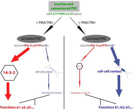
Results of this work suggest that the 3CTS sequence within PKC⑀ controls both translocation and tar-geting selectivity of this enzyme to cell-cell contact. It controls translo-cation via the␣-helix because, when the helix is destabilized, transloca-tion is inhibited. It controls selectiv-ity because of the presence of the essential Glu-374 amino acid, and it controls the functions that PKC⑀ exerts via its association with 14-3-3 because one of the binding sites involved in PKC⑀ interaction with 14-3-3 is located within 3CTS.
The fact that GFP cannot accu-mulate efficiently at the cell-cell contact when fused to 3CTS indi-cates that 3CTS contains the molecular determinants needed for selective targeting but not for accumulation at the cell-cell con-tact. This agrees well with the fact that the predicted amphipathic ␣-helix in 3CTS most probably does not mediate any strong inter-action with phospholipids but is involved in protein/protein interaction. Indeed, the amphipathic ␣-helix is involved upstream of the interaction of PKC⑀ with 14-3-3 because when it is destructured, PKC⑀ translocation is abolished. Also, our data show that overexpression of 3CTS abolishes selectivity of targeting. Because 3CTS cannot accumulate at cell-cell contacts, this argues in favor of selectivity being determined prior to accumulation at cell-cell contacts.
In the present work, we also provide substantial evidence demonstrating that PKC⑀ interacts with 14-3-3, as recently sug-gested by Saurin et al. (28) and by Durgan et al. (33), although in these two publications, the interaction of the endogenous pro-teins was not documented in contrast to our work. We also provide a model predicting the nature of this interaction via a 14-3-3-binding site located in 3CTS. Endogenous PKC⑀/14-3-3 interaction was weak in basal conditions and markedly increased in the presence of PMA or TRH, suggesting that in GH3B6 cells also, phosphorylation of PKC⑀ by a PMA-depend-ent kinase is responsible for PKC⑀/14-3-3 interaction. PKC␣ could participate in this process because its activation is neces-sary for PKC⑀ translocation (10) and because it was shown to be involved in PKC⑀ Ser-368 phosphorylation in the process of cytokinesis (28). The involvement of 3CTS in the PKC⑀/14-3-3 interaction was further supported by the fact that C1–3CTS-GFP and 3CTS-C1–3CTS-GFP both coimmunoprecipitated with 14-3-3 inactive and
sequestered PKC
activated PKC activated PKC
Functions a1, a2, a3....
Functions b1, b2, b3.... Functions a1, a2, a3....
Functions b1, b2, b3.... + PMA/TRH
14-3-3
cell-cell contact cell-cell contact14-3-3
LKELENNIRKALpSFDNRGEE
LKELENNIRKALSFDNRGEE
+ PMA/TRH
LKELENNIRKALpSFDNRGEE
FIGURE 8. Integrated model for the coordinated involvement of the amphipathic␣-helix, the
14-3-3-binding site, and the Glu-374 amino acid of 3CTS in the control of PKC⑀ translocation and function. The
␣-helix is involved very early in the translocation process because its alteration inhibits translocation. Upon PMA or TRH activation, the amount of PKC⑀ that localizes at the cell-cell contact might depend on the local amount of 14-3-3 available to bind PKC⑀. The higher the amount of 14-3-3, the lower the amount of PKC⑀ translocated to cell-cell contacts will be. This implies that two sets of functions might be regulated by PMA/TRH because both PKC⑀ translocation to cell-cell contact and phosphorylations at Ser-368 and Ser-346, which are necessary for PKC⑀/14-3-3 interaction, are PMA/TRH-dependent.
at INRA Institut National de la Recherche Agronomique on August 4, 2015
http://www.jbc.org/
and by showing that the Ser-to-Ala substitution in either of these two constructs abolished the interaction.
The data presented here show that 14-3-3 functions as an inhibitor of PKC⑀ translocation, in agreement with other pre-vious examples found in the literature (35, 36). However, our results also point to a new paradigm; PMA (or TRH) may ini-tiate two processes with seemingly opposed effects on two sep-arate pools of PKC⑀: it initiates PKC⑀ translocation, and at the same time, it initiates binding of PKC⑀ to 14-3-3, which inhibits translocation. The fact that 14-3-3 inhibits PKC⑀ translocation does not imply that it sequesters PKC⑀ to prevent its function. Instead, we propose that the interaction of PKC⑀ with 14-3-3 may initiate a function of PKC⑀ different from that exerted at the cell-cell contact, thus allowing the involvement of PKC⑀ in new aspects of cell biology, as shown by Saurin et al. (28) con-cerning cytokinesis. In this model, upon PMA stimulation, the amplitude of each PKC⑀ function could depend on the propor-tion interacting with 14-3-3 and that relocating to the cell membrane (Fig. 8). Interestingly, Par-1b membrane localiza-tion has been shown to be also negatively regulated when its binding to 14-3-3 is induced via phosphorylation of Ser-400 by protein kinase D activated by nPKC (37).
Thus, two molecular mechanisms involving 3CTS appear to be at work for PKC⑀: one that allows translocation or not and the other that determines selectivity. The former involves the amphipathic␣-helix and 14-3-3; the latter occurs via Glu-374 (Fig. 8). This does not mean that other amino acids or regions of PKC⑀ are not involved in translocation/targeting, as already demonstrated (38, 39). Nevertheless, this work identifies for the first time a previously unsuspected function for the V3 domain of PKC⑀ in the translocation/targeting process and underlines the role of this region as a platform for protein/protein interactions.
Acknowledgments—We thank Dr. Carsten Brock (Pharmacology Department, Functional Genomics Institute, Montpellier, France) for providing the anti-14-3-3 antibody. We also thank Catherine Legra-verend (Functional Genomics Institute) for critical comments on the manuscript.
REFERENCES
1. Nishizuka, Y. (2003) J. Biochem. 133, 155–158
2. Parker, P. J., and Murray-Rust, J. (2004) J. Cell Sci. 117, 131–132 3. Lehel, C., Olah, Z., Jakab, G., and Anderson, W. B. (1995) Proc. Natl. Acad.
Sci. U.S.A. 92,1406 –1410
4. Schmalz, D., Hucho, F., and Buchner, K. (1998) J. Cell Sci. 111, 1823–1830 5. Schultz, A., Ling, M., and Larsson, C. (2004) J. Biol. Chem. 279,
31750 –31760
6. Aschrafi, A., Franzen, R., Shabahang, S., Fabbro, D., Pfeilschifter, J., and Huwiler, A. (2003) Biochim. Biophys. Acta 1634, 30 –39
7. Hocevar, B. A., and Fields, A. P. (1991) J. Biol. Chem. 266, 28 –33 8. Mochly-Rosen, D., and Gordon, A. S. (1998) FASEB J. 12, 35– 42 9. Ron, D., Chen, C. H., Caldwell, J., Jamieson, L., Orr, E., and Mochly-Rosen,
D. (1994) Proc. Natl. Acad. Sci. U.S.A. 91, 839 – 843
10. Collazos, A., Diouf, B., Gue´rineau, N. C., Quittau-Pre´vostel, C., Peter, M., Coudane, F., Hollande, F., and Joubert, D. (2006) Mol. Cell. Biol. 26, 2247–2261
11. Quittau-Pre´vostel, C., Delaunay, N., Collazos, A., Vallentin, A., and Jou-bert, D. (2004) J. Cell Sci. 117, 63–72
12. Pauken, C. M., and Capco, D. G. (1999) Mol. Reprod. Dev. 54, 135–144 13. Louis, K., Gue´rineau, N., Fromigue´, O., Defamie, V., Collazos, A., Anglard,
P., Shipp, M. A., Auberger, P., Joubert, D., and Mari, B. (2005) J. Biol. Chem.
280,1272–1283
14. Larsen, E. C., DiGennaro, J. A., Saito, N., Mehta, S., Loegering, D. J., Ma-zurkiewicz, J. E., and Lennartz, M. R. (2000) J. Immunol. 165, 2809 –2817 15. Huang, J., Lo, P. F., Zal, T., Gascoigne, N. R., Smith, B. A., Levin, S. D., and
Grey, H. M. (2002) Proc. Natl. Acad. Sci. U.S.A. 99, 9369 –9373 16. Monks, C. R., Kupfer, H., Tamir, I., Barlow, A., and Kupfer, A. (1997)
Nature 385,83– 86
17. O’Keefe, J. P., Blaine, K., Alegre, M. L., and Gajewski, T. F. (2004) Proc.
Natl. Acad. Sci. U.S.A. 101,9351–9356
18. Alvaro, V., Le´vy, L., Dubray, C., Roche, A., Peillon, F., Que´rat, B., and Joubert, D. (1993) J. Clin. Endocrinol. Metab. 77, 1125–1129
19. Pre´vostel, C., Alvaro, V., de Boisvilliers, F., Martin, A., Jaffiol, C., and Jou-bert, D. (1995) Oncogene 11, 669 – 674
20. Vallentin, A., Lo, T. C., and Joubert, D. (2001) Mol. Cell. Biol. 21, 3351–3363
21. Zhu, Y., Dong, Q., Tan, B. J., Lim, W. G., Zhou, S., and Duan, W. (2005)
Cancer Res. 65,4520 – 4524
22. Gue´rineau, N. C., Bonnefont, X., Stoeckel, L., and Mollard, P. (1998) J. Biol.
Chem. 273,10389 –10395
23. Douguet, D., and Labesse, G. (2001) Bioinformatics 17, 752–753 24. Canutescu, A. A., Shelenkov, A. A., and Dunbrack, R. L., Jr. (2003) Protein
Sci. 12,2001–2014
25. Sali, A., and Blundell, T. L. (1993) J. Mol. Biol. 234, 779 – 815 26. Sippl, M. J. (1993) Proteins 17, 355–362
27. Eisenberg, D., Lu¨thy, R., and Bowie, J. U. (1997) Methods Enzymol. 277, 396 – 404
28. Saurin, A. T., Durgan, J., Cameron, A. J., Faisal, A., Marber, M. S., and Parker, P. J. (2008) Nat. Cell Biol. 10, 891–901
29. Johnson, J. E., Giorgione, J., and Newton, A. C. (2000) Biochemistry 39, 11360 –11369
30. Newton, A. C. (1993) Annu. Rev. Biophys. Biomol. Struct. 22, 1–25 31. Abes, S., Moulton, H. M., Clair, P., Prevot, P., Youngblood, D. S., Wu, R. P.,
Iversen, P. L., and Lebleu, B. (2006) J. Control Release 116, 304 –313 32. Jin, J., Smith, F. D., Stark, C., Wells, C. D., Fawcett, J. P., Kulkarni, S.,
Metalnikov, P., O’Donnell, P., Taylor, P., Taylor, L., Zougman, A., Woodgett, J. R., Langeberg, L. K., Scott, J. D., and Pawson, T. (2004) Curr.
Biol. 14,1436 –1450
33. Durgan, J., Cameron, A. J., Saurin, A. T., Hanrahan, S., Totty, N., Messing, R. O., and Parker, P. J. (2008) Biochem. J. 411, 319 –331
34. Obsil, T., Ghirlando, R., Klein, D. C., Ganguly, S., and Dyda, F. (2001) Cell
105,257–267
35. Matto-Yelin, M., Aitken, A., and Ravid, S. (1997) Mol. Biol. Cell 8, 1889 –1899
36. Meller, N., Liu, Y. C., Collins, T. L., Bonnefoy-Be´rard, N., Baier, G., Isakov, N., and Altman, A. (1996) Mol. Cell. Biol. 16, 5782–5791
37. Watkins, J. L., Lewandowski, K. T., Meek, S. E., Storz, P., Toker, A., and Piwnica-Worms, H. (2008) Proc. Natl. Acad. Sci. U.S.A. 105, 18378 –18383
38. Csukai, M., Chen, C. H., De Matteis, M. A., and Mochly-Rosen, D. (1997)
J. Biol. Chem. 272,29200 –29206
39. Takahashi, M., Mukai, H., Oishi, K., Isagawa, T., and Ono, Y. (2000) J. Biol.
Chem. 275,34592–34596
PKC
⑀ Cell-Cell Contact Targeting
at INRA Institut National de la Recherche Agronomique on August 4, 2015
http://www.jbc.org/
Hollande and Dominique Joubert
Nathalie C. Guérineau, Philippe Jay, Frédéric
Philippe Clair, Cécile Gauthier-Rouvière,
Labesse, Françoise Macari, Armelle Choquet,
Barthélémy Diouf, Alejandra Collazos, Gilles
Targeting at Cell-Cell Contacts
C? Involved in Translocation and Selective
A 20-Amino Acid Module of Protein Kinase
doi: 10.1074/jbc.M109.004614 originally published online May 8, 2009 2009, 284:18808-18815.
J. Biol. Chem.
10.1074/jbc.M109.004614 Access the most updated version of this article at doi:
. JBC Affinity Sites Find articles, minireviews, Reflections and Classics on similar topics on the
Alerts:
When a correction for this article is posted •
When this article is cited •
to choose from all of JBC's e-mail alerts Click here
Supplemental material:
http://www.jbc.org/content/suppl/2009/05/08/M109.004614.DC1.html http://www.jbc.org/content/284/28/18808.full.html#ref-list-1This article cites 39 references, 24 of which can be accessed free at
at INRA Institut National de la Recherche Agronomique on August 4, 2015
http://www.jbc.org/