Publisher’s version / Version de l'éditeur:
Vous avez des questions? Nous pouvons vous aider. Pour communiquer directement avec un auteur, consultez la
première page de la revue dans laquelle son article a été publié afin de trouver ses coordonnées. Si vous n’arrivez pas à les repérer, communiquez avec nous à [email protected].
Questions? Contact the NRC Publications Archive team at
[email protected]. If you wish to email the authors directly, please see the first page of the publication for their contact information.
https://publications-cnrc.canada.ca/fra/droits
L’accès à ce site Web et l’utilisation de son contenu sont assujettis aux conditions présentées dans le site
LISEZ CES CONDITIONS ATTENTIVEMENT AVANT D’UTILISER CE SITE WEB.
Toxins, 11, 12, pp. 714-01-714-40, 2019-12-07
READ THESE TERMS AND CONDITIONS CAREFULLY BEFORE USING THIS WEBSITE.
https://nrc-publications.canada.ca/eng/copyright
NRC Publications Archive Record / Notice des Archives des publications du CNRC :
https://nrc-publications.canada.ca/eng/view/object/?id=3a442df2-b1dd-4e84-9ead-9bcd2d43b6e8
https://publications-cnrc.canada.ca/fra/voir/objet/?id=3a442df2-b1dd-4e84-9ead-9bcd2d43b6e8
Archives des publications du CNRC
This publication could be one of several versions: author’s original, accepted manuscript or the publisher’s version. / La version de cette publication peut être l’une des suivantes : la version prépublication de l’auteur, la version acceptée du manuscrit ou la version de l’éditeur.
For the publisher’s version, please access the DOI link below./ Pour consulter la version de l’éditeur, utilisez le lien DOI ci-dessous.
https://doi.org/10.3390/toxins11120714
Access and use of this website and the material on it are subject to the Terms and Conditions set forth at
Structural diversity, characterization and toxicology of microcystins
Bouaïcha, Noureddine; Miles, Christopher O.; Beach, Daniel G.; Labidi,
Zineb; Djabri, Amina; Benayache, Naila Yasmine; Nguyen-Quang, Tri
Toxins 2019, 11, 714; doi:10.3390/toxins11120714 www.mdpi.com/journal/toxins
Review
Structural Diversity, Characterization and Toxicology
of Microcystins
Noureddine Bouaïcha
1,*, Christopher O. Miles
2, Daniel G. Beach
2, Zineb Labidi
3, Amina
Djabri
1,3, Naila Yasmine Benayache
1and Tri Nguyen-Quang
41 Écologie, Systématique et Évolution, Univ. Paris-Sud, CNRS, AgroParisTech, Université Paris-Saclay,
91405 Orsay, France; [email protected] (A.D.); [email protected] (N.Y.B.)
2 Biotoxin Metrology, National Research Council Canada, 1411 Oxford St, Halifax, NS B3H 3Z1, Canada;
[email protected] (C.O.M.); [email protected] (D.G.B.)
3 Laboratoire Biodiversité et Pollution des Écosystèmes, Faculté des Sciences de la Nature et de la Vie,
Université Chadli Bendjedid d’El Taref, 36000, El Taref, Algeria; [email protected]
4 Biofluids and Biosystems Modeling (BBML), Faculty of Agriculture, Dalhousie University, 39 Cox Road,
Truro, B2N 5E3 Nova Scotia, Canada; [email protected]
* Correspondence: [email protected]; Tel.: +33 (01)69154990; Fax: +33 (0)169155696 Received: 1 October 2019; Accepted: 4 December 2019; Published: 7 December 2019
Abstract:
Hepatotoxic microcystins (MCs) are the most widespread class of cyanotoxins and the one
that has most often been implicated in cyanobacterial toxicosis. One of the main challenges in
studying and monitoring MCs is the great structural diversity within the class. The full chemical
structure of the first MC was elucidated in the early 1980s and since then, the number of reported
structural analogues has grown steadily and continues to do so, thanks largely to advances in
analytical methodology. The structures of some of these analogues have been definitively elucidated
after chemical isolation using a combination of techniques including nuclear magnetic resonance,
amino acid analysis, and tandem mass spectrometry (MS/MS). Others have only been tentatively
identified using liquid chromatography-MS/MS without chemical isolation. An understanding of
the structural diversity of MCs, the genetic and environmental controls for this diversity and the
impact of structure on toxicity are all essential to the ongoing study of MCs across several scientific
disciplines. However, because of the diversity of MCs and the range of approaches that have been
taken for characterizing them, comprehensive information on the state of knowledge in each of these
areas can be challenging to gather. We have conducted an in-depth review of the literature
surrounding the identification and toxicity of known MCs and present here a concise review of these
topics. At present, at least 279 MCs have been reported and are tabulated here. Among these, about
20% (55 of 279) appear to be the result of chemical or biochemical transformations of MCs that can
occur in the environment or during sample handling and extraction of cyanobacteria, including
oxidation products, methyl esters, or post-biosynthetic metabolites. The toxicity of many MCs has
also been studied using a range of different approaches and a great deal of variability can be
observed between reported toxicities, even for the same congener. This review will help clarify the
current state of knowledge on the structural diversity of MCs as a class and the impacts of structure
on toxicity, as well as to identify gaps in knowledge that should be addressed in future research.
Keywords:
microcystin; cyanobacteria; cyanotoxin; structural elucidation; toxicology
Key
Contribution: An up to date review surrounding the diversity, biosynthesis, characterization,
and toxicology of MC congeners reported in the literature as identified from cyanobacterial cultures
and field samples. At present, 279 MCs have been characterized using a wide range of combinations
of analytical techniques. Modifications of each amino acid residue in their structure, especially in
the positions 5 and 6, affect both their in vivo and in vitro toxicities.
1. Introduction
There are an increasing number of warnings about toxic cyanobacterial blooms observed
worldwide and global warming is thought to stimulate their development in eutrophic waters [1–5].
These blooms are often accompanied by production of a variety of cyanotoxins generally classified
according to the target organs: hepatotoxins (liver), neurotoxins (nervous system), and dermatotoxins
(skin). Among these cyanotoxins, it appears that hepatotoxic and tumor promoting microcystins
(MCs) are more commonly found in cyanobacterial blooms and considered to be one of the most
hazardous groups throughout the world [6–10]. Despite the significant amount of available
information, interest in MCs continues to increase due to their well-known hazards to farm animals,
fisheries, aquaculture, human health, and wildlife through exposure via drinking, environmental,
and recreational waters [7,8,10–12].
Louw and Smit [13] were among the first scientists who isolated and attempted to characterize
a cyanotoxin from a cyanobacterial bloom, dominated by Microcystis toxica, which occurred in the
Vaal Dam reservoir in South Africa in 1942–1943. They concluded that it was an alkaloid of
undetermined structure with acute and chronic hepatotoxic properties. Initial attempts to
characterize the structures of MCs started in the late 1950s [14]. However, full structural identification
of the first MC congeners was achieved in 1984, when a combination of amino acid analysis, nuclear
magnetic resonance (NMR) spectroscopy, and mass spectrometry (MS) were used [15,16]. These and
subsequent studies showed the chemical structure of these toxins to consist of a cyclic heptapeptide
composed of five relatively conserved amino acids plus two variable
L-amino acids [14–20].
Two decades later, studies have shown that these toxins are biosynthesized nonribosomally via
an MC synthetase gene cluster (mcy), consisting of a combination of polyketide synthases (PKSs),
nonribosomal peptide synthetases (NRPSs) and tailoring enzymes [21–24]. Recent studies have
reported the bulk of this structural diversity is the result of genetic and/or environmental factors,
which have an impact on the functioning of enzymes encoded in the mcy gene cluster [25–32]. This
mcy
gene cluster is often spontaneously modified through point mutations, deletions and insertions,
or a series of genetic recombinations, which affect the functioning of the MC peptide synthetases and
result in the chemical diversity observed in nature [33–37].
As a result of the large number of literature reports spanning over four decades of research, it
can be difficult to glean accurate information on the total number of identified MCs. The phrase ‘more
than 100 microcystin congeners’ is still often used in the literature [12,38–41] however estimates as
high as 248 known MCs have recently been published [42]. In this review, we update this number to
279 and describe the methods by which this structural elucidation was carried out. In addition, we
provide an in-depth review of their toxic potential and a discussion of the structure–activity
relationships this information provides.
2. Nomenclature and General Chemical Structure of Microcystins
Hughes et al. [43] first described a hepatotoxic ‘fast death factor’ in an extract of the isolated
strain Microcystis aeruginosa NRC-1. This hepatotoxic factor was later renamed microcystin, derived
from the genus Microcystis [44–46]. Since, they have also been referred to in the literature as
cyanoginosin, with prefix ’cyano’ from the term cyanobacteria and ’ginosin’ derived from aeruginosa
[19,47]; and cyanoviridin, with the root ’viridin’ from the species M. viridis [48].
After almost two decades of structural analysis of toxic peptides from the colonial
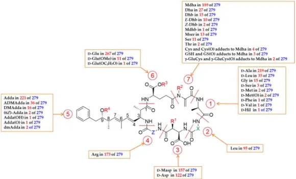
bloom-forming cyanobacterium M. aeruginosa, Botes et al. [15–19] and Santikarn et al. [20] provided
structural details of these toxins with the general structure of: cyclo-(
D-Ala
1-X
2-
D-Masp
3-Z
4-Adda
5-
D--Glu
6-Mdha
7) (Figure 1) in which X and Z are variable
L-amino acids,
D-Masp
3is
D-erythro- -
methyl-isoaspartic acid, and Mdha is N-methyldehydroalanine. The -amino acid Adda,
(2S,3S,4E,6E,8S,9S)-3-amino-9-methoxy-2,6,8-trimethyl-10-phenyldeca-4,6-dienoic acid, is the most unusual
substructure, and has not been reported elsewhere in nature apart from in the closely related group
of toxic pentapeptides, the nodularins. After a time, leading researchers in the field warned that the
continued use of multiple naming systems could generate confusion as the number of publications
on these cyclic peptides increased [49]. Therefore, a universal system of nomenclature was proposed
based on the original term microcystin-XZ, where X and Z denote the variable amino acid residues
in positions 2 and 4 [50].
The number of fully and partially characterized MCs increased significantly throughout the
1990s and structural variations have been reported in all seven amino acids, but most frequently with
substitution of the two variable
L-amino acids X and Z at positions 2 and 4, respectively, and
demethylation of amino acids at position 3 and/or 7. Multiple combinations of the variable
L-amino
acids (X and Z), and modifications to the other amino acids, lead to the high levels of structural
diversity observed in MCs which have been characterized to date from bloom samples and isolated
strains of Microcystis, Oscillatoria, Planktothrix, Anabaena (syn. Dolichospermum), and Nostoc spp. [51].
While Carmichael et al. [50] originally proposed abbreviation in the format MCYST-XZ, in recent
decades the abbreviation MC-XZ has come in to general use, where X and Z are the one-letter amino
acid abbreviations (where these exist) and with any variations at positions 1, 3, and 5–7 (relative to
D
-Ala,
D-Masp, Adda,
D-Glu, and Mdha, respectively) shown in square brackets, using 3–7-letter
amino acid abbreviations [52], in numerical order with the position indicated by a superscript, and
separated by commas without spaces in square brackets immediately prior to MC. For example, [
D-Leu
1,
D-Asp
3,Dha
7]MC-LR contains
D-Leu,
D-Asp and Dha at positions 1, 3, and 7, and
L-Leu and
L-Arg at positions 2 and 4, respectively, with Adda and
D-Glu assumed by default at positions 5 and 6
(see Figure 1). If an amino acid residue at positions 2 or 4 is not one of the 20 standard amino acids,
the three-letter (or more, where necessary) abbreviation is used; e.g., the congener containing Leu at
position 2 and homoarginine in position 4 is named MC-LHar. Ring opened MCs are designated with
the prefix [seco-a/b], where a and b are the residue numbers between which the amide bond has been
hydrolysed. For example, [seco-4/5]MC-LR indicates MC-LR hydrolysed between the Arg
4- and
Adda
5-residues. A MC name-generator is included in version 16 of the MC mass calculator tab of a
publicly available toxin mass list [53]. To facilitate ongoing efforts to maintain comprehensive lists
and databases of toxins and/or cyanobacterial metabolites, it is recommended that researchers use
these naming conventions going forward when reporting the identification of new MCs.
Figure 1. General structure of microcystins (MCs) and an overview of their observed structural
diversity. R1 = H or CH3; R2 = H or CH3; R3 = H, CH3 or C3H6OH; R4 = H, CH3 or COCH3; X and Z =
variable L-amino acids.
3. Biosynthesis of Microcystins
The nonribosomal biosynthesis of MCs has been described in detail in Microcystis [21–23,54],
Planktothrix
[55], and Anabaena [56]. This MC synthetase system is encoded by two transcribed
operons in M. aeruginosa, mcyABC, and mcyDEFGHIJ, from a central bidirectional promoter located
between mcyA and mcyD [23,54]. Insertional gene knockout experiments have demonstrated that all
MC congeners produced by a strain are synthesized by a single enzyme complex encoded by a 55 kb
gene cluster [21,55,57,58]. Therefore, strains that do not produce MCs can result from inactivation
either by point mutation, or by partial or complete deletion of mcy genes [36,57,59,60]. Recently,
Shishido et al. [61] reported that the mcy gene cluster did not encode all enzymes for the synthesis of
rare variants of MC that contain a selection of homo-amino acids by the benthic cyanobacterium
Phormidium
sp. LP904c, but may instead activate homo-amino acids produced during the synthesis
of anabaenopeptins.
Several studies have assessed the impact of environmental factors, including nutrient
availability, light, iron limitation, temperature, and pH on cellular MC content [25,31,32,62–68]. Some
of these factors are involved in enhancing or suppressing the expression of the mcy gene, leading to
an increase or decrease in MC production. For example, Kaebernick et al. [62] reported that light
quality has a significant effect on the transcription levels of the genes mcyB and mcyD. Recently, Yang
and Kong [69] reported that the exposure of M. aeruginosa cells to UV-B not only remarkably inhibited
the growth of Microcystis, but also led to a reduction in MC concentration by decreasing transcription
of the gene mcyD. Sevilla et al. [64] demonstrated that iron starvation causes an increase in mcyD
transcription and MC levels. Moreover, it was also shown that stress conditions caused by nutrient
deprivation increased mcyD transcription and MC production [68]. Horst et al. [70] combined
laboratory and field experiments and observed a correlation between nitrogen limitation and lower
MC cell quotas in a field survey of Lake Erie (situated at the international boundary between Canada
and the USA). Since MC is a nitrogen-rich metabolite, Van de Waal et al. [30] have observed an
increase in MC cell quotas after excess nitrogen supply, the nitrogen-rich variant MC-RR being the
most significantly affected. Similarly, Puddick et al. [32] recently reported that when Microcystis strain
CAWBG11 was grown in batch culture until nitrogen-depletion, the relative abundance of the MC
congeners shifted and the relative abundance of arginine-containing MCs decreased, whilst the
congeners which did not contain arginine increased. These changes coincided with a large decrease
in nitrate concentration to 0.35% of initial levels.
Therefore, the biosynthesis of MCs is strain dependent and their diversity is due to variability
in the coding of the MC synthetase genes among cyanobacterial strains [71] as well as some
environmental factors such as nitrogen concentration [29,30,32,72,73]. The large variety of amino
acids that can be incorporated and further modified by tailoring enzymes, having activities such as
the epimerization, cyclisation, N-methylation, formylation, and reduction of amino acids, accounts
for the production of highly complex peptides [74,75]. In the field, it is important to consider that
multiple strains of cyanobacteria co-exist, often leading to more complex profiles of MCs. Typically,
cyanobacterial strains in the field and in controlled conditions produce up to twenty MC congeners
but usually only one or two congeners dominate in any single strain [12,40,76–80]. However, the
number of congeners produced in a single strain can exceed 47 when all minor MC analogues are
considered [33] but in many other cases MC profiles reportedly contain only a small number of
congeners. This highlights the fact that there are still new MCs being constantly identified, with
varying degrees of certainty, in the very recent literature [81,82]. However, gaps remain in the
understanding of how environmental factors can modulate the relative abundance of MC congeners
in a bloom, requiring further research on the topic.
4. Structural Elucidation of Microcystins
Definitive structural elucidation of new MCs can only be achieved by purifying individual MCs
using preparative isolation techniques followed by comprehensive NMR, amino acid analysis and
tandem MS (MS/MS) experiments.
Proper purification, structural elucidation, and quantitation are also of critical importance to
toxicity studies, where the knowledge of structure, purity, and amount of each MC tested for toxicity
are required to ensure the validity and comparability of toxicology results. In this section we give a
brief overview of the techniques available for structural elucidation of MCs and the structural
information that each technique can provide. This will be useful in choosing appropriate methods for
structural elucidation as well as in the critical evaluation of published MC identifications and
toxicities.
4.1. Mass Spectrometry for Structural Elucidation
In addition to offering sensitive and selective targeted quantitative analysis of known MCs, MS
is an invaluable tool for tentative structural elucidation of new MCs, providing information about
their molecular formula (full scan) and structure (MS/MS). The types of MS techniques that have been
used for the identification of MCs have evolved as mass spectrometers suitable for intact analysis of
peptides have become commercially available. Early work relied heavily on direct analysis of MCs
purified off-line, using fast atom bombardment MS in a variety of low and high resolution as well as
tandem MS configurations [15,75,83–86]. Similarly, the usually high-resolution MS information from
matrix assisted laser desorption ionization-time of flight MS with and without post-source decay has
been invaluable in identifying new purified MCs over the years [87–91]. The widespread availability
of electrospray ionization (ESI) has made it commonplace to couple analytical liquid chromatography
(LC) separations directly to high resolution and/or tandem mass spectrometers. This has offered the
possibility for structural characterization and tentative identification of many MCs in complex
mixtures without the need for isolation of milligram amounts of toxin needed for full structural
elucidation. LC with sequential multi-stage MS dissociation (LC-MS
n), while usually offering only
unit mass resolution, provides a great deal of information for the determination of structure and
connectivity and has been widely used for MC identification [33,75,77,90,92,93]. Alternatively, LC
with high resolution MS/MS (LC-HRMS/MS) provides accurate mass data on precursor and product
ions suitable for determining their molecular formulae [81,82,94–97].
LC-MS alone is usually not considered definitive proof of chemical structure, but a great deal of
structural information can nevertheless be obtained. LC separation of MCs is typically carried out in
reverse phase using C18 or similar stationary phases and acidic mobile phases, resulting in
protonation of carboxyl groups, with acetonitrile as the organic modifier. The hydrophobicity and
therefore elution order of MCs in reverse-phase LC is governed primarily by the number of arginine
residues in their structure, and the relative elution order of new congeners can provide useful
information about structure. MCs with two arginine or homoarginines (e.g., MC-RR) are the most
polar and elute first, followed by MCs with a single Arg at position four (e.g., MC-LR), then those
with a single Arg at position 2 (e.g., MC-RY), and finally those with no Arg residues (e.g., MC-LA).
Though sometimes difficult to separate chromatographically [98], the relative elution order of
desmethylated MCs can also be useful for identitication of minor analogues with [DMAdda
5]-variants eluting much earlier than [
D-Asp
3]-variants, which are followed closely by [Dha
7]-variants
[82].
The nominal molecular weights for known parent MCs range from 882 Da to 1101 Da, although
the theoretical range using the known amino acids identified to date in natural MCs is significantly
wider than this [53]. These masses are too large for definitive molecular formula elucidation by
HRMS when all possible elements are considered (e.g., F, P, Cl, etc.), but when the possible elemental
composition and ring or double bond equivalents (RDBE) are limited to a reasonable range similar
to known MCs, in most cases a candidate formula can be determined. Careful evaluation of isotopic
patterns in HRMS can further help narrow down possible molecular formulae, relying less on
pre-determined limits in elements, as reported recently in Mallia et al. [99]. Known parent (unconjugated)
MCs have between 44 and 57 C atoms, 63 and 84 H atoms, 7 and 13 N atoms, 12 and 17 O atoms, and
up to one S atom in methionine-containing MCs [53]. It can also be useful to limit possible molecular
formulas by the number of RDBEs in the structure, which ranges from 17 to 27. Within these limits
there are currently no cases among known MC where [M+H]
+or [M+2H]
2+ions are within 5 ppm
mass accuracy from one another. However, there are >120 cases reported of isomeric MCs reported,
significantly limiting the utility of accurate mass measurements alone for MC identification. It should
be noted that conjugates between MCs and thiol-containing biomolecules (e.g., cysteine, glutathione)
have been reported [81,100,101] and that mass ranges and molecular formula tolerances need to be
widened accordingly when characterizing these compounds.
The ionization and MS/MS fragmentation of MCs are also generally similar between MCs with
the same number of Arg/Har residues. In positive ESI mode, MCs with two Arg/Har residues, or
conjugated to Cys or GSH, appear primarily as doubly charged ions after ESI while those with one
or no Arg/Har are predominantly singly charged. MCs with no Arg/Har ionize comparatively poorly
and therefore tend to form adducts with other cations present in the sample (e.g., [M+Na]
+or
[M+NH
4]
+). In negative ESI all MCs are observed predominantly as singly charged [M-H]
-ions. The
MS/MS dissociation of MCs has been studied extensively in both positive [102,103], and negative
[104,105] ionization modes. In most cases, MCs fragment to produce sensitive class-characteristic
fragments at m/z 135.0804 originating from the Adda
5–residue in positive mode, and m/z 128.0353
originating from the Glu
6-residue in negative mode [82,105,106]. Since MCs are cyclic peptides, a
large body of previous work on the collision induced dissociation (CID) of non-microcystin peptides
also exists [107] that can be useful in interpretation of microcystin MS/MS spectra. In addition to the
class-characteristic fragments, various “sequence ions” are formed during CID following ring
opening that represent cleavages of individual amino acids. These can be used to effectively
reconstruct the MC structure one amino acid at a time. For example, in negative mode, the B-ion
series is useful for determining the mass of the amino acid at position 4 since ions represent successive
cleavages from the B1 ion [C5H4O3-X
4-Adda-(Glu-H2O)-Mdha-Ala]
−[105]. Similarly, in positive mode,
fragments associated with each individual amino acid can be readily identified from HRMS/MS data.
This allows the amino acid at which small structural variations, such as demethylation have occurred,
to be determined but not always the definitive structure of amino acid side chains, particularly for
low resolution MS data [102].
There are two different types of CID that provide different and often complementary
information for structural elucidation; quadrupole and ion trap CID. Quadrupole CID, sometimes
termed higher energy collisional dissociation (HCD), is a single-stage process involving a smaller
number of more energetic collisions. The main advantage of this approach is that the full m/z range
of product ions can be detected simultaneously, allowing for sensitive detection of both class-specific
and structurally informative product ions in a single experiment. However, it can be more difficult
to trace the origin of lower mass product ions. Ion trap CID involves longer time scales and a larger
number of lower energy collisions, as well as offering the potential for multiple stages of MS/MS
(MS
n). Here, structural elucidation is done by constructing a fragmentation tree where the origin of
lower mass product ions can be established in higher order experiments (e.g., MS
3or MS
4) in relation
to higher-mass product ions observed in MS
2. The principal limitation of ion trap dissociation is the
1/3 rule, stating that product ions of less than 1/3 the m/z of a selected precursor ion cannot be isolated
for detection. This means that class-specific low mass product ions cannot be detected in MS
2in an
ion trap regardless of the collision energy used.
Use of established chemical degradation or modification reactions to probe the structure of
unknown chemicals is a classical approach that pre-dates the availability of modern analytical
instrumentation such as NMR or MS. This approach was an important part of the first structural
elucidation studies on MCs, where partial hydrolysis reactions followed by Edman degradation
reactions and mass spectrometric methods of characterizing the linearized products were utilised
[15,16]. However, when combined with modern LC-MS, similar approaches are even more powerful
due to the ease with which multiple products of reactions with multiple reactants can be selectively
detected. The reactivity of the double bond in Dha
7and Mdha
7of MCs towards thiols has emerged
as being particularly useful for characterizing MCs congeners [81,82,99]. In particular,
mercaptoethanol derivatization has been exploited in such a manner both to identify new MCs from
complex samples [77,108] or to quickly differentiate Mdha
7- from Dhb
7-containing MCs [109], which
is not possible using LC-MS alone unless authentic standards are available. Another useful reaction
is methyl esterification that, together with LC-MS, can be used to count the number of reactive
carboxylic acid (the CO2H group of Glu
6is reactive, but not that of Masp
3/Asp
3) and phenolic groups
in MCs [96,99]. Selective oxidation with mild oxidants can also be combined with LC-MS to identify
MCs containing sulfide and sulfoxides, such as methionine (e.g., MC-MR) [81,93]. This approach has
also proved useful in helping to identify sulfide (e.g., Cys, GSH) conjugates of MCs, and their
autoxidation products, in natural samples [81]. These conjugates are also readily semi-synthesised to
produce authentic standards for confirmation of their presence in samples and cultures by LC-MS
(e.g., [81,100]) (see Section 5.7). In addition, the identity of S-conjugates at position-7 of MCs can be
confirmed through a range of deconjugation reactions [101,110,111].
It should be noted that stereochemical information is not generally accessible from LC-MS/MS
analyses. It is therefore generally assumed in the literature that the stereochemistry of the amino acids
in MCs whose structures are established using LC-MS methods are the same as those in
well-established structures such as MC-LR unless there is evidence to the contrary, and the same
assumption is made throughout this review.
4.2. Preparative Isolation of Microsystins
While a great deal of structural information is available from LC-MS without the need for
additional purification of MCs, stereochemistry is not accessible by LC-MS. Definitive structural
elucidation by NMR currently requires isolation of 50–100 μg or more of a pure MC. This is most
often done by scaling up production of laboratory cultured cyanobacteria, or directly from naturally
occurring bloom samples. Dried cyanobacterial biomass is usually first extracted under acidic
conditions. Aqueous extracts can then be applied to a reverse phase (e.g., C18) open column or large
solid phase extraction (SFE) cartridge, depending on the complexity of the toxin profile. Elution is
then carried out in a stepwise fashion, usually with aqueous methanol. In some cases, size exclusion
chromatography has been used [83] but is often not required. For more complex toxin mixtures, a
final semi-preparative isolation step is required, again using a reverse phase column. Additional
selectivity at each step can be achieved by varying the pH of the eluent with various buffers. These
then need to be removed with an additional SPE step. Long-term storage of MCs in acidic methanol
should be avoided because of the potential for formation of methyl esters [81,112] (see Section 5.7).
4.3. Amino Acid Analysis
One of the classical approaches to structural characterization of MCs after preparative isolation
is to hydrolytically digest them to their constituent amino acids, which are then identified
chromatographically based on retention time comparison to amino acid standards. Typically,
hydrolysis is done in 6 M HCl at 105 °C for 24 h. It should be noted that under these conditions, Mdha
and Adda were not detected [18] and some amino acids (e.g., tryptophan, glutamine, serine,
threonine, tyrosine, methionine, and cysteine) are altered [113] and their presence must be inferred.
Amino acids can then be derivatized with a UV absorbent tag and analyzed by reverse phase LC-UV
[83]. Alternatively, they can be derivatized with trifluoroacetic anhydride to make them volatile and
then analyzed by gas chromatography [114]. When chiral stationary phases or derivatizing agents
are used, it is possible to determine the absolute configuration of the amino acids, which is not
possible by MS or NMR alone.
4.4. Structural Elucidation by Nuclear Magnetic Resonance Spectroscopy
Although it is most often used in combination with other approaches mentioned earlier, NMR
is usually required for definitive structural identification of MCs including relative stereochemistry.
Often CD
3OD and sometimes D
2O were used in the past as solvents for NMR spectroscopy of MCs
to reduce
1H signals arising from the solvent signals in 1- and 2-D spectra e.g., [115]. The disadvantage
is that exchangeable amide protons, and thus their correlations, cannot be detected. It is therefore
advantageous to use solvents that do not exchange the amide protons, such as (CD3)2SO or CD3OH,
and with modern NMR spectrometers and solvent suppression techniques, the use of partially
protonated solvents such as CD
3OH is not usually problematic. However, NMR spectroscopy is a
relatively insensitive technique, and significant amounts of purified compound are generally
required. With modern high-field NMR spectrometers, full structural elucidations can be performed
on less than 50 μg of an MC in CD
3OH in 5 mm NMR tubes.A comprehensive approach to assigning all protons and carbons in MCs by NMR alone was
presented nearly 3 decades ago that relied on a combination of three 2-D NMR techniques:
double-quantum filtered correlation spectroscopy (DQF-COSY),
lH-detected multiple quantum coherence
(HMQC), and heteronuclear multiple bond correlation (HMBC) NMR spectroscopy [115].
DQF-COSY was first used to assign H-2 of each constituent amino acid along with several other proton
assignments. HMQC was then used to assign C-2 of each constituent amino acid based on the H-2
resonances already assigned. Finally, HMBC NMR spectra, that can detect
2J
- and
3J
-couplings
between carbon and hydrogen atoms, were used to assign the carbonyl carbon from each amino acid
and ultimately to determine the overall amino acid sequence. Nowadays, total correlation
spectroscopy (TOCSY) or decoupling in the presence of scalar interactions (DIPSI-2) NMR spectra
would typically also be acquired to identify the individual amino acid spin systems, and edited
heteronuclear single quantum correlation (HSQC) spectra to identify methyl, methylene, and
methine
13C and
1H resonances. Rotating frame nuclear overhauser effect spectroscopy (ROESY)
NMR spectra are generally better suited for detection of through-space correlations for molecules of
the size of MCs than are nuclear overhauser effect spectroscopy (NOESY) NMR spectra. In addition
to detecting through-space correlations and thus potentially defining the relative stereochemistry of
the molecule, ROESY NMR correlations can be useful for establishing the connectivity of the amino
acids as a supplement to HMBC NMR correlations. Selective 1D-TOCSY or –DIPSI-2 NMR spectra
can often be used to obtain
1H signal multiplicities and coupling constants in regions of spectral
overlap, which can be useful for chemical shift and stereochemical assignments. The higher
resolution afforded in the
13C axis by band-selective HMBC and HSQC spectra is sometimes useful
for resolving crowded regions of the HSQC (e.g., methylene region) and HMBC (e.g., amide carbonyl
region) spectra, and data acquisition times for 2-D spectra can be substantially reduced by use of
non-uniform sampling and NMR by ordered acquisition using
1H detection techniques.
NMR spectroscopy can also be used quantitatively to measure the concentrations of algal toxin
solutions relative to reference solutions [116]. This approach has been applied to produce quantitative
certified reference materials (CRMs) for MC-RR, MC-LR, and [Dha
7]MC-LR [117] with CRMs for
other MCs including MC-LA and [
D-Leu
1]MC-LY in preparation (P. McCarron, personal
communication).
4.5. 3-Dimensional Structures of Microcystins
Two methods have been used for determining the 3-dimensional structures of MCs. X-ray
crystallography has been used for nearly a century to reveal the 3-dimensional relationships of heavy
atoms in crystallized organic molecules. Unfortunately, MCs have not so far yielded suitable crystals
for such studies. However, in recent decades the 3-dimensional structures of MCs bound to protein
phosphatases (PPs) have been determined [118–126] (see Section 6.2). In addition to providing key
information about the mode of action and structure–activity relationships of the MCs, this also
provides 3-dimensional structural information about the toxins themselves. The other approach that
has been used is NMR spectroscopy, using through-space NOESY and ROESY correlations to
estimate the relative intramolecular distances between hydrogen atoms in the MCs in solution [127–
130]. Such measurements can be supplemented with measurements of
1H–
1H coupling constants,
which provide information about the dihedral angles between spin-coupled hydrogen atoms within
the molecule. Such studies have led to several published 3-dimensional NMR-based solution
structures, something that is not available from X-ray crystal structures because the structures are
based on the solid-state structures that do not necessarily reflect the structure in solution.
Although not in and of itself proof of structure or stereochemistry, observation of high affinity
of a purified MC for PPs or MC-specific antibodies can be taken as strong supporting evidence that
the absolute stereochemistry of the MC is largely the same as that of other MCs. The reason is that
recognition of MCs by receptor biomolecules is dependent on the 3-dimensional structure of the
ligand (see Section 6.2), which is controlled by amino acids present in the MC and their
stereochemistries. Biosynthetic reasoning also suggests that most MCs likely share the same
stereochemistry, since they are assembled by closely related synthetases produced from genetically
similar gene clusters, even in separate MC-producing genera (see Section 3).
5. Diversity of Characterized Microcystin Congeners
To date, the identification of at least 279 different MC congeners have been reported in the
literature (Table 1) using various combinations of the techniques reviewed in Section 4. These
congeners include MCs biosynthesized with structural variations at every amino acid position as well
as the products of several chemical and biochemical transformations that can occur in the
environment or the laboratory, all of which are reviewed in this section.
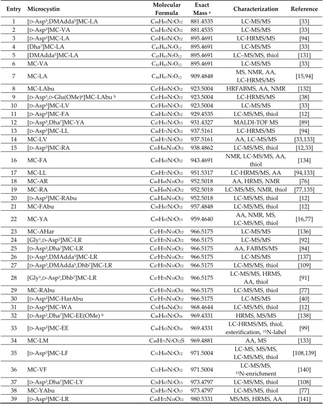
Table 1. Microcystin congeners reported in the literature as identified from cyanobacterial cultures and field samples.
Entry Microcystin Molecular
Formula
Exact
Mass a Characterization Reference 1 [D-Asp³,DMAdda5]MC-LA C44H63N7O12 881.4535 LC-MS/MS [33]
2 [D-Asp³]MC-VA C44H63N7O12 881.4535 LC-MS/MS [33]
3 [D-Asp3]MC-LA C45H65N7O12 895.4691 LC-HRMS/MS [94]
4 [Dha7]MC-LA C45H65N7O12 895.4691 LC-MS/MS [33]
5 [DMAdda5]MC-LA C45H65N7O12 895.4691 LC–MS/MS, thiol [131]
6 MC-VA C45H65N7O12 895.4691 LC-MS/MS [33]
7 MC-LA C46H67N7O12 909.4848 MS, NMR, AA,
LC-HRMS/MS [15,94] 8 MC-LAbu C47H69N7O12 923.5004 HRFABMS, AA, NMR [132]
9 [D-Asp3,D-Glu(OMe)6]MC-LAbu b C47H69N7O12 923.5004 LC-HRMS/MS [38]
10 [D-Asp³]MC-LV C47H69N7O12 923.5004 LC-MS/MS [33]
11 [D-Asp³]MC-FA C48H63N7O12 929.4535 LC-MS/MS, thiol [12]
12 [D-Asp³,Dha7]MC-YA C47H61N7O13 931.4327 MALDI-TOF MS [89]
13 [D-Asp3]MC-LL C48H71N7O12 937.5161 LC-HRMS/MS [94] 14 MC-LV C48H71N7O12 937.5161 AA, LC-MS/MS [33,133] 15 [D-Asp³]MC-RA C45H66N10O12 938.4862 LC-MS/MS, thiol [12,33] 16 MC-FA C49H65N7O12 943.4691 NMR, LC-MS/MS, AA, thiol [134] 17 MC-LL C49H73N7O12 951.5317 LC-HRMS/MS, AA [94,133] 18 MC-AR C46H68N10O12 952.5018 AA, HRMS, NMR [76] 19 MC-RA C46H68N10O12 952.5018 LC-MS/MS, NMR, thiol [77,135] 20 [D-Asp³]MC-RAbu C46H68N10O12 952.5018 LC-MS/MS, thiol [12] 21 MC-FAbu C50H67N7O12 957.4848 LC-MS/MS, thiol [12] 22 MC-YA C49H65N7O13 959.4640 AA, NMR, MS, LC-MS/MS, thiol [16,77] 23 MC-AHar C47H70N10O12 966.5175 LC-MS/MS [136] 24 [Gly1,D-Asp3]MC-LR C47H70N10O12 966.5175 LC-MS/MS [92]
25 [D-Asp3,Dha7]MC-LR C47H70N10O12 966.5175 AA, FABMS/MS [84]
26 [D-Asp³,DMAdda5]MC-LR C47H70N10O12 966.5175 LC-MS/MS [137] 27 [D-Asp³,DMAdda5,Dhb7]MC-LR C47H70N10O12 966.5175 LC-MS/MS, thiol [109] 28 [Gly¹,D-Asp3,Dhb7]MC-LR C47H70N10O12 966.5175 LC-MS/MS, HRMS, AA, thiol [91] 29 MC-RAbu C47H70N10O12 966.5175 LC-MS/MS, thiol [77] 30 [D-Asp3]MC-HarAbu C47H70N10O12 966.5175 LC-MS/MS [40] 31 [D-Asp³]MC-WA C50H64N8O12 968.4644 LC-MS/MS, thiol [12]
32 [D-Asp3,Dha7]MC-EE(OMe) b C46H63N7O16 969.4331 HRMS, MS/MS [138]
33 [D-Asp³]MC-EE C46H63N7O16 969.4331 LC-HRMS/MS, thiol, esterification, 15N-label [99] 34 MC-LM C48H71N7O12S 969.4881 AA, MS [133] 35 [D-Asp3]MC-LF C51H69N7O12 971.5004 LC-MS, MS/MS, LC-MS/MS, thiol [108,139] 36 MC-VF C51H69N7O12 971.5004 15LC-MS/MS, N-enrichment [140] 37 [D-Asp3,Dha7]MC-LY C50H67N7O13 973.4797 LC-MS/MS, thiol [108]
38 MC-YAbu C50H67N7O13 973.4797 LC-MS/MS, thiol [77]
40 [D-Asp3,(E)-Dhb7]MC-LR C48H72N10O12 980.5331 NMR, AA, HRMS [142]
41 [D-Asp3,(Z)-Dhb7]MC-LR C48H72N10O12 980.5331 NMR, AA, HRMS [142]
42 [Dha7]MC-LR C48H72N10O12 980.5331 AA, FABMS/MS [84]
43 [DMAdda5]MC-LR C48H72N10O12 980.5331 AA, HRMS, NMR [76]
44 [Gly1,D-Asp3,Dhb7]MC-LHar C48H72N10O12 980.5331 LC-MS/MS, HRMS,
AA, thiol [91] 45 MC-RApa C48H72N10O12 980.5331 LC-MS/MS, thiol [77] 46 MC-VR C48H72N10O12 980.5331 LC-MS/MS [143] 47 MC-WA C51H66N8O12 982.4800 NMR, LC-MS/MS, AA, thiol [134] 48 [D-Ser1,D-Asp³,Dha7]MC-LR C47H70N10O13 982.5124 LC-MS/MS [33] 49 [Dha7]MC-EE(OMe) b C47H65N7O16 983.4488 HRMS, MS/MS [138]
50 [D-Asp3,Dha7]MCE(OMe)E(OMe) b C47H65N7O16 983.4488 HRMS, MS/MS [138]
51 MC-FL C52H71N7O12 985.5161 LC-MS/MS, thiol [12]
52 MC-LF C52H71N7O12 985.5161 AA, MS [133]
53 MC-KynA b C50H66N8O13 986.4749 LC-MS/MS, HRMS,
thiol, semisynthesis [90] 54 [D-Asp³]MC-LY C51H69N7O13 987.4953 LC-MS/MS, thiol [108]
55 [D-Asp3,(E)-Dhb7]MC-LY C51H69N7O13 987.4953 NMR, LC-HRMS/MS,
thiol [109] 56 [Gly1,D-Asp3,ADMAdda5]MC-LR C48H70N10O13 994.5124 LC-MS/MS [92]
57 [Gly1,D-Asp³,ADMAdda5,Dhb7]MC-LR C48H70N10O13 994.5124 LC-MS/MS, HRMS,
AA, thiol [91] 58 [D-Asp³,ADMAdda5]MC-VR C48H70N10O13 994.5124 LC-MS/MS [33]
59 [D-Asp³,Dhb7]MC-AhaR C49H74N10O12 994.5488 LC-MS/MS, thiol [109]
60 [D-Asp³]MC-Hil/HleR C49H74N10O12 994.5488 LC-MS/MS [33] 61 [D-Asp³,(E)-Dhb7]MC-HilR C49H74N10O12 994.5488 NMR, HRMS, AA [144] 62 [Dha7]MC-HilR C49H74N10O12 994.5488 HRMS, NMR, AA [145] 63 [DMAdda5]MC-HilR C49H74N10O12 994.5488 LC-MS/MS [33] 64 [DMAdda5]MC-LHar C49H74N10O12 994.5488 LC-MS/MS [33] 65 [D-Asp3,D-Glu(OMe)6]MC-LR b C49H74N10O12 994.5488 HRMS, MS/MS, AA [85] 66 MC-LR C49H74N10O12 994.5488 AA, NMR, HRMS, LC-MS/MS [16,141,14 6] 67 [D-Asp3]MC-ER C47H68N10O14 996.4916 LC-HRMS/MS, thiol,
esterification, 15N-label [99] 68 [(6Z)-Adda5]MC-LR b C49H74N10O12 994.5488 NMR, AA, MS [115,147] 69 MC-RL C49H74N10O12 994.5488 LC-MS/MS, thiol [77] 70 MC-WAbu C52H68N8O12 996.4957 LC-MS/MS, thiol [90] 71 [Dha7]MC-E(OMe)E(OMe) b C48H67N7O16 997.4644 HRMS, MS/MS [138] 72 MC-OiaA b C51H66N8O13 998.4749 LC-MS/MS, HRMS, thiol, semisynthesis [90] 73 [D-Asp³]MC-MR C47H70N10O12S 998.4895 LC-MS/MS, thiol, S-oxidation [93]
74 [seco-4/5][D-Asp³]MC-LR b C48H74N10O13 998.5437 LC-MS/MS, thiol [93]
75 [D-Asp³,Mser7]MC-LR C48H74N10O13 998.5437 LC-MS/MS, MS/MS, thiol [88,93] 76 [Ser7]MC-LR C48H74N10O13 998.5437 AA, HRMS, MS/MS [84] 77 MC-LHph C53H73N7O12 999.5317 LC-MS/MS [75] 78 MC-KynAbu b C51H68N8O13 1000.4906 LC-MS/MS, thiol [90] 79 [D-Asp³,Dha7]MC-FR C50H68N10O12 1000.5018 LC-MS/MS [33] 80 [Ser7]MC-EE(OMe) b C47H67N7O17 1001.4593 HRMS, MS/MS [138]
81 [D-Asp3,Ser7]MC-E(OMe)E(OMe) b C47H67N7O17 1001.4593 HRMS, MS/MS [138]
82 [D-Asp³]MC-HilY C52H71N7O13 1001.5110 LC-MS/MS, thiol [108]
83 MC-LY C52H71N7O13 1001.5110 LC-MS/MS, NMR [38,148]
84 MC-YL C52H71N7O13 1001.5110 LC-MS/MS [38]
85 [D-Asp³,Mser7]MC-LY C51H71N7O14 1005.5059 LC-MS/MS, thiol [108]
86 [D-Asp³,ADMAdda5,Dha7]MC-HilR C49H72N10O13 1008.5280 LC-MS/MS [33]
87 [Gly1,D-Asp3,ADMAdda5,Dhb7]MC-LHar C49H72N10O13 1008.5280 LC-MS/MS, HRMS,
AA, thiol [91] 88 [Gly1,D-Asp3,ADMAdda5]MC-LHar C49H72N10O13 1008.5280 LC-MS/MS [92]
89 [D-Asp3,ADMAdda5]MC-LR C49H72N10O13 1008.5280
HRMS, NMR, AA, MS/MS, LC-MS/MS
90 [ADMAdda5,Dha7]MC-LR C49H72N10O13 1008.5280 LC-MS/MS [33] 91 [D-Asp³,ADMAdda5,Dhb7]MC-LR C49H72N10O13 1008.5280 NMR, HRMS, AA [150] 92 MC-HilR C50H76N10O12 1008.5644 MS/MS, HRMS, NMR, AA [80] 93 MC-LHar C50H76N10O12 1008.5644 AA, MS/MS, HRMS, NMR [151] 94 [D-Glu(OMe)6]MC-LR b C50H76N10O12 1008.5644 HRMS, MS/MS, AA, HRMS/MS [85,152] 95 [Mdhb7]MC-LR C50H76N10O12 1008.5644 AA, MS [79] 96 [D-Leu1,D-Asp³,DMAdda5]MC-LR C50H76N10O12 1008.5644 LC-MS/MS [153]
97 [D-Asp3,Dha7]MC-RR C47H71N13O12 1009.5345 AA, NMR, HRMS [76]
98 [D-Asp³,DMAdda5]MC-RR C47H71N13O12 1009.5345 MS, MS/MS [55] 99 [Gly1,D-Asp3]MC-RR C47H71N13O12 1009.5345 LC-MS/MS [92] 100 [Gly1,D-Asp3,Dhb7]MC-RR C47H71N13O12 1009.5345 LC-MS/MS, HRMS, AA, thiol [91] 101 [D-Asp3]MC-LW C53H70N8O12 1010.5113 LC-MS, MS/MS [139] 102 [D-Asp3,(E)-Dhb7]MC-LW C53H70N8O12 1010.5113 NMR, LC-HRMS/MS [95] 103 MC-OiaAbu b C52H68N8O13 1012.4906 LC-MS/MS, thiol [90] 104 MC-MR C48H72N10O12S 1012.5052 LC-MS/MS, thiol, S-oxidation [93] 105 [Mser7]MC-LR C49H76N10O13 1012.5593 LC-HRMS [80,94] 106 [seco-4/5]MC-LR b C49H76N10O13 1012.5593 LC-MS/MS, HRMS, thiol, NMR [93,154] 107 [seco-1/2]MC-LR b C49H76N10O13 1012.5593 MS/MS, HRMS, NMR [154] 108 MC-NfkA b C51H66N8O14 1014.4698 NMR, LC-MS/MS, HRMS, thiol, semisynthesis [90] 109 [D-Asp³]MC-M(O)R b C47H70N10O13S 1014.4845 LC-MS/MS, thiol, S-oxidation [93] 110 [D-Asp³,Dha7]MC-HphR C51H70N10O12 1014.5175 LC-MS/MS [33] 111 [D-Asp3]MC-FR C51H70N10O12 1014.5175 AA, MS, NMR [87] 112 [Dha7]MC-FR C51H70N10O12 1014.5175 AA, HRMS, MS/MS [155] 113 [DMAdda5]MC-FR C51H70N10O12 1014.5175 LC-MS/MS [137] 114 [D-Asp³]MC-RF C51H70N10O12 1014.5175 LC-MS/MS, thiol [108] 115 [Ser7]MC-E(OMe)E(OMe) b C48H69N7O17 1015.4750 HRMS, MS/MS [138] 116 MC-LHty C53H73N7O13 1015.5266 LC-MS/MS [75]
117 [D-Asp³,Dha7]MC-RY C50H68N10O13 1016.4967 LC-MS/MS, thiol [108]
118 [D-Asp³,DMAdda5]MC-RY C50H68N10O13 1016.4967 LC-MS/MS, thiol [109]
119 MC-YM C51H69N7O13S 1019.4674 AA, NMR, MS [16]
120 [D-Asp3,ADMAdda5]MC-LHar C50H74N10O13 1022.5437 HRMS, MS/MS, AA [86]
121 [ADMAdda5]MC-LR C50H74N10O13 1022.5437 HRMSNMR, AA,
MS/MS [78,79] 122 [D-Leu1,DMAdda5]MC-LR C51H78N10O12 1022.5801 LC-HRMS/MS, thiol [81]
123 [D-Leu1,dmAdda5]MC-LR (isomer 1) c C51H78N10O12 1022.5801 LC-HRMS/MS, thiol [81]
124 [D-Leu1,dmAdda5]MC-LR (isomer 2) c C51H78N10O12 1022.5801 LC-HRMS/MS, thiol [81]
125 [D-Leu1,D-Asp3]MC-LR C51H78N10O12 1022.5801 LC-MS/MS, HRMS/MS [152,153]
126 [D-Leu1,Dha7]MC-LR C51H78N10O12 1022.5801 LC-MS/MS [75]
127 [D-Val1]MC-LR C51H78N10O12 1022.5801 LC-MS/MS [75]
128 [Gly1,D-Asp³,Dhb7]MC-RHar C48H73N13O12 1023.5502 LC-MS/MS, HRMS,
AA, thiol [91] 129 [D-Asp3]MC-RR C48H73N13O12 1023.5502 AA, HRMS, NMR [76,156]
130 [Dha7]MC-RR C48H73N13O12 1023.5502 AA, HRMS, MS/MS,
NMR [157]
131 [D-Asp3,(E)-Dhb7]MC-RR C48H73N13O12 1023.5502 NMR, HRMS [158]
132 [Gly1,D-Asp3]MC-RHar C48H73N13O12 1023.5502 LC-MS/MS [92]
133 [DMAdda5]MC-RR C48H73N13O12 1023.5502 LC-HRMS/MS, thiol [82]
134 MC-WL C54H72N8O12 1024.5270 LC-MS/MS, thiol [90]
135 MC-LW C54H72N8O12 1024.5270 LC-MS/MS,
15N-enrichment [140]
136 [D-Asp3]MC-RCit C48H72N12O13 1024.5342 LC-HRMS/MS, thiol,
15N-label [99]
138 [Seco-1/2]MC-HilR b C50H78N10O13 1026.5750 MS/MS, HRMS [80]
139 [D-Asp3,Ser7]MC-RR C47H73N13O13 1027.5451 LC-MS/MS, thiol [159]
140 MC-NfkAbu b C52H68N8O14 1028.4855 LC-MS/MS, thiol [90] 141 MC-M(O)R b C48H72N10O13S 1028.5001 AA, HRMS, NMR [76] 142 MC-FR C52H72N10O12 1028.5331 AA, HRMS, NMR [76,160] 143 MC-RF C52H72N10O12 1028.5331 LC-MS/MS, thiol [77] 144 [D-Asp³]MC-HphR C52H72N10O12 1028.5331 MS/MS [88] 145 [Dha7]MC-HphR C52H72N10O12 1028.5331 AA, HRMS, MS/MS, 1H-NMR [161] 146 [D-Asp³]MC-M(O2)R b C47H70N10O14S 1030.4794 LC-MS/MS, thiol, S-oxidation [93] 147 [D-Asp3,Dha7]MC-HtyR C51H70N10O13 1030.5124 AA, HRMS, MS/MS,
NMR [161] 148 [D-Asp³,DMAdda5]MC-HtyR C51H70N10O13 1030.5124 MS/MS [88] 149 [Dha7]MC-YR C51H70N10O13 1030.5124 HRMS, MS/MS, AA [162] 150 [D-Asp3]MC-RY C51H70N10O13 1030.5124 HRMS, LC-MS/MS, thiol [108,163,1 64] 151 [Dha7]MC-RY C51H70N10O13 1030.5124 LC-MS/MS, thiol [77]
152 [D-Asp³,Dhb7]MC-RY C51H70N10O13 1030.5124 LC-MS/MS, thiol [109]
153 [D-Asp3]MC-YR C51H70N10O13 1030.5124 AA, HRMS, MS/MS [165]
154 [D-Asp3,(E)-Dhb7]MC-YR C51H70N10O13 1030.5124 NMR, LC-HRMS/MS [95]
155 [DMAdda5]MC-YR C51H70N10O13 1030.5124 LC–MS/MS, thiol [131]
156 MC-LY(OMe) C53H73N7O14 1031.5216 LC-MS/MS, thiol [108] 157 [D-Asp³]MC-(H4)YR C51H74N10O13 1034.5437 LC–MS/MS, thiol [131] 158 [Dha7]MC-(H4)YR C51H74N10O13 1034.5437 HRMS, NMR, AA [145] 159 [DMAdda5]MC-(H4)YR C51H74N10O13 1034.5437 LC-MS/MS [137] 160 MC-YM(O) b C51H69N7O14S 1035.4623 AA, NMR, MS [166] 161 [ADMAdda5]MC-HilR C51H76N10O13 1036.5593 LC-MS/MS [33]
162 [ADMAdda5]MC-LHar C51H76N10O13 1036.5593 HRMS, NMR, AA,
MS/MS [78,79] 163 MC-AnaR C52H80N10O12 1036.5957 LC–MS/MS, thiol [131]
164 [D-Leu1]MC-LR C52H80N10O12 1036.5957 NMR, HRMS, MS/MS,
AA [167,168] 165 [D-Asp³]MC-YY C54H67N7O14 1037.4746 LC-MS/MS, thiol [108]
166 [Gly1,D-Asp3,ADMAdda5,Dhb7]MC-RR C48H71N13O13 1037.5294 LC-MS/MS, HRMS,
AA, thiol [91] 167 [Gly1,D-Asp3,ADMAdda5]MC-RR C49H71N13O13 1037.5294 LC-MS/MS [92]
168 MC-RR C49H75N13O12 1037.5658 NMR, AA, MS [48]
169 [(6Z)-Adda5]MC-RR b C49H75N13O12 1037.5658 NMR, AA, MS [115,147]
170 [D-Asp3,D-Glu(OMe)6]MC-RR b C49H75N13O12 1037.5658 NMR, AA, MS/MS [169]
171 [D-Asp3]MC-RHar C49H75N13O12 1037.5658 LC-HRMS/MS [170] 172 [D-Ser1,ADMAdda5]MC-LR C50H74N10O14 1038.5386 HRMS, MS/MS, AA [86] 173 [D-Met1,D-Asp3]MC-LR C50H76N10O12S 1040.5365 LC-MS/MS [75] 174 [ADMAdda5,Mser7]MC-LR C50H76N10O14 1040.5542 HRMS, MS/MS, AA [86] 175 [Ser7]MC-RR C48H75N13O13 1041.5607 AA, HRMS, MS/MS [84] 176 [D-Asp³,Mser7]MC-RR C48H75N13O13 1041.5607 AA, HRMS, MS/MS [114] 177 [D-Asp³,Thr7]MC-RR C48H75N13O13 1041.5607 MS/MS [171]
178 [seco-1/6][D-Asp3]MC-RR b C48H75N13O13 1041.5607 NMR, AA, MS/MS [169]
179 MC-HphR C53H74N10O12 1042.5488 LC-MS/MS, thiol [34,93] 180 [D-Glu(OMe)6]MC-FR b C53H74N10O12 1042.5488 LC-MS/MS [40] 181 [D-Leu1]MC-LY C55H77N7O13 1043.5579 LC-HRMS/MS, LC-MS/MS [97,172] 182 MC-M(O2)R b C48H72N10O14S 1044.4950 LC-MS/MS, thiol, S-oxidation [93] 183 [D-Asp3]MC-HtyR C52H72N10O13 1044.5280 AA, MS, NMR [173]
184 [Dha7]MC-HtyR C52H72N10O13 1044.5280 AA, HRMS, MS/MS,
1H-NMR [161]
185 [D-Asp3,(E)-Dhb7]MC-HtyR C52H72N10O13 1044.5280 NMR, AA, HRMS [142]
186 [D-Asp3,(Z)-Dhb7]MC-HtyR C52H72N10O13 1044.5280 NMR, AA, HRMS [142]
187 MC-RY C52H72N10O13 1044.5280 LC-MS/MS, NMR, thiol [77,163]
188 MC-YR C52H72N10O13 1044.5280 AA, NMR, MS [16]
![Figure 2. Examples of chemical reactions of MCs demonstrated using the hypothetical congener [ADMdda 5 ]MC-RM](https://thumb-eu.123doks.com/thumbv2/123doknet/13998833.455727/19.892.163.736.111.501/figure-examples-chemical-reactions-demonstrated-hypothetical-congener-admdda.webp)
![Figure 3. X-ray crystallographic structure showing the interaction of microcystin-LR (MC-LR) with the PP2A catalytic subunit and its adjacent amino acid side chains (based on data provided in [257])](https://thumb-eu.123doks.com/thumbv2/123doknet/13998833.455727/25.892.120.785.111.406/figure-crystallographic-structure-interaction-microcystin-catalytic-adjacent-provided.webp)
