Publisher’s version / Version de l'éditeur:
Journal of Nutritional Biochemistry, 17, June 6, pp. 396-401, 2006
READ THESE TERMS AND CONDITIONS CAREFULLY BEFORE USING THIS WEBSITE. https://nrc-publications.canada.ca/eng/copyright
Vous avez des questions? Nous pouvons vous aider. Pour communiquer directement avec un auteur, consultez la première page de la revue dans laquelle son article a été publié afin de trouver ses coordonnées. Si vous n’arrivez pas à les repérer, communiquez avec nous à PublicationsArchive-ArchivesPublications@nrc-cnrc.gc.ca.
Questions? Contact the NRC Publications Archive team at
PublicationsArchive-ArchivesPublications@nrc-cnrc.gc.ca. If you wish to email the authors directly, please see the first page of the publication for their contact information.
NRC Publications Archive
Archives des publications du CNRC
This publication could be one of several versions: author’s original, accepted manuscript or the publisher’s version. / La version de cette publication peut être l’une des suivantes : la version prépublication de l’auteur, la version acceptée du manuscrit ou la version de l’éditeur.
For the publisher’s version, please access the DOI link below./ Pour consulter la version de l’éditeur, utilisez le lien DOI ci-dessous.
https://doi.org/10.1016/j.jnutbio.2005.08.013
Access and use of this website and the material on it are subject to the Terms and Conditions set forth at
Effects of different phytosterol analogs on colonic mucosal cell
proliferation in hamsters
Jia, Xiaoming; Ebine, Naoyuki; Wang, Yanwen; Awad, Atif B.; Jones, Peter J.
H.
https://publications-cnrc.canada.ca/fra/droits
L’accès à ce site Web et l’utilisation de son contenu sont assujettis aux conditions présentées dans le site
LISEZ CES CONDITIONS ATTENTIVEMENT AVANT D’UTILISER CE SITE WEB.
NRC Publications Record / Notice d'Archives des publications de CNRC:
https://nrc-publications.canada.ca/eng/view/object/?id=972024af-e059-46ae-8f2b-850e6ca85ac8 https://publications-cnrc.canada.ca/fra/voir/objet/?id=972024af-e059-46ae-8f2b-850e6ca85ac8Effects of different phytosterol analogs on colonic
mucosal cell proliferation in hamsters
Xiaoming Jia
a, Naoyuki Ebine
a, Yanwen Wang
b, Atif B. Awad
c, Peter J.H. Jones
a,T
a
School of Dietetics and Human Nutrition, McGill University, Macdonald Campus, QC, Canada H9X 3V9
b
Institute for Nutrisciences and Health, National Research Council of Canada, Charlottetown, PE, Canada C1A 5T1
c
Department of Exercise and Nutrition Sciences, State University of New York, Buffalo, NY 14214, USA Received 10 May 2005; received in revised form 15 July 2005; accepted 21 August 2005
Abstract
Objective: The aim of this study was to investigate the effects of different phytosterols and their analogs on colonic mucosal cell proliferation in hamsters.
Method: Hamsters (n = 70) were randomly assigned to seven groups after a 2-week acclimation and fed the experimental diet for 5 weeks. Diets included (i) the semipurified diet with no cholesterol (Con), (ii) the Con diet plus 0.25% cholesterol (Ch-con), or the Ch-con diet with (iii) 1% phytosterols (Ste), (iv) 1% phytostanols (Sta), (v) 1.76% sterol esters (esterified to fish oil, SteF), (vi) 0.71% stanol esters (esterified to ascorbic acid [disodium ascorbyl phytostanol phosphate, FM-VP4], 0.7% StaA) and (vii) 1.43% stanol esters (1.4% StaA), respectively. After 5 weeks on experimental diet, hamsters were sacrificed, and colons were collected. Colonic mucosal cell proliferation was measured by immunohistochemistry using monoclonal antibodies against antigen Ki-67.
Results: Colonic mucosal cell proliferation was 21.4% ( P b.01) lower in the 0.7%, but not 1.4%, StaA relative to the Ch-con group. In addition, a lower ( 13.9%) cell proliferation was observed in the SteF group in comparison to the Ch-con group; however, this difference achieved only a borderline level of statistical significance ( P = .069). No differences were observed between Con and Ch-con, as well as among Ste, Sta, 1.4% StaA and Ch-con treatments.
Conclusion: Plant stanols esterified to ascorbic acid may possess anticarcinogenic properties in the colon by suppressing colonic mucosa cell proliferation; however, this effect was not observed with free plant sterols or stanols.
D2006 Elsevier Inc. All rights reserved.
Keywords: Plant sterols; Plant stanols; Sterol esters; Stanol esters; n-3 Polyunsaturated fatty acid; Ascorbic acid; Colon cell proliferation; Ki-67
1. Introduction
The interest in studying plant sterols (PS) was initiated from findings that PS can reduce plasma total cholesterol and low-density lipoprotein cholesterol levels, and thus
offer a protection against cardiovascular disease [1–7].
Recent evidence indicates that consumption of PS, apart from lowering cholesterol levels, also provides protection against certain cancers, such as colon cancer [8 –10]. It has been suggested that the inhibition of colonic mucosal cell proliferation by PS is associated with a lower risk of colon
cancer development [11]. Dietary supplementation of
sitosterol resulted in a 39% reduction in the number of rats that developed methylnitrosourea-induced tumors and a
60% reduction of tumor cells per rat, in comparison to controls [8]. Consistently, in vitro studies have shown an inhibitory effect of h-sitosterol on the growth of HT-29 cells, a human colon cancer cell line, after 5 days of incubation in a medium containing h-sitosterol at 16 Amol/L
[9]. Similarly, Awad et al. [10] reported that feeding rats with a diet containing 2% PS resulted in the normalization of cholic acid-induced hyperproliferation of colonocytes. Recent studies suggest that antioxidants, such as vitamins C and E, can enhance the chemotherapeutic efficacy of antitumor agents [12,13], raising the question of whether vitamin C can work synergistically with PS to reduce cancer development in the colon.
In addition, epidemiological and experimental evidence has demonstrated that n-3 polyunsaturated fatty acids (PUFA) can protect the colon from cancer development. Alaskan, Greenland Eskimos and West-Coast fishermen
0955-2863/$ – see front matter D 2006 Elsevier Inc. All rights reserved. doi:10.1016/j.jnutbio.2005.08.013
T Corresponding author. Tel.: +1 514 398 7547; fax: +1 514 398 7739. E-mail address: peter.jones@mcgill.ca (P.J.H. Jones).
consume higher levels of n-3 PUFA than other North
Americans and have lower rates of colon cancer [14 –16].
An inverse relationship has been reported between fish or fish oil consumption and the risk of colorectal cancer
[17,18]. The inhibitory effect of n-3 PUFA on colon cancer has been confirmed by results from several laboratory studies in rodents, supplemented with either
eicosapentae-noic acid (EPA) or docosahexaeeicosapentae-noic acid (DHA)[19 –22].
However, it is not known whether plant sterols esterified to n-3 PUFA can provide added protection against colon cancer development.
The objectives of the present study were, therefore, to investigate the effects of dietary supplementation with different PS analogs, including plant sterols, stanols, sterols esterified to n-3 fatty acids of fish oil and stanols esterified to ascorbic acid on colonic mucosal cell proliferation in hamsters.
2. Materials and methods 2.1. Animals and diets
Seventy male golden Syrian hamsters, weighing 100 –120 g (Charles River Laboratories, Montreal, QC, Canada), were housed individually in stainless steel mesh cages and subjected to a 12:12 light/dark cycle. Animals were fed a regular rodent chow with free access to diet and water. After 2 weeks of adaptation, hamsters were randomly divided into seven groups and fed the experimental diets for 5 weeks. Group 1 was given a semipurified corn starch– casein–sucrose diet with no cholesterol (Con). Group 2 was given the Con diet plus 0.25% cholesterol (Ch-con). The remaining five groups were given the Ch-con diet contain-ing either 1% phytosterols (Ste), 1% phytostanols (Sta), 1.76% sterol esters (esterified to fish oil, SteF), 0.71% stanol
esters (esterified to ascorbic acid [disodium ascorbyl phytostanol phosphate, FM-VP4], 0.7% StaA) or 1.43% stanol esters (1.4% StaA). Each diet contained 5% oil, in the form of a mixture of beef tallow and safflower oil, to provide a polyunsaturated to saturated fatty acids ratio of 0.4. Composition of the experimental diets is presented in
Table 1. The SteF diet contained the same amount of plant sterols as the Ste diet. Diets containing 0.7% and 1.4% StaA provided 0.5% and 1% free plant stanols, respectively. Plant sterol analogs were mixed into the oil by heating at 558C before blending into diets. Diets were prepared every
2 weeks and stored at 208C.
After 5 weeks of experimental diet feeding, hamsters were anaesthetized with halothane and killed by decapita-tion. Colons were removed, flushed twice with phosphate-buffered saline (PBS, pH 7.4) and fixed in 10% neutral buffered formalin.
The experiment was reviewed and approved by the Animal Care and Research Ethics Committee of McGill University and was conducted in accordance with the guidelines of the Canadian Council on Animal Care. 2.2. Measurement of colon cell proliferation
The formalin-fixed colon tissues were embedded in paraffin and cut into slices at a thickness of 5 Am and dried at 608C for 1 h. Slides were deparaffinized with xylene and rehydrated using graded alcohols. Endogenous peroxidase
was quenched with 3% H2O2for 15 min and washed with
PBS containing Triton (PBST). Antigens were retrieved with citrate buffer (pH 6.0) and microwaved twice with 10 min each, allowed to cool for 15 min and washed with PBST. The slides were then placed on an autostainer, washed with PBST and blocked with PBST containing 0.03% casein at room temperature for 30 min. The primary antibody Ki-67 (Novus Biologicals, Littleton, CO, USA)
Table 1
Composition (% wt/wt) of the experimental diets
Ingredients Cona Ch-con Ste Sta SteF 0.7% StaA 1.4% StaA
Casein 20.0 20.0 19.8 19.8 19.6 19.8 19.7
Corn starch 28.0 28.0 27.7 27.7 27.5 27.7 27.6
Sucrose 36.3 36.0 35.6 35.6 35.3 35.6 35.5
Beef tallow/safflower oil 5.0 5.0 5.0 5.0 5.0 5.0 5.0
Cellulose 5.0 5.0 4.9 4.9 4.9 4.9 4.9 dl-Methionine 0.5 0.5 0.5 0.5 0.5 0.5 0.5 Mineral mixture 4 4 4 4 4 4 4 Vitamin mixture 1 1 1 1 1 1 1 Choline bitartrate 0.2 0.2 0.2 0.2 0.2 0.2 0.2 Butylated hydroxytoluene 0.02 0.02 0.02 0.02 0.02 0.02 0.02 Cholesterol – 0.25 0.25 0.25 0.25 0.25 0.25 Phytosterols – – 1.0 – – – – Phytostanols – – – 1.0 – – – SteFb – – – – 1.76 – – StaAc – – – – – 0.71 1.43
a Con: non-cholesterol control diet; Ch-con: cholesterol control diet which was the non-cholesterol control diet with 0.25% cholesterol added; Ste:
cholesterol control diet with 1% plant sterols added; Sta: cholesterol control diet with 1% plant stanols added; 1.76% SteF: cholesterol control diet with 1.76% sterol esterified to fish oil added; 0.7% and 1.4% StaA: cholesterol control diet with 0.71% or 1.43% stanol esterified with ascorbate added.
b
SteF: plant sterols esterified to fish oil; 1.76% SteF contains an equivalent amount of 1% unesterified phytosterols.
c
StaA: plant stanols esterified to ascorbate; 0.7% and 1.4% StaA contain an equivalent amount of 0.5% and 1.0% unesterified phytostanols, respectively. X. Jia et al. / Journal of Nutritional Biochemistry 17 (2006) 396 – 401 397
was diluted by 1:100 with PBS and applied to the slides, which were then incubated at room temperature for 1 h. Rabbit IgG was used as isotype-matched negative control. After washing with PBST, the slides were incubated with biotinylated secondary antibody (goat antirabbit IgG) at room temperature for 30 min. The slides were washed with PBST and treated with an avidin–biotinylated antibody complex (Vector Labs, Burlingame, CA, USA). After 30 min, the slides were washed with PBST and treated with chromogen 3,3V-diaminobenzidine tetrahydrochloride (DAKO, Carpinteria, CA, USA) for 5 min. The counterstain was performed with hematoxylin, and the slides were dehydrated, cleaned and covered with glass lids. Prolifera-tive cells are Ki-67 posiProlifera-tive showing distincProlifera-tively brown stains and were counted using a microscope. For each slide, colonocytes of 10 well-oriented crypts (longitudinal section) were counted. The colonic mucosal cell proliferation was expressed as a labeling index, i.e., the ratio of the number of Ki-67-positive cells to the total number of colonocytes[10]. 2.3. Data analysis and statistics
Data are presented as meansFS.E.M. One-way ANOVA was used to determine whether there was a significant treatment effect. When a significant ANOVA was obtained, the method of least squares means test was applied to test the differences between treatments. All statistical
computa-tions were done using the general linear model procedure of the SAS software[23], and the statistical significance level was set at P b.05.
3. Results
3.1. Body weight and food consumption
No significant difference was observed among the treatment groups for the beginning and final body weights, respectively, after the 5-week feeding period. Similarly, there were no significant differences among the groups in the average or total food intake during the last 2-week period. 3.2. Colon mucosal cell proliferation analysis
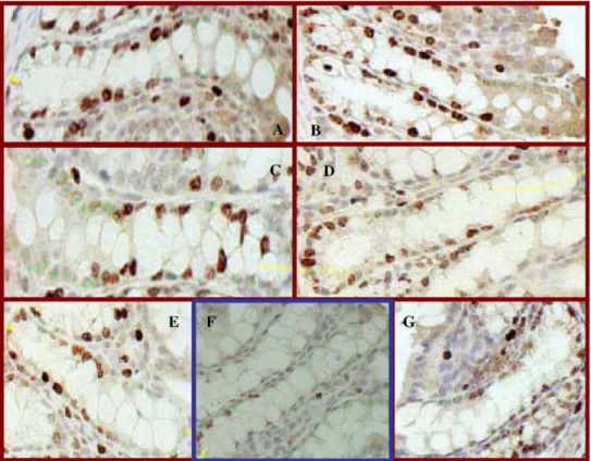
The immunohistochemistry technique for labeling pro-liferative colonocytes with antibody Ki-67 is demonstrated inFig. 1. Cells at G1, S, G2or M phases are considered to be
proliferative and identified by positive binding with Ki-67 monoclonal antibodies. Those cells bearing a dark-brown stain are proliferative cells and are distinct from the nonproliferative cells.
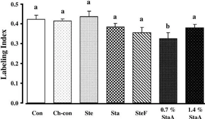
As shown inFig. 2, colon cell proliferation did not differ between Con and Ch-con groups. A decrease ( P b.01) in colon cell proliferation was observed in animals given 0.7% StaA as compared with those fed the Ch-con diet.
Fig. 1. Ki-67-positive colonocytes. Red spots represent proliferative cell, while green spots are normal cells. Labeling index was calculated as the ratio of the number of Ki-67-positive cells to the total number of cells per crypt. (A) Non-cholesterol control diet; (B) cholesterol-added diet; (C) cholesterol- and phytosterol-added diet; (D) cholesterol and phytosterols esterified to fish oil-added diets; (E) cholesterol- and phytostanol-added diet; (F) cholesterol and 0.7% phytostanols esterified to ascorbate-added diets; (G) cholesterol and 1.4% phytostanols esterified to ascorbate-added diets. Diet (F) showed a significant decrease in number of Ki-67-positive cells.
Surprisingly, animals fed 1.4% StaA did not show further inhibition of colon cell proliferation as compared to those fed 0.7% StaA. Hamsters fed SteF tended to have a lower ( 13.9%) proliferation rate of colonocytes relative to those given the Ch-con diet; however, the difference was of borderline significance ( P = .069). No differences were detected between the Con and Ch-con treatments, or among the other groups.
4. Discussion
Results of the present study demonstrate for the first time that plant stanols esterified to ascorbic acid may inhibit colonic mucosal cell proliferation under normal physiolog-ical conditions in hamsters fed an atherogenic diet. It is surprising that the lower dose (0.7%) of stanol esters showed a stronger effect than the higher dose (1.4%), since it has been anticipated that plant stanol esters would produce an inhibitory effect on colon cancer development in a dose-dependent manner. Feeding hamsters with plant sterol esters showed an inhibitory effect on colon mucosal cell prolifer-ation at a marginal level of statistical significance, which was in agreement, to a certain extent, with the results of previous studies[18,24,25]. Free sterols and stanols did not show any effect on the proliferation of colonic mucosal cells, which contradict the results of previous animal studies
[8,10,26] that PS showed significant inhibitory effects on colon mucosal cell proliferation. The discrepancy may be due to the differences in experimental conditions since early studies examined the effects of PS on the proliferation of colon tumors induced by oral administration of either chemical carcinogens[8]or cholic acid [10,26]. Results of these studies also suggest that plant sterols may inhibit the
proliferation of colon tumor cells or stimulated colon mucosal cells, but do not affect the proliferation of healthy colon mucosal cells. Although high dietary cholesterol was considered as a bstimulatorQ of colon mucosal cell prolifer-ation, no effect was indeed observed.
It is known that ascorbic acid has antioxidant activities in the presence of oxidative stress, such as inflammation[27]. Ascorbic acid enhances the chemotherapeutic effect of cyclophosphamide, vinblastine and doxorubicin in mice
when supplemented at a dose of 500 mg/kg per day [28],
while showing no effect when administered at a high dose of
2 g/kg per day [29]. Ascorbic acid possesses not only
antioxidant activity but also pro-oxidant properties, al-though the latter effect remains controversial [30 –34]. A recent study [34]showed that ascorbic acid increases pro-oxidant effect in a dose-dependent manner in macrophages. Data of these early published studies may partly explain the paradoxical results observed in the current study, i.e., ascorbate in StaA functioned as an anti-oxidant at the dose of 0.7% and enhanced the antitumor property of phytosta-nols, while acting as a pro-oxidant at the higher dose of 1.4% and resulted in the loss of inhibitory effect of StaA on colonic mucosal cell proliferation. Since ascorbic acid was not tested separately in the current study, it is impossible to elucidate whether the enhanced inhibitory effect of stanol esters was simply a result of increased bioavailability of phytostanols or due to a synergistic action of phytostanols and ascorbic acid.
Balance between proliferation and apoptosis determines
tumor development and growth. An early study [24]
assessed both proliferation and apoptosis in the colonic crypts of rats exposed to azoxymethane and fed with either a high corn-oil diet or a high fish-oil diet. Results showed that n-3 PUFA consumption suppressed tumor growth by increasing apoptosis instead of inhibiting cell proliferation.
Later, two other studies [20,25] in rats showed that n-3
PUFA in fish oil inhibited the proliferation and promoted the apoptosis of normal colonic mucosa cells and cells in 1,2-dimethylhydrazine-induced aberrant colonic crypts. Present results showed a tendency ( P = .069) of inhibiting normal colonic mucosa cell proliferation by sterols esterified to n-3 PUFA in fish oil, while PS alone did not show any effect. It is implied that fish oil possesses a potential to inhibit colon mucosal cell proliferation. Further studies are required to assess the potential of SteF as a dietary chemopreventive agent, including cell proliferation, apo-ptosis and differentiation.
High circulating cholesterol levels may favor cancer development because they increase the gene expression of cholesterol 7a-hydroxylase, the first and rate-limiting en-zyme in bile acid synthetic pathway, and thus increase the secretion of bile acids, particularly cholic acid[35]. Cholic acid has been reported to be a cancer inducer[10]. However, few studies have been conducted to examine whether cholesterol is a stimulator of colon cancer. Our current findings did not reveal any effect of cholesterol on the cell
Fig. 2. Effects of different phytosterol analogs on normal colonic mucosal cell proliferation in hamsters. Data across treatment groups were analyzed by one-way ANOVA. Where a significance level of less than .05 was achieved; differences between group means were evaluated using the least squares means test. Data are presented as meansFS.E.M. (n = 10). Values with different letters are significantly different ( P b.05). Con: non-cholesterol control diet; Ch-control: cholesterol-added diet; Ste: cholesterol- and phytosterol-added diet; Sta: cholesterol- and phytosta-nol-added diet; SteF: cholesterol and phytosterols esterified to fish oil-added diets; 0.7% and 1.4% StaA: cholesterol and phytostanols esterified to ascorbate-added diets.
proliferation of colonic mucosa; indicating that the choles-terol level used in the current study may not be high enough to increase cholic acid concentration to the level that passes the colon and stimulate the colon mucosal cell proliferation. In summary, dietary supplementation of stanol esterified with ascorbic acid at the lower dose significantly inhibited colon cell proliferation rate, and sterol esterified with n-3 PUFA in fish oil showed a trend of inhibiting colonic cell proliferation. Free sterols and stanols did not affect colonic mucosal cell proliferation under normal physiological conditions. Dietary cholesterol at the dose of the present study did not show any effect on colonic mucosal cell proliferation. Our results implicated that plant sterol esters and stanol esters may possess capabilities to protect or delay cancer development in the colon.
Acknowledgments
The authors wish to thank Mrs. Sandra Mendel, M.A., and Dr. Szczesny, T.M., of the University at Buffalo for processing the tissues of the immunohistochemistry and the technical assistance in microscopy. We also thank Mr. Gordon Bingham at McGill University for his assistance in animal care. This study was supported by a grant from the Natural Sciences and Engineering Research Council of Canada. The phytostanols esterified to ascorbic acid were a generous gift of Forbes Medi-Tech, Inc., Vancouver, BC, Canada.
References
[1] Pollack OJ. Reduction of blood cholesterol in man. Circulation 1953;7:702 – 6.
[2] Pollack OJ, Kritchevsk D. Sitosterol. Monogr Atheroscler 1981;10: 1 – 219.
[3] Jones PJH, Ntanios FY, Raeini-Sarjaz M, Vanstone CA. Cholesterol-lowering efficacy of a sitostanol-containing phytosterol mixture with a prudent diet in hyperlipidemic men. Am J Clin Nutr 1999;69: 1140 – 50.
[4] Hallikainen MA, Sarkkinen ES, Gylling H. Comparison of the effects of plant sterol ester and plant stanol ester-enriched margarines in lowering serum cholesterol concentrations in hypercholesterolaemic subjects on a low-fat diet. Eur J Clin Nutr 2000;54:715 – 25. [5] Vanstone CA, Raeini-Sarjaz M, Parsons WE, Jones PJ. Unesteried
plant sterols and stanols lower LDL-cholesterol concentrations equivalently in hypercholesterolemic persons. Am J Clin Nutr 2002;76:1272 – 8.
[6] Skeaff CM, Thoma C, Chisholm A, Mann J, Williams S. Effects on plasma lipids when plant sterol enriched fat spread or carbohydrate provide replacement energy for saturated fatty acids. Asia Pac J Clin Nutr 2004;13:80 – 7.
[7] Thomsen AB, Hansen HB, Christiansen C, Green H, Berger A. Effect of free plant sterols in low-fat milk on serum lipid profile in hypercholesterolemic subjects. Eur J Clin Nutr 2004;58:860 – 70. [8] Raicht RF, Cohen LI, Fazzini EP, Sarwal AN, Takahashi M. Protective
effect of plant sterols against chemically induced colon tumors in rats. Cancer Res 1980;40:403 – 5.
[9] Awad AB, Chen YC, Fink CS, Hennessey T. h-Sitosterol inhibits HT-29 human colon cancer cell growth and alters membrane lipids. Anticancer Res 1996;16:2797 – 804.
[10] Awad AB, Hernandez AYT, Fink CS, Mendel SL. Effect of dietary phytosterols on cell proliferation and protein kinase C activity in rat colonic mucosa. Nutr Cancer 1997;27:210 – 5.
[11] Lipkin M, Uehara K, Winawer S, Blattner WA, Fraumeni J. Seventh-day Adventist vegetarians have a quiescent proliferative activity in colonic mucosa. Cancer Lett 1985;26:139 – 44.
[12] Conklin KA. Dietary antioxidants during cancer chemotherapy: impact on chemotherapeutic effectiveness and development of side effects. Nutr Cancer 2000;37:1 – 18.
[13] Pathak AK, Singh N, Khanna N, Reddy VG, Prasad KN, Kochupillai V. Potentiation of the effect of paclitaxel and carboplatin by antioxidant mixture on human lung cancer H520 cells. J Am Coll Nutr 2002;21:416 – 21.
[14] Blot WJ, Lanier A, Fraumeni JF, Bender TR. Cancer mortality among Alaskan natives, 1960–1969. J Natl Cancer Inst 1975;55: 547 – 54.
[15] Bang HO, Dyerberg J, Hjoorne N. The composition of food consumed by Greenland Eskimos. Acta Med Scand 1976;200:69 – 73. [16] Schloss I, Kidd MS, Tichelaar HY, Young GO, O’Keefe SJ. Dietary
factors associated with a low risk of colon cancer in coloured west coast fishermen. S Afr Med J 1997;87:152 – 8.
[17] Caygill CP, Charlett A, Hill MJ. Fat, fish, fish oil and cancer. Br J Cancer 1996;74:159 – 64.
[18] Bartsch H, Nair J, Owen RW. Dietary polyunsaturated fatty acids and cancers of the breast and colorectum: emerging evidence for their role as risk modifiers. Carcinogenesis 1999;20:2209 – 18.
[19] Minoura T, Takata T, Sakaguchi M, Takada H, Yamamura M, Hioki K, et al. Effect of dietary eicosapentaenoic acid on azoxymethane-induced colon carcinogenesis in rats. Cancer Res 1988;48:4790 – 4. [20] Calviello G, Palozza P, Maggiano N, Piccioni E, Franceschelli P,
Frattucci A, et al. Cell proliferation, differentiation, and apoptosis are modified by n-3 polyunsaturated fatty acids in normal colonic mucosa. Lipids 1999;34:599 – 604.
[21] Rose DP, Connolly JM. Omega-3 fatty acids as cancer chemo-preventive agents. Pharmacol Ther 1999;83:217 – 44.
[22] Boudreau MC, Sohn KH, Rhee SH, Lee SW, Hunt JD, Hwang DH. Suppression of tumor cell growth both in nude mice and in culture by n-3 polyunsaturated fatty acids: mediation through cyclooxygenase-independent pathways. Cancer Res 2001;61:1386 – 91.
[23] SAS Institute. SAS user’s guide: statistics, version 6.12. Cary, NC7 SAS Institute; 1994.
[24] Chang WL, Chapkin RS, Lupton JR. Fish oil blocks azoxymethane-induced rat colon tumorigenesis by increasing cell differentiation and apoptosis rather than decreasing cell proliferation. J Nutr 1998; 128:491 – 7.
[25] Latham P, Lund EK, Johnson IT. Dietary n-3 PUFA increases the apoptotic response to 1,2-dimethyldrazine, reduces mitosis and suppresses the induction of carcinogenesis in the rat colon. Carcino-genesis 1999;20:645 – 50.
[26] Janezic SA, Rao AV. Dose-dependent effect of dietary phytosterols on epithelial cell proliferation of the murine colon. Food Chem Toxicol 1992;30:611 – 6.
[27] Frei B. Ascorbic acid protects lipids in human and low-density lipoprotein against oxidative damage. Am J Clin Nutr 1991;54: 1113S – 8S.
[28] Taper HS, Gerlache J, Lans M, Roberfroid M. Non-toxic potentiation of cancer chemotherapy by combined C and K vitamin pre-treatment. Int J Cancer 1987;40:575 – 9.
[29] Shimpo K, Nagatsu T, Yamada K, Sato T, Fujita K. Ascorbic acid and adriamycin toxicity. Am J Clin Nutr 1991;54:1298S – 301S. [30] Crott JW, Fenech M. Effect of vitamin C supplementation on
chromosome damage, apoptosis and necrosis ex vivo. Carcinogenesis 1999;20:1035 – 41.
[31] Chen K, Suh J, Carr AC, Morrow JD, Zeind J, Frei B. Vitamin C suppresses oxidative lipid damage in vivo, even in the presence of iron overload. Am J Physiol Endocrinol Metab 2000;279: E1406 – 12.
[32] Proteggente AR, Rehman A, Halliwell B, Rice-Evans CA. Potential problems of ascorbate and iron supplementation: pro-oxidant effect in vivo? Biophys Res Commun 2000;277:535 – 40.
[33] Suh J, Zhu BZ, Frei B. Ascorbate does not act as a pro-oxidant towards lipids and proteins in human plasma exposed to redox-active transition metal ions and hydrogen peroxide. Free Radic Biol Med 2003;34:1306 – 14.
[34] Ashidate K, Kawamura M, Tohda H, Miyazaki S, Hayashi H, Teramoto T, et al. Ascorbic acid augments cytotoxicity induced by oxidized low-density lipoprotein. J Atheroscler Thromb 2003;10: 7 – 12.
[35] Horton JD, Cuthbert JA, Spady DK. Regulation of hepatic 7a-hydroxylase expression and response to dietary cholesterol in the rat and hamster. J Biol Chem 1995;270:5381 – 7.