NEUROSCIENCE
Large-conductance calcium-dependent potassium
channels prevent dendritic excitability in neocortical
pyramidal neurons
Narimane Benhassine&Thomas Berger
Received: 30 January 2008 / Revised: 25 June 2008 / Accepted: 29 July 2008 / Published online: 2 September 2008 # Springer-Verlag 2008
Abstract Large-conductance calcium-dependent potassium channels (BK channels) are homogeneously distributed along the somatodendritic axis of layer 5 pyramidal neurons of the rat somatosensory cortex. The relevance of this conductance for dendritic calcium electrogenesis was studied in acute brain slices using somatodendritic patch clamp recordings and calcium imaging. BK channel activation reduces the occurrence of dendritic calcium spikes. This is reflected in an increased critical frequency of somatic spikes necessary to activate the distal initiation zone. Whilst BK channels repolarise the somatic spike, they dampen it only in the distal dendrite. Their activation reduces dendritic calcium influx via glutamate receptors. Furthermore, they prevent dendritic calcium electrogenesis and subsequent somatic burst discharges. However, the time window for coincident somatic action potential and dendritic input to elicit dendritic calcium events is not influenced by BK channels. Thus, BK channel activation in layer 5 pyramidal neurons affects cellular excitability
primarily by establishing a high threshold at the distal action potential initiation zone.
Keywords BK channels . Neocortical pyramidal neurons . Dendrite . Critical frequency . Rat . Calcium-dependent potassium channels . Calcium action potential . Pyramidal neuron . Sensorimotor cortex
Introduction
The apical dendrite of layer 5 pyramidal neurons is an excitable structure, characterised by a high threshold action potential (AP) initiation zone in the distal tuft [43]. Neocor-tical layer 5 neurons receive qualitatively different inputs along their somatodendritic axis [1]. In the trigeminal system, tactile information is primarily relayed in the thalamus which projects via different pathways to the deep granular layers of the neocortex. These thalamic inputs are directly and indirectly projecting onto the soma and the basal dendrites of the layer 5 pyramidal neurons [8]. This sensory information can give rise to a sodium spike at the somatic AP initiation zone [28] which is not only propagated along the axon but also backpropagated into the apical dendrite [47]. Associative intracortical inputs impinge on the distal portion of the apical dendrite. Neither a backpropagating somatic AP alone nor isolated associative inputs to the distal tuft are usually sufficient to elicit a dendritic calcium AP. However, the coincidence of a backpropagating AP with an excitatory input to the distal dendrite effectively halves the threshold for a dendritic AP (BAC firing; [25]). Once a calcium AP is initiated in the distal dendrite, it propagates forward to the soma where it can induce in turn a short burst of sodium APs. The mutual activation of both AP initiation zones (somatodendritic interaction) switches the DOI 10.1007/s00424-008-0569-3
Electronic supplementary material The online version of this article (doi:10.1007/s00424-008-0569-3) contains supplementary material, which is available to authorized users.
N. Benhassine
:
T. BergerInstitute of Physiology, University of Bern, Bühlplatz 5,
CH-3012 Bern, Switzerland N. Benhassine (*)
Brain Research Institute, University of Zürich, Winterthurerstrasse 190,
CH-8057 Zürich, Switzerland e-mail: [email protected] T. Berger
Institute of Physiology and Pathophysiology, University of Mainz, Duesbergweg 6,
spiking pattern of the cell from regular spiking to burst discharge [27].
Dendritic conductances are decisive for the integrative properties of pyramidal cells in the hippocampus and neo-cortex. Dendritic A-type potassium and hyperpolarisation-activated conductances [5,19,32] determine spike shape and passive membrane properties, respectively. Thus, these conductances have an impact on suprathreshold and sub-threshold signals. Large-conductance calcium-activated po-tassium channels (BK channels; [31,39,41]) participate in the AP repolarisation [12,46]. In CA1 pyramidal cells, BK channels shape the somatic AP but do not influence its backpropagation [37]. In contrast, in the same cell type, Golding and coworkers [15] showed that BK channel activation curtails dendritic calcium events. Functional BK channels were found to be homogeneously distributed all along the somatodendritic axis of layer 5 pyramidal neurons [3]. The fast kinetics of BK channels (1 ms activation time constant and 3 ms deactivation time constant under 10μM free calcium), the high opening probability (0.69 under 10 μM free calcium) and the absence of inactivation [3] suggest a possible opening of these channels in the time course of a single sodium AP. Furthermore, the voltage and calcium dependence of these channels suggest that the emergence of a calcium AP in the distal dendrite could massively open dendritic BK channels. Their strong cal-cium dependence suggests in addition that subthreshold depolarisations can activate BK channels only if this depolarisation is accompanied by a local but massive increase in intracellular calcium (half-maximal activation of the BK channels at −65 and +48 mV for 10 and 1 μM calcium, respectively; [3]). This study presents evidence that BK channels activated by depolarisation and calcium influx of both distal dendritic inputs and backpropagating APs enhance the threshold for the initiation of the dendritic calcium spike without affecting the temporal window of its activation by coincident dendritic and somatic inputs. This effect is due to a hyperpolarisation of the distal dendrite after the activation of BK channels, whilst a reduced AP propagation seems to be of less importance. Thus, BK channel activation is in the position to reduce the dendritic excitability in layer 5 pyramidal neurons.
Materials and methods
Brain slice preparation and cell identification Three hundred-micrometer-thick parasagittal slices of the somato-sensory cortex were prepared from 28- to 37-day-old Wistar rats. This was carried out according to national and institutional guidelines to minimise pain and discomfort of the animals. Rats were deeply anaesthetised with halothane and killed by decapitation. The brain was removed rapidly,
chilled in 0–4°C extracellular solution and then sliced using a vibratome (Dosaka Microslicer DTK-1000, Kyoto, Japan). Slices were incubated at 37°C for 15 min, and then left at room temperature until recording. Layer 5 pyramidal neurons from the somatosensory area with a thick apical dendrite were visualised by infrared differential interference contrast video microscopy utilising a Newvicon camera (C2400, Hamamatsu, Hamamatsu City, Japan) and an infrared filter (RG9, Schott, Mainz, Germany) mounted on an upright microscope (Axioskop FS, Zeiss, Oberkochen, Germany).
Electrophysiology Current clamp recordings were made from the soma or simultaneously from the soma and the apical dendrite of rat neocortical layer 5 pyramidal neurons in the whole-cell configuration (Figs. 1, 2, 3 and 4). An Axoprobe 1A amplifier (Axon Instruments, Union City, CA, USA) or a BVC-700A amplifier (Dagan, Minneapolis, MN, USA) were used. Electrodes were made from borosilicate glass tubing (Hilgenberg, Malsfeld, Germany) using a micropipette puller (P-87; Sutter Instrument,
Fig. 1 BK channels reduce the likelihood of occurrence and the amplitude of dendritic calcium APs following somatic spike trains. a Four consecutive APs were induced in the soma with DC current injection (6 nA, 2 ms). Their frequency was either insufficient (below the critical frequency; left traces) or sufficient (beyond the critical frequency; right traces) to elicit a dendritic calcium AP under control conditions (black traces). Data were simultaneously recorded at soma (top traces) and dendrite (lower traces; 550μm from the soma). In the somatic recording, the dendritic calcium AP is reflected as an afterdepolarisation. The blockade of BK channels with 1μM paxilline (gray traces) increases the amplitude (right traces) and decreases the threshold (left traces) of the dendritic calcium AP as seen in a decrease of the critical frequency. b Upper part relative reduction in the critical frequency obtained from the somatic afterdepolarisation, following BK channel blockade. Lower part the area under the curve of the dendritic signal from eight cells is plotted against the somatic AP frequency and normalised to the value obtained under control conditions at 143 Hz. c Same as in a, following blockade of BK channels with 100 nM iberiotoxin (gray traces). Dendritic recording at 370μm from the soma. d As in a, a train of four spikes was induced in the soma followed by a single spike with a 1-s delay (upper traces). The spike train had frequencies of 83, 111 and 166 Hz. Single spikes and spike trains below a critical frequency induce only small changes in intracellular calcium concentration monitored with calcium imaging at 600 μm from the soma (OGB-1, 200 μM). Beyond the critical frequency under control conditions (black traces), a large dendritic calcium event is evoked under control (166 Hz). One micromolar paxilline decreases this critical frequency to 111 Hz (gray traces). The calcium increase due to a spike train beyond the critical frequency becomes even larger under paxilline. e Left part the dendritic change in fluorescence due to the spike train was plotted against the spiking frequency. This plot reflects the larger increase in intracellular calcium concentration following a stimulation capable to activate the distal calcium AP initiation zone. Right part relative change in the critical frequency of somatic APs, recorded with calcium imaging at the distal apical dendrite (between 690 and 750μm from the soma) following BK channel blockade
Novato, CA, USA). Their resistance was 10–16 MΩ for dendritic and 6–8 MΩ for somatic recordings when filled with the intracellular solution. The access resistance, measured after gigaseal formation and every 10 min, was stable during the course of the experiment. The access resistance was 26.0±6.7 and 34.7±5.0 MΩ for somatic and dendritic recordings, respectively, at the beginning of the experiment and 27.0±7.3 and 34.7±4.7 MΩ for somatic and dendritic recordings, respectively, at the end of the
experiment (mean recording time=52±14 min, n=7 somato-dendritic recordings). Recordings were rejected if the access resistance increased by more than 10% of the initial value. Data were low-pass filtered at 3 kHz using the internal filter of the amplifier. The sampling frequency was 10–20 kHz. Data were digitised and stored on-line using Clampex9 (Axon Instruments). Data were analysed off-line with Clampfit9. Bridge balance and capacitance compensation was used in all recordings. Single APs were induced by
f = 83 Hz f = 111 Hz f = 166 Hz 80% ∆F / F
d
e
Spiking frequency [Hz] ∆ F / F [%] 100 150 100 150 250 50 200 300 50 0 Somaa
0 0.2 0.4 0.6 0.8 1 Control PaxillineNorm. critical frequency
b
20 mV 500 ms Control 1 M Paxilline 0 0.2 0.4 0.6 0.8 1 Control PaxillineNorm. critical frequency
**
* *
f = 95 Hz f = 133 Hz Dendrite 20 mV 10 ms 0 0.5 1.0 1.5 2.0 60 80 100 120 140 160 180Norm. dendritic integral
Spiking frequency [Hz]
c
Soma Dendrite Control 100 nM Iberiotoxin f = 98 Hz f = 76 Hz 0 0.5 1 1.5 2 40 60 80 100 Spiking frequency [Hz]Norm. dendritic integral
20 mV 10 ms
Control 1 M Paxilline
suprathreshold short DC current pulses (5–6 nA, 2–4 ms duration) injected into the soma (Fig.2a). In order to subtract the depolarisation due to the passive membrane properties and stimulation artefacts, the cell was then hyperpolarised until the same current injection failed to elicit an action potential. Ten traces obtained either at the original or at the hyperpolarised membrane potential were averaged. Average traces obtained by DC pulse injection from a hyperpolarised potential were subtracted from the average traces obtained by
DC pulse injection from the resting membrane potential. Averaged action potentials under control conditions and BK channel blockade were aligned to the AP threshold defined by the initial rise of the AP derivative. The values for AP half-width under control conditions and BK channel blockade were normalised and compared for statistical significance using ANOVA for within-group comparison with significance levels set to 0.05 or 0.01. The same statistical test was used for the analysis of the fluorescent
1 ms 10 mV 0 0.2 0.4 0.6 0.8 1 1.2 1.4 Control Paxilline
Norm. AP half width
a
10 ms 10 % ∆F / Fb
Control Paxilline Norm. ∆ F / F 0 0.2 0.4 0.6 0.8 1 1.2 1.4 Control 1 M Paxilline*
c
*
* *
5 ms Soma 250 m 450 m 550 m 620 mNorm. Peak Amplitude
Distance from Soma ( m)
0 0.2 0.4 0.6 0.8 1 1.2 0 100 200 300 400 500 600 700
Distance from Soma ( m)
Norm. Half Width
0 1 3 4 4 6 8 0 100 200 300 400 500 600 700 7 2 600 m 680 m 10 % ∆F / F 100 ms 1 ms 10 mV
Fig. 2 BK channels participate in the somatic AP repolarisation and contribute weakly to its dendritic attenuation. a Single APs were induced with a somatic DC current injection (5 nA, 4 ms). Traces were aligned and stimulation artefacts removed. Relative increase in somatic AP width at half peak amplitude under control and with BK channel blockade (n=6 cells). b The change in somatic calcium concentration during an AP is increased following blockade of BK channels (OGB-1, 200μM). Relative change under paxilline (n=7 cells). c Top panel averaged APs evoked by DC current pulse at the soma (400 ms; 0.2–0.5 nA) and recorded simultaneously in the soma (left traces) and in the dendrite at different locations from the soma (right traces). The dendritic recordings were obtained from four cells at different dendritic recording distances indicated. Middle panel dendritic calcium transients evoked by a single backpropagated AP are larger after BK channel blockade (data taken from two cells imaged at different dendritic locations). Lower panel AP peak amplitude and half-width are normalised to the somatic AP and plotted as a function of the dendritic location under control and paxilline con-ditions. The blockade of BK channels reduces slightly the peak attenuation at distal dendritic locations, whilst the half-width is less affected
peak induced by a somatic AP (Fig.2b) and for the analysis of the minimal spiking frequency of a train of somatic APs capable to elicit a dendritic calcium AP (critical frequency; Fig.1b). This same test was also used for the analysis of the fluorescent peak in the dendrite induced by a somatically triggered dendritic calcium AP (Fig.1e) and for the analysis of the fluorescence peak induced by glutamate iontophoresis on the dendrite (Fig.3). Dendritic excitability was studied by injection of 400 ms long DC pulses (0.2–1.6 nA in 0.1 nA steps) in the dendrite (Fig.4). The AP threshold was defined as the first current step able to induce a somatic AP. Pooled data are expressed as the mean±standard deviation (SD). All experiments were done at 34°C.
Calcium imaging The hexapotassium salt of the calcium-sensitive dye Oregon Green BAPTA-1 (Kdvalue 170 nM; Molecular Probes, Eugene OR, USA) was used to monitor changes in cytosolic calcium concentration, whilst mem-brane voltage changes were recorded simultaneously. Two hundred micromolar of Oregon Green BAPTA was added to the patch pipette solution and diffused freely into the cell interior. This concentration allowed the filling of the dendritic structures of a layer 5 pyramidal cell within 30– 45 min. Changes in intracellular calcium were imaged with a Zeiss Axioskop 2FS+ microscope equipped with ×40/0.8 NA optics. The light source was a 75-W Xenon short arc lamp in a monochromator (Deltaram V, PTI, Lawrenceville NJ, USA) equipped with a shutter. Excitation of the dye was obtained at 480/15 nm. A dichroic mirror (510 nm) and an emission filter BP540/25 nm were used to detect the emitted light. The increase in fluorescence of the non-ratiometric dye Oregon Green BAPTA reflecting an increase in the intracellular calcium concentration could be well-detected without any binning or averaging with a low-resolution, fast CCD camera (Redshirt NeuroCCD, RedShirt Imaging, Fairfield CT, USA). The CCD camera was running under Neuroplex with an un-binned full frame rate of 1 kHz and a field of view of 280×280 μm
(un-binned pixel field 3.5×3.5 μm, 80×80 pixels). Imaging data were digitised and stored on a PC using the Neuroplex software (Figs.1d,2b,c, 3and 5).
Glutamate iontophoresis In iontophoresis experiments, the cytosol of layer 5 pyramidal cells was filled with Oregon
a
Glutamate Iontophoresis Pipette Patch Pipette 100 µ m 0 1 2 3 4 5 6 Norm. ∆ F/F Control Paxilline 10 %∆F/Fb
Control 1 µM Paxilline**
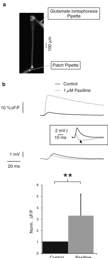
20 ms 1 mV 2 mV 10 ms Fig. 3 Postsynaptic BK channels reduce glutamate-induced calciuminflux in the distal dendrite. a Fluorescence image of a layer 5 pyramidal neuron visualised using excitation of the calcium-insensitive dye Alexa 594. The calcium imaging location is marked by a white rectangle. The positions of the glutamate iontophoresis electrode and of the recording electrode are indicated. b Top panel averaged dendritic [Ca2+]isignals
(OGB-1, 200 μM) induced by dendritic glutamate iontophoresis (500 mM, 0.1 ms, 3 V pulse; top traces) and corresponding electrophysiological averaged trace recorded from the soma (lower traces) under control (black traces) and bath application of 1 μM paxilline (gray traces). In few cases, the postsynaptic potential showed an increased inhibitory component after paxilline application (inset, arrowhead). Lower panel relative peak values of [Ca2+]isignals induced
by glutamate iontophoresis before and after BK channel blockade. Paxilline increases the glutamate-induced dendritic calcium influx (p=0.004; n=3 cells)
Green BAPTA-1 and the calcium-insensitive dye Alexa 594 (Molecular Probes; excitation 560 nm, dichroic mirror 580 nm, emission filter LP590 nm). A iontophoretic glass pipette with the tip size of a somatic patch pipette was advanced onto the distal dendritic arbor of the recorded layer 5 pyramidal neuron at a distance of 700–750 μm from the soma. Five hundred millimolar glutamate was applied iontophoretically with 0.1 ms long pulses ranging from +2 to +20 V (Figs.3 and5).
Chemicals and solutions Slices were continuously super-fused with a physiological extracellular solution containing (in millimolar): 125 NaCl, 25 NaHCO3, 2.5 KCl, 1.25 NaH2PO4, 2 CaCl2, 1 MgCl2, 20 glucose, 5 pyruvate, bubbled with 95% O2 and 5% CO2. For current clamp experiments without additional calcium imaging (Figs.1a–
c,2a,4, Supplementary Figs.1a and2), the pipette solution contained (in millimolar): 110 K-gluconate, 30 KCl, 10 EGTA, 10 HEPES, 5 CaCl2, 4 Mg-ATP, 0.3 Na2-GTP, 10 Na2-phosphocreatine (free calcium concentration of 100 nM). Pipette solution for combined current clamp recording and calcium imaging (Figs. 1d, 2b,c, 3 and 5) contained: 135 K-gluconate, 5 KCl, 4 Mg-ATP, 0.3 Na2 -GTP, 10 Na2-phosphocreatine, 10 HEPES, 0.2 Oregon Green BAPTA-1. For iontophoretic application experiments (Figs.3 and 5), the patch pipette solution contained in addi-tion the calcium-insensitive dye Alexa Fluor 594 (15 μM; Molecular Probes). The iontophoresis pipette contained 500 mM glutamate and 15μM Alexa Fluor 594 diluted in extracellular solution. The pH was adjusted in the extracellu-lar and the intracelluextracellu-lar solutions to 7.3. Osmoextracellu-larity of the intracellular solutions was in the range of 315 to 320 mOsm.
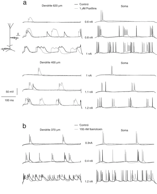
Soma Dendrite 620 µm 0.6 nA 0.8 nA 1 nA Soma Dendrite 400 µm 1 nA 1.1 nA 1.2 nA Control 1 M Paxilline 50 mV 100 ms
a
b
0.3nA 0.4 nA 1.2 nA Control 100 nM Iberiotoxin Soma Dendrite 370 µmFig. 4 BK channels impair dendritically evoked burst discharges. a DC current pulses were injected in the dendrites of two pyramidal cells (400 ms duration; 640 and 400μm from the soma). Recordings from the soma (right traces) and from the dendrite (left traces) were obtained under control (black traces) or with 1μM paxilline (gray traces). b Same as in a, but BK channels were blocked by 100 nM iberiotoxin (dendrit-ic recording at 370μm from the soma). BK channel blockade enables the induction of dendritic calcium events resulting in somatic bursts discharges
The high affinity and selective BK channel blockers paxilline [22] and iberiotoxin [14] were purchased from Alomone Labs (Jerusalem, Israel). All other drugs were from Sigma or Merck. Paxilline and iberiotoxin were first dissolved in dimethyl sulphoxide (DMSO) at 1 mM and bidistillated water at 100 μM, respectively. Aliquots of these stock solution were kept at−20°C and diluted 1:1,000 in the external solution directly before use. Paxilline or iberiotoxin at final concentrations of 1 μM and 100 nM, respectively, were bath applied with the extracellular solution. In the paxilline experiments, the final concentra-tion of DMSO was 0.1%, a concentraconcentra-tion that was found to have no effect on BK channels in control experiments. The effective BK channel blockade with paxilline occurred between 2 and 4 min after the change of the perfusion (Supplementary Fig. 3). A similar time course was observed for the perfusion of extracellular solution con-taining iberiotoxin. Data obtained in the first 10 min following change of perfusate were not used for evaluation.
Results
BK channels limit dendritic excitability following somatic action potentials
A dendritic calcium AP can be initiated by coincident dendritic and somatic input (BAC firing mechanism; [25]) or alternatively by a train of backpropagating somatic APs at a frequency higher than a critical value (critical frequency, CF; [24]). To study the impact of BK channel activation on dendritic excitability, whole-cell current clamp experiments were performed using a potassium-based intracellular solution containing a free calcium concentration of 100 nM mimicking physiological calcium levels [18]. We induced dendritic calcium APs with trains of four somatic spikes triggered by somatic current injections (6 nA, 2 ms, frequency range from 49 to 400 Hz). This was done in control conditions and after BK channel blockade with the specific blockers paxilline or iberiotoxin (Fig. 1a,c) [14, 22]. In the somatic recording, the dendritic calcium AP is reflected as a prominent afterdepolarisation (Fig.1a; compare to [24]). The dendritic calcium AP appears at a significantly lower CF under paxilline (1 μM) in comparison to control conditions. In somatodendritic patch clamp recordings (mean distance 540μm from the soma), paxilline reduces the CF to 56.6% of the control (127.1 ±35.7 Hz under control conditions and 71.9±13.9 Hz under paxilline; p=0.003; n=7 cells; Fig.1a, b). In addition, paxilline increases the amplitude and integral of the calcium AP recorded in the dendrite (Fig. 1a,b). Similarly, the blockade of BK channels with 100 nM iberiotoxin lowers the CF to 76.4±1.6% of the con-trol (n=3 cells) and increases the integral of the dendritic calcium AP (Fig. 1c). Application of 1 μM paxilline in addition to 100 nM iberiotoxin does not intensify the effects observed under iberiotoxin alone.
In additional experiments, we have combined somatic current clamp recordings with calcium imaging experiments in the soma and apical dendrite. Changes in the intracellular calcium concentration were induced by trains of four somatic action potentials below and above the CF as well as single spikes (Fig.1d). The somatic calcium concentra-tion increases with the number of spikes and the spiking frequency (control values; single spike =11.5±7.9% ΔF/F; spike trains below the CF=26.4±14.5%ΔF/F; spike trains above the CF=35.7±18.1%ΔF/F; n=7 cells, not shown). The calcium transients at the calcium AP initiation zone (580±172 μm distal to the soma; compare to [26]) are small for single backpropagating spikes (mean 6.1%ΔF/F, range between 0.6%ΔF/F at 900 μm and 12.24% ΔF/F at 400μm; n=5 imaging positions in three cells) and for trains of backpropagating spikes below the CF (mean 11.3%ΔF/ F, range between 0.8%ΔF/F at 900 μm and 22.1% ΔF/F ∆t (ms) Norm. ∆ F/F 0 1 2 3 -80 -60 -40 -20 0 20 40 60 80 10% ∆F/F 25 mV 20 ms Control 1 µM Paxilline
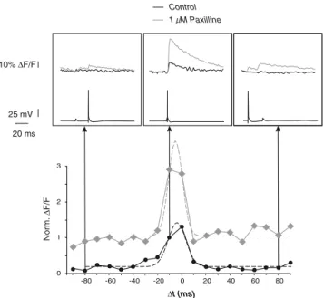
Fig. 5 BK channels reduce the amplitude but not the time window for induction of the calcium spike triggered by coincident somatic and dendritic inputs. Upper panel change in intradendritic calcium concentration (dendritic calcium imaging at 670μm from the soma, top traces in insets) and electrophysiological traces (lower traces in insets) induced by a combination of a single backpropagating AP (DC current pulse 1 nA, 1 ms) and a dendritic EPSP induced by glutamate iontophoresis on the distal dendrite (700μm from the soma) under control (black traces) and paxilline (gray traces) conditions. Lower panel all ΔF/F values for both conditions are normalised to that obtained under control conditions with aΔt of −10 ms. Δt represents the time between the onset of the somatic current injection triggering the backpropagated AP and the onset of the glutamate iontophoresis stimulation. Changes in fluorescence are fitted by Gaussian functions (dashed lines) of different peak amplitude but similar mean and sigma (mean−3.8 ms and sigma 7.4 ms under control; mean −5.1 ms and sigma 4.7 ms under paxilline for the cell shown here)
at 400μm; n=5 imaging positions in three cells). However, calcium transients due to spike trains above the CF are large
(mean 61.0% ΔF/F, range between 22.6% ΔF/F and
102.2% ΔF/F; n=5 imaging positions in three cells) (Fig. 1d). The small calcium transients are due to single backpropagating spikes or groups of backpropagating spikes with a low frequency insufficient to activate the dendritic calcium AP zone, whilst the large transients are due to an activation of the calcium AP zone. In this set of experiments, under control condition, a frequency of 116.6± 23.1 Hz is found to be critical for the initiation of calcium spikes (Fig. 1d,e; n=8 cells). Blockade of BK channels with 1 μM paxilline resulted in a large increase of the intracellular calcium for spike trains above the CF (151.2 ± 4.3% of the control at somatic and 202.7±37.4% of the control at dendritic imaging sites; n=2 somatic and n=5 dendritic imaging positions; p=0.017 for all seven record-ings; Fig. 1d,e). The CF in the distal apical dendrite is reduced to 77.6% of the control (116.6±23.1 Hz under control conditions and 90.6±8.4 Hz under paxilline; p= 0.007; n = 8 cells; Fig. 1e). Also, under iberiotoxin application, the amplitude of the dendritic calcium event following somatic spikes above the CF increases to 188.1± 63.6% of the control and reduced the CF to 76.4% of the control (n=3 cells; not shown). These data show a clear role of BK channels in setting a high threshold for dendritic calcium action potentials induced by somatic activity. BK channels shorten the somatic action potential and tend to reduce its backpropagation in layer 5 pyramidal cells Different mechanisms may underlie the reduced impact of somatic spike trains on dendritic calcium AP generation following BK channel activation. One possibility is the modulation of the somatic AP shape. A sodium spike of a reduced half-width undergoes stronger attenuation during its backpropagation (compare to [6]) and is less likely to elicit a calcium spike in the distal dendrite. In order to test this, we induced somatic APs under control conditions and with BK channel blockade. Figure2a shows averaged AP waveforms from the same cell following stimulus artefact and leak subtraction under both conditions. The spike under 1 μM paxilline shows a significant prolongation (half-width =0.71±0.03 ms under control, 0.87±0.05 ms with paxilline; p=0.004; n=6 cells). This is mainly due to a reduction of the fast afterhyperpolarisation of the AP underlying the repolarisation slope. In addition, paxilline application increases the somatic intracellular calcium concentration accompanying isolated APs (Fig.2b). These data demonstrate the physiological activation of BK channels during a single somatic AP, an effect already expected due to the fast activation kinetics found on the single channel level [3].
In a next step, we have investigated whether BK channel activation could affect the ability of the somatic spike to backpropagate into the apical dendrite. We used simulta-neous whole-cell recordings on the soma and the apical dendrite of layer 5 pyramidal neurons and compared the dendritic changes in AP peak amplitude and half-width due to BK channel blockade (Fig. 2c). Backpropagating APs were evoked by DC pulse current injection at the soma (400 ms; 0.2 to 0.5 nA, four dendritic recording locations
between 250 and 620 μm). The backpropagating AP
undergoes a distance-dependent peak attenuation and increase in half-width. In the distal dendrite (620μm from soma), the AP peak amplitude was 13 mV under control and 21 mV under BK blockade. This depolarisation should be insufficient to open high-voltage calcium channels and, therefore, should fail to activate dendritic BK channels. However, in the distal dendrite only (>400 μm), the blockade of BK channels tends to reduce the peak amplitude attenuation. Changes in the half-width of the backpropagating AP in the distal portion of the apical dendrite are less clear (Fig. 2c). In another set of experi-ments, the calcium transients induced by the backpropaga-tion of single APs or trains of APs below the CF were studied in the distal dendrite. Calcium transients due to single spikes increase to 182.1±88.0% of the control after BK channel blockade (Fig.2c middle panel). Similarly, the calcium transients for spike trains below the CF increase to 175.8±61.7% of the control (n=5 dendritic recordings in three cells; distance 400 to 900 μm from the soma; not shown). The dendritic calcium transients have comparable half-widths under control and in paxilline. But one has to have in mind that the kinetics of imaged calcium transients are primarily a function of the unbinding of calcium from the dye and not of the underlying changes in membrane potential or calcium concentration [16]. In addition, results obtained with calcium imaging are always hampered by the fact that calcium-sensitive dyes do not respond in a linear fashion to the changes in membrane potential [4]. Taken together, these results show that BK channels tend to reduce the AP backpropagation in the distal apical dendrite. BK channels reduce dendritic calcium influx
via ligand-gated conductances
In addition to backpropagating APs in the apical dendrite, BK channels may also be activated by dendritic inputs opening glutamate-gated NMDA receptor channels. BK channels would prevent the depolarisation necessary to activate dendritic calcium channels and thereby the den-dritic calcium action potential discharge. We mimicked an excitatory input on the distal apical dendrite of layer 5 pyramidal neurons (690–750 μm from the soma; Fig.3a). Following iontophoretic glutamate application (500 mM;
0.1 ms duration; +2 to +20 V pulse, n=3 cells; compare to [44]), we imaged the changes in dendritic calcium concentration (Fig.3b). Glutamate iontophoresis induces a rise in intradendritic calcium concentration which is significantly enhanced following an overall blockade of BK channels (ΔF/F=1.9±0.9% under control, 3.7±2.4% under paxilline, p=0.004; Fig.3b and insets of Fig.5). Due to the amplitude of these evoked calcium transients in comparison to those obtained with a dendritic calcium AP (range between 22.6% ΔF/F and 102.2% ΔF/F under control conditions; Fig. 1d), it seems probable that this procedure evokes only a local calcium transient but is insufficient to evoke directly a dendritic calcium AP. Whilst subthreshold activity without increase in intracellular calcium is insufficient to activate dendritic BK channels (Supplementary Fig. 1), the local depolarisation due to distal glutamatergic inputs and the concomitant rise in intradendritic calcium due to the NMDA-type glutamate receptor activation seem to be sufficient to activate dendritic BK channels. In four out of 11 applications, the postsynaptic potentials evoked by the distal glutamate ejection and recorded at the soma had smaller amplitude after paxilline application than under control conditions, suggesting an increased inhibitory component (Fig. 3b, inset, arrowhead). The global blockade of BK channels by bath application of paxilline may have changed the inhibitory postsynaptic component evoked by glutamate iontophoresis. Even though the blockade of BK channels in neocortical GABAergic interneurons does not affect their discharge properties [11], BK channels have been pre-dominantly localised in axons and presynaptic terminals, including those of GABAergic interneurons [33]. Thus, the BK channel blockade might have facilitated the neurotrans-mitter release in this cell type. However, even in these cases, the BK channel blockade led to a bigger increase in distal dendritic fluorescence. In addition, the latency be-tween the glutamate iontophoresis stimulation and the onset of the somatically recorded subsequent excitatory post-synaptic potential (EPSP) was identical for all cells between control and BK blockade conditions, suggesting that the increased change in intracellular calcium concentration could not be due to a variation of the iontophoresis pipette position after paxilline treatment. Dendritic BK channels dampen the depolarisation following distal glutamatergic inputs, preventing the activation of calcium conductances and, therefore, setting a high threshold for the distal action potential initiation zone.
BK channels prevent dendritically evoked burst discharges DC current pulses (400 ms, 0.2–1.6 nA) were injected in the apical dendrite of layer 5 pyramidal neurons (450– 620 μm from the soma) and the voltage responses were
simultaneously recorded at both somatic and dendritic locations (Fig. 4). Following BK channel blockade, a dendritic current injection is more likely to induce somatic spikes compared to control conditions (somatic spike threshold reduced to 82.5±0.13% of the control; n=5 cells). Moreover, in four out of six cells, this dendritic current injection leads to tonic somatic spike discharges under control, but induces doublets or high-frequency burst discharges due to a BAC firing mechanism when BK channels are blocked by paxilline or iberiotoxin (Fig. 4). We saw no evidence of a favoured propagation of the dendritic depolarisation to the soma following BK channel blockade. Indeed, if EPSP-like waveforms (peak amplitude 1,000 pA, time to peak 2.5 ms, decay time constant 5.7 ms; Supplementary Fig. 1a, upper trace) are injected in the dendrite (300 μm from the soma), the blockade of BK channels does not affect the EPSP propagation to the soma (Supplementary Fig. 1a, middle trace). In addition, this result cannot be explained by a modification of the somatic discharge threshold if the BK conductance is blocked. Indeed, neither the somatic nor the dendritic input resis-tance was changed after BK channel blockade (somatic input resistance=26.2±8.0 MΩ under control and 27.1± 8.7 MΩ under paxilline; p=0.88; n=5 cells; dendritic input resistance=19.4±4.7 MΩ under control and 19.2±4.0 MΩ under paxilline; p=0.74; n=6 cells). In addition, 1 μM paxilline does not affect the mean spiking frequency of the cells following somatic DC current (400–1,200 pA, 18 s) or Gaussian-distributed noisy current injection (mean=800– 1,200 pA; sigma=200 pA, 18 s) (Supplementary Fig.2; n= 4 cells). No change was found in the instantaneous frequency or in the spiking frequency adaptation under paxilline.
In the presence of paxilline, the same dendritic current injection changes the spiking pattern of some cells from a regular spiking discharge to a burst discharge (Fig. 4). BK channel blockade prevents the deactivation of calcium conductances allowing a lower threshold at the dendritic AP initiation zone. This result suggests a functional importance of BK channels in limiting the switch from a low level of excitability to a burst discharge pattern in layer 5 pyramidal neurons.
BK channels do not change the time window for BAC firing
The critical frequency paradigm used in Fig. 1 elicits a calcium spike in the distal dendrite using high-frequency somatic spikes discharges [24]. Such burst discharges of three to five APs at frequencies between 200 and 400 Hz have been reported in layer 5 pyramidal neurons in vivo firing spontaneously or following sensory stimulation [17]. The temporal coincidence of a backpropagating somatic AP
and a distal dendritic depolarisation within an optimal time window (the distal depolarisation arriving 3 to 7 ms after the backpropagating AP) is the most probable way to induce a calcium AP (BAC firing; [25]). We studied this mechanism in layer 5 pyramidal neurons with somatic patch clamp recordings and simultaneous calcium imaging of the distal dendrite (600–680 μm from the soma). A combination of a backpropagating AP triggered by somatic current injection (DC pulse, 0.6–1 nA, 1 ms) and glutamate iontophoresis at the dendrite (500 mM; 0.1 ms; +2 to +20V pulse) mimics a distal excitatory input. Using this, we have tested whether BK channels could affect the coincidence detection mechanism in layer 5 pyramidal neurons (Fig.5).
As expected, the peak amplitude of calcium concentra-tion rise in the dendrite is much bigger after BK channel blockade compared to control conditions (for coincident timing between the dendritic input and the somatic AP, the dendritic ΔF/F was 6.3±1.1% under control and 14.2± 4.3% under paxilline conditions; n=3 cells). This pro-nounced increase in the distal dendritic calcium influx reinforces the idea of an activation of local BK channels by glutamate-gated conductances, preventing the opening of voltage-gated calcium conductances. In order to compare the time window for calcium AP induction under control and BK channel blockade conditions, the profile of the intradendritic change in calcium concentration was fitted with Gaussian functions (Fig.5, lower part, dashed lines). Similar parameters can be used to fit the change in dendritic calcium concentration under both conditions (mean −4.2 ms and sigma 10.1 ms under control; mean −5.1 ms and sigma 8.2 ms under paxilline; n=3 cells). This result suggests that BK channels do not affect the time window for the induction of calcium APs in the case of coincident somatic and dendritic inputs. Despite a slight attenuation of AP propagation in the distal dendrite (Fig.2), BK channel activation has no influence on the time window for the emergence of calcium spikes. Evidence shows BK channels in the distal dendrite act likely primarily locally to set a high threshold for the dendritic AP initiation zone. The temporal window of calcium AP activation by coincident dendritic and somatic inputs observed is consistent with previous reports [24]. The averaged latency between the onset of the glutamate iontophoresis stimulation and the rise of the corresponding EPSP observed at the soma was 8.5±0.3 ms (n=14 in three cells). Taking into account an approximate conduction velocity of 0.5 m s−1in the apical dendrite of layer 5 pyramidal neurons [27,48], the EPSP is expected to peak in the distal dendrite about 7.1 ms after the onset of the iontophoresis stimulation. Using these corrections, we could observe, in accordance with previous reports [24], a maximal calcium rise in the distal dendrite occurring for a latency of 2.9 ms between the back-propagating AP and the distal EPSP under control
conditions. We observe a real boost of the calcium transient amplitude when BK channels are blocked primarily due to a local dendritic effect on ligand- and voltage-gated calcium conductances. Thus, BK channels act in layer 5 pyramidal cells as effective filters under high levels of cellular activity. They dampen the dendritic excitability and prevent bursting in this cell type.
Discussion
BK channel activation in layer 5 pyramidal cells of the somatosensory cortex depends on two biophysical parame-ters, namely, depolarisation and increased intracellular calcium [3]. These properties limit the influence of this conductance to suprathreshold events like somatic sodium spikes and dendritic calcium action potentials if the calcium increase is moderate (about 1μM). The specific activation pattern of BK channels, in combination with their even expression along the somatodendritic axis, shortens APs at the soma and reduces their ability to initiate dendritic action potentials during a rise in cellular excitability. If the calcium increase is localised and strong enough (about 10μM), moderate depolarisations around the threshold are also sufficient for the activation of BK channels [3].
In layer 5 pyramidal neurons, as in other cell types, BK channels participate in spike repolarisation [12,23,46, this study]. Like BK channels [3], high-voltage-activated calcium channels are expressed at the soma and dendrite of layer 5 pyramidal neurons [21,50]. Furthermore, a co-localisation [7] and co-activation of BK channels and N-type calcium channels has been reported in hippocampal and neocortical pyramidal neurons [29,49]. As Sun et al. [49] have shown in mouse neocortical neurons, the opening of high-voltage-activated calcium channels during the rising phase of the AP provides a sufficient calcium influx for the activation of neighbouring BK channels (see also [34]). BK channel activation leads to the closing of voltage-dependent depolarising sodium and calcium conductances and, therefore, hastens repolarisation. This acceleration of the AP decay was shown not to interfere with repetitive spiking of the cell because BK channels remain open only for a few milliseconds [3,45].
In hippocampal pyramidal neurons, Poolos and Johnston [37] report an effect of BK channels on the AP repolarisa-tion which is restricted to the somatic compartment and conclude that the distribution of BK channels is not uniform along the somatodendritic axis. In contrast, immuno-localisation of BK channels showed their presence in both postsynaptic and presynaptic compartments of hippocampal pyramidal neurons, i.e. also in dendritic spines [40]. BK channels in neocortical pyramidal cells show a uniform distribution along the somatodendritic axis. In
addition, their biophysical properties such as their voltage and calcium dependence or their kinetics are similar in both compartments [3]. This suggests that BK channel activation during somatic and dendritic suprathreshold activity is not only able to shorten sodium but also calcium action potentials. In hippocampal CA1 pyramidal cells, dendritic calcium AP duration and initiation is controlled by BK channels among other potassium conductances [15]. In this study, we show that, in neocortical pyramidal neurons, BK channel activation prevents the generation and reduces the amplitude and duration of dendritic calcium APs.
Which are the mechanisms underlying the effect of BK channels on dendritic excitability? A faster repolarisation of the somatic AP reduces the AP backpropagation into the dendrite and, therefore, the excitability of the dendritic calcium AP initiation zone due to somatic activity (compare to [6]). In the distal dendrite, the AP filtering tends to be stronger under control conditions than under BK channel blockade. Furthermore, the calcium transients corre-sponding to the backpropagated spikes are larger following BK channel blockade. These findings suggest that BK channel activation tends to reduce the AP backpropagation, but this effect is of minor importance in regard of calcium spike generation.
Upon activation, the primary effect of BK channels is to change the membrane potential and thereby to affect the currents underlying the calcium AP. Indeed, BK channel activation reduced the overall integral of calcium APs. This finding is comparable to Purkinje cells where dendritic excitability is enhanced and the spread of dendritic calcium spikes is improved by the block of dendritic BK channels [9, 38, 51]. Following BK channel activation, we have shown a strong reduction of distal calcium influx triggered by glutamate application. Excitatory associative inputs arriving at distal positions on the apical dendrite of layer 5 pyramidal neurons activate dendritic glutamate receptors of the AMPA- and NMDA-type. In the olfactory bulb, NMDA receptors and BK channels are linked and their co-activation leads to membrane potential hyperpolarisation [20]. The calcium influx and the membrane depolarisation resulting from NMDA receptor activation during a dendritic EPSP presumably activate BK channels. This activation can lead consecutively to a closure of the NMDA channels via an increased magnesium block. In contrast, no activation of BK channels is found during dendritic EPSP-like wave-forms. In this case, the depolarisation is not accompanied by a calcium influx. This lack of a calcium increase at the vicinity of the dendritic BK channels leads to the failure of BK channel activation. Another possible mechanism for the glutamatergic activation of BK channels is the activation of metabotropic glutamate receptors (mGluR). Indeed, the mGluR1 subtype is expressed on cell bodies and dendrites of neocortical neurons [30]. In cerebellar neurons, these
receptors are found to activate BK channels [10]. Further-more, other studies report an inhibition of the calcium signaling in hippocampal or neocortical pyramidal neurons following the activation of mGluR1 [13,42]. This subtype of glutamatergic receptors may, therefore, modulate the excitability of the calcium AP initiation zone through mechanisms involving BK conductances.
In layer 5 pyramidal neurons, other voltage- and ligand-gated conductances influence the emergence of calcium APs or BAC firing. Bekkers and Delaney [2] have shown that D-type potassium channels are expressed near the site of AP initiation. This strategic site of expression allows this conductance to regulate the somatic excitability and consequently the excitability of the dendritic AP initiation zone as well. Hyperpolarisation-activated channels have a higher density in the distal apical dendrite than the proximal region [5]. This Ihconductance limits the backpropagation of sodium APs as well as the summation of coincident dendritic inputs [35]. In this way, Ih increases the electrotonic distance between both AP initiation zone and impairs the induction of BAC firing in neocortical pyramidal neurons [6]. In contrast to BK channels, these leak conductances act at the resting membrane potential and can influence thereby not only APs but also the cellular integration at the subthreshold level. A ligand-gated con-ductance, which shunts dendritic activity, acts via GABAA and GABAB receptors. Concomitant activation of distal dendritic GABAergic inputs leads to the so-called veto effect preventing the generation of calcium APs during a long time window [25, 36]. In this respect, BK channels and GABA receptors act comparably because both con-ductances directly impair the generation of the dendritic AP via a hyperpolarisation of the membrane potential.
The functional role of dendritic BK channels is of particular interest in layer 5 pyramidal neurons in the somatosensory cortex. These cells receive qualitatively different inputs along their somatodendritic axis. Tactile information is relayed via the thalamus, and excitatory spiny stellate cells in layer 4, to the soma and the proximal dendrites of layer 5 pyramidal neurons. This sensory input can give rise to a sodium spike at the somatic AP initiation zone, which is backpropagated into the dendrite. On the other hand, inputs from associative cortical areas project on the distal apical dendrite of these neurons [1]. It is likely that neither backpropagated sodium APs nor associative inputs on their own induce a sufficient depolarisation to reach the high threshold of the dendritic AP zone ([27], but see [17, 26] for the generation of calcium APs by distal inputs alone in vivo). However, the coincidence of a back-propagated AP, together with an input on the distal dendrite, is the most likely condition to pass the dendritic threshold. This will induce a calcium AP and switch the output pattern of the cell from a regular spiking pattern to a
burst discharge pattern. The triggering of calcium APs in these cells corresponds to a mechanism detecting the temporal coincidence of sensory and associative inputs. BK channels participate in setting a high threshold for the dendritic AP initiation zone by a direct curtailing of sodium and calcium APs. The activation of BK channels leads to an active decoupling of the somatic and the dendritic AP initiation zones during high-frequency input at somatic and/ or dendritic locations. BK channels would, therefore, modulate the switch between a low-excitability and a high-excitability mode and act as an active dampening filter defining the significance of coincident sensory and associative inputs.
Acknowledgements The results were extensively discussed with Drs. F. Helmchen, M. Giugliano, M. Larkum, H.R. Lüscher and D. Ulrich who, in addition, have read earlier versions of this manuscript. Supported by the Swiss National Science Foundation (grant 3100-107529/1), as well as the Novartis Foundation for Medical–Biological Research and the Bonizzi–Theler Foundation to T.B.
References
1. Abeles M (1984) Corticonics—neural circuits of the cerebral cortex. Cambridge University Press, Cambridge
2. Bekkers JM, Delaney AJ (2001) Modulation of excitability by a-dendrotoxin-sensitive potassium channels in neocortical pyramidal neurons. J Neurosci 21:6553–6560
3. Benhassine N, Berger T (2005) Homogeneous distribution of large-conductance calcium-dependent potassium channels on soma and apical dendrite of rat neocortical layer 5 pyramidal neurons. Eur J Neurosci 21:914–926
4. Berger T, Borgdorff A, Crochet S, Neubauer FB, Lefort S, Fauvet B, Ferezou I, Carleton A, Lüscher HR, Petersen CC (2007) Combined voltage and calcium epifluorescence imaging in vitro and in vivo reveals subthreshold and suprathreshold dynamics of mouse barrel cortex. J Neurophysiol 97:3751–3762
5. Berger T, Larkum ME, Lüscher HR (2001) High Ih channel
density in the distal apical dendrite of layer 5 pyramidal cells increases bidirectional attenuation of EPSPs. J Neurophysiol 85: 855–868
6. Berger T, Senn W, Lüscher HR (2003) Hyperpolarization-activated current Ih disconnects somatic and dendritic spike
initiation zones in layer V pyramidal neurons. J Neurophysiol 4:2428–2437
7. Berkefeld H, Sailer CA, Bildl W, Rohde V, Thumfart JO, Eble S, Klugbauer N, Reisinger E, Bischofberger J, Oliver D, Knaus HG, Schulte U, Fakler B (2006) BKCa-Cav channel complexes mediate
rapid and localized Ca2+-activated K+ signaling. Science 5799: 615–620
8. Bureau I, von Saint Paul F, Svoboda K (2006) Interdigitated paralemniscal and lemniscal pathways in the mouse barrel cortex. PLoS Biol 4:2361–2371
9. Cavelier P, Pouille F, Desplantez T, Beekenkamp H, Bossu JL (2002) Control of the propagation of dendritic low-threshold Ca2+ spikes in Purkinje cells from rat cerebellar slice cultures. J Physiol (Lond) 540:57–72
10. Chavis P, Ango F, Michel JM, Bockaert J, Fagni L (1998) Modulation of big K+channel activity by ryanodine receptors and
L-type Ca2+channels in neurons. Eur J Neurosci 10:2322–2327
11. Erisir A, Lau D, Rudy B, Leonard CS (1999) Function of specific K+ channels in sustained high-frequency firing of fast-spiking neocortical interneurons. J Neurophysiol 82:2476–2489
12. Faber ES, Sah P (2002) Physiological role of calcium-activated potassium currents in the rat lateral amygdala. J Neurosci 22: 1618–1628
13. Frenguelli BG, Potier B, Slater NT, Alford S, Collingridge GL (1993) Metabotropic glutamate receptors and calcium signalling in dendrites of hippocampal CA1 neurones. Neuropharmacology 32:1229–1237
14. Galvez A, Gimenez-Gallego G, Reuben JP, Roy-Contancin L, Feigenbaum P, Kaczorowski GJ, Garcia ML (1990) Purification and characterization of a unique, potent, peptidyl probe for the high conductance calcium-activated potassium channel from venom of the scorpion Buthus tamulus. J Biol Chem 265:11083–11090 15. Golding NL, Jung HY, Mickus T, Spruston N (1999) Dendritic
calcium spike initiation and repolarization are controlled by distinct potassium channel subtypes in CA1 pyramidal neurons. J Neurosci 19:8789–8798
16. Helmchen F, Imoto K, Sakmann B (1996) Ca2+ buffering and action potential-evoked Ca2+signaling in dendrites of pyramidal neurons. Biophys J 70:1069–1081
17. Helmchen F, Svoboda K, Denk W, Tank DW (1999) In vivo dendritic calcium dynamics in deep-layer cortical pyramidal neurons. Nat Neurosci 11:989–996
18. Hille B (2001) Ionic channels of excitable membranes. Sinauer, Sunderland, MA
19. Hoffman DA, Magee JC, Colbert CM, Johnston D (1997) Potassium channel regulation of signal propagation in dendrites of hippocampal pyramidal neurons. Nature 387:869–875 20. Isaacson JS, Murphy GJ (2001) Glutamate-mediated extrasynaptic
inhibition: direct coupling of NMDA receptors to Ca2+-activated K+channels. Neuron 6:877–879
21. Kelly KM, Kume A, Albin RL, Macdonald RL (2001) Auto-radiography of L-type and N-type calcium channels in aged rat hippocampus, entorhinal cortex, and neocortex. Neurobiol Aging 22:17–23
22. Knaus HG, McManus OB, Lee SH, Schmalhofer WA, Garcia-Calvo M, Helms LM, Sanchez M, Giangiacomo K, Reuben JP, Smith AB 3rd (1994) Tremorgenic indole alkaloids potently inhibit smooth muscle high-conductance calcium-activated potas-sium channels. Biochemistry 33:5819–5828
23. Lancaster B, Nicoll RA (1987) Properties of two calcium-activated hyperpolarizations in rat hippocampal neurons. J Physiol (Lond) 389:187–203
24. Larkum ME, Kaiser KM, Sakmann B (1999a) Calcium electro-genesis in distal apical dendrites of layer 5 pyramidal cells at a critical frequency of back-propagating action potentials. Proc Natl Acad Sci USA 25:14600–14604
25. Larkum ME, Zhu JJ, Sakmann B (1999b) A new mechanism for coupling inputs arriving at different cortical layers. Nature 398: 338–341
26. Larkum ME, Zhu JJ (2002) Signaling of layer 1 and whisker-evoked Ca2+ and Na+ action potentials in distal and terminal dendrites of rat neocortical pyramidal neurons in vitro and in vivo. J Neurosci 22:6991–7005
27. Larkum ME, Zhu JJ, Sakmann B (2001) Dendritic mechanisms underlying the coupling of the dendritic with the axonal action potential initiation zone of adult rat layer 5 pyramidal neurons. J Physiol (Lond) 533:447–466
28. Manns ID, Sakmann B, Brecht M (2004) Sub- and suprathreshold receptive field properties of pyramidal neurones in layers 5A and 5B of rat somatosensory barrel cortex. J Physiol (Lond) 15:601–622 29. Marrion NV, Tavalin J (1998) Selective activation of Ca2+
-activated K+ channels by co-localized Ca2+ channels in
30. Martin LJK, Blackstone CD, Huganir RL, Price DL (1992) Cellular localization of a metabotropic glutamate receptor in brain. Neuron 9:259–270
31. Marty A (1981) Calcium-dependent potassium channels with large unitary conductance in chromaffin cell membrane. Nature 291:497–500
32. Migliore M, Shepherd GM (2002) Emerging rules for the distributions of active dendritic conductances. Nat Rev Neurosci 3:362–370
33. Misonou H, Menegola M, Buchwalder L, Park EW, Meredith A, Rhodes KJ, Aldrich RW, Trimmer JS (2006) Immunolocalization of the Ca2+-activated K+ channel Slo1 in axons and nerve
terminals of mammalian brain and cultured neurons. J Comp Neurol 496:289–302
34. Müller A, Kukley M, Uebachs M, Beck H, Dietrich D (2007) Nanodomains of single Ca2+channels contribute to action potential repolarization in cortical neurons. J Neurosci 27:483–495 35. Nicoll A, Larkman A, Blakemore C (1993) Modulation of EPSP
shape and efficacy by intrinsic membrane conductances in rat neocortical pyramidal neurons in vitro. J Physiol (Lond) 468:693– 710
36. Perez-Garci E, Gassmann M, Bettler B, Larkum ME (2006) The GABAB1b isoform mediates long-lasting inhibition of dendritic Ca2+spikes in layer 5 somatosensory pyramidal neurons. Neuron 50:603–616
37. Poolos NP, Johnston D (1999) Calcium-activated potassium conductances contribute to action potential repolarization at the soma but not the dendrites of hippocampal CA1 pyramidal neurons. J Neurosci 19:5205–5212
38. Rancz EA, Häusser M (2006) Dendritic calcium spikes are tunable triggers of cannabinoid release and short-term synaptic plasticity in cerebellar Purkinje neurons. J Neurosci 26:5428–5437 39. Sah P, Davies P (2000) Calcium-activated potassium currents in
mammalian neurons. Clin Exp Pharmacol Physiol 27:657–663 40. Sailer CA, Kaufmann WA, Kogler M, Chen L, Sausbier U,
Ottersen OP, Ruth P, Shipston MJ, naus HG (2006)
Immuno-localization of BK channels in hippocampal pyramidal neurons. Eur J Neurosci 24:442–454
41. Sausbier U, Sausbier M, Sailer CA, Arntz C, Knaus HG, Neuhuber W, Ruth P (2006) Ca2+-activated K+ channels of the BK-type in the mouse brain. Histochem Cell Biol 125:725–741 42. Sayer RJ (1998) Group I metabotropic glutamate receptors
mediate slow inhibition of calcium current in neocortical neurons. J Neurophysiol 80:1981–1988
43. Schiller J, Schiller Y, Stuart G, Sakmann B (1997) Calcium action potentials restricted to distal apical dendrites of rat neocortical pyramidal neurons. J Physiol (Lond) 505:605–616
44. Schwindt PC, Crill WE (1995) Amplification of synaptic current by persistent sodium conductance in apical dendrite of neocortical neurons. J Neurophysiol 74:2220–2224
45. Shao LR, Halvorsrud R, Borg-Graham L, Storm JF (1999) The role of BK-type Ca2+-dependent K+channels in spike broadening during repetitive firing in rat hippocampal pyramidal cells. J Physiol (Lond) 521:135–146
46. Storm JF (1987) Action potential repolarization and a fast after-hyperpolarization in rat hippocampal pyramidal cells. J Physiol (Lond) 385:733–759
47. Stuart GJ, Sakmann B (1994) Active propagation of somatic action potentials into neocortical pyramidal cell dendrites. Nature 367:69–72
48. Stuart G, Schiller J, Sakmann B (1997) Action potential initiation and propagation in rat neocortical pyramidal neurons. J Physiol (Lond) 505:617–632
49. Sun X, Gu XQ, Haddad GG (2003) Calcium influx via L- and N-type calcium channels activates a transient large-conductance Ca2+-activated K+current in mouse neocortical pyramidal neurons.
J Neurosci 23:3639–3648
50. Westenbroek RE, Hell JW, Warner C, Dubel SJ, Snutch TP, Catterall WA (1992) Biochemical properties and subcellular distribu-tion of an N-type calcium channelα1 subunit. Neuron 9:1099–1115 51. Womack MD, Khodakhah K (2004) Dendritic control of spontane-ous bursting in cerebellar Purkinje cells. J Neurosci 24:3511–3521