552
J. Endocrinol. Invest.26: 552-555, 2003
ABSTRACT. A 71-yr-old man was admitted for fur-ther evaluation and trans-sphenoidal surgery of a pituitary tumor. He complained of impotence and decreased libido over a period of about 40 yr. Thirty-eight yr ago he was treated for bilateral gy-necomastia with galactorrhea. Endocrinological in-vestigation at presentation revealed only mild hy-perprolactinemia and hypogonadotropic hypogo-nadism. Pituitary magnetic resonance imaging (MRI) showed a tumor up to 2.5 cm in diameter with infiltration of the sphenoid sinus and right cav-ernous sinus. The tumor exhibited a heteroge-neous hyperintense signal on T1-weighted images and hypointense signal on T2-weighted images. Standard trans-sphenoidal surgery was performed and a brownish mass was found inside the sella, which was removed. Histological examination of
the mass revealed extensive spherical amyloid de-posits with strongly positive immunohistochemical staining for prolactin. Therefore, a prolactinoma with extensive spherical amyloid deposition was diagnosed. Extensive spherical amyloid deposition is a rare finding in prolactin-secreting pituitary ade-nomas. So far, characteristic radiological findings by MRI have been described only twice. Due to characteristic MRI findings, the diagnosis of ex-tensive intrasellar amyloid deposition can be en-tertained pre-operatively. Trans-sphenoidal surgi-cal resection is essential to confirm the diagnosis histologically and because of the potential lack of tumor shrinkage under dopaminagonist therapy in this type of prolactinoma.
(J. Endocrinol. Invest. 26: 552-555, 2003) ©2003, Editrice Kurtis
INTRODUCTION
Extensive intrasellar spherical amyloid deposition is a rare finding in pituitary adenomas, with less than 20 cases reported up to today (1). Spherical amyloid deposition is almost exclusively encountered in PRL producing pituitary adenomas and only exception-ally occurs in GH and ACTH secreting or inactive adenomas (1). Abnormal processing of a hormone or prohormone by the adenoma cells has been sug-gested as the origin of the spherical amyloid forma-tion (2). Amyloid deposits do not cause any
charac-teristic clinical or biochemical features. Therefore, in-trasellar amyloid deposition is not usually recognized pre-operatively. Magnetic resonance imaging (MRI) of intrasellar amyloid deposits have been described only twice (3, 4).
We report a patient who was admitted for further evaluation and trans-sphenoidal surgery of a pitu-itary tumor. Intra-operatively, parts of the sphenoid sinus and the sella were filled with a brownish wax-like mass of soft consistency. The histological in-vestigation of this mass revealed almost exclusive-ly spherical amyloid bodies.
CASE REPORT
A 71-yr-old man was referred for further evaluation for an incidentally found pituitary tumor on a com-puted tomography (CT), which was performed due to a syncope. The patient had had impotence and decreased libido for about 40 yr, but attributed them
Key-words: Amyloid, pituitary, prolactin.
Correspondence: Dr. Peter Wiesli. Department of Internal Medicine,
Division of Endocrinology and Diabetology, University Hospital of Zurich, CH-8091 Zurich, Switzerland.
E-mail: [email protected]
Accepted October 29, 2002.
CASE REPORT
Extensive spherical amyloid deposition presenting as a
pituitary tumor
P. Wiesli*, M. Brändle*
,*****, S. Brandner**, S.S. Kollias***, and R.L. Bernays****
*Department of Internal Medicine, Division of Endocrinology and Diabetology, **Department of Pathology, ***Institute of Neuroradiology, ****Department of Neurosurgery, University Hospital of Zurich, Switzerland, *****Division of Endocrinology and Metabolism, Department of Internal Medicine, Taubman Center, University of Michigan, USA
Amyloid tumor of the pituitary
553
serum cortisol level of 480 nmol/l (reference 280-690 nmol/l), drawn at 08:00 h confirmed a normal adrenal function. The IGF-I was low at 48 +g/l (ref-erence for adults 100-300 +g/l). Routine hemato-logical and chemical parameters including inflam-matory markers were within the normal range. Pituitary MRI demonstrated a sellar mass of 2.5 x 1.8 x 1.8 cm diameter, without suprasellar exten-sion but with infiltration of the sphenoid sinus and right cavernous sinus (Fig. 1). The pituitary stalk was shifted to the left side. This tumor mass showed heterogeneous hyperintensity on T1-weighted im-ages without iv contrast (Fig. 1A) and was hy-pointense on T2-weighted scans (Fig. 1D). After a trans-nasal/trans-sphenoidal access to the sphenoid sinus, the sphenoid mucosa was found to be normal, but the sellar floor was penetrat-ed by a brownish, soft and wax-like, slightly dot-ted mass. Exploration of the sella revealed no classical pituitary adenoma. The tumor was
re-A
B
C
Fig.1 - MRI findings. Sagittal (A) and coronal (B) T1-weighted MR images without iv contrast showing the heterogeneously hyperin-tense mass (arrow). On coronal T1-weighted MR images with iv contrast, the mass is hypoinhyperin-tense in comparison with the hypophy-seal tissue (arrow) (C). On T2 coronal image (D) the mass is strongly hypointense as compared to the brain parenchyma.
to psychosocial factors. Bilateral mastectomy due to gynecomastia and galactorrhea was performed 38 yr ago. Laboratory results from that time were not avail-able. Arterial hypertension was treated with a `-blocking agent. Physical examination disclosed pe-ripheral neuropathy with absence of ankle jerks and impaired vibratory sense. The rest of neurological examination was normal including normal visual fields. Testes were soft, 14 and 10 ml in volume. Skin was pale and showed fine wrinkles in the corners of the eyes and mouth. Body and facial hair was sparse, and pubic hair showed a female distribution pattern (Tanner P4-P5) suggesting hypogonadism.
Laboratory investigations showed mildly elevated prolactin levels of 72.5 +g/l (normal 2.2-18.5). Total T was low at 2.5 nmol/l (reference 8.2-35) and free T was 7.2 pmol/l (reference 19-66 for men above 60 yr). FSH and LH serum levels were in the normal range (LH 2.4 IE/l, reference 2-12 and FSH 4.9 IE/l, reference 2-12). Thyroid indices were normal and
P. Wiesli, M. Brändle, S. Brandner, et al.
554 moved by ring curettes. The compressed pitu-itary gland was pushed to the left side of the sel-la turcica. Tuberculosis, bacterial and mycotic in-fections were ruled out by negative microbio-logical investigations of the material (Gram-stain-ing, culture and PCR). The post-operative course was uneventful.
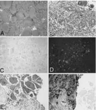
Paraffin-embedded tissue sections stained with hematoxylin and eosin revealed that the removed mass was composed almost entirely of a spherical eosinophilic material. This amorphous material stained positively and showed birefringence un-der polarized light with Congo red, indicating that it represented amyloid (Fig. 2 A-D). Immunohisto-chemically, the extracellular amyloid spheroids were positive for prolactin (Fig. 2E). Staining for growth hormone, FSH, LH, TSH, ACTH and _-subunit as well as for `-A4-amyloid was negative. Further investigation for systemic amyloidosis was
negative (transabdominal us, chest radiography and immunoelectrophoresis). Replacement ther-apy with testosterone was initiated postopera-tively, and resulted in improved well-being. The serum prolactin level slightly decreased (63.5 +g/l, normal 2.2-18.5) postoperatively and re-mained in this range at 1-yr follow-up. Repeat MRI of the sellar region showed tumor remnant. DISCUSSION
We report a man with a large pituitary tumor con-taining mainly spherical amyloid deposits. The long history of impotence and decreased libido and the early occurrence of gynecomastia with galactorrhea were suggestive for hypogonadism due to prolactinoma. Compression and apoptosis of adenoma cells due to a local mass effect of the extensive intrasellar amyloid seem a possible ex-planation for the only mild hyperprolactinemia at diagnosis in described patient with long-standing disease. Immunohistochemical staining of the spherical amyloid deposits was strongly positive for PRL, indicating that the amyloid deposits were secreted directly by prolactin-secreting cells. In addition, extensive spherical amyloid deposition occurs typically in patients suffering from pro-lactinoma (5).
Two different histological patterns of amyloid posits in pituitary adenomas have been de-scribed, the stellate and the spherical, respec-tively (6). Stellate amyloid is found perivascularly in a fibrillary pattern and small amounts may be frequently encountered in pituitary adenomas. No immunoreactive cytokeratin can be detected in these deposits. The type of spherical amyloid is rare. It is characterized by amorphous spheres ad-jacent to adenoma cells and containing im-munoreactive cytokeratin fibrils. Extensive depo-sition of amyloid spheroids in a pituitary adenoma is uncommon. In our patient, the amyloid mass penetrated the sellar floor and extended into the sphenoid sinus. This behavior would be expect-ed rather from a pituitary macroadenoma than from an amyloid mass.
Abnormal processing of prolactin or the prohor-mone produced by the adenoma cells rather than a mesenchymal origin as shown in other forms of systemic amyloidosis have been suggested as ori-gin of the pituitary amyloid (2). This was conclud-ed from the ultrastructural demonstration of amy-loid fibrils within adenoma cells and the im-munohistochemical positivity for prolactin of the amyloid deposits in some reported cases includ-ing described patient (7). Isolation and biochem-Fig. 2 - Pathology findings. The specimen consisted of several
fragments of amorphous material. (A) Hematoxylin and eosin stained fragments of amorphous material consisting of nu-merous small concentric portions. In addition, there are small fragments of hypercellular tissue, which contain compressed pituitary as it can be appreciated upon reticulin silver staining (B). Congo red (C) strongly stains all the amorphous material that appears with green birefringence in polarized light (D). The amyloid stains positively for prolactin and negative for all other pituitary hormones (E) while the compressed parts of the pituitary are positive for ACTH and negative for pro-lactin (F).
Amyloid tumor of the pituitary
555 ical analysis of protein derived from spherical amyloid deposits in a prolactin-secreting pituitary adenoma disclosed an abundance of a 4-kDa peptide (8). Amino acid sequencing revealed that this amyloidogenic peptide represents the first 34 amino acids of the intact PRL protein.
On MRI, amyloid depositions in systemic amyloi-dosis usually show signal intensity similar to that of muscle on T1-weighted images and low to in-termediate signal on T2-weighted images. The latter is the most characteristic feature of amyloid deposition (9). We found the same hypointense signal on T2-weighted images in our patient with intrasellar amyloid deposition, as described twice earlier (3, 4). These typical findings allow now sus-pecting intrasellar amyloid deposition even pre-operatively. Patients with suspected intrasellar amyloid deposition in prolactin-secreting pituitary adenomas should be treated surgically in order to confirm the diagnosis histologically and to re-move the intrasellar amyloid deposits due to the potential lack of tumor shrinkage under dopamin-agonist therapy. Dopamin-dopamin-agonist therapy may even increase the amyloid deposition (4, 5). ACKNOWLEDGMENTS
We thank Professor Christoph Schmid for valuable discussion.
REFERENCES
1. Bononi P.L., Martinez A.J., Nelson P.B., Amico J.A. Amyloid deposits in a prolactin-producing pituitary ade-noma. J. Endocrinol. Invest. 1993, 16: 339-431.
2. Kubota T., Kuroda E., Yamashima T., Tachibana O., Kabuto M., Yamamoto S. Amyloid formation in prolactinoma. Arch. Pathol. Lab. Med. 1986, 110: 72-75.
3. Sakai K., Tsutsui T., Sonobe H., Ohtsuki Y., Sawada A. MRI of pituitary adenoma with extensive amyloid formation. Neuroradiology 1999, 41: 358-359.
4. Martin S.W., Lefton D.R., Pinto R.S., Rosenblum M., Elowitz E. MR Iimaging characteristics of amyloid deposits in pi-tuitary adenoma. Am. J. Neuroradiol. 2002, 23: 368-370. 5. Saitoh Y., Mori H., Matsumoto K., et al. Accumulation of amyloid in pituitary adenomas. Acta. Neuropathol. 1985,
68: 87-92.
6. Landolt A.M., Kleihues P., Heitz P.U. Amyloid deposits in pituitary adenomas. Differentiation of two types. Arch. Pathol. Lab. Med. 1987, 111 :453-458.
7. Filippi E., Cornaggia M., Riva C., Turolla E. Spherical de-posits of amyloid in prolactin-secreting pituitary adeno-mas. Pathologica 1992, 84: 205-214.
8. Hinton D.R., Polk R.K., Linse K.D., Weiss M.H., Kovacs K., Garner J.A. Characterization of spherical amyloid protein from a prolactin-producing pituitary adenoma. Acta Neuropathol. 1997, 93: 43-49.
9. Kransdorf M.J., Murpey M.D. In: Imaging of soft tissue tu-mors. W.B. Saunders, Philadelphia, 1997, pp. 385-387.