1 3
CorrespondenCe
Introduction
spinal epidural hematoma (seH) is a rare clinical entity with an incidence of approximately 0.1 per 100,000 patients per year [1]. The etiology of spinal epidural hematoma is commonly divided into three categories “idiopathic,”“secondary,” and “spontaneous” [2]. seHs are considered idiopathic when no cause for the bleeding or a specific risk factor can be determined—secondary forms of SEH have an obvious underlying risk factors such as a neoplasm, trauma, medical intervention, i.e., spinal surgery or epidural catheterization, coagulopathy or anticoagulants [2]. Spontaneous SEHs arise in the wake of “co-risk fac-tors”. These include structural changes of the spine such as rheumatoid arthritis, ankylosing spondylitis, Paget’s dis-ease, or minor trauma [2].
In addition, two forms of seH are to be distinguished: acute and chronic seH. Acute seH is the most common and usually presents with an acute onset of symptoms including severe pain and signs of spinal cord or nerve root compres-sion. In the rare reported cases of chronic seH [2], symp-toms evolve slowly over a longer period of time. The seH is usually located in the lumbar spine below the level of the conus medullaris, most likely because the cauda equina tol-erates more pressure than the medulla spinalis [3].
Case Report
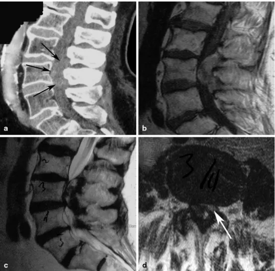
A 76-year-old man with a 1-month history of lower back pain radiating into his right upper thigh and with symptoms resembling spinal claudication was referred to our institution by his general care practitioner. His past medical history was positive for grade I obesity (BMI: 32.3), Copd, osteoarthri-tis on non steroidal anti-inflammatory drugs (NSAIDs), and atrial fibrillation treated with oral anticoagulation. Symp-toms were acute in onset and initially immobilizing due to sudden attacks of muscle weakness of his right quadriceps femoris on exertion. There were no sensory deficits and no bladder or bowel dysfunction – sphincter tone was normal. The patient was compliant with his oral anticoagulation, and international normalized ratio (Inr) levels had been stable within therapeutic range at all times. This acute initial pre-sentation was followed by some improvement of the radiat-ing pain; hence, imagradiat-ing was not obtained until one month later and after the symptoms had failed to improve with conservative management including physiotherapy and an extended analgetic regimen. Initial nonenhanced computed tomography (CT) and magnetic resonance imaging (MrI) (Fig. 1a–d) showed a 43 × 23 × 19 mm space occupying pos-terior epidural lesion at L3 compatible with a late subacute (2–4 weeks old) hematoma. The patient was then referred to our institution. on physical examination, he had no pare-sis or sensory deficits, but a decreased patellar reflex on the right side. He still complained of exertional muscle weak-ness of his right quadriceps, which could not be objectified on examination. At this time, considering the risk/benefit ratio and suspecting an epidural hematoma based on the clinical and neuroradiological findings we saw no indication for an urgent neurosurgical intervention and recommended a clinical follow-up as well as another contrast-enhanced lumbar MRI. Four weeks later, the gadolinium-enhanced
Clin neuroradiol (2013) 23:305–308 DOI 10.1007/s00062-012-0187-5
Tumor or Hematoma?
An Unusual Case of an Extradural Lesion of the Lumbar Spine
M. C. Neidert · P. Prömmel · B. Schuknecht · O. SürücüM. C. neidert, Md () · p. prömmel · o. sürücü department of neurosurgery, University Hospital Zurich, Frauenklinikstrasse 10, 8091 Zurich, Switzerland e-mail: marian.neidert@usz.ch
B. Schuknecht
Medical radiological Institute Zurich, Bahnhofsplatz 3, 8001 Zurich, switzerland
Received: 11 September 2012 / Accepted: 11 November 2012 / Published online: 4 December 2012 © Springer-Verlag Berlin Heidelberg 2012
M. C. Neidert et al.
1 3
306
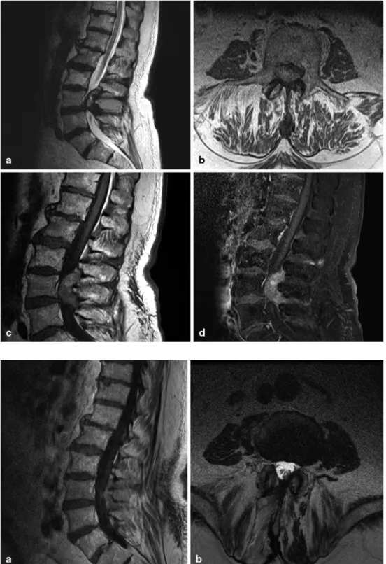
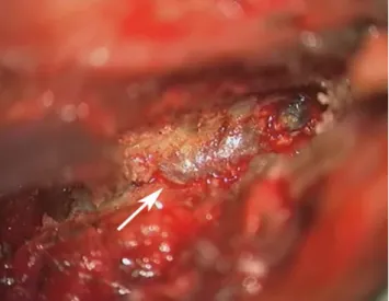
MrI (Fig. 2a–d) showed the lesion to be faint, though in the major part homogeneous contrast-enhanced. Driven by the neuroradiological suspicion of an extradural intraspinal tumor, presumably a synovial chondrosarcoma or prolif-erative hyperreactive synovial chondromatosis, we decided to perform surgery. A right-sided hemilaminectomy on the L3 level was performed by the senior author [a.s.] and a bluish encapsulated lesion appeared (Fig. 4). The capsula was strongly adherent to the dura as well as to the ligamen-tum flavum and contained greasy fluid with crankcase oil appearance. Both, the capsula and the fluid were sent for histological examination and revealed an organized chronic hematoma with no signs of neoplasia or inflammation. The postoperative hospital course was uncomplicated and the patient had complete resolution of his symptoms. At follow-up after 6 months, the patient was still asymptomatic and follow-up imaging showed no signs of a tumor or recurrent hematoma (Fig. 3a) as well as a completely decompressed spinal canal (Fig. 3b).
Discussion
Imaging features of spinal epidural hematomas using MrI depend on the age of the hematoma due to different states of oxygenation, localization and degradation of hemoglo-bin and thus may be difficult to interpret in the hyperacute, subacute, and chronic stages [4]. Intraoperative appearance and histological examination in this case revealed a chronic organized hematoma. Although it was suggested that spinal epidural hematomas undergo changes in signal intensity on both T1- and T2-weighted images analogous to intracra-nial hematomas [5], it has to be taken into consideration that blood degradation products and oxygenation states of hemoglobin might be following a different time course in encapsulated spaces lacking a capillary network compared to hematoma degradation in parenchymatous organs such as the brain.
Contrast enhancement of chronic spinal epidural hematomas has been described in the literature as either being peripheral [6, 7] or central [8, 9]. While contrast enhancement in the acute setting is often focal and only rarely homogeneous (most likely representing active
extrav-Fig. 1 Initial findings. a slightly hyperdens hardly
visible lesion on sagittal postcontrast CT (delineation is shown by black arrows).
b Midsagittal T1-weighted
image shows a slightly hyperintense mass compared with myelon. c Midsagittal T2-weighted image depicts mixed intensity. d An axial T2-weighted image showing subtotal obstruction of the spi-nal caspi-nal with residual dural sac on the left (white arrow)
Tumor or Hematoma?
1 3
307
asation), homogeneous contrast enhancement of a chronic spinal epidural hematoma has not been described in the lit-erature before. In our opinion, this unusual combination of chronic clinical symptoms and relatively homogenous con-trast enhancement is important, as it is suggestive of neopla-sia. We suspect that highly vascularized granulation tissue was the main reason for this contrast enhancement pattern.
In the literature, spontaneous SEH are linked to antico-agulation in 25–75 % of the cases [10, 11], often patients are within therapeutic range as in the reported case [10], sug-gesting that epidural bleeding might be triggered by other factors, but favored by anticoagulation.
In this case, the atypical appearance of a chronic epi-dural hematoma as well as the lack of significant clinical
Fig. 2 Follow-up after 4
weeks. a Midsagittal T2-weighted MrI showing the extension of the epidural lesion at L3 with hyper- and hypointense components.
b Axial and c sagittal
T1-weighted images without contrast show the lesion to be slightly hyperintense. Follow-ing contrast application there is moderate homogeneous increase in signal intensity on
d sagittal T1-weighted images
with fat suppression
Fig. 3 Postoperative findings. a Midsagittal
gadolinium-enhanced T1-weighted MR image showing the postopera-tive result with no contrast enhancement and a completely decompressed spinal canal, which is also seen in the b axial T2-weighted image
M. C. Neidert et al.
1 3
308
improvement led to the presumptive diagnosis of a spinal tumor. The acute onset and slight improvement of symp-toms could have been explained by an intratumoral hem-orrhage. In our opinion, surgical treatment in this patient with partial initial improvement and the histological diag-nosis of a chronic epidural hematoma should not be viewed as “avoidable” surgery because contrast-enhancement and stable residual symptoms made it impossible to rule out neoplasia as the underlying diagnosis. Additionally, other structural pathologies such as vascular malformations may be obscured by the hematoma at the time of initial imag-ing. Therefore, in the reported case, surgical management did not only provide immediate symptomatic relief but also allowed achievement of a histological diagnosis and thereby ruling out neoplasia or vascular malformation as the under-lying cause.
Conclusion
A homogeneously contrast-enhancing extradural lesion of the spine can be consistent with a chronic epidural hema-toma. If clinical symptoms fail to improve, surgery not only offers symptom relief but also provides a definite histologi-cal diagnosis.
Conflict of Interest All authors report no conflict of interest.
References
1. Holtas s, Heiling M, Lonntoft M. spontaneous spinal epidural hematoma: findings at MR imaging and clinical correlation. Radi-ology. 1996;199(2):409–13.
2. sarubbo s, Garofano F, Maida G, Fainardi e, Granieri e, Cavallo MA. spontaneous and idiopathic chronic spinal epidural hema-toma: two case reports and review of the literature. eur spine J. 2009;18(11):1055–61. doi:10.1007/s00586-009-1175-6. 3. Boyd Hr, pear BL. Chronic spontaneous spinal epidural
hema-toma. report of two cases. J neurosurg. 1972;36(2):239–42. doi:10.3171/jns.1972.36.2.0239.
4. Braun P, Kazmi K, Nogues-Melendez P, Mas-Estelles F, Aparici-Robles F. MRI findings in spinal subdural and epidu-ral hematomas. Eur J Radiol. 2007;64(1):119–25. doi:S0720-048X(07)00091-5 [pii] 10.1016/j.ejrad.2007.02.014.
5. Vazquez-Barquero A, Abascal F, Garcia-Valtuille R, Pinto JI, Figols FJ, Cerezal L. Chronic nontraumatic spinal epidu-ral hematoma of the lumbar spine: MrI diagnosis. eur radiol. 2000;10(10):1602–5.
6. Crisi G, sorgato p, Colombo A, scarpa M, Falasca A, Angiari P. Gadolinium-DTPA-enhanced MR imaging in the diagnosis of spinal epidural haematoma. report of a case. neuroradiology. 1990;32(1):64–6.
7. Watanabe n, ogura T, Kimori K, Hase H, Hirasawa Y. epidu-ral hematoma of the lumbar spine, simulating extruded lumbar disk herniation: clinical, discographic, and enhanced magnetic resonance imaging features. A case report. spine (phila pa 1976). 1997;22(1):105–9.
8. Caldemeyer Ks, Mocharla r, Moran CC, smith rr. Gadolinium enhancement in the center of a spinal epidural hematoma in a hemophiliac. J Comput Assist Tomogr. 1993;17(2):321–3. 9. Chen CJ, ro Ls. Central gadolinium enhancement of an acute
spontaneous spinal epidural haematoma. neuroradiology. 1996;38(suppl 1):s114–6.
10. Kirazli Y, Akkoc Y, Kanyilmaz S. Spinal epidural hematoma associated with oral anticoagulation therapy. Am J phys Med Rehabil/Assoc Acad Physiatr. 2004;83(3):220–3.
11. Lederle FA, Cundy KV, Farinha P, McCormick DP. Spinal epi-dural hematoma associated with warfarin therapy. Am J Med. 1996;100(2):237–8.
Fig. 4 Intraoperative image showing the bluish appearing
encapsu-lated lesion, strongly adherent to the ligamentum flavum