Invertebrate Neuroscience, 2, 245-251 (1997)
Temporal correlation between neuronal tail ganglion
activity and locomotion in the leech,
Hirudo medicinalis
A. P. BAADER and D. BekCHTOLD
University of Ziirich, Dept Zoology, Winterthurer Str. 190, CH-8057 Ziirich, Switzerland
ABSTRACT Intracellular and extracellular recordings were performed in the posterior ventral nerve cord of restrained crawling preparations of the medicinal leech, Hirudo medicinalis. Short-latency neuronal activities in the tail ganglion nerves correlated with different phases of crawling behavior. Eight neurons with characteristic activation pat- terns during crawling were identified morphologically and physiologically in the tail ganglia of 23 preparations. The axons of four of these neurons projected through posterior tail brain nerves; four ascending interneurons had projections in the connectives or in Faivre's nerve. These interneurons are suitable candidates for carrying information between the front end and the tail end of the animal to coordinate the behavioral components during a crawling step.
KEY WORDS:
Hirudo medicinalis;
leech; locomotion; motor pattern; intracellular recordings
Introduction
In the medicinal leech, the control of simple behaviors such as the local shortening reflex or the local bending reflex occurs mainly locally, on the level of individual midbody segments. In contrast, the circuitry for swim- ming consists of several hierarchically organized neu- ronal levels (Brodfuehrer and Friesen, 1986a) which are located in all body parts. Segmental pattern gener- ators (CPGs) which drive the motor output (Friesen, 1989) are regulated by descending pathways from the head brain. Several descending interneurons have s w i m - a c t i v a t i n g or s w i m - i n a c t i v a t i n g effects (Brodfuehrer and Burns, 1995), that is, they either trigger swimming (Brodfuehrer and Friesen, 1986b, c, d) or they terminate an ongoing swim episode (O'Gara and Friesen, 1995). Moreover, excitatory drive from the tail brain helps both to lower the threshold for swim i n i t i a t i o n and to prolong swim episodes (Brodfuehrer et al., 1993, 1995). The neuronal cir- cuitry for crawling behavior has only recently begun to be e l u c i d a t e d (Baader and Kristan, 1992, 1995; Eisenhart et al., 1995; Baader, 1997). Behavioral obser- vations in leeches on which surgical manipulations of the nervous system had been performed, suggested that both terminal brains were important to produce com- plete crawling cycles (Gray et al., 1938; Baader and Kristan, 1995). Therefore, both locomotor behaviors, swimming and crawling, seem to need additional regu- latory drive deriving from the head ganglion and from the tail ganglion. This paper describes neurons in the tail ganglion that participate in crawling behavior. Extracellular recordings from tail brain nerves were
performed during crawling. Neural activity was mea- sured with short latencies to behavioral events occur- ring at the front end of the animal. Eight neurons in the tail brain are described morphologically and physi- ologically. Their spike activities were characteristically modulated during different phases of crawling.
Methods
Animals and preparation
Adult leeches (Hirudo medicinalis) were obtained from commercial suppliers (Leeches USA, Westbury, NY; Moser Blutegelhandel, Schorndorf, Germany), and kept in laboratory colonies at 12 ~ Animals weighing between 2.5 and 3.5 g were anesthetized in a solution of 8% ethanol in leech saline (Muller et al., 1981 ), and pinned down with insect pins dorsal side up on a syl- gard-covered dish filled with leech saline. To dissect the posterior part of the animal containing the tail ganglion, an incision was made through the body wall along the dorsal midline between segment 18 and the tail end. After the gut and overlying tissue had been removed, the ventral blood sinus around the ganglia chain was opened. The blood sinus around the tail brain tightly adheres to the ganglion sheath. It was carefully pulled off the ganglion sheath with a sharp forceps before the sheath was removed from only those neuromeres where intracellular recordings were per- formed. The tail sucker was dissected leaving some connective tissue around the tail nerves intact, which was later used to fixate the tail brain. All nerves from ganglion 18 through the tail brain were cut at their Corresponding author: A. P. Baader. e-mail: baader@zool.unizh.ch
distal ends before the posterior body wall from segment 19 to the tail sucker was removed. Depending on the type of experiment, those nerves from which extracel- lular recordings were to be obtained were left at suffi- ctent lengths.
A n a t o m y of the posterior nervous system
T h e gross morphology of the posterior ventral nerve cord is illustrated in Fig. 1. Six animals were stamed by injecting Methylene Blue (3% in saline) into the pos- terior body cavity. Animals were then anesthetized, the body wall was opened, and after rinsing the body cavtty several times with saline, the individual nerves were traced through a dissection microscope. Fig. 1 shows the dorsal aspects of the last three midbody gan- glia (M19-M21) and of the tail ganglion, which are linked by a pair of connectives and a single Faivre's nerve. In Hirudo, each connective contains about 2800 a x o n s , a n d 97 a x o n s run in t h e F a i v r e ' s n e r v e (Wilkinson and Coggeshall, 1975). The tail ganglion is divided into seven (caudal) neuromeres (C1-C7). Two lateral roots emerge from each midbody ganglion and divide into several branches. Using the descrip- tion of Ort et al. (1974), the dorsal (DP) and the pos- terior (PP) b r a n c h of the posterior nerve could be
dorsal sucker Faivre's nerve dorsal
, midline outline ~d midline ;
, cAa
Fig. 1. Dorsal aspect of the posterior ventral nerve cord of the leech, Hirudo medicinalis. Schematic drawing of the last three midbody ganglia ( M I 9-M21) and of the tail ganglion. The latter consists of seven (caudal) neuromeres (C1-C7). For clar- W , only the right nerves of the midbody ganglia and the left and posterior nerves of the tail ganglion are drawn The dorsal mid- line of the body wall is indicated by dashed lines, the underlying sucker is outlined by the grey circle. A = anterior. DP = dorsal posterior, and PP = posterior root of the midbody ganglia; C A = anterior and CP = posterior root of the tail brain, d. = dorsal, v. = ventral.
readily re-identified in M19 to M21. However, in con- trast to anterior body segments, the anterior root (A) in M20 and M21 was often in close association to the PP nerve. Each neuromere of the tail brain has a pair of nerves on each side that run into the periphery towards the musculature of the tail sucker (grey circle designates sucker outline in Fig. 1). T h e posterior nerve in each neuromere was usually slightly thicker than the anterior one. By analogy to the rnidbody gan- glia these nerves were named according to their posi- tion in the corresponding tail neuromere (for example, CP1, for the posterior nerve tn the first caudal neu- romere, and so on).
Experimental setup
Modified versions of a previously described crawling apparatus (Baader and Kristan, 1992) were used for extracellular and intracellular recordings from the tail ganglion during crawling behavior. Animals were teth- ered in a saline-filled tank that contained a floating ball. and an extended horizontal platform with a small glass window in the center (Fig. 2). A circular ring of wax was formed around the glass window. After the dissection of the animaL, the body wall of denervated segment 18 was pinned down to the wax base with insect pins that were inserted through the muscula- ture. T h e tail ganglion was centered above the glass window and fixed with insect pins which penetrated the remainmg tissue surrounding the tail nerves. The ganglion was illuminated with a halogen light source
Fig. 2. Apparatus to record neuronal activity in the tail brain during crawling behavior. The animal ~s suspended in a
saline-filled tank which contains a ball and a honzontal platform. While its tail end is tethered to the stage ~t can produce extensions and contractmns, using the floating ball as substrate for its front sucker. The tail ganglion (TG) ~s exposed, and centered in the middle of a small glass plate. It is illuminated by a halogen light source (L) ~hrough a mzrror (MI and a dark field condensor (DK). A metal screen (S) prevents the animal from touching the recording electrode (R).
and a darkfield condensor (MBL 12010, Nikon) which was centered under the horizontal platform below the glass window. For some experiments leeches were pinned down on a small wax covered plate (15 x 30 ram) (Fig. 3). The tail ganglion was fixed to the wax plate dorsal or ventral side up with insect pins using the connective tissue around the nerves. A small metal spoon which had the shape of the tail ganglion provided additional stability for t h e ganglion. Instead of using the darkfield illumination, individual cell bod- ies were visualized by illuminating the tail ganglion with a small light guide from above.
Tail ganglion activity during leech crawling 247 morphologically, electrodes were filled with 5% of the dye Lucifer Yellow (Stewart, 1978) and with 1 m LiCk After the iontophoretical application of the dye the ganglia were processed histologically using conven- tional techniques. The neurons were drawn from whole mounts with a fluorescence compound micro- scope and camera lucid& Extracellular recordings were obtained either en p a s s a n t from the c o n n e c t i v e s between M19 and M20 or from the cut ends of periph- eral nerves using suction electrodes. Electrophysio- logical data were displayed on the screen of a storage oscilloscope and recorded on a tape recorder. A video
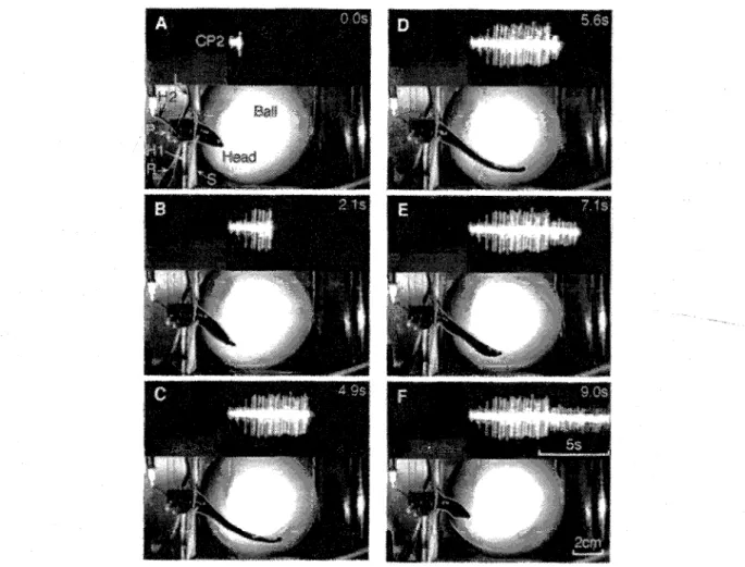
Fig. 3. Leech crawling behavior together with the extracellular recording from tail brain nerve CP2. Apparatus is slightly diffe> ent from the one shown in Fig. 2, see Methods. Video images were taken from the top at six events during a step at the times indicated. Some objects were later outlined by white hair lines to improve visibility. The animal's posterior end is tethered to a square platform (P) at the end of a stainless steel rod (H1). The ganglion is additionally supported by a metal spoon (H2) and illuminated by a small light guide (L). A metal screen (S) prevents the animal from touching the recording electrode (R). The animal inititated the step by lifting its front sucker from the ball (A) and extending its body (B). Then it re-attached with its front sucker to the substrate which terminated the elonga- tion phase (C, D). The animal shortened with a wave of segmental contractions travelling along its body from anterior to posterior (D-F), while its front sucker remained attached to the ball. After full contraction (F) it released its front sucker to inititate a new step. During the crawling cycle two main units could be distinguished in the extracellular recording: a large unit was active during the elongation phase (A-D), and a small unit fired during the contraction phase (D-F).
Intracellular recordings were obtained with glass microelectrodes filled with 3 M KAc, which had final resistances of 20-60 M ~ . To characterize neurons
camera system (PV535, Panasonic) recorded the image of the oscilloscope screen together with the behavior of the animal. Additionally, four behavioral events
during each crawling step (the onset and offset of the elongation phase, 50% and 100% contraction) were recorded with an electrical event recorder on tape together with the physiological data.
Evaluation of the physiological data
The technique for correlating neuronal events with b e h a v i o r has been described previously (Baader, 1997). In brief, responses of individual neurons during crawling episodes were evaluated by measuring their firing patterns during six behavioral events at each crawling step: during the elongation from front sucker release (fs-) to one second later, one second before the re-attachment of the front sucker (fs+), and during one s e c o n d at the m i d - p o i n t b e t w e e n these two events; during the contraction for one second after fs+ (beginning of contraction), during a one second period just before maximum contraction had occurred (end of contraction), and during one second at the mid-point of these events. The inter-step intervals during crawl- ing episodes of some animals were 500 ms and longer. In these cases neuron firing frequencies at rest were evaluated as well. An additional event was calculated when the apparent minimum or maximum of the crawling-correlated firing patterns occurred at neither of the behavioral fix-points mentioned above. The mean firing frequencies during several steps at each of the behavioral events produced the activity profiles for the individual neurons. The attachment of the front sucker was found to consist of several behavioral ele- ments, and could take more than 100 ms. The sucker becomes bowl-shaped shortly before its first physical contact with the substrate, and before the actual suck- ing occurs. Therefore. for the evaluation of the video images and for the measurements shown in Fig. 4 the moment in which the sucker became bowl shaped was taken as the onset of the attachment (fs+), and as the end of the elongation phase.
Results
With their rear end tethered in the crawling apparatus. the animals produced complete crawling episodes. Fig. 3 shows video images of a typical crawling step, together with the extracellular recording of tail brain nerve CP2. The tmages were taken at six time points during the step. The animal initiated a step by lifting its head sucker off the ball (Fig. 3A) and extending its body (Fig. 3B). A large unit in the extracellular recording was activated shortly before the beginning of the e l o n g a t i o n phase (see also Fig.4). This untt remained active throughout the elongation, and until
the front sucker re-attached to the substrate (Fig. 3C). With the onset of the contraction phase (Fig. 3D) the large unit ceased to fire, and a tonic unit of smaller amplitude became active instead. The animal short- ened with its front sucker attached to the ball (Fig. 3E, F). The smaller extracellular unit remained excited throughout the contraction phase (Fig. 3F). In extra- cellular recordings from posterior tail brain nerves in 15 animals these two units could easily be distin- guished by their different amplitudes, and by their dis- tinct activanon pattern during crawling. Both units were quiet in n o n - m o v i n g animals, and they were found in all posterior tail nerves. During crawling, their activity patterns were strongly correlated with the elongation and contraction phase (Fig. 4A), and their firing frequencies were rather independent from the actual length of the animal during crawling. Fig. 4B quantifies the temporal relationship between the occurrence of the large umt and the elongation phase of the animal. In the CPl-recording, the first action potential occurred 125 + 57 ms (mean + S.E.M.. n = 23 steps) before the first visible forward movement of the head of the animal, i.e. the initiation of the elon- gation phase. The spike train ended 266 z 51 ms after the front sucker became bowl-shaped, i.e. the begin- ning of the front sucker attachment. The small umt was turned on immediately after the large unit ceased to fire. and when the animal began to contract. The large unit was not only active during the elongation
A
C P 1 .,iJ *. IJIJlJlJLillIHlflllIJlll~liiHJHi~iliilHill~ll~iith.,~n+*, , J~.r.~lL*,z . aiitnu,uE~aannm]=l,liH~lh/lqL]lHiHI]tli~]Jiilll~ll~d[[ l s I II~,~IIIIIII II]llIIl II~rIl~llli{llfll~llFIllIIH . . . I]~ ]I~[}~ ll~IIHlilillllll~HI~![li[Ii![l
I I - - i Elongation Contraction
B
C ~
~c
' ~
~
~
s~tim_ula]!pn 2 0 0 m s n = 2 3 steps ~t l sFig. 4. Temporal correlation between neural activities in tail brain nerves and behavior A. Recording o] nerve CP1
(upper trace) together with :rawling steps (lower trace) The behavioral components (Elongatton. Contraction. Pause) are marked by the rectangles shaded in grey and white, T~ o umts are visible in the recording (see also F~g. 3): ~ large unit was acrzve during elongattons and a small unit was visible during contrac- trans. B. Temporal analysis of the large umt of CPI :luring 23 steps. The cell began to fire before the onset of elongauons, and continued until the elongations were terminated. The arrows show the mean delays with the horizontal bars indicating ~_ S.E.M. C. Response of the large unrt in a CP2-recording to mechanically touching the tip of the head of the animal which elicited the whole body shortening reflex.
phase of crawling, but also when the animal's head was mechanically stimulated by a brief stroke with a glass rod (Fig. 4C) to induce a shortening reflex (Kristan, 1982). In this case, the first response in the CP2-nerve of the tail end occurred with a delay of 280 + 9.6 ms (mean _+ SEM, n = 6 stimulations) after the first visible withdrawal of the front end.
IN902 ,
- -]~liN601o
f[Fig. 5. Reconstructions of four ascending interneurons on the ventral aspect of the tail ganglion which were acti- vated during crawling. Camera lucida drawings of the Lucifer Yellow.stained neurons. The examples shown for IN601 and IN902 were stained in different preparations. IN902 and IN904 projected into the Faivre's nerve, IN401 and IN601 ascended through the connectives. Bar 100 ~*m.
Tail ganglion activity during leech crawling 249
Firing patterns of tail ganglion n e u r o n s
Intracellular recordings in the tail brain were per- formed to trace neurons with characteristic activation patterns during crawling behavior. Four ascending interneurons on the dorsal aspect of the tail brain were identified morphologically and physiologically in a total of 14 preparations (Figs. 5, 6). IN401 and IN601 (found on both sides of the second and third neu- romere) ascended through the connectives (Fig. 5). The axons of IN902 and IN904 projected into the Faivre's nerve. IN902 was recorded in the third and seventh neuromere. It had bilaterally distributed arborizations w i t h i n the ganglion while those of IN904, IN401 and IN601 remained mainly within the ipsilateral half of the ganglion. The branches of all neurons e x t e n d e d into neighboring neuromeres. During crawling, IN902 was increasingly active at the late contraction phase and it had its firing maximum at the beginning of the elongation phase (Fig. 6A, B). The responses of the other three neurons occurred with opposite phase lags: all three were maximally excited at the end of the contraction phase and least active during e l o n g a t i o n s . In three of the four interneurons their ascending axons were traced with extracellular suction electrodes in the connectives between ganglion 19 and 20 (Fig. 6C). Corresponding action potentials in the connectives were recorded with constant delays of 4 ms to 8 ms. Three o f these interneurons (IN401, IN601 and IN904) could be
0 !lNgO2(n=16) " "'"~"-" :
1
r
IN904 - ~ [Hz] :" 20 ..[_ 1 I I / 1 0 , - - IN904 (n=4) ..,T,...a~-" I IN601 _ _ 1 30q[Hz] J
,' '
2o],...,~../
.- C I N 9 0 2 ~ " ~ , - - n=lO . _ ~ . . ~,~;~,.~z,;." . . a ~,0{
IN401 (n=16) ; " IN904 , ~ h . . n=40 0 I _..L. n=lO l,~'g'"- - -~r,. " IN401 ~ ]:;IN601 (n=20)- _, " I Fig. 6. I n t r a c e l l u l a r l y r e c o r d e d r e s p o n s e s of the ascending inter- neurons shown in Fig. 5. A. Repre- sentative activity patterns of each cell during a single step. Bars 2 s, 4 inV. B. Mean activity profiles (• S.E.M.) du~ing the number of steps indicated. C. Simul- taneous intracellular recordings from the cell bodies in the tail ganglion and extracel- lular suction electrode recordings from the connectives/Faivre's nerve between gan- glion 19 and 20. Bars i ms, 4 mV. Superimposed recordings with the number of trials indicated. For color coding of the bars see Fig. 4A.recorded when the animals started to swim but no change in activity during the swim bouts was observed (data not shown). Additionally, the tip of the head was stimulated with a glass rod to induce the shorten- ing reflex. There was no change in activity in IN401 and IN601, and a slight increase in excitation in IN902 and IN904 (data not shown).
Four neurons with axons leaving the ganglion through the posterior nerves were identified in nine preparations (Fig. 7A). N31 was most strongly excited at the beginning of the elongation phase (Fig. 7B, C). This excitation gradually decreased to a minimum when the animal contracted. The other three cells, N32, N33 and N41, were only weakly excited during the elongation phase and received stronger drive dur- ing contractions. kl31 ./ ~ J~J~l - - B
m
_ _ ]
N32 ,__1
N33~
, __1 N41 ,__1
C
_& ,-#,7" I
[Hz"
N32 (n=12). ~"[, 9
Fig. 7. Morphological and physiological characteristics of
four tail ganglion neurons which had axons projecting through the posterior nerves. Bar 100 l~m. A. Recon- structions of the neurons on the ventral side of the tail ganglion which were stained with Lucifer Yellow. Bar 100 >m. B. Neuron responses during single crawling steps. C. Averaged firing fre. quencies (+ S.E.M.) during the number of crawling cycles indi. cared. Bar 2 s, 4 mV. For color coding of the bars see Fig. 4A.
D i s c u s s i o n
Restrained leeches performed repeated crawling episodes in the apparatus although their tail suckers were tethered to the platform, and their posterior gan- glia were exposed and denervated (Fig. 3). For nor- mally crawling animals, the a t t a c h m e n t and the release of the rear sucker are important components of the crawling cycle. A failure of the rear sucker to release would prevent the animal from pulling forward. Likewise, the animal normally releases its front sucker to begin a new step after it has firmly attached its rear sucker. But the experiments with tethered leeches
show that tail sucker activities are not necessary com- ponents for the principal production of rhythmic crawling movements. If tail sucker activity is pre- vented, the animals nevertheless produce continuous crawling cycles. These findings support previous results where animals which had their tail suckers covered to p r e v e n t a t t a c h m e n t and release (Baader and Kristan, 1995) or which had denervated suckers (Gray et al., 1938) nevertheless produced cyclic crawling movements.
Neuronal activity recorded extracellularly in tail brain nerves and intracellularly from various neurons occurred temporally linked to the crawling rhythm. In posterior tail ganglion nerves, two different units were easily distinguishable. Although it was not possible to identify the two units morphologically, their firing pat- terns were clearly linked to behavior. The activation of the larger unit occurred about 125 ms before the beginning of elongation. This activity was correlated to the initiation of the elongation and not to the end of the contraction of the preceding step since it was also observed at the b e g i n n i n g of new crawling sequences with no immediate preceding contractions. Can this delay between neural activity and the onset of the extension phase help to suggest possible sites for the generation of the crawling rhythm? Previous dis- section studies have suggested that crawling is under the control of the head brain and of the tail brain (Baader and Kristan, 1995). If it is generated in the head brain, a signal must be initiated in the head, travel down the ventral nerve cord and cause the observed excitation in tail ganglion nerves, about 125 ms before the beginning of the first elongation is actu- ally observed at the head end. The existence of a prin- cipal pathway from the head to the tail is documented by the responses in CP-nerves after mechanical head stimulation (Fig. 4C), but the significance of this path- way for crawling has yet to be shown. On the other hand, if a step is initiated in the tail brain, an ascend- ing signal must travel up to the head and trigger the onset of elongation in the tip of the head, with the subsequent extension of the body. The necessary delay for such a signal travelling through the ventral nerve cord could well be in the range of what was measured between the onset of the large unit in the CP-nerves and the first observed extension of the tip of the head (=125 ms).
The activity patterns of the ascending intemeurons and their extracellular recordings from the connectives confirmed a possible importance of the tail ganglion for crawling. The delays of the action potentials of IN902, IN904 and IN401 in the connectives at the level of ganglion 19 were in the range of 4 to 8 ms. Under the assumption that the axons of these neurons
project further along t h e ventral nerve cord, their sig- nals could t h e o r e t i c a l l y arrive in the head brain after a b o u t 50 to 100 ms. T h e s e v a l u e s c o m p a r e w e l l to t h o s e o f a n o t h e r i d e n t i f i e d i n t e r n e u r o n , t h e S - c e l l (Peterson, 1984). T h i s cell has t h e largest d i a m e t e r axon in t h e ventral nerve cord and is, with an inter- segmental travel time of 2 ms, the fastest n e u r o n in the leech nervous system. A n a c t i o n p o t e n t i a l of this cell generated at one end of the a n i m a l would arrive at the o t h e r end after around 40 to 50 ms. Interestingly, the S-cell can also produce bursts during the c o n t r a c t i o n phase of crawling (Baader, 1997).
T h e o t h e r group of neurons excited during crawl- ing were cells N 3 1 - N 3 3 and N41. T h e i r morphology suggests t h a t t h e y are m o t o r neurons. However, the i d e n t i f i c a t i o n of m o t o r neurons requires simultaneous intracellular recordings from the n e u r o n and the mus- cle fiber (Stuart, 1970). T h e type of preparation used here did n o t allow muscle fiber recordings, nor was it p o s s i b l e to trigger c o r r e l a t e d m u s c l e t w i t c h e s u p o n intracellular s t i m u l a t i o n of the neuron, an alternative t h o u g h less conclusive method.
I n conclusion, the temporally organized activation of the two suckers during crawling requires fast path- ways b e t w e e n the h e a d and the tail. T h e recorded cor- r e l a t i o n b e t w e e n e x t r a c e l l u l a r l y recorded a c t i v i t y in the tail ganglion and the m o v e m e n t of the tip of the h e a d i n d i c a t e d i n d i r e c t l y t h e p r e s e n c e of such fast c o n n e c t i o n s . A d d i t i o n a l l y , the ascending interneurons had short c o n d u c t i o n times in the connectives poste- rior to ganglion 19. Moreover, they were strongly mod- ulated during crawling and n o n e of these neurons was f o u n d to be a c t i v e d u r i n g s w i m m i n g . T h e s e d a t a emphasize t h e i m p o r t a n t role of the tail ganglion dur- ing crawling and make t h e described interneurons good c a n d i d a t e s for t h e c o m m u n i c a t i o n b e t w e e n t h e two ends of t h e a n i m a l during this locomotory behavior.
Acknowledgement
S o m e of the experiments were performed in the labora- tory of W . B. Kristan Jr., to w h o m we are very grateful.
References
Baader, A. P. and Kristan, W. B., Jr. (1992) Monitoring neuronal activity during discrete behaviors: a crawling, swimming and
shortening device for tethered leeches. J. Neurosci. Meth., 43,
215-223.
Baader, A. P. and Kristan, W. B., Jr. (1995) Parallel pathways coor-
dinate crawling in the medicinal leech, Hirudo medicnialis.
J. Comp. Physiol. A, 176, 715-726.
Baader, A. P. (1997) Interneuronal and motor patterns during
crawling behavior of semi-intact leeches. J. Exp. Biol., 200,
1369-1381.
Brodfuehrer, P. D. and Friesen, W. O. (1986a) From stimulation to undulation: a neuronal pathway for the control of swimming in
the leech. Science, 234, 1002-1004.
Tail ganglion activity during leech crawling 251 Brodfuehrer, P. D. and Friesen, W. O. (1986b) Control of leech swimming activity by cephalic ganglia. J. Neurobiol., 17, 697- 705.
Brodfuehrer, P. D. and Friesen, W. O. (1986c) Initiation of leech swimming by trigger neurons in the leech subesophageal gan-
glia. I. Output connections of Trl and Tr2. J. Comp. Physiol.
A, 159, 489-502.
Brodfuehrer, P. D. and Friesen, W. O. (1986d) Initiation of leech swimming by trigger neurons in the leech subesophageal gan- glia. II. Role of segmental swim-initiating interneurons. J.
Comp. Physiol. A, 159, 503-510.
Brodfuehrer, P. D., Kogelnik, A. M., Friesen, W. O. and Cohen, A. H. (1993) Effect of the tail ganglion on swimming activity in
the leech. Behav. Neural Biol., 59, 162-166.
Brodfuehrer, P. D. and Bums, A. (1995) Neuronal factors influenc- ing the decision to swim in the medicinal leech. Neurobio[.
Learning Memory, 63,192-199.
Brodfuehrer, P. D., Debski, E. A., O'Gara, B. A. and Friesen, W. O.
(1995) Neuronal control of leech swimming. J. Neurobiol., 27,
403-418.
Eisenhart, F. J., Cacciatore, T. W., Kristan, W. B. Jr. and Wessel, R. F. (1995) Crawling in the medicinal leech is produced by a
central pattern generator. Soc. Neurosci. Abstr., 21,149.
Friesen, W. O. (1989) Neuronal control of leech swimming move-
ments. In: Neuronal and Cellular Oscillators, ed. J. W. Jacklet, pp
269-316. Basel: Marcel Dekker.
Gray, J., Lissmann, H. W. and Pumphrey, R. J. (1938) The mecha-
nism of locomotion in the leech (Hirudo medicinalis Ray). J.
Exp. Biol., 15,408-430.
Kristan, W. B. Jr. (1982) Sensory and motor neurons responsible for
the local bending response in leeches. J. Exp. Biol., 96, 161-
180.
Muller, K., Nicholls, J. G. and Stent, G. S. (1981) Neurobiology of
the leech. Cold Spring Harbor: Cold Spring Harbor Laboratories.
O'Gara, B. A. and Friesen, W. O. (1995) Termination of leech swimming activity by a previously identified swim trigger neu-
ron. J. Comp. Physiol. A, 172,627-636.
Ort, C. A., Kristan, W. B. Jr. and Stent, G. S. (1974) Neuronal con- trol of swimming in the medicinal leech. II. Identification and
connections of motor neurons. J. Comp. Physiol., 94, 121-154.
Peterson, E. L. (1984) The fast conducting system of the leech: a
network of 93 dye-coupled interneurons. J. Comp. Physiol.,
154, 781-788.
Stewart, W. W. (1978) Functional connections between cells as revealed by dye-coupling with a highly fluorescent naphtalim-
ide tracer. Cell, 14, 741-759.
Stuart, A. E. (1970) Physiological and morphological properties of motoneurones in the central nervous system of the leech. J.
Physiol. (Lond.), 209,627-646.
Wilkinson, J. M. and Coggeshall, R. E. (1975) Axonal numbers and sizes in the connectives and peripheral nerves of the leech. J.
Comp. Neurol., 162,387-396.